Edward's Syndrome
•Descargar como PPT, PDF•
10 recomendaciones•15,093 vistas

Edwards syndrome is a genetic disorder caused by the presence of all or part of an extra 18th chromosome. It is the second most common autosomal trisomy after Down syndrome. The majority of fetuses with Edwards syndrome die before birth and those that survive often have heart abnormalities, kidney malformations, and other organ disorders. Prenatal testing like amniocentesis or chorionic villus sampling is used to diagnose Edwards syndrome by analyzing fetal chromosomal material to detect the additional copy of chromosome 18.

Recomendados
Más contenido relacionado
La actualidad más candente
La actualidad más candente (20)
Similar a Edward's Syndrome
Similar a Edward's Syndrome (20)
Más de Janine Rumbaoa
Más de Janine Rumbaoa (13)
Último
Último (20)
Edward's Syndrome
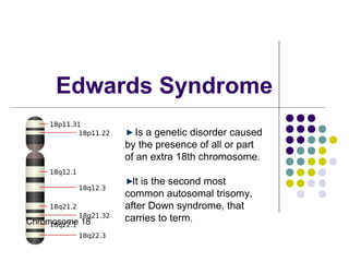
- 1. Edwards Syndrome Is a genetic disorder caused by the presence of all or part of an extra 18th chromosome. It is the second most common autosomal trisomy, after Down syndrome, that Chromosome 18 carries to term.
- 2. Elliott Greenfield suffering from Edwards syndrome Edwards syndrome occurs in around one in 6,000 live births and around 80 percent of those affected are female. The majority of fetuses with the syndrome die before birth The syndrome has a very low rate of survival, resulting from heart abnormalities, kidney malformations, and other internal organ disorders.
- 3. Signs and Symptoms kidney malformations structural heart defects at birth (i.e., ventricular septal defect, atrial septal defect, patent ductus arteriosus) intestines protruding outside the body (omphalocele) esophageal atresia mental retardation,
- 4. developmental delays growth deficiency feeding difficulties breathing difficulties and arthrogryposis (a muscle disorder that causes multiple joint contractures at birth)
- 5. Characteristics small head (microcephaly) accompanied by a prominent back portion of the head (occiput) low-set, malformed ears abnormally small jaw (micrognathia) cleft lip/cleft palate upturned nose narrow eyelid folds (palpebral fissures) widely spaced eyes (ocular hypertelorism)
- 6. drooping of the upper eyelids (ptosis) a short breast bone clenched hands choroid plexus cysts underdeveloped thumbs and or nails absent radius, webbing of the second and third toes clubfoot or Rocker bottom feet; and in males undescended testicles
- 8. Diagnostic Test Prenatal testing Triple screen-measurement of alpha fetoprotein (AFP) levels . It creens the mother's blood for AFP, hCG (human chorionic gonadotropin, the so-called “pregnancy hormone”), and estriol (a type of estrogen). The test is performed between the 15th and 17th week of pregnancy.
- 9. Ultrasound is another commonly used screening test Amniocentesis or chorionic villus sampling- Analysis of fetal chromosomal material obtained in this test is necessary to prove that the additional copy of chromosome 18 is present. Performed at 15-18 weeks of pregnancy and is the most commonly used test for the prenatal diagnosis of trisomy 18. During the procedure a thin needle is inserted through the abdominal wall and a small sample of amniotic fluid is taken.
- 10. Chorionic villus sampling is another type of test that allows the examination of fetal genetic material. It is performed earlier in pregnancy (at 10-12 weeks after the last menstrual period) and therefore carries the advantage of allowing for an earlier diagnosis. This procedure involves the collection a chorionic villus cell sample from the placenta either through the insertion of a needle in the abdominal wall or through a catheter in the vagina.