Lung cancer overview-JTL
•Descargar como PPT, PDF•
11 recomendaciones•4,477 vistas

An Overview of Lung Cancer: Epidemiology Screening Histology/Pathology Pathogenesis Invasive and Noninvasive Staging Management
![[object Object],Outline](data:image/gif;base64,R0lGODlhAQABAIAAAAAAAP///yH5BAEAAAAALAAAAAABAAEAAAIBRAA7)
Recomendados
Más contenido relacionado
La actualidad más candente
La actualidad más candente (20)
Destacado
Destacado (20)
Similar a Lung cancer overview-JTL
Similar a Lung cancer overview-JTL (20)
Último
Último (20)
Lung cancer overview-JTL
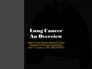
- 1. Lung Cancer An Overview Wake Forest Baptist Medical Center Radiation Oncology Department John T. Lucas Jr. MD, MSCR PGY2
- 10. Results Is there a role for screening?- NLST
- 11. Results Is there a role for screening?- NLST
- 18. Lung Cancer Herbst, Heymach, and Lippman, M.D. N Engl J Med 2008; 359:1367-1380 Etiology - Genetics
- 27. Workup - Paraneoplastic - Endocrine
- 28. Workup - Paraneoplastic - Neurologic
- 29. Workup - Paraneoplastic - Neurologic
- 30. Pathology
- 31. Pathology - Adenocarcinoma Diffuse TTF1+ Focal p63+ CK7+/CK20(-) Multiple Histologic Subtypes: mixed, acinar, papillary, nonmucinous, mucinous, solid, fetal, signet, clear cell Each w/varying genetics of unclear significance Nonsmoker EGFR mutations + f Smoker KRAS mutations + f Both TP53 mutations >70% Smokers>Nonsmokers ALK mut 7%
- 32. Pathology – Squamous Cell Carcinoma CK5/6+ Keratin + <25% TTF1 <25% CK7+ EGFR amp in 30% (-) f of KDR mut KRAS (-) p53 mut + in >60%
- 33. Pathology – Large Cell NE Carcinoma Chromogranin or Synaptophysin or NCAM + 50% TTF1+ <25% CK1/5/10/14/20 *High propensity for CNS metastasis
- 45. Current Consensus Nodal Map Regional Lymph Node Classification for Lung Cancer Staging Clifton F. Mountain and Carolyn M. Dresler CHEST June 1997 vol. 111 no. 6 1718-1723
- 46. Regional Lymph Node Classification for Lung Cancer Staging Clifton F. Mountain and Carolyn M. Dresler CHEST June 1997 vol. 111 no. 6 1718-1723
- 48. Stage Grouping
- 50. RU Mediastinum - R pre/paratracheal LN chain (4R, 2R, Sup 10R) - Tracheo-esophageal chain (LN Lvl 3p) - R. Phrenic n. chain (LN Lvl 3a) LU Mediastinum - Preaorto-carotid chain (LN Lvl 5,6) - L. Superior bronchial recurrent chain (Lvl 10L, 4L, 2L) - L. Phrenic n. Chain (Lvl 3a) Lower Mediastinum - Common inter-tracheobronchial chain (Lvl 7, 8, 10R, 10L) - Inf Pulm l. chain (LN not relevant-not pictured) M. Riquet, A. Arame, C. Foucault and F. Le Pimpec Barthes, Prognostic classifications of lymph node involvement in lung cancer and current International Association for the Study of Lung Cancer descriptive classification in zones. Interact Cardiovasc Thorac Surg , 11 (2010), pp. 260–264. Pattern of Spread
- 51. Re-appraisal of N2 disease by lymphatic drainage pattern for non-small-cell lung cancers: By terms of nodal stations, zones, chains, and a composite. Zheng H, Wang LM, Bao F, Jiang GN, Xie HK, Ding JA, Hu XF, Chen C. Lung Cancer . 2011 Apr 27.
- 52. Patterns of Spread RUL (black nodes) RML (dark gray nodes) RLL (striped nodes) LUL (crosshatched nodes) LLL (dotted nodes) Radiographics. 2004 Mar-Apr;24(2):419-34. Patterns of lymphadenopathy in thoracic malignancies. Sharma A, Fidias P, Hayman LA, Loomis SL, Taber KH, Aquino SL.
- 55. Summary ROC curve for imaging mediastinal lymph nodes that are > 1 cm diameter w/standard CT scanning Chest. 2003 Jan;123(1 Suppl):137S-146S. Noninvasive staging of non-small cell lung cancer: a review of the current evidence. Toloza EM , Harpole L , McCrory DC . Mediastinal Staging - Noninvasive
- 56. Summary ROC curve for imaging mediastinal lymph nodes that are > 1 cm diameter w/FDG PET Chest. 2003 Jan;123(1 Suppl):137S-146S. Noninvasive staging of non-small cell lung cancer: a review of the current evidence. Toloza EM , Harpole L , McCrory DC . Mediastinal Staging - Noninvasive
- 57. Sensitivity ~ 100% & Specificity ~ 84% for USPIO in predicting LAD J Magn Reson Imaging. 2000 Dec;12(6):899-904. MR imaging of mediastinal lymph nodes: evaluation using a superparamagnetic contrast agent. Pannu HK , Wang KP , Borman TL , Bluemke DA . Mediastinal Staging - Noninvasive
- 61. Mediastinal Staging – Invasive - TBNA Chest. 2011 Feb;139(2):395-401. 2010 Oct 28. Rapid on-site evaluation of transbronchial aspirates in the diagnosis of hilar and mediastinal adenopathy: a randomized trial. Trisolini R, Cancellieri A, Tinelli C, Paioli D, Scudeller L, Casadei GP, Parri SF, Livi V, Bondi A, Boaron M, Patelli M.
- 63. Mediastinal Staging – Invasive J Thorac Cardiovasc Surg. 2011 Sep 30. A prospective controlled trial of endobronchial ultrasound-guided transbronchial needle aspiration compared with mediastinoscopy for mediastinal lymph node staging of lung cancer. Yasufuku K, Pierre A, Darling G, de Perrot M, Waddell T, Johnston M, da Cunha Santos G, Geddie W, Boerner S, Le LW, Keshavjee S. EBUS & mediastinoscopy concordance rate for staging in 136 patients (91%; Kappa, 0.8; 95% confidence interval, 0.7-0.9). No significant differences were found between EBUS-TBNA & mediastinoscopy in determining the true pathologic N stage (McNemar's test, P = .78). Prospective Trial EBUS vs. Mediastinoscopy 2.6%% 0% Complications Med EBUS 100% 100% PPV 93% 93% Accuracy 90% 91% NPV 100% 100% Spec 79% 81% Sensitivity Med EBUS 153 NSCLC patients EBUS TBNA Mediastinoscopy
- 67. Role for PET – Staging Int J Radiat Oncol Biol Phys. 2004 May 1;59(1):78-86. Impact of FDG-PET on radiation therapy volume delineation in non-small-cell lung cancer. Bradley J, Thorstad WL, Mutic S, Miller TR, Dehdashti F, Siegel BA, Bosch W, Bertrand RJ.
- 68. Role for PET – Target Delineation Int J Radiat Oncol Biol Phys. 2004 May 1;59(1):78-86. Impact of FDG-PET on radiation therapy volume delineation in non-small-cell lung cancer. Bradley J, Thorstad WL, Mutic S, Miller TR, Dehdashti F, Siegel BA, Bosch W, Bertrand RJ.
- 78. The frequency of exacerbations (P=0.01): -Azithro: 1.48 per patient-yr -Placebo: 1.83 per patient-yr -HR for having an acute COPD exacerbation per patient-yr in the azithromycin group was 0.73 (95% CI, 0.63 to 0.84) (P<0.001). Lung Cancer Management - Dx & Tx of Exacerbations Prospective Trial -Azithromycin vs. Placebo for 1 yr Endpoint: - # of exacerbations - QOL
Notas del editor
- Figure 1 | Epidemiological and histological features of lung cancer in never smokers. a | Estimated nonsmoking related lung cancer deaths worldwide . Global statistics indicate that 1.18 million lung cancer deaths occurred in 2002 (REF. 1). An estimated 15% of lung cancer in men and 53% of lung cancer in women (25% of all cases) are not attributable to tobacco use1, making lung cancer in never smokers the seventh leading cause of cancer death for both sexes worldwide
- b | Lung cancers in never smokers show geographic and gender variations. A review of published studies over the past 25 years was performed using search terms ‘lung cancer’, ‘epidemiology’, ‘never smokers’ and ‘smokers’. Only studies of larger series of patients that reported the number of lung cancer cases in never smokers versus smokers, and men versus women were included. These data were pooled and categorized by geographic region (18 reports; n = 82,037)4,5,7,11,13–15,33–42, and the proportionof lung cancer cases arising in never smokers for all male and female cases were calculated and compared. These include studies from the US, Europe (Germany, Sweden, Poland, Hungary, Czech Republic, Slovakia, Romania and Russia), East Asia (China, Taiwan, Korea, Japan and Singapore), and South Asia (India and Pakistan). A marked gender bias was observed whereby lung cancer in never smokers appears to affect women more frequently than men, irrespective of geography (P <0.0001 using the Cochran-Mantel-Haenszel test). The proportion of female lung cancer cases in never smokers is particularly high in East and South Asia.
- Mayo Project - Incidence of lung cancer, the resectability of the tumor, and the survival was greater in the exp arm, but there was no significant difference in mortality (either all-cause mortality or lung cancer-specific) between 2 groups Length Time Bias Faster growing tumors have a shorter asymptomatic phase and are less likely to be detected at screening Slowly growing malignancies are over represented in screening trials Difficulty in determining who is high risk when some NSCLC/Adeno can be present in never smokers Ideally want a RCT demonstrating Number of deaths from lung cancer in the two arms Number of deaths from any cause in the two arms Stage shift (increase in the number of patients with early stage disease AND reduction in the number of patients with advanced disease) Decreased death from Lung Ca (cause specific mortality)
- 24.2% (low-dose CT) vs. 6.9% (radiography); False positive: 96.4% vs. 94.5%
- -60 days into NLST, 21 patients died 2/2 invasive eval for a abnl finding - 6 of these didn’t have lung ca
- 20% decrease in deaths is the single most dramatic decrease ever reported for deaths from lung cancer, with the possible exception of smoking cessation Only 15% of patients with lung cancer in the United States are diagnosed with early-stage (stage I or II) disease, which is usually discovered incidentally on chest imaging studies done for other reasons (3, 4) . Five-year survival with localized (early-stage) disease is 50% but only 4% in those with distant (stage IV) disease (3) . NLST also demonstrated an all-cause mortality reduction of 6.7%, although this predominantly resulted from reducing deaths from lung cancer. Lung cancer caused 60% of the 121 excess deaths in the chest radiography group
- deaths from lung cancer surgery in the NLST trial were only about 1%, but in the general population it can be as high as 4% radiation dose associated with CT screening of the chest is generally less than 2 mSv, whereas the dose of standard non–contrast-enhanced chest CT is 7 mSv risk for radiation exposure from LDCT screening in 55-year-old smokers is 1 to 3 deaths from lung cancer per 10 000 persons cumulative mortality reduction in the NLST was 30 cases of lung cancer per 10 000 persons screened. benefit–risk ratio clearly demonstrates benefit
- screening is not going to occur unless the cost of each additional quality-adjusted life-year (QALY) gained is reasonable, he said. In the U.S., the informal dividing line between cost-effective and not cost-effective is somewhere around $50,000 spent per QALY. it's a huge assumption that the overall cost of treatment would be the same in the two [screening and nonscreening] arms 39% w/+ scan first go round = amount to three CT scans per positive patient ($1,200), and much less frequently the cost of biopsy and surgery ($320 per patient) -- since only about 3% of screening patients end up getting an invasive procedure, he said -- for a total of $1,520 per patient. Current risk prediction models are approximately 70% accurate -risk model recently developed on the basis of the PLCO (Prostate, Lung, Colorectal, and Ovarian Cancer Screening Trial) accounts for the age, education level, body mass index, family history of lung cancer in first-degree relatives, history of chronic obstructive pulmonary disease, recent history of chest radiography, smoking status (current or former), pack-years smoked, and smoking duration -receiver-operating characteristic curve was 0.784 for predicting the 9-year risk for lung cancer -model doesn’t work at all for nonsmokers
- Risk of developing lung cancer in former smokers is around 1/2 (9x vs. 20x) that of current smokers (ACS cohort study). There is an RR of 1.2–1.3 for developing lung cancer from passive smoke exposure. <20% of smokers actually develop lung cancer (in the Carotene and Retinol Efficacy Trial, 10-yr cancer risk was 1%–15%). c | Distinct histological features of lung cancer in never smokers. The histological distribution of lung cancers in never smokers versus smokers was estimated using pooled data available from 17 reports published worldwide using the search criteria above4–20. Histological subtypes were classified as adenocarcinoma (ADC), squamous cell carcinoma (SCC) or ‘other’ (not shown; these included unknown histology, large cell carcinoma, mixed, not otherwise specified nonsmall- cell lung cancer (NSCLC) and small cell lung cancer (SCLC)). Cases of bronchioloalveolar carcinoma (BAC) (n = 109) were combined with ADCs. The ratio of thenumber of ADC to SCC cases are reversed in never smokers and smokers, reflecting the predominance of ADC in never smokers (P <0.0001 using the Chi-square test).
- Figure 3 | Distinct features of EGFR, KRAS and TP53 mutations in lung cancers in never smokers. a | Frequency of EGFR, KRAS and TP53 mutations. Significant differences in the frequency of mutations in the EGFR, KRAS and TP53 genes have been observed in lung cancers arising in smokers and never smokers: - EGFR mutations are more frequently found in adenocarcinomas arising in never smokers KRAS mutations are more frequently found in adenocarcinomas arising in smokers TP53 mutations are frequent in both, but are more common in non-small-cell lung cancers (NSCLCs) in smokers b | TP53 mutation signature and spectrum. *** Although EGFR and KRAS mutations appear to be mutually exclusive , TP53 mutations are found in NSCLCs, independent of EGFR or KRAS mutation status. - Distinct features in the TP53 mutation patterns in lung cancers arising in smokers and never smokers have been observed. Specifically, G:C to T:A (G–T) transversions and A:T to G:C (A–G) transitions at CpG sites are characteristic of tobacco-related carcinogens131. By contrast, G:C to A:T (G–A) transitions at non-CpG sites are associated with lung cancers in never smokers . Five codons in the central DNA binding domain of the TP53 gene (codons 157, 158, 245, 248 and 273) have been identified as preferred sites for DNA adduct formation, and are the sites of tobaccoassociated mutations131. As less than 10% of TP53 mutations occur at any individual site, we have termed these ‘warm spots’157. Mutations at these codons (except for codon 273) occur less frequently in lung cancers arising in never smokers131. In addition, the relationship of KRAS, EGFR and TP53 mutations is more complex. In a study of Japanese adenocarcinomas119, mutations in TP53 occurred independently of EGFR or KRAS mutations . However, the TP53 mutation spectrum and signature were different in EGFR-mutant and non-mutant tumours, TP53 mutations at the tobacco-related warm spots and G:T transversions occurred almost exclusively in patients with EGFR mutations . By implication, the mutation patterns of all three genes, their frequencies and interrelationships are strongly related to tobacco exposure.
- Environmental risk factors: 1. Radon 2. Asbestos 3. Occupational toxins Viral etiology may be restricted to certain subtypes of HPV- significance unclear Polymorphisms in DNA repair genes involved in base excision repair ( XRCC1 and OGG1 ), nucleotide excision repair ( ERCC1 , ERCC2 and XPA ), DNA double-strand break repair ( XRCC3 ) and mismatch repair pathways ( MLH1 and MSH2 ) have been studied in association with lung cancer risk102. The role of polymorphisms in DNA repair genes could be dependent on smoking status. Three polymorphisms in the genes XRCC1 and ERCC2 , which are associated with decreased adduct removal, have been found to be risk factors in never or light smokers, but tended to be protective in heavy smokers105. Although the reasons for these differences are not clear, it is possible that in heavy smokers, the effects of the DNA repair polymorphisms are overwhelmed. The effects of several other DNA repair genes on lung cancer risk have also been evaluated, and the studies so far have not identified any consistent associations for any specific polymorphisms102.
- ALK initially described in lymphomas, neuroblastomas - novel fusion oncogene in non-small cell lung cancer (NSCLC). The fusion results from a small inversion within chromosome 2p, leading to expression of a constitutively activated, chimeric tyrosine kinase. Target of crizotinib- originally developed as a c-Met inhibitor Present primarily in light/never smokers w/out EGFR mutation ALK and EGFR Mutually exclusive EGFR and KRAS mutations appear to be mutually exclusive , TP53 mutations are found in NSCLCs, independent of EGFR or KRAS EGFR Met T790M associated w/resitance ot TKIs Ex 19 &21 mutation are associated w/excellent response to gefitinib
- Figure 2. Mutant EGFR-expressing cell lines show dissimilar kinetics of DNA repair at 1 Gy as WT-expressing NSCLC cell lines. Cell lines were irradiated at 1 Gy, and samples were fixed at various time points and probed with anti-phosphorylated histone gH2AX antibody that was detected with a Cy5-labeled secondary antibody by fluorescence microscopy. A, images of H2AX foci in nuclei at 40 magnification from three representative NSCLC cell lines. B, graphical representation of results from four WT (solid lines ) and six mutant (broken lines ) EGFR NSCLCs. Points, mean values of >6,000 nuclear events taken from 30 fields; bars, SD
- Fig. 1. Clonogenic survival of HT1080 cells is reduced after loss of activated N-ras. Cells in log phase growth were plated and irradiated for clonogenic survival at the doses indicated. After 14 –21 days, plates were stained and scored for colony formation. CIRCLE: SG-1 (E), HT1080 cells expressing both activated and wild-type endogenous N-ras alleles. TRIANGLE: SG-2 (D), HT1080 expressing only the WT endogenous N-ras allele. BOX: SG-6 (M), HT1080 expressing the wild-type endogenous N-ras allele and an activated N-ras introduced by transfection. Bars, SD.
- Figure 2 | Pathogenesis of lung cancer in never smokers: targeting a specific anatomical compartment. The putative stem cell of the bronchus is the basal cell, which is believed to give rise to the differentiated ciliated, mucous and neuroendocrine cells. The terminal bronchioles and alveoli are believed to share a common stem cell, and a putative stem cell has recently been identified in the mouse. Peripheral airway stem cell is difficult to identify morphologically, and the cells capable of dividing in these locations are: -- Clara cells (in the bronchioles BRONCHOALVEOLAR TYPE) -- Type II pneumocytes (surfactant secreting) (in the alveoli ACINAR TYPE). Thyroid transcription factor 1 (TTF1), a transcription factor important in lung morphogenesis, is expressed by peripheral airway cells, by their putative stem cell. The secretory products of peripheral airway cells include Clara cell protein 10 (CC10) and surfactant (from type II pneumocytes). Thus, region-specific stem cell niches function to maintain epithelial diversity. Lung cancer may be considered to arise from either the central airway compartment (predominantly SCLC and squamous cell carcinoma) or from the peripheral airway compartment (predominantly adenocarcinoma). Carcinogens from tobacco smoke appear to target both central and peripheral airways, whereas the poorly understood factors leading to lung cancers in never smokers appear to specifically target the peripheral compartment.
- Currently medically inoperable based on trials listed as 50% FEV1 and/or 50% DLCO If the FEV1 is <80% predicted for the age and size of the pt or the DLCO is <80% predicted, then the pt may need quantitative lung scans/exercise testing to carefully predict postop pulmonary function Pneumonectomy: >2 L Lobectomy: >1.5 L Subsets at high risk for surgical morbidity: 1. pCO2 <45 mm Hg 2. pO2 <50 mm Hg 3. Preop FEV1 <40% of predicted value 4. Poor exercise tolerance 5. DLCO <50% of predicted value 6. Postop FEV1 <0.71 or <30% of predicted value 7. Cardiac problems (left ventricle ejection fraction <40%, myocardial infarction within 6 mos, arrhythmias) 8. Obesity
- Horners Syndrome: Ptosis, miosis, ipsilateral anhidrosis
- TTF1 is a homeodomain containing nuclear transcription protein of the Nkx2 gene gamily in epithelial cells of the embryonal and mature lung and thyroid ** Very important in differentiating Primary Adeno vs. Metastatic Adeno of the colon is CK7(-) and CK20+ EGFR and KRAS mutations appear to be mutually exclusive , TP53 mutations are found in NSCLCs, independent of EGFR or KRAS *** Least associated w/smoking FIGURE 10. Adenocarcinoma in small biopsy and cytology. Poorly differentiated non-small cell carcinoma, favor adenocarcinoma. A, This core biopsy shows a solid pattern of growth, and morphologically, it lacks any acinar, papillary, or lepidic patterns. The mucin stain was also negative. B, The TTF-1 stain is strongly positive. C, The p63 stain is very focally positive. The strongly and diffusely positive TTF-1 and only focal p63 staining favor adenocarcinoma. In this case, EGFR mutation was positive. D, Cytology from different adenocarcinoma shows large malignant cells with abundant cytoplasm and prominent nuclei growing in an acinar structure. EGFR, epidermal growth factor receptor; TTF, thyroid transcription factor.
- ** Most associated w/smoking SCC Cytology A) Lots of keratinization, pap stain B) No keratinization present, dysplastic malignant cells, pap stain C) No keratinization, gaps b/n cells and distinct cell borders, sheet apperance, Pap stain SCC Histo/path Invasive fibrous stroma w/keratin pearls, atypia w/hyperchromatic nuclei Squamous pearls, distinct intercellular bridges Layers of keratinization
- SCLC is negative for Chromogranin and Synaptophysin High power photomicrograph showing cells with an organoid architecture. The cells have a modest amount of eosinophilic cytoplasm and nuclei with coarse chromatin and prominent nucleoli. An area of necrosis is seen in the lower right hand portion of the photomicrograph, which is characteristic of large cell neuroendocrine carcinoma. Low power photomicrograph showing tumor nests with an organoid appearance characteristic of a neuroendocrine neoplasm. There is an abundant necrosis associated with many of the tumor cell nests.
- Fig 1.16 Small Cell Carcinoma Densly Packed, small, scant cytplasm, finely granular nuclear chromatin, no nucleoli, frequent mitoses Spingle shape/fusiform, nuclear chromatin is granular/nucleoli are absent D) Extensive necrosis E) Combined Small cell and Adenocarcinoma F) Combined SCLC and Large Cell Carcinoma (Large cell part has more cytoplasm and more prominent nucleoli
- 10 yrs b/n 4&5th 5 yrs b/n 5&6 th 7 yrs b/n 6 th &7th
- Fig. 5. After permutation and combination with nodal “stations”, “zones”, and “chains”, two subgroups indicated discrepant survival: One nodal chain involvement and multiple ( p < 0.0001). 5YS: 5-year survival rate, MST (median survival time).
- Fig. 5. After permutation and combination with nodal “stations”, “zones”, and “chains”, two subgroups indicated discrepant survival: One nodal chain involvement and multiple ( p < 0.0001). 5YS: 5-year survival rate, MST (median survival time).
- Fig. 5. After permutation and combination with nodal “stations”, “zones”, and “chains”, two subgroups indicated discrepant survival: One nodal chain involvement and multiple ( p < 0.0001). 5YS: 5-year survival rate, MST (median survival time).
- Ant & Lateral View Fig. 1. Showing the relation between each lymph node chain, lymph node stations and the zones of current IASLC mapping: supraclavicular zone: 1R and 1L (two chains); upper zone: 2R and 4R (one chain); 2L and 4L (one chain); 3a (one chain); AP zone: 5 and 6 (one chain); subcarinal zone: 7 and 8 (one chain); lower zone: 9 (one chain); hilaryinterlobar zone: 10 and 11. IASLC, International Association for the Study of Lung Cancer; AP, aortopulmonary RU Mediastinum -- The right pre- or paratracheal LNs chain has abundant LNs, whose size decreases from below to upward and whose number varies from one individual to another. It corresponds to the stations 4R, 2R, and superior 10R w3x or LN of the azygos vein arch of Rouvie`re w10x. The chain lies in the pretracheal space. The pretracheal space is bounded anterolaterally by the superior vena cava, right brachiocephalic vein and mediastinal pleura on the right, the aorta and pericardium on the left, and posteriorly by the trachea . -- The tracheo-esophageal chain w5x consists of a lymph vessel with nodes at the thoracic inlet of the mediastinum; rarely, a retrobronchial node is present. It corresponds to station 3p w3x. The chain lies in the right tracheo-esophageal groove. -- The right phrenic nerve chain runs along the right phrenic nerve and is equivalent to station 3a. It rarely participates in the lymphatic drainage of the lung w3, 10x. In the left upper mediastinum, there are also three LN chains. -- The pre-aorto-carotid chain : begins in front of the aortopulmonary space with a node commonly located in front of the ligamentum arteriosum, and ascends subpleurally in the triangle formed by the hilum of the lung inferiorly, the phrenic nerve anteriorly, and the vagus nerve posteriorly, the whole being covered by the mediastinal pleura w25x. LNs are not numerous. It corresponds to stations 5 and 6 w3, 15x. -- The left superior bronchial-recurrent chain w5x usually consists of one or two lymphatics ascending in the left tracheo-esophageal groove. It rarely contains nodes after the left superior bronchial (10L) and lower tracheal (4L) ones. It corresponds to stations (superior) 10L, 4L, and 2L w3, 15x. 4L and 10L are located in the aorto-pulmonary space, commonly called as aorto-pulmonary window. It is bounded superiorly by the aortic arch, medially by the trachea and esophagus, inferiorly by the superior border of the main left bronchus, anteriorly and laterally by the pulmonary artery and ligamentum arteriosum w25x. -- The left phrenic nerve chain runs along the left phrenic nerve and is equivalent to station 3a. It rarely participates in the pulmonary lymphatic drainage, but does so more often than the right side as a result of its close connection with the pre-aorto-carotidian chain In the lower mediastinum, there are three chains. -- A common chain for both lungs of intertracheobronchial LNs w5x: has a variable number of nodes whose size varies from one individual to another and are arranged in three clusters: one median subcarinal cluster flanked by right and left clusters of nodes situated under the corresponding bronchus. These clusters correspond to stations 7, 8, and inferior 10R and 10L w3, 15x. The chain is located within the subcarinal space w25x, which is bounded superiorly by the carina, laterally by the mainstem bronchi, anteriorly by the pulmonary artery and the posterior aspect of the pericardium as well as left atrium, and posteriorly by the esophagus. -- The inferior pulmonary ligament has a LN chain in each side. LNs are neither frequent nor numerous, but lymphatic vessels are always present at this level. These chains are near the lateral aspect of the esophagus, which is surrounded by juxtaesophageal LN which mainly receives the lymph from the esophagus w26, 27x. However, the juxtaesophageal LNs do not normally participate in the lymph drainage of the lungs Discrepancies b/n anatomy and staging anatomical zones ** LN station description resembles anatomy but is inaccurate: -- For instance - The discrepancies between stations 2 and 4 have not been completely resolved. In fact, 2R and 4R pertain to the preor paratracheal LN chain, and 2L and 4L to the left superior bronchial-recurrent chain: there are four stations, but two chains from a strictly anatomical point of view. **The arbitrary division along the midline of the trachea created by the ATS has been rightly eliminated: it was also not anatomical, and all LN in the pretracheal space pertain to the pre- or paratracheal chain, obviously including the Naruke station 3 overlying the midline of the trachea. Fig. 2. Lateral view showing the 3p and 3a lymph node stations within their chain (upper zone of the IASLC mapping: 3p and 3a, one chain each). IASLC, International Association for the Study of Lung Cancer.
- LN Mets pattern of spread as indicated by zone (% of Total ) PURPOSE: N2 non-small-cell lung cancer (NSCLC) is a heterogeneous disease with an extremely wide range of 5-year survival rates. A composite method of sub-classification for N2 is likely to provide a more accurate method to more finely differentiate prognosis of N2 disease. METHODS: A total of 720 pN2 (T1-4N2M0) NSCLC cases were enrolled in our retrospective analysis of the proposed composite method. Survival rates were respectively calculated according to the N2 stratification methods: singly by &quot;nodal stations&quot;, &quot;nodal zones&quot;, or &quot;nodal chains&quot;, or by combination of all three. Statistical analysis was carried out by Kaplan-Meier and Cox regression models. RESULTS: A total of 10,199 lymph nodes (8059 mediastinal; 2140 hilar and intra-lobar) were removed. By nodal station, there were 173 cases of single-station involvement and 547 multi-stations. By nodal zone, there were 413 single-zone involvement and 307 with multiple zones. By nodal chain, there were 311 cases with single-chain and 409 multi-chain involvements. The overall 5-year survival was 20% and median survival time was 27.52 months. The 5-year survival was significantly better for cases of single-zone involvement, as compared to multi-zones (29% vs. 6%, p<0.0001). The 5-year survival rates of single- and multi-chains involvement were 36% and 8%, respectively (p<0.0001). When taking all of the above grouping methods into consideration, the N2 disease state could be further sub-classified into two subgroups with respective survival rates of 36% and 7% (p<0.0001). Subgroup I was composed of individuals with single-chain involvement and having either one or two station metastasis; individuals with any other metastasis combinations formed Subgroup II. Multivariate analysis revealed that the composite sub-classification method, number of positive lymph nodes, ratio of nodal metastasis, and pT information were the most important risk factors of 5-year survival. CONCLUSIONS: By combining the three N2 stratification methods based on &quot;stations&quot;, &quot;zones&quot;, and &quot;chains&quot; into one composite method, prognosis prediction was more accurate for N2 NSCLC disease. Single nodal chain involvement, which may be either one or two nodal stations metastasis, is associated with best outcome for pN2 patients.
- Figure 1. Diagrams of the axial anatomy of the chest show the lymph nodes and drainage pathways for lung cancer in different lobes of the lung. All tumors drain to the interlobar and hilar nodes. The separate drainage pathways for tumors of the right upper lobe (black nodes), middle lobe (dark gray nodes), right lower lobe (striped nodes), left upper lobe (crosshatched nodes), and left lower lobe (dotted nodes) are shown. The corresponding American Thoracic Society (ATS) stations are labeled. E esophagus, TD thoracic duct. Diagram shows the left paratracheal nodes (LPTN) (ATS station 2L) and right paratracheal nodes (RPTN) (ATS station 2R). LBCV left brachiocephalic vein, LCCA left common carotid artery, LSCA left subclavian artery, RBCA right brachiocephalic artery, RBCV right brachiocephalic vein, Tr trachea. (b) Diagram shows the left paratracheal nodes (LPTN) (ATS station 4L), right paratracheal nodes (RPTN) (ATS station 4R), and subaortic nodes (SAN) (ATS station 5). AA ascending aorta, Ao aorta, SVC superior vena cava, Tr trachea. (c) Diagram shows the hilar nodes (HN) (ATS station 10), interlobar nodes (ILN) (ATS station 11), lobar nodes (LN) (ATS station 12), and subcarinal nodes (SCN) (ATS station 7). Ao aorta, BI bronchus intermedius, LMB left main bronchus, LPA left pulmonary artery, LULB left upper lobe bronchus, PA pulmonary artery, RPA right pulmonary artery, SPV superior pulmonary vein, SVC superior vena cava. (d) Diagram shows the left inferior pulmonary ligament node (LIPLN) (ATS station 9) and right inferior pulmonary ligament node (RIPLN) (ATS station 9). Ao aorta, CS coronary sinus, ECF epicardial fat, LV left ventricle, RA right atrium, RV right ventricle.
- Noninvasive Staging of Non-small Cell Lung Cancer A Review of the Current Evidence CHEST January 2003 vol. 123 no. 1 suppl 137S-146S Summary ROC curve for imaging mediastinal lymph nodes that are > 1 cm diameter with standard CT scanning. The gray ellipses indicate individual study estimates of sensitivity and 1 − specificity. A study showing the highest accuracy will appear in the top left corner of the graph. The size of the gray ellipse is proportional to the statistical weight of the study in the analysis (roughly proportional to its size). The “X” indicates the summary point estimates for sensitivity and 1 − specificity from meta-analyses of sensitivity and specificity, independent of one another. The outlined box indicates 95% CIs about the summary sensitivity and 1 − specificity estimates. The dark line represents the summary ROC curve.
- Summary ROC curve for imaging mediastinal lymph nodes that are > 1 cm diameter with PET using 18F-2-fluoro-2-deoxy-D-glucose. The gray ellipses indicate individual study estimates of sensitivity and 1 − specificity. The size of the gray ellipse is proportional to the weight of the study in the analysis. The “X” indicates the summary point estimates for sensitivity and 1 − specificity from meta-analyses of sensitivity and specificity, independent of one another. The outlined box indicates 95% CIs about the summary sensitivity and 1 − specificity estimates. The dark line represents the summary ROC curve. Est of Falp + N2 nodes based on PET ∼10%–20%. PPV ∼80%. +N2 nodes by PET/CT need pathologic confirmation before deferring to potentially curative surgery. Est of FN N2 nodes based on PET ∼5%–16%. NPV ∼95%. N2 nodes by PET/CT for clinical T1 lesions may not need mediastinoscopic evaluation (this is controversial). Rate of occult mets detected from PET- In many series, the range is ∼6%–18%.
- Figure 1. T2-weighted fast spin-echo image in a patient with non-small cell lung cancer. A node is present in the right paratracheal region . It is slightly hyperintense to muscle both before (arrow) and after (arrowhead) administration of ferumoxtran-10. Therefore it does not change in signal intensity, indicating lack of iron uptake. Biopsy was positive for tumor. A right pleural effusion is also noted. Figure 2. T2-weighted fast spin-echo image in a patient with non-small cell lung cancer. A node is present in the right paratracheal region. It is hyperintense to muscle before administration of ferumoxtran-10 (arrow) and decreases in signal intensity after contrast (arrowhead). Transbronchial biopsy and subsequent lymphadenectomy were both negative for tumor . The primary tumor in the left lung is also visualized. A small node in the aortopulmonic window is also present that decreased in signal intensity, but biopsy confirmation of this node was not obtained Figure 3. T2-weighted fast spin-echo image with fat suppression in a patient with a single pulmonary nodule, biopsy proven as paraganglioma. A precarinal node is present. It is hyperintense to muscle on precontrast images (arrow). On postcontrast images, the node shows hyperintense signal (arrowhead). Lack of decreased signal indicates lack of ferumoxtran-10 uptake . Transbronchial biopsy was negative for tumor and positive for inflammatory chang
- Procedure by Level X indicates accessible, (-) indicates not accesible P indicates only posterior accessible A indicates only anterior accessible Some form of extended Mediastinoscopy is required for Lvls 5&6
- Overall Sensitivity and Specificity is high for EUS and EBUS, w/>88% reported by Dr. Bellinger her in our clinic Most accurate is still Mediastinal LN exam
- The samples obtained by transbronchial needle aspiration (TBNA) during fiberoptic bronchoscopy (FOB) are suitable for rapid on-site evaluation (ROSE). This study prospectively investigated the diagnostic range, yield and practical value of this technique . METHODS AND RESULTS: Consecutive patients with radiologically suitable mediastinal mass lesions or lymph nodes on conventional chest x-rays and lung CT scans were investigated with FOB. TBNA-ROSE was performed during FOB. A cytopathologist prepared unstained slides, stained them and evaluated the aspirates on-site and notified the bronchoscopist about the necessity of further sampling. If adequate diagnostic material was collected with TBNA, the provisional diagnosis was noted and the procedure ended. Fifty patients with mediastinal masses or lymph nodes were included. In 37 (74%) patients, the diagnosis was made on-site: non small cell lung cancer in 16, small cell lung cancer in 4, metastatic cancer in 6, granulomatous disease in 9 and lymphoma in 2 patients. TBNA was not diagnostic in 9 (18%) patients with reactive lymphoid hyperplasia that required additional studies. Overall, ROSE shortened bronchoscopic sampling in 74% of patients . The mean intervention time was 25 minutes . No side effects of TBNA were observed. CONCLUSION: TBNA combined with ROSE is safe and highly effective. ROSE increased the sensitivity of TBNA. Complication rate was low at 16% and only minor noted as 20-50ml of blood loss was noted
- making an incision approximately 1 cm above the suprasternal notch of the sternum , or breast bone. Dissection is carried out down to the pretracheal space and down to the carina . A scope ( mediastinoscope ) is then advanced into the created tunnel which provides a view of the mediastinum. The scope may provide direct visualization or may be attached to a video monitor.
- OBJECTIVE: The study objective was to compare endobronchial ultrasound-guided transbronchial needle aspiration (EBUS-TBNA) with mediastinoscopy for mediastinal lymph node staging of potentially resectable non-small cell lung cancer. METHODS: Patients with confirmed or suspected non-small cell lung cancer who required mediastinoscopy to determine suitability for lung cancer resection were entered into the trial. All patients underwent EBUS-TBNA followed by mediastinoscopy under general anesthesia. If both were negative for N2 or N3 disease, the patient underwent pulmonary resection and mediastinal lymphadenectomy. RESULTS: Between July 2006 and August 2010, 190 patients were registered in the study, 159 enrolled, and 153 were eligible for analysis. EBUS-TBNA andmediastinoscopy sampled an average of 3 and 4 lymph node stations per patient, respectively. The mean short axis of the lymph node biopsied by EBUS-TBNA was 6.9 ± 2.9 mm. The prevalence of N2/N3 disease was 35% (53/153). There was excellent agreement between EBUS-TBNA and mediastinoscopy for mediastinal staging in 136 patients (91%; Kappa, 0.8; 95% confidence interval, 0.7-0.9). Specificity and positive predictive value for both techniques were 100%. The sensitivity, negative predictive value, and diagnostic accuracy for mediastinal lymph node staging for EBUS-TBNA and mediastinoscopy were 81%, 91%, 93%, and 79%, 90%, 93%, respectively. No significant differences were found between EBUS-TBNA and mediastinoscopy in determining the true pathologic N stage (McNemar's test, P = .78). There were no complications from EBUS-TBNA. Minor complications from mediastinoscopy were observed in 4 patients (2.6%). CONCLUSIONS: EBUS-TBNA and mediastinoscopy achieve similar results for the mediastinal staging of lung cancer. As performed in this study, EBUS-TBNA can replacemediastinoscopy in patients with potentially resectable non-small cell lung cancer.
- Remeber that PPV/NPV are a function of prevalence so low prevalence will result in low PPV and high NPV. Incidence of asymptomatic Neuro, Abd and bone mets is so low, adding additional imaging w/out a symptom is of low utility The present meta-analysis also updates the meta-analysis performed by Silvestri et al 4 As with his study, we found the calculated summary estimates for the NPV to be ≥ 90% for the clinical evaluation of asymptomatic patients for brain, abdominal, or bone metastases. These findings are consistent with the findings of a retrospective analysis 75 of 755 patients with clinical stage T1-2N0, which found only five sites of silent metastasis after extensive imaging for extrathoracic disease in all presenting patients. However, within the studies combined in our meta-analyses, there was considerable heterogeneity with respect to their individual NPV. The greatest range was found for the evaluation for bone metastases, in which the NPVs ranged from 0.7 to 1.0 . Some of the heterogeneity may be due to the imperfect reference standard, in which abnormal bone scan findings were not followed up with histologic confirmation of disease. For example, in the study in which 30% of patients had a false-negative clinical evaluation, 50 only 4 of 17 patients with abnormal bone scan findings received confirmations of lung cancer that was metastatic to bone. There was also significant variation in the stage and cell type of disease in the patients who were evaluated in the included studies. For the evaluation of abdominal metastases, the NPVs also ranged broadly between 0.82 and 1.0 . In this case, (1) the studies did not control for stage and/or size of the primary tumor, (2) the reference standard was suboptimal, as abnormal findings on CT scanning were not always confirmed with biopsy, and (3) patient populations varied between studies. For the evaluation of brain metastases, with the exception of one very poorly designed study, 57 the NPV, although heterogeneous, ranged more narrowly between 0.91 and 1.0. In contrast, although the PPV varied among the studies included in this meta-analysis of the clinical evaluation for detecting metastatic disease, the resultant following conclusion remains the same: further diagnostic testing is necessary to confirm the presence of disease in patients with abnormal findings on clinical evaluation. A separate issue that was not evaluated by this meta-analysis is the role that whole-body PET scanning may play in the workup of asymptomatic patients for metastatic disease. A recent randomized trial 79 suggests that the addition of PET scanning to a conventional workup identified more patients with distant metastases among those with suspected NSCLC.
- ET findings altered the TNM stage in 15 (44%) of 32 patients compared with CT alone. However, only 7 of these 15 alterations were confirmed by pathologic examination
- Purpose: Locoregional failure remains a significant problem for patients receiving definitive radiation therapy alone or combined with chemotherapy for non–small-cell lung cancer (NSCLC). Positron emission tomography (PET) with [18 F]fluoro-2-deoxy-D-glucose (FDG) has proven to be a valuable diagnostic and staging tool for NSCLC. This prospective study was performed to determine the impact of treatment simulation with FDG-PET and CT on radiation therapy target volume definition and toxicity profiles by comparison to simulation with computed tomography (CT) scanning alone. Methods: Twenty-six patients with Stages I–III NSCLC were studied . Each patient underwent sequential CT and FDG-PET simulation on the same day. Immobilization devices used for both simulations included an alpha cradle, a flat tabletop, 6 external fiducial markers, and a laser positioning system. A radiation therapist participated in both simulations to reproduce the treatment setup. Both the CT and fused PET/CT image data sets were transferred to the radiation treatment planning workstation for contouring. Each FDG-PET study was reviewed with the interpreting nuclear radiologist before tumor volumes were contoured. The fused PET/CT images were used to develop the three-dimensional conformal radiation therapy (3DCRT) plan. A second physician, blinded to the results of PET, contoured the gross tumor volumes (GTV) and planning target volumes (PTV) from the CT data sets, and these volumes were used to generate mock 3DCRT plans. The PTV was defined by a 10-mm margin around the GTV. The two 3DCRT plans for each patient were compared with respect to the GTV, PTV, mean lung dose, volume of normal lung receiving >20 Gy (V20), and mean esophageal dose. Results: The FDG-PET findings altered the AJCC TNM stage in 8 of 26 (31%) patients; 2 patients were diagnosed with metastatic disease based on FDG-PET and received palliative radiation therapy. Of the 24 patients who were planned with 3DCRT, PET clearly altered the radiation therapy volume in 14 (58%), as follows. PET helped to distinguish tumor from atelectasis in all 3 patients with atelectasis . Unsuspected nodal disease was detected by PET in 10 patients, and 1 patient had a separate tumor focus detected within the same lobe of the lung. Increases in the target volumes led to increases in the mean lung dose, V20, and mean esophageal dose. Decreases in the target volumes in the patients with atelectasis led to decreases in these normal-tissue toxicity parameters. Conclusions: Radiation targeting with fused FDG-PET and CT images resulted in alterations in radiation therapy planning in over 50% of patients by comparison with CT targeting. The increasing availability of integrated PET/CT units will facilitate the use of this technology for radiation treatment planning. A confirmatory multicenter, cooperative group trial is planned within the Radiation Therapy Oncology Group. Figure 1 PET/CT alters radiation therapy volume delineation. In the left panel, CT demonstrated a right lower lobe spiculated mass. A PET/CT was subsequently performed demonstrating additional involvement in a subcarinal lymph node. PET/CT, positron emission tomography-computed tomography.
- Purpose: Routine assessment was made of tumor metabolic activity as measured by 18F-fluorodeoxyglucose (FDG) positron emission tomography (PET) in Stage I non–small-cell lung cancer (NSCLC). This report describes PET correlates prospectively collected after stereotactic body radiotherapy (SBRT) for patients with medically inoperable NSCLC . Methods and Materials: 14 consecutive patients with medically inoperable Stage I NSCLC were enrolled . All patients received SBRT to 60–66 Gy in three fractions. Patients underwent serial planned FDG-PET/computed tomography fusion imaging before SBRT and at 2, 26, and 52 weeks after SBRT. Results: With median follow-up of 30.2 months, no patients experienced local failure . One patient developed regional failure, 1 developed distant failure, and 1 developed a second primary. The median tumor maximum standardized uptake value (SUVmax ) before SBRT was 8.70. The median SUVmax values at 2, 26, and 52 weeks after SBRT were 6.04, 2.80, and 3.58, respectively. Patients with low pre-SBRT SUV were more likely to experience initial 2-week rises in SUV, whereas patients with high pre-SBRT SUV commonly had SUV declines 2 weeks after treatment (p = 0.036). Six of 13 patients had primary tumor SUVmax >3.5 at 12 months after SBRT but remained without evidence of local disease failure on further follow-up. Conclusions: A substantial proportion of patients may have moderately elevated FDG-PET SUVmax at 12 months without evidence of local failure on further follow-up. Thus, slightly elevated PET SUVmax should not be considered a surrogate for local treatment failure. Our data do not support routine serial FDG-PET/computed tomography for follow-up of patients receiving SBRT for Stage I NSCLC. Fig. 1. Maximum standardized uptake volume (SUVmax) over time. The SUVmax of the 14 study patients at the time of their positron emission tomography/computed tomography (SBRT) scans before and after stereotactic body radiation therapy is graphed. At 12months after SBRT, many patients still had SUVmax values of 3.5 or above, despite a lack of local failure seen on extended follow-up.
- PURPOSE: The aim of this study was to assess the outcomes of patients treated with stereotactic body radiation therapy (SBRT) in patients with primary, recurrent, or metastatic lung lesions, with a focus on positron emission tomography (PET)/computed tomography (CT)-based management . PATIENTS AND METHODS: Fifty-one patients with primary stage I non-small-cell lung cancer (NSCLC; n = 26), recurrent lung cancer after definitive treatment (n = 12), or solitary lung metastases (n = 13) were treated with SBRT between 2005 and 2007. Patients were treated with the CyberKnife Robotic Radiosurgery System with Synchrony respiratory tracking. A dose of 60 Gy was delivered in 3 fractions. All patients had CT or PET/CT performed at approximately 3-month intervals after treatment. RESULTS: The median follow-up was 12 months . Local control at median follow-up was 85% in patients with stage I NSCLC, 92% in patients with recurrent lung cancer, and 62% in the patients with solitary lung metastasis. Analysis of the 28 patients with pre- and post-treatment PET/CT scans demonstrated that those with stable disease (n = 4) had a mean standardized uptake value (SUV) decrease of 28%, partial responders (n = 11) had a decrease of 48%, and patients with a complete response (n = 11) had a decrease of 94%. Patients with progressive disease (n = 2) had an SUV decrease of only 0.4%. Only 2 patients (7%) who had reduced fluorodeoxyglucose avidity later progressed locally . No correlations were found between pretreatment SUV and tumor response, disease progression, or survival . Overall 1-year survival rates were 81%, 67%, and 85% among the patients with primary NSCLC, recurrent lung cancer, and solitary lung metastases, respectively. CONCLUSION: Stereotactic body radiation therapy with CyberKnife is an effective treatment for patients with medically inoperable recurrent or metastatic lung cancer. Positron emission tomography/CT is valuable in staging, planning, and evaluating treatment response and might predict long-term outcome.
- PURPOSE: To study the relationship between fluorodeoxyglucose (FDG) uptake in pulmonary tissue after radical radiation therapy (RT) and the presence and severity of radiation pneumonitis. METHODS AND MATERIALS: In 88 consecutive patients, (18)F-FDG-positron emission tomography was performed at a median of 70 days after completion of RT. Patients received 60 Gy in 30 fractions, and all but 15 had concurrent platinum-based chemotherapy. RT-induced pulmonary inflammatory changes occurring within the radiation treatment volume were scored, using a visual (0 to 3) radiotoxicity grading scale, by an observer blinded to the presence or absence of clinical radiation pneumonitis. Radiation pneumonitis was retrospectively graded using the Radiation Therapy Oncology Group (RTOG) scale by an observer blinded to the PET radiotoxicity score . RESULTS: There was a significant association between the worst RTOG pneumonitis grade occurring at any time after RT and the positron emission tomograph (PET) radiotoxicity grade (one-sided p = 0.033). The worst RTOG pneumonitis grade occurring after the PET scan was also associated with the PET radiotoxicity grade (one-sided p = 0.035). For every one-level increase in the PET toxicity scale, the risk of a higher RTOG radiation pneumonitis score increased by approximately 40%. The PET radiotoxicity score showed no significant correlation with the duration of radiation pneumonitis. CONCLUSIONS: The intensity of FDG uptake in pulmonary tissue after RT determined using a simple visual scoring system showed significant correlation with the presence and severity of radiation pneumonitis. (18)F-FDG-PET may be useful in the prediction, diagnosis and therapeutic monitoring of radiation pneumonitis. 88 patients – backwards stepwise elimination Covariates not specified No interactions were tested for (i.e. tumor size not accounted for)
- - A consistent reduction in tumor volume (range, 14%-34%) and an increase in maximum SUV (range, 7.5%-159%) was observed. - Addition of matching 4D-CT images for attenuation correction to the 4D-PET imaged improved image coregistration accuracy by 41% in comparison to 4D-PET with a nonrespiratory correlated clinical CT. - Integration of respiratory-motion corrected PET/CT may aid in further delineation of tumor size, allow for a more precise localization of lesions, and improve the detection of additional nodules. Figure 2 An improvement in tumor delineation and measurement of SUV is seen with gated FDG-PET in (A) compared to the corresponding ungated image in (B). The attenuation corrected SUV scale is shown to the right of each image. A plot of the SUV values with each plane of view is demonstrated in (C).40 FDG, 2-deoxy-2-(18F)fluoro-D-glucose; PET, positron emission tomography; SUV, standardized uptake values.
- INTRODUCTION: Few validated prognostic factors are available for survival in patients with lung cancer. [F]-fluoro-2-deoxy-d-glucose positron emission tomography has been shown to be of additional value to conventional imaging for staging lung cancer. The prognostic value of this lung tumor metabolic activity was studied in a first systematic review of studies published until 2006. METHODS: As further studies have appeared since 2006, this report has as objective to confirm and to estimate with less variability the prognostic value of primary tumor standardized uptake value (SUV) measured with [F]-fluoro-2-deoxy-d-glucose positron emission tomography on the basis of an updated search of eligible studies. RESULTS: Ten additional studies were eligible for the updated review and eight of them provided, in the publication, data allowing survival results aggregation. All together, 21 studies were analyzed . Comparing patients with low and high SUV, using preferentially the median SUV value of each study as threshold, we obtained a poor prognostic value for high SUV compared with low SUV with an overall combined hazard ratio of 2.08, significantly different from one with a 95% confidence interval ranging from 1.69 to 2.56. No interaction between older and newer studies was detectable (P = 0.60) as well as between studies having selected non metastatic patients or studies without selection criterion related to stage (P = 0.46). CONCLUSIONS: We confirmed the results of our previous review showing that SUV is potentially a very interesting factor for predicting patient outcome. We believe that a meta-analysis based on individual patient data would be of great value as allowing to assess the independent prognostic value, to take into account some factors responsible for heterogeneity between studies (SUV assessment method, disease stage, and histology), and to update survival data. We are planning to conduct such a meta-analysis on behalf of the International Association for the Study of Lung Cancer Staging Project. FIGURE 3. Combined HRs for survival results aggregation using different subsets of studies. Combined hazard ratios with 95% confidence intervals obtained from random-effects models Last 2 lines were in chemo only studies – JCO paper
- BACKGROUND: Acute exacerbations adversely affect patients with chronic obstructive pulmonary disease (COPD). Macrolide antibiotics benefit patients with a variety of inflammatory airway diseases. METHODS: We performed a randomized trial to determine whether azithromycin decreased the frequency of exacerbations in participants with COPD who had an increased risk of exacerbations but no hearing impairment, resting tachycardia, or apparent risk of prolongation of the corrected QT interval. RESULTS: A total of 1577 subjects were screened; 1142 (72%) were randomly assigned to receive azithromycin, at a dose of 250 mg daily (570 participants), or placebo (572 participants) for 1 year in addition to their usual care. The rate of 1-year follow-up was 89% in the azithromycin group and 90% in the placebo group. The median time to the first exacerbation was 266 days (95% confidence interval [CI], 227 to 313) among participants receiving azithromycin, as compared with 174 days (95% CI, 143 to 215) among participants receiving placebo (P<0.001). The frequency of exacerbations was 1.48 exacerbations per patient-year in the azithromycin group, as compared with 1.83 per patient-year in the placebo group (P=0.01), and the hazard ratio for having an acute exacerbation of COPD per patient-year in the azithromycin group was 0.73 (95% CI, 0.63 to 0.84; P<0.001). The scores on the St. George's Respiratory Questionnaire (on a scale of 0 to 100, with lower scores indicating better functioning) improved more in the azithromycin group than in the placebo group (a mean [±SD] decrease of 2.8±12.8 vs. 0.6±11.4, P=0.004); the percentage of participants with more than the minimal clinically important difference of -4 units was 43% in the azithromycin group, as compared with 36% in the placebo group (P=0.03). Hearing decrements were more common in the azithromycin group than in the placebo group (25% vs. 20%, P=0.04). CONCLUSIONS: Among selected subjects with COPD, azithromycin taken daily for 1 year, when added to usual treatment, decreased the frequency of exacerbations and improved quality of life but caused hearing decrements in a small percentage of subjects. Although this intervention could change microbial resistance patterns, the effect of this change is not known. (Funded by the National Institutes of Health; ClinicalTrials.gov number, NCT00325897.).
- Fig. 3. Illustration of nodal levels which can be reached by cervical mediastinoscopy and EBUS-TBNA versus nodal levels which are accessible by EUS-FNA.