Síndrome coronario agudo presentacion
•
3 recomendaciones•662 vistas

Actualización 2016 de síndrome coronario agudo realizada por Juan Guillermo Buitrago Md.

Recomendados
Más contenido relacionado
La actualidad más candente
La actualidad más candente (20)
Similar a Síndrome coronario agudo presentacion
Similar a Síndrome coronario agudo presentacion (20)
Más de Juan Buitrago
Último
Último (20)
Síndrome coronario agudo presentacion
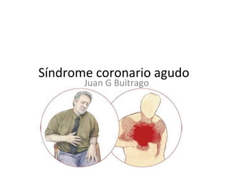
- 1. Síndrome coronario agudo Juan G Buitrago
- 2. Acute coronary Syndrome: • “ Any group of clinical symptoms compatible with acute myocardial ischemia and includes unstable angina (UA), non–ST-segment elevation myocardial infarction (NSTEMI), and ST-segment elevation myocardial infarction (STEMI).” Mayo Clin Proc. 2009;84(10):917-938
- 3. A Tale of Coronary Artery Disease and Myocardial Infarction. Nabel EG. Braunwald E. N Engl J Med 2012;366:54-63.
- 4. Epidemiología • La pandemia mas importante del s XXI • 15% - 20% de todas las muertes EC • 2020 25 millones de muertes al año aprox. 36% • 1 muerte por minuto; 30% en 1as 24h • 20% puede debutar con muerte súbita (IAM) • Hasta el 30% presentaciones atípicas, pte espera 2h • 1960 : 30% • 2012 : 2 – 4% McManus DD, Gore J, Yarzebski J, Spencer F, Lessard D, Goldberg RJ. Recent trends in the incidence, treatment, and outcomes of patients with STEMI and NSTEMI. Am J Med 2011;124:40–47.
- 5. El 1er mundo y el 3er mundo Los procedimientos de revascularización son 15 veces mas frecuentes en países de alto PIB Revista Colombiana de Cardiología ,127 Vol. 17 Suplemento 3 ISSN 0120-5633
- 8. Roffi, M et al. (2015). 2015 ESC Guidelines for the management of acute coronary syndromes in patients presenting without persistent ST-segment elevation. European heart journal, ehv320.
- 10. Definiciones Según la tercera definición universal:Según la tercera definición universal: • IAM : evidencia de necrosis miocárdica en unIAM : evidencia de necrosis miocárdica en un contexto clínico consistente con isquemia miocárdicacontexto clínico consistente con isquemia miocárdica • Aumento o caída de biomarcadores (cTn) con al menos un valor por encima del percentil 99 y que cumpla uno de los siguientes Síntomas isquemia Cambios de la T y el ST o nuevo BCRI Ondas Q Evidencia imagenológica de nueva perdida de miocardio viable o nueva anormalidad de la movilidad de la pared regional Identificacione de un trombo intracoronario por angiografia o biopsia European Heart Journal (2012) 33, 2551–2567
- 11. • Muerte cardiaca que ocurre antes de demostrar elevación de biomarcadores en contexto de: síntomas + cambios nuevos en EKG • PCI: aumento de 5 veces el nivel normal de las troponinas+ síntomas isquémicos o cambios ECG o hallazgo angiográfico o imagenológico Third universal definition of myocardial infarction. European Heart Journal (2012) 33, 2551–2567
- 12. • Trombosis del Stent con IAM detectado por autopsia o angiografía asociado a síntomas o elevación de biomarcadores. • IAM relacionado a revascularización quirúrgica (CABG): elevación de marcadores > 10 veces el nivel normal asociado Third universal definition of myocardial infarction. European Heart Journal (2012) 33, 2551–2567
- 15. Thygesen K, Alpert JS, JaffeAS, et al: Third universal definition of myocardial infarction. J Am Coll Cardiol 60:1581, 2012.)
- 16. SCASEST • El dolor anginoso del SCASEST se puede presentar como: Prolongado – duración de 20 minutos en reposo De Novo – Angina clase II-III Canadian Cardiovascular Society De reciente desestabilización en angina estable previa (angina en crescendo) Clase III Angina Post Infarto Roffi, M et al. (2015). 2015 ESC Guidelines for the management of acute coronary syndromes in patients presenting without persistent ST-segment elevation. European heart journal, ehv320.
- 18. BIOMARCADORES
- 19. Biomarcadores marcador Tiempo inicial elevación pico retorno mioglobina 1-4 horas 6-7 horas 24 horas CK-MB 3-6 horas 18-24 horas 48-72 horas Troponina I 3- 12 horas 24 horas 5-10 días
- 20. Curva enzimática
- 21. Roffi, M et al. (2015). 2015 ESC Guidelines for the management of acute coronary syndromes in patients presenting without persistent ST-segment elevation. European heart journal, ehv320.
- 22. Roffi, M et al. (2015). 2015 ESC Guidelines for the management of acute coronary syndromes in patients presenting without persistent ST-segment elevation. European heart journal, ehv320.
- 23. • ¿ Entonces en pacientes con menos de 3 horas de dolor torácico típico y sin cambios en ECG se debe esperar para hacer troponinas y/o hacer curva enzimática?
- 24. Neumann, J. T et al. . (2016). Diagnosis of Myocardial Infarction Using a High-Sensitivity Troponin I 1-Hour Algorithm. JAMA Cardiology.
- 25. • No toda elevación de troponinas es Infarto y no todo Infarto secundario a enfermedad aterosclerótica.
- 27. Mann, D. L., Zipes, D. P., Libby, P., & Bonow, R. O. (2014). Braunwald's heart disease: a textbook of cardiovascular medicine. Elsevier Health Sciences.
- 29. FISIOPATOLOGÍA
- 31. fisiopatología • El SCA se precipita por una trombosis aguda inducida por rotura o erosión de una placa coronaria aterosclerótica, con o sin vasoconstricción concomitante, lo que causa una reducción brusca y crítica del flujo sanguíneo Rev Esp Cardiol. 2012;65(2):173.e1-e55
- 32. N Engl J Med 2013; 368:2004-2013 May 23, 2013
- 33. N Engl J Med 2013; 368:2004-2013 May 23, 2013
- 34. N Engl J Med 2013; 368:2004-2013 May 23, 2013
- 35. Fisiopatología N Engl J Med 2005;352:1685-95.
- 38. N Engl J Med 2012;366:54-63.
- 41. NecrosisNecrosis Miocardiosano Miocardio Aturido Miocardio Hibernado Defectos reversibles Defectos irreversiblesDefectos irreversibles
- 42. Mann, D. L., Zipes, D. P., Libby, P., & Bonow, R. O. (2014). Braunwald's heart disease: a textbook of cardiovascular medicine. Elsevier Health Sciences.
- 43. Mann, D. L., Zipes, D. P., Libby, P., & Bonow, R. O. (2014). Braunwald's heart disease: a textbook of cardiovascular medicine. Elsevier Health Sciences.
- 44. Diagnóstico inicial • “El Tiempo es miocardio” • Los FR son menos importantes que la clínica, el ECG y los biomarcadores 10 mins • 30 mins • 90 mins
- 47. Dolor torácicoDolor torácico Dolor típico y atípico, Equivalentes anginosos Edad y género •Sensibilidad 67% •7% reproducibles palpación •30% atípicos 5-10% serán IAMCEST 15-20% serán IAMSEST 10% Angina inestable 15% otras enfermedades CV 50% no CV.
- 51. ECG• 1er ECG en 1ros 10 mins • Seriados c/15 a 30 mins si el 1º no es Dx • ST elevado 90% trombo , 90% cTn + • Monitoría continua • V3R, V4R, V7, V8, V9
- 52. Mann, D. L., Zipes, D. P., Libby, P., & Bonow, R. O. (2014). Braunwald's heart disease: a textbook of cardiovascular medicine. Elsevier Health Sciences.
- 53. Mann, D. L., Zipes, D. P., Libby, P., & Bonow, R. O. (2014). Braunwald's heart disease: a textbook of cardiovascular medicine. Elsevier Health Sciences.
- 54. Smith, S. W., Dodd, K. W., Henry, T. D., Dvorak, D. M., & Pearce, L. A. (2012). Diagnosis of ST-elevation myocardial infarction in the presence of left bundle branch block with the ST-elevation to S-wave ratio in a modified Sgarbossa rule. Annals of emergency medicine, 60(6), 766-776.
- 55. Smith, S. W., Dodd, K. W., Henry, T. D., Dvorak, D. M., & Pearce, L. A. (2012). Diagnosis of ST- elevation myocardial infarction in the presence of left bundle branch block with the ST-elevation to S-wave ratio in a modified Sgarbossa rule. Annals of emergency medicine, 60(6), 766-776.
- 56. Dx Rx de tórax Ecocardiograma TSH otros Troponina I Sens 97% 1ª neg, tomar c/8h por 24h >Pc99, elevación y posterior descenso
- 57. Mann, D. L., Zipes, D. P., Libby, P., & Bonow, R. O. (2014). Braunwald's heart disease: a textbook of cardiovascular medicine. Elsevier Health Sciences.
- 58. Scores TIMI Risk Score Calculator •Age ≥ 65 years? Yes (+1) •≥ 3 Risk Factors for CAD? Yes (+1) •Known CAD (stenosis ≥ 50%)? Yes (+1) •ASA Use in Past 7d? Yes (+1) •Severe angina (≥ 2 episodes w/in 24 hrs)? Yes (+1) •ST changes ≥ 0.5mm? Yes (+1) •+ Cardiac Marker? Yes (+1)
- 60. Roffi, M et al. (2015). 2015 ESC Guidelines for the management of acute coronary syndromes in patients presenting without persistent ST-segment elevation. European heart journal, ehv320.
- 62. MANEJO INICIAL
- 63. O2 y SueroFisiológico • La hipoxemia Sat O2 <90% aumenta la mortalidad a corto plazo • El oxigeno no debe ser usado de rutina en pacientes no hipoxémicos, pues causa vasoconstricciòn y <GC • En SCA existe evidencia del beneficio del Oxígeno C • VMNI es evidencia IA en edema pulmonar cardiogénico • CPAP es otra opción de primera línea en edema pulmonar cardiogénico • En SCA que lleva a shock cardiogénico, existe evidencia a favor de IOT • Líquidos no están totalmente restringidos, se pueden aplicar infusiones bajas, para mantener la precarga y mejorar la diuresis
- 64. -RCT, prospectivo: 441 pacientes con IAMCEST -Punto final primario: Tamaño del infarto Puntos secundarios: IAM recurrente, Arritmias y tamaño de infarto a 6 meses evaluado por Cardio RMN . 218 pacientes recibieron O2 a 8 litros por min y 223 no recibieron oxígeno. -Troponinas similares en ambos grupos, aumento significativo en el pico de CK en el grupo de oxígeno, aumento de recurrencia Infarto (5.5%vs.0.9%, P=0.006) y aumento de arritmias cardiacas (40.4% vs. 31.4%; P=0.05). Y amento del tamaño del infarto en el grupo O2 (n=139; 20.3 grams vs. 13.1 grams; P=0.04). Stub, D., Smith, K., Bernard, S., Nehme, Z., Stephenson, M., Bray, J. E., ... & Meredith, I. T. (2015). Air versus oxygen in ST-segment elevation myocardial infarction. Circulation, CIRCULATIONAHA-114.
- 65. Vasodilatadores < precarga < poscarga > volumen/latido Beneficia paciente hipertenso y con sobrecarga hídrica B y C Siempre PAS>110mmHg MonitoríaTA si PAS <90mmHg suspender Hipotensión aumenta mortalidad en IC aguda Nitroprusiato: disminuye tono venoso, <poscarga der e izq dosis inicial 0,1 – 0,2 mcg/kg/min proteger de la luz, precaución intoxicación tiocianato y cianuro ERC > % intox por cianuro (dolor abdominal y cambios mentales) : adm tiosulfato de sodio 1ml por c/ amp NTP > mortalidad en IAM sin IC aguda Debería, si se requiere, ir acompañado de nitroglicerina para evitar el fenómeno del «robo coronario»
- 66. Nitroglicerina: activa GMP cíclico dosis 0,25 – 5 mcg/kg/min produce hipotensión cefalea y mareos tolerancia en el 20% luego de 24 a 48h Se inicia con NTP si estÁ hipertenso. Si tiene SCA con NTG, sino responde a ninguno de los dos, neseritide. También como coadyuvante
- 67. Solo usarla cuando el umbral de dolor sea inaceptable- 2 – 5 mg /iv. 57,039 del CRUSADE se hizo un estudio NR retrospectivo en SCASEST, y se encontró que Tto con morfina tuvo mayor riesgo de muerte (29.8 percent) (OR1.48, 95% CI 1.33-1.64) Puede interactuar P2Y12 Meine, T. J. et al & CRUSADE Investigators. (2005). Association of intravenous morphine use and outcomes in acute coronary syndromes: results from the CRUSADE Quality Improvement Initiative. American heart journal, 149(6), 1043-1049.
- 69. SCASEST Muy alto riesgo Alto riesgo GRACE > 140 Riesgo intermedio GRACE 109-140 Bajo riesgo 1. DM 2. ERC TFG<60 3. FEVI < 40% 4. Agina Post IM 5. PCI-CABG previo 6. Grace 109-140 1. Aumento o Caida de troponinas 2. Cambios dinámicos ECG asintomáticos 3. Grace>140 1. Inestable 2. Dolor refractario 3. Complicaciones mecánicas 4. Cambios dinámicos con EST intermitente Roffi, M et al. (2015). 2015 ESC Guidelines for the management of acute coronary syndromes in patients presenting without persistent ST-segment elevation. European heart journal, ehv320.
- 70. Antiisquémicos: BB, nitratos Antiplaquetarios: AAS, clopidogrel, ticagrelor, cangrelor, plasugrel, GP IIb/IIIa Anticoagulantes: HNF, Enoxaparina, fondaparinux Roffi, M et al. (2015). 2015 ESC Guidelines for the management of acute coronary syndromes in patients presenting without persistent ST-segment elevation. European heart journal, ehv320.
- 71. Reducen el consumo miocárdico de oxígeno al disminuir la frecuencia cardiaca, la presión arterial y la contractilidad. Estudio CRUSADE , disminución mortalidad 34% Estudio COMMIT aumentó shock cardiogénico disminuyó reinfarto y Arritmias V. Roffi, M et al. (2015). 2015 ESC Guidelines for the management of acute coronary syndromes in patients presenting without persistent ST-segment elevation. European heart journal, ehv320.
- 72. From Chen ZM, Pan HC, Chen YP, et al: Early intravenous then oral metoprolol in 45,852 patients with acute myocardial infarction: Randomised placebo-controlled trial. Lancet 366:1622,2005
- 73. Producen reducción en la precarga miocárdica y el volumen telediastólico delVI y < consumo miocárdico de O2 y dilatación de Art. Coronarias. Registro GRACE demostró que uso crónico, puede cambiar de IAMCEST a IAMSEST. Mejor IV que oral. Ambrosio G, Del Pinto M, Tritto I, Agnelli G, Bentivoglio M, Zuchi C, et al. Chronic nitrate therapy is associated with different presentation and evolution of acute coronary syndromes: insights from 52,693 patients in the Global Registry of Acute Coronary Events. Eur Heart J. 2010;31:430-8.
- 74. La ivabradina inhibe puede utilizarse en pacientes seleccionados con contraindicaciones para bloqueadores beta. La ranolazina ejerce efectos antianginosos al inhibir la corriente tardía de sodio. Reduce tasa de izquemia recurrente ( Estudio MERLIN) Rev Esp Cardiol. 2012;65:173.e1-e55 - Vol. 65 Núm.02 DOI: 10.1016/j.recesp.2011.11.006
- 75. Rev Esp Cardiol.2012;65:173.e1- e55 - Vol. 65 Núm.02 DOI: 10.1016/j.recesp.2011.11.006
- 76. Reduce la incidencia de IAM recurrente o muerte en pacientes con Angina inestable. N Engl J Med 1988; 319:1105–11. dosis de carga masticable entre 150 y 300 mg, dosis diaria de 100 mg. Rev Esp Cardiol.2012;65:173.e1-e55 - Vol. 65 Núm.02 DOI:10.1016/j.recesp.2011.11.006
- 77. Roffi, M et al. (2015). 2015 ESC Guidelines for the management of acute coronary syndromes in patients presenting without persistent ST-segment elevation. European heart journal, ehv320.
- 78. El estudio CURE: ASA+ Clopidogrel redujo la incidencia de muerte por causas cardiovasculares e IAM no fatal o accidente cerebrovascular, comparada con ácido acetilsalicílico solo. Dosis altas = a dosis convencionales ( 300 mg de carga y 75 mg diarios) Estudio CURRENT-OASIS. > tasas de sangrado con dosis más altas. No hay evidencia que demuestre que IBP+ Clopidogrel aumente episodios isquemia. Rev Esp Cardiol. 2012;65:173.e1-e55 - Vol. 65 Núm.02 DOI: 10.1016/j.recesp.2011.11.006
- 79. EstudioTRITON comparó plasugrel y clopidogrel. Menor tasa de trombosis del stent con plasugrel , > tasa de sangrado en plasugrel. Mayor beneficio de plasugrel a quienes van a ICP, en diabéticos jóvenes. Roffi, M et al. (2015). 2015 ESC Guidelines for the management of acute coronary syndromes in patients presenting without persistent ST-segment elevation. European heart journal, ehv320.
- 80. Wiviott, S. D., Braunwald, E., McCabe, C. H., Montalescot, G., Ruzyllo, W., Gottlieb, S., ... & Riesmeyer, J. (2007). Prasugrel versus clopidogrel in patients with acute coronary syndromes. New England Journal of Medicine,357(20), 2001-2015.
- 81. redujo significativamente el punto final primario con ticagrelor 10% versus 12.3% HR 0.83 p: 0.0013 con igual reducción de mortalidad y todas las causas de mortalidad las tasas de sangrado fueron similares. Mayor disnea con ticagrelor Roffi, M et al. (2015). 2015 ESC Guidelines for the management of acute coronary syndromes in patients presenting without persistent ST-segment elevation. European heart journal, ehv320.
- 82. Wallentin, L., Becker, R. C., Budaj, A., Cannon, C. P., Emanuelsson, H., Held, C., ... & Mahaffey, K. W. (2009). Ticagrelor versus clopidogrel in patients with acute coronary
- 83. Roffi, M et al. (2015). 2015 ESC Guidelines for the management of acute coronary syndromes in patients presenting without persistent ST-segment elevation. European heart journal, ehv320.
- 84. Roffi, M et al. (2015). 2015 ESC Guidelines for the management of acute coronary syndromes in patients presenting without persistent ST-segment elevation. European heart journal, ehv320.
- 85. ¿3 a 6 meses ? ¿1 año ? ¿Más de un año?
- 86. Bonaca, M. P., Bhatt, D. L., Cohen, M., Steg, P. G., Storey, R. F., Jensen, E. C., ... & Bengtsson, O. (2015). Long-term use of ticagrelor in patients with prior myocardial infarction. New England Journal of Medicine, 372(19), 1791-1800.
- 87. Fondaparinux: dosis de 2,5 mg/día según estudios PENTUA, OASIS-5, OASIS-6. superioridad a enoxaparina respecto a muerte,no inferioridad respecto a isquemia. ( OASIS-5 )- de primera línea HBPM: disminuyen mortalidad frente a placebo No diferencia entre HBPM y HNF en cuanto a mortalidad. Objetivo combinado de muerte e IAM a favor de enoxaparina. Roffi, M et al. (2015). 2015 ESC Guidelines for the management of acute coronary syndromes in patients presenting without persistent ST-segment elevation. European heart journal, ehv320.
- 88. Fifth Organization to Assess Strategies in Acute Ischemic Syndromes Investigators. (2006). Comparison of fondaparinux and enoxaparin in acute coronary syndromes. N engl j med, 2006(354), 1464-1476.
- 89. El estudio SINERGY con 10.027 ptes se comparó Enoxaparina y HNF en pacientes que iban a ICP, sin diferencias en la mortalidad. Dosis de 1 mg/kg. HNF: reduccion del riesgo del 33% en muerte e IAM frente a placebo. Rev Esp Cardiol. 2012;65:173.e1-e55 - Vol. 65 Núm.02 DOI: 10.1016/j.recesp.2011.11.006
- 90. Bivalirudina: Estudio REPLACE-2 Bivalirudina + Inh Gp IIB/IIIA no es inferior a HNF+ bivalirudina pero si < sangrado. E igual en el estudio ACUITY. Bivalirudina como monoterapia no inferior. Es no inferior a HNF o HBPM frente a tasa de complicaciones isquémicas (7.8 versus 7.3 percent, relative risk 1.08, 95% CI 0.93-1.24), Roffi, M et al. (2015). 2015 ESC Guidelines for the management of acute coronary syndromes in patients presenting without persistent ST-segment elevation. European heart journal, ehv320.
- 91. rivaroxabán 2,5 mg dos veces al día en pacientes con IAMSEST con alto riesgo isquémico y bajo riesgo de sangrado que reciben Aspirina y clopidogrel No ancianos, bajo de peso y ACV. Roffi, M et al. (2015). 2015 ESC Guidelines for the management of acute coronary syndromes in patients presenting without persistent ST-segment elevation. European heart journal, ehv320.
- 92. Roffi, M et al. (2015). 2015 ESC Guidelines for the management of acute coronary syndromes in patients presenting without persistent ST-segment elevation. European heart journal, ehv320.
- 93. No reducción de muerte por IAM en pacientes tratados con inh. GPIIb/IIIa frente a placebo. El único beneficio significativo se observó cuando el paciente iba para angioplastia. EstudioTARGET : abciximab superior a tirofibán en dosis estándar en reducción de riesgo de muerte, IAM y revascularización urgente a los 30 dias. Un metanálisis demostró que eran =. Roffi, M et al. (2015). 2015 ESC Guidelines for the management of acute coronary syndromes in patients presenting without persistent ST-segment elevation. European heart journal, ehv320.
- 94. Topol EJ, Moliterno DJ, Herrmann HC, et al; the TARGET Investigators. Comparison of two platelet glycoprotein IIb/IIIa inhibitors, tirofiban and abciximab, for the prevention of ischemic events with percutaneous coronary revascularization. N Engl J Med. 2001;344:1888-94.
- 95. No reducción de muertes o IAM en pacientes tratados Md.Y con Inh. GPIIb/IIIa frente a placebo. El único beneficio significativo de los Inh. GPIIb/IIIa cuando se mantenían durante la angioplastia. > riesgo de sangrado. Roffi, M et al. (2015). 2015 ESC Guidelines for the management of acute coronary syndromes in patients presenting without persistent ST-segment elevation. European heart journal, ehv320.
- 96. Rev Esp Cardiol.2012;65:173.e1- e55 - Vol. 65 Núm.02 DOI: 10.1016/j.recesp.2011.11.006
- 97. Rev Esp Cardiol.2012;65:173.e1- e55 - Vol. 65 Núm.02 DOI: 10.1016/j.recesp.2011.11.006
- 98. Mayor riesgo de sangrado mayor a un año con Doble antiagregación y ACO. No usar Plasugrel yTicagrelor Mantener INR entre 2-2,5 en documarínicos Doble antiagregación al menos un mes en Stent convencional y 6 en liberador de Mdto. Roffi, M et al. (2015). 2015 ESC Guidelines for the management of acute coronary syndromes in patients presenting without persistent ST-segment elevation. European heart journal, ehv320.
- 99. Roffi, M et al. (2015). 2015 ESC Guidelines for the management of acute coronary syndromes in patients presenting without persistent ST-segment elevation. European heart journal, ehv320.
- 101. Bavry AA,et al. Benefit of early invasive therapy in acute coronary syndromes: a meta- analysis of contemporary randomized clinical trials. J Am Coll Cardiol 2006;48:1319– 1325.
- 102. Fox KA, et al. Long-term outcome of a routine versus selective invasive strategy in patients with non-ST-segment elevation acute coronary syndrome a meta-analysis of individual patient data. J Am Coll Cardiol 2010;55:2435–2445.
- 103. La estrategia invasiva de rutina en SCASEST Ha demostrado mejorar los desenlaces clínicos y reducir SCA recurrentes, rehospitalizaciones y revascularizaciones. Roffi, M et al. (2015). 2015 ESC Guidelines for the management of acute coronary syndromes in patients presenting without persistent ST-segment elevation. European heart journal, ehv320.
- 104. Roffi, M et al. (2015). 2015 ESC Guidelines for the management of acute coronary syndromes in patients presenting without persistent ST-segment elevation. European heart journal, ehv320.
- 105. Roffi, M et al. (2015). 2015 ESC Guidelines for the management of acute coronary syndromes in patients presenting without persistent ST-segment elevation. European heart journal, ehv320.
- 106. Roffi, M et al. (2015). 2015 ESC Guidelines for the management of acute coronary syndromes in patients presenting without persistent ST-segment elevation. European heart journal, ehv320.
- 107. Roffi, M et al. (2015). 2015 ESC Guidelines for the management of acute coronary syndromes in patients presenting without persistent ST-segment elevation. European heart journal, ehv320.
- 108. Roffi, M et al. (2015). 2015 ESC Guidelines for the management of acute coronary syndromes in patients presenting without persistent ST-segment elevation. European heart journal, ehv320.
- 109. Roffi, M et al. (2015). 2015 ESC Guidelines for the management of acute coronary syndromes in patients presenting without persistent ST-segment elevation. European heart journal, ehv320.
- 112. Roffi, M et al. (2015). 2015 ESC Guidelines for the management of acute coronary syndromes in patients presenting without persistent ST-segment elevation. European heart journal, ehv320.
- 113. Ohman, E. M. (2016). Chronic Stable Angina. N Engl J Med, 2016(374), 1167- 1176.
- 114. Ohman, E. M. (2016). Chronic Stable Angina. N Engl J Med, 2016(374), 1167-1176.
- 115. IECA en FEvi < 40 despues de estabilizar para reducir mortalidad ( IA ) BB en FEVi <40% despues de estabilizar para reducir mortalidad y recurrencia de isquemia (IA ) ARA ( espironolactona - eplerenona ) en NYHA II-IV FEVI < 35% a pesar de tratamiento con IECA ARA II y BB. ( IA ) Roffi, M et al. (2015). 2015 ESC Guidelines for the management of acute coronary syndromes in patients presenting without persistent ST-segment elevation. European heart journal, ehv320.
- 116. eplerenone se recomienda en reducir riesgo de hospitalizacion y mortalidad en FEVI <40% ( IB) Roffi, M et al. (2015). 2015 ESC Guidelines for the management of acute coronary syndromes in patients presenting without persistent ST-segment elevation. European heart journal, ehv320.
- 117. Iniciado en las primeras 24-96 horas, en SCASEST, reduce la incidencia de eventos isquémicos en las primeras 16 semanas, independientemente de los niveles de colesterol. El inicio precoz se asocia además a mayor adherencia al tratamiento a largo plazo. Atorvastatina 40 mg/día Simvastatina 20-40 mg/día Pravastatina 20-40 mg/día Roffi, M et al. (2015). 2015 ESC Guidelines for the management of acute coronary syndromes in patients presenting without persistent ST-segment elevation. European heart journal, ehv320.
- 118. ( CRT o DAI) paciente sintomático con disfunción delVI FEVI < 35% a pesar de terapia óptima por mas de 40 días después del evento agudo sin opciones de RVM en pacientes con expectativa de vida mayor a un año. Roffi, M et al. (2015). 2015 ESC Guidelines for the management of acute coronary syndromes in patients presenting without persistent ST-segment elevation. European heart journal, ehv320.
- 119. Fevi menor de 35% hacer test de isquemia residual y revascularización subsecuente en quienes se las va a hacerTRC o DAI, después de la RVM evaluar el remodelado del VI a los 6 meses pata el implante de dispositivo profiláctico. Roffi, M et al. (2015). 2015 ESC Guidelines for the management of acute coronary syndromes in patients presenting without persistent ST-segment elevation. European heart journal, ehv320.
- 123. Mann, D. L., Zipes, D. P., Libby, P., & Bonow, R. O. (2014). Braunwald's heart disease: a textbook of cardiovascular medicine. Elsevier Health Sciences.
- 124. Mann, D. L., Zipes, D. P., Libby, P., & Bonow, R. O. (2014). Braunwald's heart disease: a textbook of cardiovascular medicine. Elsevier Health Sciences.
- 125. Mann, D. L., Zipes, D. P., Libby, P., & Bonow, R. O. (2014). Braunwald's heart disease: a textbook of cardiovascular medicine. Elsevier Health Sciences.
- 127. O2 Nt Morfina Bb IECA AAS (162-325 mg) Clopidogrel (300-600mg)
- 128. Se evitan 18 muertes por cada 1000 pacientes sometidos a fibrinolíticos Mann, D. L., Zipes, D. P., Libby, P., & Bonow, R. O. (2014). Braunwald's heart disease: a textbook of cardiovascular medicine. Elsevier Health Sciences.
- 130. (Modificado de Boersma E, Mercado N, Poldermans, etal: Acute myocardial infarction. Lancet361:851, 2003.)
- 131. Tiempo= ICP mejor en pacientes con más de 12 h Mayor riesgo de muerte= ICP mejor en pte con alto riesgo de muerte. Shock Cardiogénico= Se benficia > de ICP. > riesgo de Sangrado I.C. = se beneficia >ICP. Si no se dispone de ICP= trombolisis. Mann, D. L., Zipes, D. P., Libby, P., & Bonow, R. O. (2014). Braunwald's heart disease: a textbook of cardiovascular medicine. Elsevier Health Sciences. Pag 1107
- 132. Mann, D. L., Zipes, D. P., Libby, P., & Bonow, R. O. (2014). Braunwald's heart disease: a textbook of cardiovascular medicine. Elsevier Health Sciences.
- 134. Angioplastia debe hacerse los primeros 120 minutos. Ha demostrado ser superior a fibrinólisis. Paciente que se someta a Fibrinólisis debe ser seguido por angiografía de rutina. Rev Esp Cardiol. 2012;65:173.e1-e55 - Vol. 65 Núm.02 DOI: 10.1016/j.recesp.2011.11.006
- 135. Solo tratar el vaso obstruido relacionado con el infarto, no hay evidencia que apoyeTto de otras lesiones. Angioplastia múltiple en shock cardiogénico, isquemia persistente, o trombos. Mejores desenlaces abordaje radial que femoral.
- 136. 3507 patients were randomly assigned to radial access and 3514 to femoral access. lower rate of local vascular complications inthe radial approach olly SS, Yusuf S, Cairns , et al. Radial vs. femoral access for coronary angiography and intervention in patients with acute coronary syndromes (RIVAL):
- 143. Mann, D. L., Zipes, D. P., Libby, P., & Bonow, R. O. (2014). Braunwald's heart disease: a textbook of cardiovascular medicine. Elsevier Health Sciences. Pag 1152
- 147. Mann, D. L., Zipes, D. P., Libby, P., & Bonow, R. O. (2014). Braunwald's heart disease: a textbook of cardiovascular medicine. Elsevier Health Sciences.
- 151. Cooper, H. A., & Panza, J. A. (2013). Cardiogenic Shock. Cardiology clinics,31(4), 567-580.
- 152. Cooper, H. A., & Panza, J. A. (2013). Cardiogenic Shock. Cardiology clinics,31(4), 567-580.
- 155. Thiele, H., Zeymer, U., Neumann, F. J, et al. (2012). Intraaortic balloon support for myocardial infarction with cardiogenic shock. New England Journal of Medicine, 367(14), 1287-1296.
- 156. randomized controlled trials versus IABP are required to prove a clinical benefit in the setting of CS. The pVADs provided superior hemodynamic support when compared with IABP. However, 30-day survival was similar in the pVAD and IABP groups (55% vs 57%, relative risk 1.06, 95% confidence interval 0.68–1.66, P 5 .8). Cooper, H. A., & Panza, J. A. (2013). Cardiogenic Shock. Cardiology clinics,31(4), 567-580.
- 162. Complicaciones del IAM Mecánicas eléctricas pericárdicas Mann, D. L., Zipes, D. P., Libby, P., & Bonow, R. O. (2014). Braunwald's heart disease: a textbook of cardiovascular medicine. Elsevier Health Sciences. Pag 1127 trombóticas
- 163. 2% de los IAM – 10% de las muertes súbitas por IAM – característica de IAMCEST Desde el primer día del IAM hasta 3 semanas post IAM- es característica los primeros 4-5 días. Síncope – AESP –Taponamiento Mann, D. L., Zipes, D. P., Libby, P., & Bonow, R. O. (2014). Braunwald's heart disease: a textbook of cardiovascular medicine. Elsevier Health Sciences. Pag 1127
- 164. > en mujeres ancianas- 7 veces más enVI que VD ( Zona de la ADA ) EC crónica con Colaterales protege Ruptura subaguda – 25% casos con paso sangre pericardio ECOTTderrame Pericárdico con fibrina, trombos densos Simula reinfarto, eleva ST - tto = QX. Mann, D. L., Zipes, D. P., Libby, P., & Bonow, R. O. (2014). Braunwald's heart disease: a textbook of cardiovascular medicine. Elsevier Health Sciences. Pag 1127
- 165. Mann, D. L., Zipes, D. P., Libby, P., & Bonow, R. O. (2014). Braunwald's heart disease: a textbook of cardiovascular medicine. Elsevier Health Sciences. Pag 1129
- 166. Si es septo anterior = IAM anterior Si es septo-posterior= IAM inferior – Mal pronóstico. Mujeres ancianas sin colaterales y ERC Poco probable en DM, Isquemia crónica por preacondicionamiento Mann, D. L., Zipes, D. P., Libby, P., & Bonow, R. O. (2014). Braunwald's heart disease: a textbook of cardiovascular medicine. Elsevier Health Sciences. Pag 1127
- 167. Deterioro Brusco- edema pulmonar – Shock cardiogénico + soplo pansistólico con frémito paraesternal Dx ECOTT en CAT: Salto oximétrico del VD Qx. Tto med: Nitroprusiato - < postcarga- BCPA Mann, D. L., Zipes, D. P., Libby, P., & Bonow, R. O. (2014). Braunwald's heart disease: a textbook of cardiovascular medicine. Elsevier Health Sciences. Pag 1127
- 168. Mann, D. L., Zipes, D. P., Libby, P., & Bonow, R. O. (2014). Braunwald's heart disease: a textbook of cardiovascular medicine. Elsevier Health Sciences. Pag 1129
- 169. En la primera semana post IAM por isquemia de músculo papilar Se asocia a IAM inferior ( por ruptura de músculo papilar inferior) ya que el anterior tiene doble irrigación – ADA-CX Falla cardiaca aguda+ edema pulmonar – soplo sistólico que puede atenuarse por = de presiones.Tto < postcarga NTG grave QX Mann, D. L., Zipes, D. P., Libby, P., & Bonow, R. O. (2014). Braunwald's heart disease: a textbook of cardiovascular medicine. Elsevier Health Sciences. Pag 1129
- 170. Mann, D. L., Zipes, D. P., Libby, P., & Bonow, R. O. (2014). Braunwald's heart disease: a textbook of cardiovascular medicine. Elsevier Health Sciences. Pag 1129
- 171. Mann, D. L., Zipes, D. P., Libby, P., & Bonow, R. O. (2014). Braunwald's heart disease: a textbook of cardiovascular medicine. Elsevier Health Sciences. Pag 1129
- 172. AneurismaVI : en zona cicatricial con discinesia – ocurre en 15 % de los pacientes Aparece porque Presión IV hace expansión de tejido necrótico IAM apical 80% casos EAC con colaterales protege. Doble impulso apical a la palpación – EST persistente Dx ECO-tt Mann, D. L., Zipes, D. P., Libby, P., & Bonow, R. O. (2014). Braunwald's heart disease: a textbook of cardiovascular medicine. Elsevier Health Sciences. Pag 1129
- 173. No predispone a Ruptura- no síntomas fomenta aparición de trombo-embolias y arritmias > mortalidad Tto QX en arritmia o ICC refractaria. Mann, D. L., Zipes, D. P., Libby, P., & Bonow, R. O. (2014). Braunwald's heart disease: a textbook of cardiovascular medicine. Elsevier Health Sciences. Pag 1129
- 174. Ruptura de la pared delVI contenida por un trombo que se organiza .Tto QUirúrgico Mann, D. L., Zipes, D. P., Libby, P., & Bonow, R. O. (2014). Braunwald's heart disease: a textbook of cardiovascular medicine. Elsevier Health Sciences. Pag 1129
- 175. Mann, D. L., Zipes, D. P., Libby, P., & Bonow, R. O. (2014). Braunwald's heart disease: a textbook of cardiovascular medicine. Elsevier Health Sciences. Pag 1128
- 176. Mann, D. L., Zipes, D. P., Libby, P., & Bonow, R. O. (2014). Braunwald's heart disease: a textbook of cardiovascular medicine. Elsevier Health Sciences. Pag 1128
- 180. Arritmias ventriculares: mecanismos de arritmias en fase aguda ( primarias) es diferente a C. isquémica crónica ( reentrada en cicatriz ) FV 20% de los pacientes con IAM ( 48h) causa más frecuente de muerte en IAM extenso- reperfusión ineficaz Torsade pointesmultifactorial: hipoxemia hipokalemia hipomagnesemia tto acortar Qt Mann, D. L., Zipes, D. P., Libby, P., & Bonow, R. O. (2014). Braunwald's heart disease: a textbook of cardiovascular medicine. Elsevier Health Sciences. Pag 1128
- 181. TV monomorfa sostenida: es infrecuente en la fase aguda de un IAM – precisa desarrollo de sustrato anatómico 3% casos. ExtrasístolesVentricularesTVMNS no requiereTto en especial BB previene aparición RIVA en IAM anterior e inferior depués de reperfusión. Mann, D. L., Zipes, D. P., Libby, P., & Bonow, R. O. (2014). Braunwald's heart disease: a textbook of cardiovascular medicine. Elsevier Health Sciences. Pag 1128
- 182. Taquicardia sinusal: en IAM anterior extenso con difunción sistólica Bradicardia sinusal: fase aguda de un IAM inferior por hipertonía vagal 35% de los casos. Atropina 0,3 mg cada 30 min Fibrilación auricular: 10-20% de los pacientes con IAM extenso con disfunción deVI- > en Ancianos tto con BB. No dar digoxina. Mann, D. L., Zipes, D. P., Libby, P., & Bonow, R. O. (2014). Braunwald's heart disease: a textbook of cardiovascular medicine. Elsevier Health Sciences. Pag 1128
- 183. suprahisiano infrahisianO Bloqueo AV post IAM Mobitz I – Qrs Estrecho IAM inferior Ritmo 40-60 Responde a atropina- Mejor pronóstico Mobitz II – QRs ancho IAM anterior Ritmo <40 No Responde a atropina Peor pronóstico
- 184. Bloqueos de Rama: 20 % desarrolla bloqueo de rama transitoria, persiste 5%. El BRIHH nuevo se asocia a infartos extensos con gran disfunción sistólica y mal pronóstico. BRDHH --> puede producir BAV se asocia a infarto anteroseptal aumento de riesgo de mortalidad bloqueo bifascicular --> alto riesgo de BAV completo y fallo de bomba Mann, D. L., Zipes, D. P., Libby, P., & Bonow, R. O. (2014). Braunwald's heart disease: a textbook of cardiovascular medicine. Elsevier Health Sciences. Pag 1128
- 185. Mann, D. L., Zipes, D. P., Libby, P., & Bonow, R. O. (2014). Braunwald's heart disease: a textbook of cardiovascular medicine. Elsevier Health Sciences. Pag 1128
- 186. síndrome de Dressler : 1-8 semanas post IAM 3-4% de los IM malestar general - fiebre - leucocitosis - dolor torácico Puntada aumento deVSG -derrame pericárdico. Pericarditis fibrinosa. Tto 650 mg de AAS. cada 4 horas. No dar corticoides ni AINES alteran la cicatrización del infarto. inducen rotura ventricular y aumentan RVcoronaria Mann, D. L., Zipes, D. P., Libby, P., & Bonow, R. O. (2014). Braunwald's heart disease: a textbook of cardiovascular medicine. Elsevier Health Sciences. Pag 1128
Notas del editor
- Typical chest pain is characterized by a retrosternal sensation of pressure or heaviness (‘angina’) radiating to the left arm (less frequently to both arms or to the right arm), neck or jaw, which may be intermittent (usually lasting several minutes) or persistent. Atípicos en diabéticos, mujeres, ancianos y demencia. La exacerbación con la actividad y mejora con el reposo aumentan la probabilidad de que sea un dolor isquémico EL dolor que mejora con nitratos no es típico de la angina y puede también mejorar en otras condiciones.
- Clasificación de Braunwald de la Angina.
- Sensibilidad de 77% and 93% for troponin I and 80% and 91% for troponin T Estos marcadores en la actualidad están en desuso con la aparición de las troponinas de alta sensibilidad y de la copeptina la cual tiene alto valor predictivo negativo.
- Algoritmo del “Rule-in” y del “Rule-Out” recomendado por las guías.
- Algoritmo del “Rule-in” y del “Rule-Out” recomendado por las guías. Para hacer en pacientes con dolor torácico de menos de una hora de evolución en donde se ven los puntos de corte para las diferentes troponinas de alta sensibilidad deacuerdo a la marca de esta.
- Of the 1040 patients included from the study cohort, 673 (64.7%) were male and had a median age of 65 (interquartile range, 52-75) years. With application of a low troponin I cutoff value of 6 ng/L, the rule-out algorithm showed a high negative predictive value of 99.8%(95%CI, 98.6%-100.0%) after 1 hour for non–ST-segment elevation MI type 1. The 1-hour approach was comparable to a 3-hour approach. Similarly, a rule-in algorithm based on troponin I levels provided a high positive predictive value with 82.8%(95%CI, 73.2%-90.0%). Moreover, application of the cutoff of 6 ng/L resulted in lower follow-up mortality (1.0%) compared with the routinely used 99th percentile (3.7%) for this assay. Two independent cohorts further validated the performance of this algorithm with high negative and positive predictive values.
- Caño cristales colombia- EL río de los 7 colores
- Ruptur de placa de &lt; de 55 micrometros producía sca fatales
- A cross-section of an atheromatous plaque at the bottom of the figure shows the central lipid core that contains macrophage foam cells (yellow) and T cells (blue). The intima and media also contain arterial smooth-muscle cells (red), which are the source of arterial collagen (depicted as triple helical coiled structures). Activated T cells (of the type-1 helper T-cell subtype) secrete cytokine interferon-γ, which inhibits the production of the new, interstitial collagen that is required to repair and maintain the plaque&apos;s protective fibrous cap (upper left). The T cells can also activate the macrophages in the intimal lesion by expressing CD40 ligand (CD154), which engages its cognate receptor (CD40) on the phagocyte. This inflammatory signaling causes overproduction of interstitial collagenases (matrix metalloproteinases [MMPs] 1, 8, and 13) that catalyze the initial rate-limiting step in collagen breakdown (top right). CD40 ligation also causes macrophages to overproduce tissue-factor procoagulant. Thus, inflammatory signaling puts the collagen in the plaque&apos;s fibrous cap in double jeopardy — decreasing synthesis and increasing breakdown — rendering the cap susceptible to rupture. Inflammatory activation also boosts tissue-factor production, which triggers thrombus formation in the disrupted plaque. These are the mechanisms through which inflammation in the plaque can precipitate the thrombotic complications of atherosclerosis, including acute coronary syndromes
- Inducir la deleción de los genes de metaloproteinasas en ratón demostró que se morían menos de infarto.
- Sensbilidad del 20% con especificidad del 98%
- Los criterios de Smith aumentan la baja sensibilidad que tenían los criterios de Sgarbossa.
- Valle de cocora – Salento Quindío Colombia Palma de cera – árbol nacional de colombia – Hogar de especie en extensión: Loro orejiamarillo
- We conducted a multicenter, prospective, randomized, controlled trial comparing oxygen (8 L/min) with no supplemental oxygen in patients with STEMI diagnosed on paramedic 12-lead electrocardiogram. Of 638 patients randomized, 441 were confirmed STEMI patients who underwent primary endpoint analysis. The primary endpoint was myocardial infarct size as assessed by cardiac enzymes, troponin (cTnI) and creatine kinase (CK). Secondary endpoints included recurrent myocardial infarction, cardiac arrhythmia and myocardial infarct size assessed by cardiac magnetic resonance (CMR) imaging at 6 months. Mean peak troponin was similar in the oxygen and no oxygen groups (57.4 mcg/L vs. 48.0 mcg/L; ratio, 1.20; 95% confidence interval [CI], 0.92 to 1.56; P=0.18). There was a significant increase in mean peak CK in the oxygen group compared to the no oxygen group (1948 U/L vs. 1543 U/L; means ratio, 1.27; 95% CI, 1.04 to 1.52; P= 0.01). There was an increase in the rate of recurrent myocardial infarction in the oxygen group compared to the no oxygen group (5.5%vs.0.9%, P=0.006) and an increase in frequency of cardiac arrhythmia (40.4% vs. 31.4%; P=0.05). At 6-months the oxygen group had an increase in myocardial infarct size on CMR (n=139; 20.3 grams vs. 13.1 grams; P=0.04). Conclusions—Supplemental oxygen therapy in patients with STEMI but without hypoxia may increase early myocardial injury and was associated with larger myocardial infarct size assessed at six months.
- In the IMPRESSION trial, 70 acute MI patients treated with ticagrelor were randomly assigned to receive intravenous morphine (5 mg) or placebo [22]. Morphine lowered (active) ticagrelor plasma concentration and impaired its antiplatelet effect. ●In a study of 24 healthy subjects who received a loading dose of 600 mg of clopidogrel and either 5 mg of intravenous morphine or placebo, morphine significantly delayed clopidogrel resorption and reduced the area under the curve levels of its active metabolite by 52 percent [23]. Platelet inhibition, as measured by multiple tests, was less pronounced in those given morphine. ●In a study of 50 patients with STEMI undergoing primary percutaneous coronary intervention who were randomly assigned to either prasugrel or ticagrelor, morphine was an independent predictor of high residual platelet reactivity at two hours (odds ratio 5.29, 95% CI 1.44-19.49) [24].
- Controlled trials have repeatedly documented the beneficial effects of beta blockers in patients with acute MI; however, there have been no randomized trials specifically addressing the efficacy of these drugs in non-ST elevation ACS.
- meta-analysis of 27 early studies showing that beta-blocker treatment was associated with a significant 13% relative risk reduction (RRR) of mortality in the first week following MI. ESTUDIO COMMIT – 2005 Chen Et al The Lancet : 45,852 patients admitted to 1250 hospitals within 24 h of suspected acute MI onset were randomly allocated metoprolol (up to 15 mg intravenous then 200 mg oral daily; n=22,929) or matching placebo (n=22,923). 93% had ST-segment elevation or bundle branch block, and 7% had ST-segment depression. Treatment was to continue until discharge or up to 4 weeks in hospital (mean 15 days in survivors) and 89% completed it. The two prespecified co-primary outcomes were: (1) composite of death, reinfarction, or cardiac arrest; and (2) death from any cause during the scheduled treatment period. Comparisons were by intention to treat, and used the log-rank method. This study is registered with ClinicalTrials.gov, number NCT 00222573. FINDINGS: Neither of the co-primary outcomes was significantly reduced by allocation to metoprolol. For death, reinfarction, or cardiac arrest, 2166 (9.4%) patients allocated metoprolol had at least one such event compared with 2261 (9.9%) allocated placebo (odds ratio [OR] 0.96, 95% CI 0.90-1.01; p=0.1). For death alone, there were 1774 (7.7%) deaths in the metoprolol group versus 1797 (7.8%) in the placebo group (OR 0.99, 0.92-1.05; p=0.69). Allocation to metoprolol was associated with five fewer people having reinfarction (464 [2.0%] metoprolol vs 568 [2.5%] placebo; OR 0.82, 0.72-0.92; p=0.001) and five fewer having ventricular fibrillation (581 [2.5%] vs 698 [3.0%]; OR 0.83, 0.75-0.93; p=0.001) per 1000 treated. Overall, these reductions were counterbalanced by 11 more per 1000 developing cardiogenic shock (1141 [5.0%] vs 885 [3.9%]; OR 1.30, 1.19-1.41; p&lt;0.00001). This excess of cardiogenic shock was mainly during days 0-1 after admission, whereas the reductions in reinfarction and ventricular fibrillation emerged more gradually. Consequently, the overall effect on death, reinfarction, arrest, or shock was significantly adverse during days 0-1 and significantly beneficial thereafter. There was substantial net hazard in haemodynamically unstable patients, and moderate net benefit in those who were relatively stable (particularly after days 0-1). INTERPRETATION: The use of early beta-blocker therapy in acute MI reduces the risks of reinfarction and ventricular fibrillation, but increases the risk of cardiogenic shock, especially during the first day or so after admission. Consequently, it might generally be prudent to consider starting beta-blocker therapy in hospital only when the haemodynamic condition after MI has stabilised. ESTUDIO MIAMI: 1985 -European Heart Journal The effect of metoprolol on mortality and morbidity after 15 days, was compared with that of placebo in a double-blind randomised international trial (the MIAMI trial) in patients with definite or suspected acute myocardial infarction (AMI). Treatment with intravenous metoprolol (15mg) or placebo was started shortly after the patient&apos;s arrival in hospital within 24 h of the onset of symptoms, and then oral treatment (200 mg daily) was continued for the study period (15 days). Of the 5778 patients included, 2901 were allocated to placebo and 2877 to metoprolol. Definite AMI was confirmed in 4127 patients. There were 142 deaths in the placebo group (4.9%) and 123 deaths in the metoprolol group (4.3%), a difference of 13 per cent with 95 per cent confidence limits of −8 to +33 per cent, not statistically significant (P=0.29). Previously recorded risk indicators of mortality were analysed in retrospect. These indicated that there was a category which showed higher risk which contained approximately 30% of all randomized patients. In these, the mortality rate in the metoprolol treated group was 29% less than in the placebo group. In the remaining lower risk categories there was no difference between the treatment groups. This subset analysis must be interpreted with caution in view of the findings from other similar studies. Positive effects were observed on the incidence of definite AMI and on serum enzyme activity in patients treated early ( &lt;7h). There was no significant effect on ventricular fibrillation but the number of episodes tended to be lower in the metoprolol treated patients during the later phase (6–15 days; 24 vs 54 episodes). The incidence of supraventricular tachyarrhythmias, the use of cardiac glycosides and other antiarrhythmics, and the need for pain-relieving treatment were significantly diminished by metoprolol amongst all randomised patients. Adverse events associated with metoprolol were infrequent, expected, and relatively mild. ESTUDIO ISIS-1 – 1986 - The Lancet Between mid-1981 and Jan 1, 1985, 16 027 patients entering 245 coronary care units at a mean of 5.0 h after the onset of suspected acute myocardial infarction were randomised either to a control group or to a group receiving atenolol (5-10 mg iv immediately, followed by 100 mg/day orally for 7 days). Vascular mortality during the treatment period (days 0-7) was significantly lower (2p less than 0.4) in the treated group, 313/8037 (3.89%) versus 365/7990 (4.57%), but this 15% difference has wide 95% confidence limits (from about zero to about a quarter). No subgroups were identified in which the proportional difference in days 0-7 was clearly better, or clearly worse, than 15%. After the treatment period, there was only a slight further divergence (691 vs 703 additional vascular deaths by Jan 1, 1985). Thus, overall vascular mortality was significantly lower in the atenolol group at one year (life-table estimates: 10.7% atenolol vs 12.0% control; 2p less than 0.01) but not at Jan 1, 1985 (crude percentages: 12.5% vs 13.4%; 2p less than 0.07). However, atenolol patients were more likely than controls to be discharged on beta-blockers, which can account for much of the additional difference in vascular mortality after day 7. Immediate beta-blockade increased the extent of inotropic drug use (5.0% vs 3.4%, 2p less than 0.0001), chiefly on days 0-1, but despite this most of the improvement in vascular mortality was seen during days 0-1 (121 vs 171 deaths). Treatment did not appear to decrease the number in whom cardiac enzymes rose to above twice the local upper limit of normal. Slightly fewer non-fatal cardiac arrests (189 vs 198) and reinfarctions (148 vs 161) were recorded in the atenolol group, neither difference being significant. Systematic review of fatal and of non-fatal events in ISIS-1 and in all other randomised trials of iv beta-blockade reinforces the suggestion that treatment reduces mortality in the first week by about 15%, but with a rather less extreme effect in days 0-1 than was observed in ISIS-1 alone. It also provides highly significant (2p less than 0.0002) evidence of an effect on the combined end-point of death, arrest, or reinfarction, suggesting that treatment of about 200 patients would lead to the avoidance of 1 reinfarction, 1 arrest, and 1 death during days 0-7. ISIS-1 suggests these early gains will persist.
- ANTIAGREGANTES PLAQUETARIOS – Uptodate 2016 ASPIRIN FOR ALL PATIENTS — Aspirin has an established benefit in a variety of cardiovascular disorders, including primary and secondary prevention of coronary heart disease, transient ischemic attack, and stroke, and in the acute therapy of patients with an acute coronary syndrome (ACS). (See &quot;Benefits and risks of aspirin in secondary and primary prevention of cardiovascular disease&quot;.) The loading dose should be given as soon as possible to any patient with a non-ST elevation myocardial infarction (MI), irrespective of treatment strategy. The loading dose is 162 to 325 mg of uncoated aspirin; the first tablet should be chewed or crushed to establish a high blood level quickly. There is no evidence that higher doses are more effective and they may lead to greater gastric irritation. Aspirin (75 to 100 mg once a day) should be continued indefinitely for secondary prevention [2]. Early evidence of benefit from aspirin came from the Antithrombotic Trialists&apos; Collaboration, which reviewed the effect of antiplatelet therapy, mostly aspirin (in doses ranging from 75 to 1500 mg daily) in nearly 200,000 patients [3]. Antiplatelet therapy produced a significant 46 percent reduction in the combined end point of subsequent nonfatal MI, nonfatal stroke, or vascular death (8.0 versus 13.3 percent) in patients with unstable angina, and a 30 percent reduction in patients with acute MI (10.4 versus 14.2 percent) (table 1). There was no significant difference in efficacy between lower and higher daily doses (75 to 325 versus 500 to 1500 mg). Our recommended maintenance dose is based on the following three studies: ●CURRENT OASIS 7 trial, in which there was no significant difference in cardiovascular outcomes, but lower risk of gastrointestinal bleeding, with low-dose (75 to 100 mg daily) aspirin (compared with higher dose of 300 to 325 mg daily) [4]. This finding was irrespective of whether or not ACS patients received percutaneous coronary intervention. (See &quot;Antiplatelet agents in acute ST elevation myocardial infarction&quot;, section on &apos;P2Y12 receptor blockers&apos;.) ●The TRANSLATE-ACS study evaluated outcomes in a nonrandomized series of 10,123 patients with MI in the United States who underwent percutaneous coronary intervention and were discharged on either 325 or 81 mg ofaspirin [5]. The rate of major adverse cardiovascular events (death, MI, stroke, or unplanned revascularization) was similar between the two groups by six months (adjusted hazard ratio 0.99, 95% CI 0.85-1.17) but the rate of Bleeding Academic Research Consortium-defined bleeding (table 2) was higher with high-dose aspirin (24.2 versus 19.5 percent; adjusted odds ratio 1.19, 95% CI 1.05-1.34). The higher rate of bleeding with higher-dose aspirin was mostly due to bleeding not requiring hospitalization. ●The upper limit of 100 mg is also based in part on the observation of a dose-dependent increase in the rate of major bleeding in the CURE trial: 2, 2.3, and 4 percent at doses of less than 100, 100 to 200, and greater than 200 mg daily [6]. (See &quot;Benefits and risks of aspirin in secondary and primary prevention of cardiovascular disease&quot;, section on &apos;Dosing and cardiovascular benefit&apos; and &quot;Benefits and risks of aspirin in secondary and primary prevention of cardiovascular disease&quot;, section on &apos;Dosing and bleeding risk&apos;.) For patients taking ticagrelor (see &quot;Antiplatelet agents in acute ST elevation myocardial infarction&quot;, section on &apos;P2Y12 receptor blockers&apos;), aspirin must be given at a dose ≤100 mg daily, based on results in the PLATO study [7,8] and as listed in the United States Food and Drug Administration package insert for the drug. Aspirin therapy may be associated with gastrointestinal intolerance, including bleeding, or worsening of preexistent bleeding outside the gastrointestinal tract. Gastrointestinal side effects such as dyspepsia and nausea are infrequent with the low doses used in this setting. Patients who develop gastrointestinal side effects should be treated with a proton pump inhibitor and are usually then able to tolerate a lower dose (75 to 100 mg of aspirin per day) [9]. Enteric-coated aspirin may be of some benefit, but it does not protect against gastrointestinal bleeding. (See &quot;Benefits and risks of aspirin in secondary and primary prevention of cardiovascular disease&quot;, section on &apos;Aspirin formulation&apos; and &quot;Periprocedural and long-term gastrointestinal bleeding in patients undergoing percutaneous coronary intervention&quot;.) For patients with a history of possible aspirin allergy, the optimal approach is not known. For patients who have stabilized and in whom an invasive approach can be deferred, we obtain a consultation with an allergy specialist with the intent of having immediate desensitization to aspirin. (See &quot;Diagnostic challenge and desensitization protocols for NSAID reactions&quot;, section on &apos;Aspirin desensitization following a procedure&apos;.) We use one of the following three approaches, which have not been formally evaluated, in patients who cannot be desensitized before the procedure (desensitization should be attempted at some point after revascularization): ●Administer a P2Y12 receptor blocker alone. With this approach, some experts prefer the more potent agentsprasugrel or ticagrelor (if available) to clopidogrel for those patients in whom clopidogrel would ordinarily be chosen. (See &quot;Antiplatelet agents in acute ST elevation myocardial infarction&quot;, section on &apos;With fibrinolytic therapy&apos;.) ●Substitute cilostazol for aspirin. (See &quot;Antithrombotic therapy for elective percutaneous coronary intervention: Clinical studies&quot;, section on &apos;Cilostazol&apos;.) ●Substitute vorapaxar for aspirin. (See &quot;Prevention of cardiovascular disease events in those with established disease or at high risk&quot;, section on &apos;Vorapaxar&apos;.) P2Y12 INHIBITORS FOR ALL PATIENTS — All patients with non-ST elevation acute coronary syndrome (NSTEACS) should be treated with a P2Y12 receptor blocker as well as aspirin (see &apos;Aspirin for all patients&apos; above), which is referred to as dual antiplatelet therapy (DAPT). The choice of the P2Y12 receptor blocker and the timing of administration depends on the choice between invasive ischemia-guided management strategies, as discussed below. (See &apos;Invasive management&apos; below and &apos;Ischemia-guided management&apos; below.) The rationale for the use of early oral DAPT is that platelet adhesion and aggregation are early steps in the formation of occlusive coronary artery thrombus. (See &quot;The role of platelets in coronary heart disease&quot;.) DAPT is directed at limiting these early steps, which might result in thrombus occlusion of a coronary artery or stent thrombosis in those patients who are stented. (See &quot;Long-term antiplatelet therapy after coronary artery stenting in stable patients&quot;.) The benefit of DAPT was established in the CURE trial, which randomly assigned 12,562 patients who presented within 24 hours after the onset of a NSTEACS to aspirin alone (75 to 325 mg/day) or with clopidogrel (300 mg loading dose immediately followed by 75 mg/day) for 3 to 12 months; both were given immediately on presentation. The majority of patients were at increased risk of an adverse outcome because of electrocardiogram changes (mostly ST depression ≥1 mm or T wave inversion ≥2 mm) or elevated cardiac enzymes [6]. Over 60 percent did not receive revascularization. The primary end point was cardiovascular death, myocardial infarction (MI), or stroke. At an average follow-up of nine months, combination therapy led to a significant reduction in the combined primary end point of cardiovascular death, nonfatal MI, or stroke (9.3 versus 11.4 percent), which was largely due to fewer MIs (5.2 versus 6.7 percent) (figure 3). However, clopidogrel therapy significantly increased the rate of major bleeding (3.7 versus 2.7 percent) but not life-threatening bleeding or hemorrhagic stroke. This risk (5 per 1000) was much smaller than the cardiovascular benefit (22 per 1000). Clopidogrel therapy produced a similar relative risk reduction in patients who were treated medically or underwent revascularization [10] and in low-, intermediate-, and high-risk patients as defined by the TIMI risk score (calculator 1) [11]. High-risk patients derived the greatest absolute benefit. INVASIVE MANAGEMENT — Most patients with a non-ST elevation acute coronary syndrome (NSTEACS) will be managed with an invasive strategy with which revascularization will likely be performed. Percutaneous coronary intervention (PCI) is more commonly used than coronary artery bypass graft surgery as the revascularization strategy. (See &quot;Coronary angiography and revascularization for unstable angina or non-ST elevation acute myocardial infarction&quot;, section on &apos;Summary and recommendations&apos;.) All patients undergoing PCI with stenting should receive dual antiplatelet therapy (DAPT) with aspirin and a P2Y12receptor blocker to prevent stent thrombosis. (See &quot;Long-term antiplatelet therapy after coronary artery stenting in stable patients&quot;, section on &apos;Summary and recommendations&apos;.) This recommendation applies to patients with NSTEACS. For these patients undergoing PCI, we give ticagrelor at the time of diagnosis in most cases. If the P2Y12 receptor blocker is given after coronary angiography, we prefer either ticagrelor or prasugrel to clopidogrel. Glycoprotein (GP) IIb/IIIa receptor blocker therapy may be considered for high-risk patients. The evidence supporting the early use of P2Y12 receptor blocker therapy in NSTEACS patients undergoing PCI comes from multiple sources. Subgroup analyses of the CURE (PCI-CURE) [10] and the CREDO trials [12] have confirmed a benefit of therapy with aspirin and clopidogrel. (See &quot;Antithrombotic therapy for elective percutaneous coronary intervention: Clinical studies&quot;, section on &apos;Timing and dose&apos;.) In addition, two large randomized trials in high-risk ACS patients undergoing PCI have found that more potent platelet inhibition with prasugrel or ticagrelor is superior to clopidogrel: ●The TRITON-TIMI 38 trial compared prasugrel with clopidogrel in 13,608 moderate- to high-risk ACS patients undergoing PCI, including 10,074 with NSTEACS [13]. In the majority of cases, both clopidogrel and prasugrel were given after coronary angiography. At 15-month follow-up, the primary efficacy end point (cardiovascular death, nonfatal myocardial infarction [MI], or nonfatal stroke) occurred significantly less often in patients treated with prasugrel (9.9 versus 12.1 percent; hazard ratio [HR] 0.81; 95% CI 0.73-0.90) [13,14]. This was driven primarily by a significant reduction in nonfatal MI (7.4 versus 9.7 percent). The safety end point of a major bleeding event not associated with coronary artery bypass graft surgery (CABG) occurred significantly more often in patients treated with prasugrel (2.4 versus 1.8 percent; HR 1.32; 95% CI 1.03-1.68). This difference was attributable to an increase in bleeding events with prasugrel after (but not before) the first three days [15]. The rate of life-threatening bleeding was also significantly increased with prasugrel. In addition, the rates of transfusion and reoperation after CABG were significantly increased with prasugrel. Post-hoc analysis identified three predictors of less net clinical efficacy and greater absolute levels of bleeding with prasugrel: a history of stroke or transient ischemic attack (TIA), age ≥75 years of age, and body weight ≤60 kg. In those with stroke or TIA, the use of prasugrel was associated with harm. The United States Food and Drug administration states the following: “Prasugrel use is contraindicated in patients with active pathologic bleeding or history of TIA or stroke.” ●The PLATO trial compared ticagrelor with clopidogrel in 18,624 ACS patients (43 percent had non-ST elevation MI) randomly assigned on presentation, regardless of revascularization strategy [7]. Unlike the protocol in the TRITON-TIMI 38 trial, treatment was started as soon as possible after hospital admission (median of five hours), but many patients received the drug after coronary angiography. The median time from the first dose of the study drug to PCI was approximately four hours (interquartile range [approximately] 0.45 to 50.8 hours). PLATO included patients who underwent PCI, were referred for CABG, or were managed medically. At 12 months, the composite primary end point (first event of death from vascular causes, MI, or stroke) occurred significantly less often in patients receiving ticagrelor (9.8 versus 11.7 percent, HR 0.84, 95% CI 0.77-0.92). There was no significant difference in the rates of major bleeding between the ticagrelor and clopidogrel groups (11.6 versus 11.2 percent, respectively), nor were there differences in transfusion rates, although ticagrelor was associated with a significantly higher rate of major bleeding not related to CABG (4.5 versus 3.8 percent). GP IIb/IIIa inhibitors — Most patients with NSTEACS scheduled for PCI do not require GP IIb/IIIa inhibitor therapy. This is particularly true if they have received oral DAPT. In patients for whom cardiac surgery is likely to be required urgently after coronary angiography, acute administration of a GP IIb/IIIa inhibitor may be substituted for a P2Y12 inhibitor until operative indications have been defined. Otherwise, we make the following recommendations for NSTEACS patients scheduled for PCI: ●For patients who have received DAPT (aspirin plus a P2Y12 inhibitor), we believe that the evidence for additional benefit of GP IIb/IIIa blockade is weak (the ISAR-REACT 2 trial) and do not recommend its routine use. ●For patients who have received DAPT and who have evidence of ongoing ischemia (eg, persistent chest pain and electrocardiographic evidence of ischemia), we suggest adding a GP IIb/IIIa inhibitor. ●GP IIb/IIIa blockade may be indicated in high-risk patients who have a large thrombus burden seen at the time of angiography or as bailout for intraprocedural thrombotic complications. This is particularly true if they have not received prasugrel or ticagrelor. The role of GP IIb/IIIa inhibitors in NSTEACS patients undergoing an invasive strategy is not well studied. The early randomized trials of GP IIb/IIIa inhibitors have limited applicability to patients treated today with the routine use of potent P2Y12 receptor blockers and PCI with stenting [16]. Clopidogrel was often used in these studies, which are discussed separately. (See &quot;Early trials of platelet glycoprotein IIb/IIIa receptor inhibitors in coronary heart disease&quot;.) More studies evaluating the role of this class of drugs in patients undergoing an invasive strategy are as follows: ●In the ISAR-REACT 2 trial, 2022 patients with a NSTEACS undergoing PCI with stenting were treated withclopidogrel (600 mg loading dose at least two hours before PCI) and then randomly assigned to abciximab or placebo in the catheterization laboratory [17]. All patients received heparin as the anticoagulant. Abciximab therapy was associated with a significant reduction in the rate of the primary end point of death, MI, or urgent target vessel revascularization at 30 days (8.9 versus 11.9 percent; relative risk 0.75, 95% CI 0.58-0.97). The lower rate was driven principally by a lower rate of MI. There were no significant differences in the two groups in the risk of major bleeding. The benefits provided by abciximab appeared to be confined to patients with an elevated troponin. ●In the ACUITY Timing trial, 9207 patients with moderate- and high-risk ACS were randomly assigned to routine upstream or deferred selective GP IIb/IIIa treatment [18]. The composite ischemic event end point at 30 days occurred in 7.1 compared with 7.9 percent of the two groups, respectively (relative risk 1.12, 95% CI 0.97-1.29). Upstream use resulted in a higher rate of major bleeding (6.1 versus 4.9 percent). The EARLY ACS trial randomly assigned 9492 NSTEACS patients to receive early eptifibatide or placebo with provisional use after angiography [19]. The primary efficacy end point (death, MI, recurrent ischemia requiring urgent revascularization, or the occurrence of a thrombotic complication that required bolus therapy opposite to the initial study group assignment at 96 hours) occurred in 9.3 and 10.0 percent of the two groups, respectively (odds ratio 0.92, 95% CI 0.80-1.06). Patients in the early eptifibatide group had significantly higher rates of bleeding and red cell transfusion. ISCHEMIA-GUIDED MANAGEMENT — Ischemia-guided management refers to the strategy of an initial attempt at medical management rather than invasive management. (See &apos;Invasive management&apos; above.) Some of these patients are switched to an invasive management strategy for ongoing or repetitive episodes of ischemia. For non-ST elevation acute coronary syndrome patients not undergoing an invasive approach, we give ticagrelor at the time of diagnosis in most cases. Glycoprotein (GP) IIb/IIIa receptor is not used in these patients, unless a decision has been made to switch to an invasive strategy. In patients not undergoing an invasive approach, the relative efficacy and safety of ticagrelor compared with clopidogreland prasugrel compared with clopidogrel were compared in the PLATO and TRILOGY trials, respectively. In the former, ticagrelor was superior to clopidogrel, while in the latter, prasugrel was not shown to be superior to clopidogrel. Based on these studies, we suggest ticagrelor instead of clopidogrel if the bleeding risk is felt to be reasonable. Many of these patients have multiple significant comorbidities, making them at high risk for bleeding complications. For patients in whom ticagrelor cannot be used, we suggest using clopidogrel. This preference is based on a trend toward lower rates of bleeding with clopidogrel compared with prasugrel in TRILOGY. The TRILOGY ACS trial directly compared prasugrel with clopidogrel in 9326 patients treated with aspirin with unstable angina or non-ST elevation myocardial infarction (MI) in whom percutaneous coronary intervention was not performed [20]. Randomization was carried out more than four days after admission. Prasugrel was given with a loading dose of 30 mg and a maintenance dose of 10 mg/day in patients less than 75 years old or 5 mg/day for those 75 years or older or weighed less than 60 kg; clopidogrel was given with a 300 mg loading dose and a 75 mg/day maintenance dose. The primary efficacy end point was a composite of death from cardiovascular causes, nonfatal MI, or nonfatal stroke among patients under the age of 75 years. The median duration of exposure to a study drug was 14.8 months. During a median follow-up of 17.1 months, the following findings were noted: ●There was no statistically significant difference in the rate of the primary end point in the 7243 patients under the age of 75 years between those who received prasugrel and those who received clopidogrel (13.9 versus 16.0 percent; hazard ratio [HR] 0.91, 95% CI 0.79-1.05). ●The rates of severe and intracranial bleeding were not significantly different. However, irrespective of which bleeding criteria were applied, the event rates at 30 days tended to be higher with prasugrel and the confidence intervals for the hazard ratios for bleeding often included upper boundaries that indicated as much as a doubling of the risk. ●A separate analysis of the 2083 individuals ≥75 years of age found that the risks of the primary end point and of TIMI major bleeding increased progressively with age and were two– to threefold higher in older individuals [21]. Similar to the entire study population, there was no significant difference in these outcomes between the two interventions in these older patients. In the PLATO trial (see &apos;Invasive management&apos; above), the incidence of the primary end point was lower with ticagrelorthan clopidogrel in the subgroup of patients with a planned noninvasive management (12.0 versus 14.3 percent; HR 0.85, 95% CI 0.73-1.00) [22]. While there is some evidence to recommend GP IIb/IIIa inhibitor therapy in high-risk patients treated with a conservative approach, the evidence comes from studies performed before the routine use of P2Y12 receptor blockers. Thus, we suggest not adding a GP IIb/IIIa inhibitor for most high-risk patients on dual oral antiplatelet therapy, unless there is evidence of recurrent ischemia and a plan to convert to an invasive strategy. EARLY CABG — For non-ST elevation (ACS) patients who undergo early coronary artery bypass graft surgery (CABG) and who are taking dual antiplatelet therapy (with or without a glycoprotein [GP] IIb/IIIa inhibitor), there is a concern for an increased risk of bleeding. Any excess bleeding risk due to recent P2Y12 receptor blocker use in the minority of patients who will or might require urgent/immediate CABG must be weighed against the potentially deleterious effect of not administering such therapy early to the vast majority of patients who will not require immediate surgery [23]. For most patients, we suggest discontinuing such therapy before CABG (at least five days for ticagrelor and clopidogrel and at least seven days for prasugrel) [24,25]. There is some evidence to suggest that discontinuation of ticagrelor within three days of surgery may be a reasonable strategy [24]. In patients taking a GP IIb/IIIa inhibitor, eptifibatide or tirofiban should be discontinued for at least two to four hours before surgery and that abciximab for at least 12 hours [26]. The following studies inform our recommendation regarding the timing of discontinuation of P2Y12 receptor blocker: ●The likelihood of CURE-defined major bleeding within seven days of CABG was nonsignificantly increased in patients in the CURE trial who had received clopidogrel within the five days prior to CABG (9.6 versus 6.3 percent with placebo), but not in those who had discontinued clopidogrel ≥5 days prior to CABG (4.4 versus 5.3 percent) [27]. This finding is supported by several observational studies [28-32]. (See &quot;Early noncardiac complications of coronary artery bypass graft surgery&quot;, section on &apos;Bleeding&apos;.) ●A 2011 systematic review and meta-analysis evaluated outcomes from over 10 heterogenous studies that included ACS patients who underwent CABG on or off clopidogrel [33]. In those with clopidogrel exposure within five days of CABG, the rates of death, stroke, and the combined rate of death, stroke, and myocardial infarction were nonsignificantly higher (odds ratios 1.44, 95% CI 0.97-2.13; 1.23, 95% CI 0.66-2.29; and 1.10, 95% CI 0.87-1.41, respectively). ●In the TRITON-TIMI 38 of P2Y12 receptor blocker therapy, the risk of CABG-related major bleeding was significantly increased with prasugrel (13.4 versus 3.2 percent) [13]. (See &apos;Invasive management&apos; above.) ●In the PLATO trial of P2Y12 receptor blocker therapy, the risk of CABG-related major bleeding was not significantly increased with ticagrelor (7.4 versus 7.9 percent) [7,34]. (See &apos;Invasive management&apos; above.) ●In subgroup analyses of patients undergoing CABG in the CURE trial [27], patients with ACS who receivedclopidogrel had a lower rate of adverse outcomes than those who did not. (See &quot;Anticoagulant therapy in non-ST elevation acute coronary syndromes&quot;, section on &apos;Bivalirudin&apos;.) SPECIFICS OF P2Y12 THERAPY — We prefer ticagrelor or prasugrel to clopidogrel and we do not give prasugrel prior to coronary angiography. However, issues of cost or availability may influence the choice of P2Y12 receptor blocker. Dose — We use the following P2Y12 receptor blocker dosing regimens in patients with non-ST elevation acute coronary syndrome (NSTEACS). These regimens are generally consistent with the doses used in the randomized trials: ●Prasugrel: We give a loading dose of 60 mg and maintenance dose of 10 mg/day. ●Ticagrelor: We give a loading dose of 180 mg and a maintenance dose of 90 mg twice daily. ●Clopidogrel: We give a loading dose of 600 mg in patients who will undergo angiography and possible revascularization within 24 hours of diagnosis. We give a maintenance dose of 75 mg daily. The standard clopidogrel regimen in patients with an NSTEACS, which was used in CURE and CREDO, has been a 300 mg loading dose followed by a maintenance dose of 75 mg/day [6,10,12]. However, the benefit in CREDO was only seen in patients who had been treated at ≥15 hours before percutaneous coronary intervention (PCI) [35]. This concern existed for non-ACS patients as well and led to the following studies that compared a 300 mg with a 600 mg loading dose (see &quot;Antithrombotic therapy for elective percutaneous coronary intervention: Clinical studies&quot;, section on &apos;Timing and dose&apos;): In the ARMYDA-trial in which 255 patients (75 percent stable angina, 25 percent NSTEACS) scheduled to undergo PCI were randomly assigned to a 300 or 600 mg loading dose four to eight hours before the procedure [36]. The primary end point (death, myocardial infarction [MI], or target vessel revascularization) occurred significantly less often with the 600 mg loading dose (4 versus 12 percent). The optimal loading dose of clopidogrel in patients with ACS was best addressed in the CURRENT-OASIS 7 trial, which randomly assigned 25,086 patients with an ACS (70.8 percent unstable angina or non-ST elevation MI [NSTEMI]) referred for an invasive strategy to either clopidogrel 600 mg loading dose on day one followed by 150 mg daily for six days (and 75 mg thereafter) or clopidogrel 300 mg loading dose followed by 75 mg daily [4]. The interval between randomization and PCI was 3.4 hours. The following findings were noted: ●The rate of the primary outcome (cardiovascular death, MI, or stroke at 30 days) was not statistically different (4.2 versus 4.4 percent, respectively). However, in the subgroup of patients who underwent PCI (n = 17,263), the higher dose of clopidogrel significantly reduced the rate of the primary outcome (3.9 versus 4.5 percent; adjusted hazard ratio 0.86, 95% CI 0.74-0.99). ●Major bleeding occurred significantly more often in patients who received the higher clopidogrel dose (2.5 versus 2.0 percent in the overall population and 1.6 versus 1.1 in the PCI subgroup). ●The higher dose of clopidogrel was associated with a significant reduction in the secondary outcome of definite or probable stent thrombosis among the 17,263 patients who underwent PCI (1.6 versus 2.3 percent) [37]. (See&quot;Long-term antiplatelet therapy after coronary artery stenting in stable patients&quot;.) We prefer the maintenance dose of 75 mg daily, as opposed to the higher dose of 150 mg for six days. As the evidence supporting the 150 mg dose (for one week) comes from a subgroup analysis of a negative trial (CURRENT-OASIS 7), the lower dose is preferred until further evidence is available. We are concerned with an increase in the frequency of bleeding with the higher dose. Timing — For most NSTEACS patients, irrespective of whether they are to be managed by an invasive or an ischemia-guided management strategy, we recommend that the platelet P2Y12 receptor blocker (either ticagrelor or clopidogrel) be given at the time of diagnosis rather than given later. However, it is reasonable to withhold treatment until after diagnostic coronary angiography in some patients undergoing an invasive approach, such as those at very high bleeding risk, those who have a high likelihood of needing urgent open heart surgery (eg, prior coronary anatomy known, mechanical complications of MI, or clinically-important concomitant valvular disease), or those at low risk of a cardiovascular event in the short term (eg, negative troponin, nondiagnostic changes on an electrocardiogram, and less than a few hours to catheterization). There are no studies of clopidogrel or ticagrelor that directly compare preangiography with postangiography administration in patients with NSTEACS. Thus, the rationale for the recommendation for early treatment with either ticagrelor or clopidogrel in NSTEACS patients undergoing an invasive approach is based principally on three indirect pieces of evidence: ●The finding of an early reduction in events in the CURE trial. (See &apos;P2Y12 inhibitors for all patients&apos; above.) In the CURE trial, which compared clopidogrel plus aspirin with aspirin alone, the event rates in the two arms diverged within a few hours [38]. The absolute event rates were low during the first few hours and thus the absolute number of events prevented by dual antiplatelet therapy was small. ●A 2012 meta-analysis indirectly evaluated the optimal timing of clopidogrel in patients undergoing PCI using data from six randomized trials and nine observational studies of both ACS and stable patients [39]. The principal analysis was limited to the randomized trials, in which 75 percent of patients had NSTEACS. Clopidogrel pretreatment, compared with treatment after catheterization, was not associated with a reduction in mortality (absolute risk 1.54 versus 1.97 percent, respectively, odds ratio [OR] 0.80, 95% CI 0.57-1.11) but was associated with a lower risk of a secondary composite end point of death, MI, stroke, or urgent revascularization (9.83 versus 12.35 percent, OR 0.77, 95% CI 0.66-0.89). Bleeding rates were comparable (3.57 versus 3.08 percent). ●In the PLATO trial, which compared ticagrelor with clopidogrel in ACS patients, most individuals received ticagrelor or clopidogrel before angiography. (See &apos;P2Y12 inhibitors for all patients&apos; above.) A 2014 meta-analysis of the CURE and CREDO trials (both discussed above) as well as the ACCOAST trial (discussed in the next paragraph) found no benefit (all-cause mortality) and an increase in major bleeding with pretreatment withclopidogrel and prasugrel [40]. However, as we prefer ticagrelor to clopidogrel or prasugrel, and as the meta-analysis included studies that used a 300 mg dose of clopidogrel (rather than 600 mg), we do not believe it provides important evidence against pretreatment. With regard to prasugrel, we do not recommend that it be given prior to angiography. In the TRITON-TIMI 38 trial, which established the superiority of prasugrel to clopidogrel, both were given after angiography. The randomized ACCOAST trial evaluated the effect of prasugrel pretreatment. ACCOAST assigned 4033 patients with NSTEMI who were scheduled to undergo an invasive approach within 2 to 48 hours to prasugrel 30 mg before angiography or placebo [41]. For patients undergoing PCI after angiography, an additional 30 mg was given to those in the pretreatment group and 60 mg to those given placebo, and all patients received prasugrel 10 mg daily thereafter; P2Y12 therapy for those treated medically or with CABG was left to the discretion of the investigator. PCI was performed at a mean time of 4.3 hours after the initial loading dose. The trial was stopped early because of the finding of harm. At seven days, the rate of the primary composite efficacy end point (death from cardiovascular causes, MI, stroke, urgent revascularization, or glycoproteinIIb/IIIa inhibitor rescue therapy) was similar in the two groups (10.0 versus 9.9 percent, respectively; hazard ratio 1.02, 95% CI 0.84-1.25). However, the rate of the key safety end point of all TIMI major bleeding (table 3) episodes was greater with pretreatment (2.6 versus 1.4 percent; hazard ratio 1.90, 95% CI 1.19-3.02). The results were similar in the subgroup of patients undergoing PCI. In an analysis of only those patients who received PCI (68 percent), the results were similar: There was no difference in the primary end point (13.1 percent), while the rate of major bleeding was greater with pretreatment (1.4 versus 0.5 percent) [42]. For patients undergoing a conservative approach (no intended revascularization), we suggest that either clopidogrel or ticagrelor be given as soon as possible after the diagnosis. (See&apos;Ischemia-guided management&apos; above.) Method of administration — Ticagrelor and prasugrel, which are our preferred P2Y12 receptor blockers, are given orally (tablet). There is evidence that, compared with whole tablets, administration of crushed ticagrelor tablets leads to significantly higher plasma concentrations at earlier time points and earlier platelet inhibition [43-45]. No study has shown evidence of an improvement in clinical outcomes. We believe it is reasonable to administer crushed ticagrelor to patients who are unable to swallow tablets, such as those who are intubated. Most of our experts have not begun the routine administration of crushed ticagrelor. Duration — We treat all NSTEACS patients treated with stenting with dual antiplatelet therapy (DAPT), the combination of aspirin plus a P2Y12 receptor blocker, for at least 12 months unle
- Estudio clarity, oasis 7
- In the 10 074 NSTE-ACS patients included, recurrent CV events were reduced in prasugrel-treated patients at the 15-month follow-up [from 11.2% to 9.3%; relative risk (RR) 0.82 (95% CI 0.73, 0.93), P ¼ 0.002], driven by a significant reduction in MI [from 9.2% to 7.1%; RRR 23.9% (95% CI 12.7, 33.7), P , 0.001]. Severe bleeding complications were more common with prasugrel
- Los autores del estudio comparan la administración de ticagrelor (180 mg en dosis de carga y, posteriormente, 90 mg dos veces al día) vsclopidogrel (300 a 600 mg en dosis de carga y, posteriormente, 75 mg al día) en 18.624 pacientes ingresados en el hospital con un síndrome coronario agudo. El objetivo primario del estudio es el combinado de muerte por causas vasculares, infarto de miocardio o accidente cerebrovascular. DOI: 10.1056/NEJMoa0904327 A los 12 meses, el objetivo primario del estudio se presentó en el 9,8% de los pacientes que recibieron ticagrelor en comparación con el 11,7% de los que recibieron clopidogrel (HR 0,84; 95%CI 0,77 – 0,92, P&lt;0,001). Otras diferencias que se demostraron fueron en la presencia de infarto de miocardio (5,8% en el grupo ticagrelor vs 6,9% en el grupo de clopidogrel, p = 0,005) y la muerte por causas vasculares (4,0% vs 5,1%, P = 0,001), no mostrando diferencias significativas en accidente cerebrovascular (1,5% frente a 1,3%, P = 0,22). La tasa de muerte por cualquier causa se redujo también con ticagrelor (4,5%, frente al 5,9% con clopidogrel, P &lt;0,001). No hubo diferencias significativas en las tasas de hemorragia mayor entre ticagrelor y clopidogrel (11,6% y 11,2%, respectivamente, p = 0,43), pero ticagrelor fue asociado con una mayor tasa de hemorragias graves no relacionadas con cirugía de revascularización coronaria (4,5% vs 3,8%, P = 0,03). Los autores concluyen que en pacientes con un síndrome coronario agudo con o sin elevación del segmento ST, el tratamiento con ticagrelor en comparación con clopidogrel redujo significativamente la tasa de muerte por causas vasculares, infarto de miocardio o accidente cerebrovascular, sin un aumento en la tasa de hemorragia mayor general, pero con un aumento en la tasa de sangrado no relacionada con la cirugía de revascularización coronaria. Comentarios El estudio PLATO demuestra que el tratamiento con ticagrelor, al compararlo con clopidogrel, reduce la incidencia de muerte de causa vascular, infarto de miocardio o accidente cerebrovascular en pacientes con síndromes coronarios agudos. Los beneficios de ticagrelor se obtuvieron en pacientes con síndrome coronario agudo con o sin elevación del segmento ST, sin aumento de las tasas de hemorragias mayores, y fueron independientes de la estrategia seguida en el tratamiento del SCA, invasiva o conservadora. Además, dicho beneficio se observó al mes y se mantuvo a los 360 días. En cuanto a los efectos secundarios del ticagrelor, hubo una mayor incidencia de disnea comparado con clopidogrel (14,2% vs 9,2%; p&lt;0,001), algo que ya se había observado en los estudios previos. Por otra parte, los pacientes tratados con ticagrelor presentaron pausas ventriculares más frecuentes, sobre todo la primera semana de tratamiento. Otro aspecto importante es que aunque el tratamiento con ticagrelor globalmente no aumentó las tasas de hemorragia, si se apreció una tasa de sangrado mayor no relacionada con la cirugía de revascularización coronaria, claramente superior comparado con el tratamiento con clopidogrel.
- ESTUDIO PEGASUS: Ensayo clínico que aleatoriza pacientes que habían tenido un infarto de miocardio 1 a 3 años antes a ticagrelor a una dosis de 90 mg dos veces al día, ticagrelor a dosis de 60 mg dos veces al día, o a placebo evaluando un compuesto de muerte cardiovascular, infarto o ictus.El estudio PEGASUS-TIMI 54 (Prevention of Cardiovascular Events in Patients with Prior Heart Attack Using Ticagrelor Compared to Placebo on a Background of Aspirin–Thrombolysis in Myocardial Infarction 54) parte de que potencial beneficio de la terapia con doble antiagregación más allá del año tras el infarto de miocardio no ha sido establecido todavía. Los autores investigaron la eficacia y seguridad del ticagrelor, un antagonista del receptor P2Y12 con eficacia establecida tras un síndrome coronario agudo (SCA) en este contexto.La metodología fue la siguiente: se asignaron aleatoriamente, con doble ciego en un reparto 1:1:1, 21.162 pacientes que habían tenido un infarto de miocardio 1 a 3 años antes a ticagrelor a una dosis de 90 mg dos veces al día, ticagrelor a dosis de 60 mg dos veces al día, o a placebo. Todos los paciente estaban recibiendo una dosis baja de aspirina y fueron seguidos durante una mediana de 33 meses. El objetivo de eficacia primaria fue un compuesto de muerte cardiovascular, infarto de miocardio, o ictus. El objetivo primario de seguridad fue un sangrado mayor según la clasificación TIMI.Os recuerdo brevemente que es un sangrado mayor según TIMI: 1) cualquier sangrado intracreaneal; o 2) signos clínicamente manifiestos de hemorragia asociados con una caída en la hemoglobina ≥5 g/dL (o del hematocrito ≥15%), o 3) sangrado mortal (un evento de sangrado que llevó a la muerte en un período de 7 días). Los resultados fueron los siguientes: las dos dosis de ticagrelor redujeron, si se comparaban con placebo, la tasa del objetivo primario de eficacia, con tasas de Kaplan-Meieir a 3 años de 7,85% en el grupo de ticagrelor 90 mg dos veces al día, 7,77% en el grupo que recibió 60 mg de ticagrelor dos veces al día, y 9,04% en el grupo placebo (hazard ratio para 90 mg de ticagrelor vs. placebo 0,85; intervalo de confianza [IC] 95% 0,75-0,96; P=0,008; hazard ratio para 60 mg de ticagrelor vs. placebo 0,84; IC 95% 0,74-0,95; P=0,004). Las tasas de sangrado mayor TIMI fueron más altas con ticagrelor (2,60% con 90 mg [HR 2,69 (1,96–3,70)] y 2,30% [HR 2,32 (1,68-3,21)] con 60 mg) que con placebo (1,06%) (P&lt;0,001 para cada dosis vs. placebo); las tasas de hemorragia o sangrado mortal en los tres grupos fueron 0,63%, 0,71,% y 0,60%, respectivamente.Los autores ante estos hallazgos concluyeron que en pacientes con un infarto de miocardio de más de un año de evolución, el tratamiento con ticagrelor reducía significativamente el riesgo de muerte cardiovascular, infarto de miocardio, o ictus, e incrementaba el riesgo de sangrado mayor elevado para episodios recurrentes isquémicos, por lo que se esperaría que una prevención secundaria intensiva derivase en un beneficio en dicha población. Un elemento clave en la fisiopatología de los episodios cardiovasculares isquémicos es la plaqueta activada, la aspirina reduce ese riesgo en el SCA y en la fase de prevención secundaria en pacientes con infarto antiguo. La duda que existía era si la suma de un antagonista de los receptores P2Y12 en pacientes en prevención secundaria (no en SCA donde en primer año es claro el beneficio) de infarto de miocardio podía ser beneficiosa. Su análisis será complejo y buena prueba de ello son las 45 páginas de material suplementario que lo acompañan.Los datos de DAPT ya nos apuntaron que la prolongación de la doble antiagregación tras un año del evento agudo podía aportar un beneficio, aunque en el caso de PEGASUS la inclusión fue distinta ya que los pacientes podían ser incluidos tras 1 a 3 años del infarto agudo y se fue menos restrictivo con la historia de sangrado según los autores y los pacientes incluidos eran de un perfil peor. Pero veamos cuales han sido los criterios de inclusión del estudio para conocer ese &quot;paciente apropiado&quot;: mayores de 50 años, que tolerasen aspirina diaria (75-150 mg) con historia previa de infarto de miocardio hace 1 a 3 años y con al menos uno de estos factores: Edad ≥65 años Diabetes mellitus con medicación Historia de un segundo infarto de miocardio espontáneo Enfermedad multivaso Enfermedad renal crónica no terminal. En cuanto a los criterios de exclusión destacan:Necesidad de anticoagulación oral crónica Pacientes con historia conocida de alteración en la coagulación o diátesis hemorrágica Pacientes con historia de sangrado intracraneal, anomalía vascular intracraneal o sangrado gastrointestinal en los últimos 6 meses Ictus isquémico Cirugía cardiaca en los últimos 5 años En cuanto a los resultados interpretados para la dosis de 90 mg, para prevenir el objetivo primario el NNT fue de 85 con una reducción del riesgo absoluto del 1,2% y del riesgo relativo del 15%; en cuanto a la dosis de 60 mg tenemos un NNT de 54, reducción absoluta del 1,86 y relativa del 16%. Otra forma de presentar esto que consta en el artículo es que por cada 10.000 pacientes que empiezan el tratamiento se van a prevenir 40 objetivos primarios al año con la dosis de 90 mg y 42 con la dosis de 60 mg.Y en cuanto a sangrados, según los autores, se estima que por cada 10.000 pacientes habrá 41 sangrados TIMI mayores al año con la dosis de 90 mg y 31 con la dosis de 60 mg. La aparición de disnea fue más frecuente en el grupo de ticagrelor con tasas a 3 años de 18,9% con 90 mg y 15,8% con 60 mg, siendo en placebo del 6,3%, obligando a la suspensión del tratamiento en solo el 6,5% o 4,5% en 90 mg y 60 mg respectivamente.Si buscáis en el análisis por subgrupos en lo que se refiere a sangrado no hay ninguna característica que confiera un menor riesgo de sangrado claramente; cosa diferente es el análisis para prevención de episodios isquémicos que puede mostrar matizaciones como los menores de 75 años o lo que se sometieron a intervencionismo por ejemplo donde el beneficio fue mayorEn un editorial que acompaña al artículo, John F. Keaney Jr., con un título muy adecuado, Balanceando los riesgos y beneficios de la doble antiagregación, afirma que los datos presentados le llevan a especular inmediatamente al respecto de si la doble antiagregación con agentes de alta potencia se está acercando a un punto donde el retorno que se obtiene es cada vez más bajo (the point of diminishing returns), argumento que comparto ya que es muy difícil ofrecer un beneficio adicional sin presentar algún riesgo añadido como en este caso es el sangrado. En lo del sangrado habría que discutir, como dice el editorialista, si es lo mismo sufrir un nuevo episodio isquémico o un episodio de sangrado.Para concluir, me quedaría con la conclusión que cerró Sabatine su presentación Long-Term Dual Antiplatelet Therapy With Low-Dose Aspirin and Ticagrelor Should be Considered in Appropriate Patients With a Myocardial Infarction. Ahora el problema o reto que nos queda es definir y encontrar ese paciente apropiado que se va a beneficiar de prolongar doble antiagregación sin un excesivo riesgo de sangrado.
- Anticoagulant therapy in non-ST elevation acute coronary syndromes-Uptodate 2016 ANTICOAGULANT VERSUS PLACEBO — We recommend anticoagulant therapy for all patients with non-ST elevation ACS. This is based on studies with UFH and dalteparin (an LMWH), which found that anticoagulant therapy is superior to no anticoagulant therapy in these patients. ●A pooled analysis of 10 small trials of patients with non-ST elevation ACS demonstrated a significant short-term (seven days) reduction in death or MI with either UFH or LMWH plus aspirin compared to aspirin alone (summary odds ratio 0.53, 95% CI 0.38-0.73) [5]. Subgroup analysis based on the presence or absence of myocardial infarction was not reported. ●The FRISC trial evaluated 1506 patients who were randomly assigned to either placebo or dalteparin [6]. The rate of death or new MI was significantly lower in patients treated with dalteparin (1.8 versus 4.8 percent) and there was a reduced frequency of the need for revascularization. No large trials comparing direct thrombin inhibitors or fondaparinux to placebo have been performed. HEPARINS — The evidence provided in the section above confirms a benefit from anticoagulant therapy with eitherunfractionated heparin (UFH) or a LMWH compared to placebo. A number of trials have compared an LMWH, usuallyenoxaparin, to unfractionated heparin in patients with unstable angina or NSTEMI and found that enoxaparin leads to better outcomes in patients managed with a conservative strategy (medical therapy), whereas other LMWHs have generally not been found to be superior to UFH in this setting. Enoxaparin versus UFH — The efficacy of enoxaparin, compared to UFH, has been evaluated in four large randomized trials. In these trials, the usual dose of enoxaparin was 1 mg/kg every 12 hours and for UFH was 60 U/kg initial bolus, followed by an infusion of 12 U/kg per hour (adjusted to a goal activated partial thromboplastin time [aPTT] of 1.5 to 2.0 times the upper limit of normal or 50 to 70 seconds). Treatment was generally started soon after the diagnosis and continued for two and eight days, unless otherwise noted. The following four trials had differing populations and co-interventions: ●The ESSENCE trial compared the effectiveness enoxaparin to continuous intravenous UFH in 3171 aspirin treated patients with unstable angina (angina at rest) or acute NSTEMI; therapy was given for a minimum of 48 hours to a maximum of eight days [7]. Revascularization was not intended in this trial. At 30 days, enoxaparin therapy had a significant lower rate of a combined end point of death, MI, and recurrent angina (19.8 versus 23.3 percent with UFH) or a revascularization procedure (27.0 versus 32.2 percent). There was no difference between the two groups in the rates of major bleeding (6.5 versus 7.0 percent) or severe thrombocytopenia (0.4 versus 0.6 percent) [8]. These benefits were maintained at one year for both the combined end point (32 versus 36 percent) and the need for repeat revascularization (36 versus 41 percent) [9]. Approximately one-third of patients underwent percutaneous coronary revascularization or coronary artery bypass graft surgery. ●The benefits of enoxaparin compared to UFH in UA or NSTEMI were confirmed in the TIMI 11B trial of 3910 aspirintreated patients for whom revascularization was not intended [10]. The incidence of the primary end point (death, MI, or urgent revascularization) was significantly lower with enoxaparin at eight days (12.4 versus 14.5 for heparin). The benefit of enoxaparin was limited to patients with elevated levels of troponin I [11]. ●In Phase A of the A to Z trial, 3987 patients with a non-ST elevation ACS (74 percent NSTEMI) were treated withtirofiban and aspirin and then randomly assigned to enoxaparin or UFH [12]. An early invasive strategy was declared in 55 percent of the study subjects, at the discretion of the physician. The following findings were noted: •At seven days, there was no significant difference in the incidence of the primary end point (death, MI, or refractory ischemia) between the enoxaparin and UFH arms (8.4 versus 9.4 percent, respectively; hazard ratio 0.88; 95% CI 0.71-1.08). These findings met the prespecified criteria for non-inferiority and, although not significant, the 1 percent absolute reduction in the primary end point was consistent with the findings in the above trials performed without GP IIb/IIIa inhibitors. •An analysis of prespecified subgroups found no difference in outcome in the 55 percent of patients treated with an intended early invasive strategy (8.8 versus 8.5 percent). In contrast, there was a significant reduction in the primary end point with enoxaparin (7.7 versus 10.6 percent) in the remaining patients who were treated with an intended early conservative strategy. •The incidence of major bleeding was greater with enoxaparin (0.9 versus 0.4 percent with UFH; p = 05), but there was no increase in transfusion rates. ●Most patients with non-ST ACS are treated with dual antiplatelet therapy (aspirin plus a platelet P2Y12 receptor antagonist or GP IIb/IIIa inhibitor) in addition to anticoagulant. Thus, the three trials presented above are not directly applicable to current care. (See &quot;Antiplatelet agents in acute non-ST elevation acute coronary syndromes&quot;, section on &apos;Summary and recommendations&apos;.)The SYNERGY trial provides the best evidence with which to compare UFH to enoxaparin in patients who receive contemporary antiplatelet therapy [13,14]. This trial randomly assigned 10,027 patients with a non-ST elevation ACS for whom an early invasive management strategy was planned to open label enoxaparin or UFH. Concomitant medications in SYNERGY included aspirin (95 percent), clopidogrel or ticlopidine (66 percent), and a GP IIb/IIIainhibitor (57 percent). Coronary angiography was performed in 92 percent of the SYNERGY patients; 47 percent underwent PCI and 19 percent underwent surgical revascularization. In many patients, anticoagulant therapy was stopped after PCI.The following findings were noted: •There was no significant difference in the rate of the primary end point of death or nonfatal MI at 30 days or at six months with enoxaparin (14.0 versus 14.5 percent and 17.6 versus 17.8 percent, enoxaparin compared to UFH). There was also no difference in death or nonfatal MI or in all-cause mortality at one year (7.4 versus 7.8 percent). •There was a significant increase in in-hospital major bleeding with enoxaparin (9.1 versus 7.6 percent for UFH). •Both cardiovascular and bleeding outcomes were worse in patients initially treated with either enoxaparin or UFH and then switched to the other agent compared to patients who did not switch. The results from SYNERGY suggest that, in patients with a non-ST elevation ACS who receive an oral platelet P2Y12receptor blocker and aspirin (and a GP IIb/IIIa inhibitor in some cases) and undergo PCI, enoxaparin and UFH have comparable efficacy. However, enoxaparin is associated with a significant increase in major bleeding. A 2012 meta-analysis comparing enoxaparin to UFH included studies of patients with ST- and non-ST elevation ACS and stable angina and evaluated outcomes of mortality and major bleeding [15]. Although there were trends toward lower rates of death and major bleeding with enoxaparin, we do not feel this meta-analysis should be used in the formulation of our recommendations due to the inclusion of STEMI patients and many stable patients, as well as other limitations. (See&quot;Anticoagulant therapy in acute ST elevation myocardial infarction&quot;, section on &apos;UFH compared to enoxaparin&apos;.) Other LMWHs — Other LMWHs, including nadroparin, tinzaparin, and dalteparin have been compared to placebo, UFH, or enoxaparin. These drugs appear to have equivalent efficacy to UFH, may be less effective than enoxaparin, and may be associated with higher rates of major bleeding. Nadroparin — In the FRAXIS trial of over 2300 patients with a non-ST elevation ACS, nadroparin was compared to intravenous UFH, both given for six days given for either six or fourteen days [16]. There was no advantage to nadroparin compared to UFH in the incidence of the primary end point (cardiac death, MI, refractory angina, or recurrence of unstable angina at day 14). Furthermore, there was a significantly increased risk of major hemorrhage among those receiving nadroparin for 14 days (3.5 versus 1.6 percent for UFH). Tinzaparin — Enoxaparin and tinzaparin given for seven days were directly compared in the EVET trial of 438 patients with a non-ST elevation ACS [17]. Enoxaparin therapy was associated with a significantly greater reduction in the recurrence of unstable angina at seven days (11.8 versus 19.3 percent) and the incidence at 30 days of recurrent angina (17.3 versus 26.1 percent) or MI (0.5 versus 2.8 percent). The benefit persisted at six months, as the enoxaparin group had significantly lower rates of death, MI, or recurrent angina (26 versus 44 percent) and death or MI (2.7 versus 6.9 percent) [18]. Dalteparin — The efficacy of longer-term therapy with dalteparin was evaluated in two trials: in FRIC, in which patients received either dalteparin or placebo for 45 days after six days of dalteparin or UFH; and in FRISC II, in which patients received dalteparin acutely and then dalteparin or placebo for three months [19,20]. There was no benefit of prolonged therapy in FRIC, which also failed to demonstrate better outcomes with dalteparin than with heparin. FRISC II found no difference in death or MI at three or six months. There was a reduction in the combined end point of death, MI, or revascularization at three months, but not at six months. However, there was an increased incidence of intracranial hemorrhage with dalteparin (5 versus 0 with placebo). Heparin-induced thrombocytopenia — The incidence of immune-mediated heparin-induced thrombocytopenia (HIT) is approximately 2.5 to 3.0 percent in patients exposed to UFH for more than four days [21]. The incidence is much lower in patients treated with UFH for less than four days (0.2 percent) and in those treated with LMWH (0.2 versus 2.6 percent with UFH in a meta-analysis of studies that mostly consisted of patients who had undergone orthopedic surgery) [21]. (See &quot;Management of heparin-induced thrombocytopenia&quot;.) Based upon these observations, we suggest that the platelet count be measured serially in patients treated with UFH. In patients with a history of HIT, or in whom HIT develops or is suspected while on UFH therapy, the preferred anticoagulant is bivalirudin. (See &quot;Management of heparin-induced thrombocytopenia&quot; and &apos;Bivalirudin&apos; below.) Heparins summary — Enoxaparin and UFH appear to be of equal efficacy when patients with UA and NSTEMI are evaluated in the aggregate. However, patients who are managed by a conservative strategy (ESSENCE, TIMI 11B, and Phase A of the A to Z trials) appear to have fewer adverse cardiovascular events when treated with enoxaparin compared to UFH. In addition, enoxaparin is easier to administer (subcutaneous as opposed the intravenous route with UFH) and requires no laboratory monitoring. On the other hand, for patients undergoing an early invasive strategy, UFH may be preferable in patients at high bleeding risk due to the increased risk of bleeding (table 1) with enoxaparin seen in the SYNERGY trial. There is no evidence to support the use of other LMWHs in preference to enoxaparin. DIRECT THROMBIN INHIBITORS — The efficacy and safety of the direct thrombin inhibitors (argatroban, inogatran, efegatran, hirudin, and bivalirudin [Hirulog]), have been evaluated in trials of patients with non-ST elevation acute coronary syndrome (ACS). A meta-analysis of early trials found a significantly lower risk of death or MI at 30 days with direct thrombin inhibitors compared to heparin (7.4 versus 8.2 percent with unfractionated heparin [UFH], odds ratio 0.91, 95% CI 0.84-0.99), which was primarily due to a reduction in MI [22]. The treatment benefit was seen with hirudin and bivalirudin, but not with inogatran or argatroban. The better outcome was significant only in patients who underwent early PCI [23]. Compared to UFH, the risk of major bleeding (table 1) was increased with hirudin and reduced with bivalirudin; there was no difference in intracranial hemorrhage. Bivalirudin — The efficacy of bivalirudin, a synthetic congener of hirudin, was initially evaluated in trials of patients with unstable angina (TIMI 7 trial and the Bivalirudin Angioplasty Study, both of which were included in the meta-analysis discussed directly above) [24,25]. A majority of patients enrolled in these early trials did not receive either glycoprotein (GP) IIb/IIIa inhibitors or a platelet P2Y12 receptor blocker such as clopidogrel. Bivalirudin has been compared to heparin in patients with non-ST elevation ACS in three large randomized trials: ●ACUITY - Bivalirudin (alone or with a glycoprotein [GP] IIb/IIIa inhibitor) was compared to unfractionated heparin(UFH) or enoxaparin (both with a GP IIb/IIIa inhibitor) in the ACUITY trial of almost 14,000 patients with moderate- to high-risk acute coronary syndromes (non-ST elevation myocardial infarction [MI] and unstable angina) undergoing percutaneous coronary intervention (PCI) [26,27]. Prior to randomization, about 64 percent of patients were receiving either UFH or enoxaparin, while about 36 percent were receiving neither. Nearly 65 percent of patients were taking a P2Y12 receptor blocker (either clopidogrel or ticlopidine) at the time of angiography. Primary end points included a composite of ischemic complications (death, MI, or unplanned revascularization for ischemia at 30 days) and major bleeding. The following were noted: •Bivalirudin alone was noninferior to UFH or enoxaparin for the rate of ischemic complications (7.8 versus 7.3 percent, relative risk 1.08, 95% CI 0.93-1.24), while the rate of major bleeding was significantly lower with bivalirudin monotherapy (3.0 versus 5.7 percent, relative risk 0.53, 95% CI 0.43-0.65). •A pre-specified analysis of 30-day and one-year major bleeding rates revealed a progressive increase in the frequency of bleeding with age irrespective of therapy [28]. However, bivalirudin was associated with a significantly lower rate of non-coronary artery bypass graft surgery major bleeding at all ages with the magnitude of the absolute difference greatest in patients ≥75 years (5.8 versus 10.1 percent). •ACUITY also emphasized the importance of clopidogrel pretreatment in patients given bivalirudin who are not treated with a GP IIb/IIIa inhibitor. On subset analysis, ischemic outcomes tended to be worse if clopidogrel was not given before angiography or PCI. (See &quot;Antithrombotic therapy for elective percutaneous coronary intervention: Clinical studies&quot;, section on &apos;Bivalirudin&apos;.) ●ISAR-REACT 4 - The benefits of bivalirudin compared to UFH in patients with non-ST elevation MI were also evaluated in the ISAR-REACT 4 trial, which randomly assigned 1721 patients with non-ST elevation MI to heparin plus GP IIb/IIIa inhibitor or bivalirudin monotherapy immediately before PCI [29]. At 30 days, the following findings were noted: •There was no significant difference in the rate of the primary composite end point (death, large recurrent myocardial infarction, urgent target–vessel revascularization [TVR], or major bleeding) between heparin plus GP IIb/IIIa inhibitor and bivalirudin (10.9 versus 11.0 percent; relative risk 0.99, 95% CI 0.74-1.32). •There was no significant difference in the rate of the secondary end point of death, any recurrent MI, or urgent TVR (12.8 versus 13.4 percent, respectively). •There was significantly more major bleeding with UFH plus GP IIb/IIIa inhibitor (4.6 versus 2.6 percent). ●MATRIX - The MATRIX trial randomly assigned 2713 patients with an acute coronary syndrome (non-ST elevation ACS in 44 percent) to receive either bivalirudin (initial bolus of 0.75 mg/kg followed by an intravenous infusion of 1.75 mg/kg per hour that was discontinued after percutaneous coronary intervention [PCI]) or unfractionated heparin (UFH; 70 to 100 units/kg in patients not receiving glycoprotein [GP] IIb/IIIa inhibitors and 50 to 70 units per kg in those receiving them) [30]. In patients receiving bivalirudin, GP IIb/IIIa inhibitors could only be given to patients with periprocedural ischemic complications; in patients receiving heparin, GP IIb/IIIa inhibitor (at the discretion of treating physician) could be given before PCI. All patients received a P2Y12 inhibitor (clopidogrel,ticagrelor, or prasugrel at the discretion of the treating physician). The following findings were noted: •Clopidogrel, ticagrelor, or prasugrel was given in 46, 24, and 13 percent of patients, respectively. •GP IIb/IIIa inhibitor was given in 4.6 percent of patients in the bivalirudin group and 25.9 percent of patients in the heparin group. •Comparing bivalirudin to heparin, there was no difference in the rates of the primary end points of major adverse cardiovascular events, a composite of death, myocardial infarction, or stroke; or net adverse clinical events, a composite of major bleeding or major adverse cardiovascular event (10.3 versus 10.9 percent; relative risk 0.94, 95% CI 0.81-1.09 and 11.2 versus 12.4 percent; relative risk 0.89, 95% CI 0.78-1.03, respectively). •Post-PCI bivalirudin infusion, as compared with no infusion, did not decrease the rate of urgent target-vessel revascularization, definite stent thrombosis, or net adverse clinical events. •The rate of definite stent thrombosis was borderline higher with bivalirudin (1.0 versus 0.6 percent; rate ratio 1.71, 95% CI 1.00-2.93). However, there was no significant difference in the rate of definite or probable stent thrombosis. •The rate of Bleeding Academic Research Consortium 3 or 5 bleeding (table 2) was lower in the bivalirudingroup (1.4 versus 2.5 percent; rate ratio 0.55, 95% CI 0.39-0.78). The impact of switching from a heparin to bivalirudin was evaluated in a substudy of the ACUITY trial [31]. Patients who were switched had similar outcomes compared to those not switched. This information is relevant to the extent that in many hospitals, UFH or enoxaparin is used as the first anticoagulant in most patients with non-ST elevation ACS, often due to the high cost of prolonged bivalirudin therapy. Direct thrombin inhibitors summary — Among the different direct thrombin inhibitors, bivalirudin is the best studied agent and appears to have the best efficacy to safety profile. When compared to UFH (or enoxaparin) plus a GP IIb/IIIainhibitor in patients with moderate to high risk acute coronary syndromes undergoing PCI, it is as effective and is associated with a lower risk of bleeding (table 1). The benefit of bivalirudin is preserved even in patients switched from heparin or enoxaparin. Bivalirudin has not been compared to fondaparinux. Bivalirudin has not been evaluated in patients managed with a conservative strategy and is thus not recommended in this setting. FACTOR XA INHIBITORS — Fondaparinux and otamixaban are two factor Xa inhibitors that have been evaluated for efficacy and safety in patients with non-ST elevation coronary syndrome (NSTEACS). The former has an established clinical role while the latter does not. (See &quot;Direct oral anticoagulants: Dosing and adverse effects&quot;, section on &apos;Direct factor Xa inhibitors&apos;.) Fondaparinux — Fondaparinux is a reasonable anticoagulant choice in ACS patients, particularly those at increased risk of bleeding (table 1), based principally on the results of the OASIS-5 trial. In OASIS-5, 20,078 NSTEACS patients were randomly assigned to fondaparinux (2.5 mg/day) or enoxaparin (1 mg/kg twice daily) for a mean of six days (anticoagulant was generally stopped after PCI, if it was performed) [32-35]. Over 60 percent of patients underwent catheterization and over 30 percent had percutaneous coronary intervention (PCI). Patients received weight-adjusted UFH during PCI if the last dose of enoxaparin was greater than six hours before the procedure. For patients who received an initial dose of subcutaneous fondaparinux and then underwent PCI, an additional dose was given intravenously (table 3). After isolated reports of catheter thrombosis, a protocol amendment allowed for the addition of open-label UFH in patients receiving fondaparinux at the discretion of the investigator. The following findings were noted: ●The two groups had similar rates of the primary end point, defined as death, myocardial infarction (MI), or refractory ischemia at nine days (5.8 versus 5.7 percent with enoxaparin), and the secondary end point of death or MI (4.1 percent in both groups). ●At longer term follow-up, there was a reduction in the primary end point with fondaparinux at 30 days (2.9 versus 3.5 percent, hazard ratio 0.83, 95% CI 0.71-0.97) and six months (5.8 versus 6.5 percent, hazard ratio 0.89, 95% CI 0.81-1.00). These differences were entirely due to a significantly lower rate of death. ●The rate of major bleeding was significantly reduced with fondaparinux (2.2 versus 4.1 percent, HR 0.52, 95% CI 0.44-0.61). The benefits and risks were consistent among various subgroups, including age and sex, the spectrum of renal function, and whether or not revascularization was performed within nine days [32,33]. The safety of fondaparinux relative to enoxaparin was confirmed in two important prespecified subgroups: ●In the 6238 patients who underwent PCI, fondaparinux significantly reduced major bleeding at day nine (2.4 versus 5.1 percent, hazard ratio 0.46, 95% CI 0.35-0.61), with comparable rates of the combined end point of death, MI or stroke [34]. ●Fondaparinux significantly reduced major bleeding in the 3630 patients who received glycoprotein IIb/IIIa inhibitors and the 13,531 patients who received platelet P2Y12 receptor blockers (5.2 versus 8.2 percent, hazard ratio 0.46, 95% CI 0.35-0.61 and 3.4 versus 5.4 percent, hazard ratio 0.62, 95% CI 0.52-0.73 respectively) [35]. Ischemic events were similar between the two treatment arms in these two subsets, as in the entire OASIS-5 population. In addition, these findings regarding safety and efficacy were similar in the 3246 patients who received both antiplatelet therapies. However, fondaparinux was associated with a small but significant increase in catheter-related thrombi (in patients undergoing PCI) compared to enoxaparin (0.9 versus 0.4 percent). The frequency of catheter related thrombi was significantly reduced in both groups in those patients who received open-label UFH before the procedure. The observation in the OASIS-5 trial of a small but significant increase in catheter-related thrombi, which was mitigated by the use of UFH, raises the question as to the optimal dose of UFH in such patients. This issue was directly addressed in the FUTURA/OASIS 8 trial in which 2026 high-risk patients with non-ST elevation acute coronary syndromes treated with fondaparinux and scheduled to undergo PCI within 72 hours were randomly assigned to either a standard dose UFH regimen (85 units/kg bolus with additional boluses based on an activated clotting time dosing algorithm) or a fixed low dose (50 units/kg without activated clotting time) [36]. For patients receiving a glycoprotein IIb/IIIa inhibitor, the UFH bolus in the standard-dose group was lowered to 50 units/kg. At 48 hours after PCI, there was no significant difference between low-dose and standard regimens in the rate of the primary composite endpoint of major bleeding, minor bleeding, or major vascular access-site complications (4.7 versus 5.8 percent, respectively). The composite secondary outcome of major bleeding at 48 hours with death, myocardial infarction, or target vessel revascularization within 30 days occurred more often in the low-dose group, nearly attaining statistical significance (5.8 versus 3.9 percent; odds ratio 1.51, 95% CI 1.00-2.28). Catheter thrombus rates were very low and not statistically different in the two groups (0.5 and 0.1 percent, respectively). (See &quot;Periprocedural bleeding in patients undergoing percutaneous coronary intervention&quot;.) Otamixaban — The TAO trial randomly assigned 13,229 patients with NSTEACS scheduled to undergo an early invasive strategy to the investigational drug otamixaban or unfractionated heparin plus eptifibatide [37]. There was no significant difference between the two strategies in the rate of the primary composite efficacy outcome of all-cause death or new MI through day seven, but the rate of the bleeding was greater with otamixaban. Factor Xa summary — Fondaparinux is a reasonable anticoagulant option to UFH, enoxaparin, or bivalirudin for patients with non-ST elevation ACS: ●For patients managed with a conservative strategy, we prefer fondaparinux or enoxaparin to unfractionated heparinor bivalirudin. Issues of cost and local practice may influence the choice between the two. We prefer fondaparinux to enoxaparin in patients at increased risk of bleeding (table 1), based on the findings in OASIS-5. ●For those patients undergoing PCI in whom fondaparinux was chosen as the initial anticoagulant, we recommend switching to standard dose UFH, with further dosing of the latter based on the activated clotting time, as was done in the FUTURA/OASIS 8 trial. Although bivalirudin has not been evaluated in this setting, we believe that switching to bivalirudin is also a reasonable anticoagulant strategy. We do not recommend using otamixaban for these patients. APPROACH TO ANTICOAGULATION — Anticoagulant therapy should be given as soon as possible after diagnosis to all patients with non-ST elevation ACS. This recommendation for early use is based on the fact that this therapy was started within a few hours of diagnosis in all of the randomized trials. The choice between UFH, enoxaparin, bivalirudin, or fondaparinux is determined by whether the patient is managed by an early invasive or a conservative approach. (See&quot;Risk stratification after non-ST elevation acute coronary syndrome&quot;, section on &apos;Early risk stratification tools&apos;.) ●For patients managed with a conservative approach, we prefer either fondaparinux or enoxaparin to eitherbivalirudin or UFH. Bivalirudin has not been adequately assessed in this population, and enoxaparin leads to fewer adverse cardiovascular events when compared to UFH. In addition, enoxaparin is easier to administer (subcutaneous as opposed to the intravenous route with UFH) and requires no laboratory monitoring. (See&apos;Heparins summary&apos; above.) The choice between fondaparinux and enoxaparin may be determined by issues of cost and local practice. For patients at higher risk of bleeding, we prefer fondaparinux to enoxaparin. ●For patients managed with an early invasive strategy (angiography within 4 to 48 hours), bivalirudin, fondaparinux, UFH, and enoxaparin are all effective at reducing the rates of ischemic events. However, the risk of bleeding appears to be greater with enoxaparin than each of the other anticoagulants. The choice between bivalirudin, fondaparinux, enoxaparin, and UFH should be made based on issues of cost, risk of bleeding, and local practice. When fondaparinux is chosen, UFH (or bivalirudin) should be given before percutaneous coronary intervention. ●For patients who will be referred to the catheterization laboratory for catheterization within four hours, usually due to patient instability for reasons such as refractory angina, heart failure, arrhythmia, or hemodynamic instability, we prefer UFH or bivalirudin to other anticoagulants. Fondaparinux makes less sense in this setting as heparin or bivalirudin would need to be added to fondaparinux soon after starting the latter. (See &apos;Factor Xa summary&apos; above.) We prefer UFH to bivalirudin in patients receiving ticagrelor or prasugrel for reasons of a lower rate of definite stent thrombosis and lower cost. For patients receiving clopidogrel, either bivalirudin or UFH is reasonable. For patients at particularly high risk of bleeding, bivalirudin is a reasonable choice. ●For patients who undergo an invasive approach, and who were started on fondaparinux, we prefer switching to heparin or bivalirudin (See &apos;Bivalirudin&apos; above.). We acknowledge that the use of bivalirudin after fondaparinux (which otherwise requires the use of UFH at the time of PCI) has not been formally studied. ANTICOAGULANT REGIMENS Dose — The initial dosing schedules are as follows: ●UFH – Intravenous bolus of 60-70 units/kg (maximum dose 5000 units) followed by 12 units/kg per hour intravenously to achieve an activated partial thromboplastin time of 50 to 75 seconds. ●Enoxaparin – No loading dose is necessary. Dosing is 1 mg/kg subcutaneously every 12 hours or for patients with an estimated creatinine clearance less than 30 mL/min, 1 mg/kg subcutaneously daily. ●Fondaparinux – 2.5 mg subcutaneously once daily in patients managed with a non-invasive strategy. (See&apos;Fondaparinux&apos; above.) ●Bivalirudin – Intravenous bolus of 0.1 mg/kg and an infusion of 0.25 mg/kg per hour before angiography; if PCI is performed, an additional 0.5 mg/kg bolus is given and the infusion rate is increased to 1.75 mg/kg per hour. For patients who undergo PCI after initial medical therapy, subsequent dosing depends upon which anticoagulant was given initially: ●If UFH was given, we continue with an additional bolus of UFH after sheath insertion with an activated clotting time (ACT) of 200 to 250 seconds if the patient is treated with a GP IIb/IIIa inhibitor and 250 to 300 seconds if a GPIIb/IIIa inhibitor has not been given and is not planned during PCI. ●If enoxaparin was initiated and two or more doses were given and the last dose was given less than eight hours earlier, we proceed without additional enoxaparin or UFH. If a patient was given only one dose or the last dose was more than eight hours earlier, we give an additional bolus of enoxaparin (0.3 mg) or give UFH according to the above protocol. It may be better to continue with enoxaparin, especially if anticoagulation may be continued after the procedure, since extrapolation from SYNERGY suggests that switching to UFH may be associated with an increased risk of death or MI at 30 days. (See &apos;Enoxaparin versus UFH&apos; above.) ●If fondaparinux was initiated, UFH in standard dose (50 U/kg bolus, with an ACT goal of greater than 200 seconds) or bivalirudin in standard dose is given. (See &apos;Fondaparinux&apos; above.) Duration — The duration of anticoagulant therapy depends on the initial management strategy. Although the optimal treatment length has not been determined, the following represent commonly employed regimens in clinical practice (which we consider reasonable): ●For patients undergoing PCI, anticoagulant therapy is stopped at the end of the procedure in uncomplicated cases. Continuation of anticoagulation beyond the times suggested above should be undertaken only if: •The PCI is complicated and there is an ongoing risk or recurrent ischemia. •There is evidence of high risk for systemic or venous thromboembolism (anterior STEMI, severe left ventricular dysfunction, heart failure, history of systemic or pulmonary embolus, or echocardiographic evidence of mitral or left ventricular thrombus) or a preexistent rational for long term anticoagulation, such as patients with prosthetic heart valves or atrial fibrillation. •In cases where a platelet P2Y12 receptor blocker has been given just before or after PCI and there is a concern about less than optimal antithrombotic effect. Under these circumstances, it is reasonable to continue a full infusion dose of bivalirudin for up to four hours after the procedure. This extended use of bivalirudin was used in the first randomized trial of Hirulog (bivalirudin) in patients with unstable angina and allowed for in the ACUITY, HORIZONS-AMI trials, and REPLACE-2 trials [4,27,38,39]. (See &apos;Bivalirudin&apos; above and&quot;Antithrombotic therapy for elective percutaneous coronary intervention: Clinical studies&quot;, section on &apos;Bivalirudin&apos; and &quot;Anticoagulant therapy in acute ST elevation myocardial infarction&quot;, section on &apos;UFH compared to bivalirudin&apos;.) ●For patients undergoing a non-invasive (conservative) strategy, UFH has been continued two to five days in most trials. We suggest a minimum of 48 hours for the duration of UFH; longer is reasonable in patients at low risk of bleeding (table 1). Fondaparinux and enoxaparin have been continued for the duration of hospitalization (up to eight days).
- Fondaparinux — Fondaparinux is a reasonable anticoagulant choice in ACS patients, particularly those at increased risk of bleeding (table 1), based principally on the results of the OASIS-5 trial. In OASIS-5, 20,078 NSTEACS patients were randomly assigned to fondaparinux (2.5 mg/day) or enoxaparin (1 mg/kg twice daily) for a mean of six days (anticoagulant was generally stopped after PCI, if it was performed) [32-35]. Over 60 percent of patients underwent catheterization and over 30 percent had percutaneous coronary intervention (PCI). Patients received weight-adjusted UFH during PCI if the last dose of enoxaparin was greater than six hours before the procedure. For patients who received an initial dose of subcutaneous fondaparinux and then underwent PCI, an additional dose was given intravenously (table 3). After isolated reports of catheter thrombosis, a protocol amendment allowed for the addition of open-label UFH in patients receiving fondaparinux at the discretion of the investigator. The following findings were noted: ●The two groups had similar rates of the primary end point, defined as death, myocardial infarction (MI), or refractory ischemia at nine days (5.8 versus 5.7 percent with enoxaparin), and the secondary end point of death or MI (4.1 percent in both groups). ●At longer term follow-up, there was a reduction in the primary end point with fondaparinux at 30 days (2.9 versus 3.5 percent, hazard ratio 0.83, 95% CI 0.71-0.97) and six months (5.8 versus 6.5 percent, hazard ratio 0.89, 95% CI 0.81-1.00). These differences were entirely due to a significantly lower rate of death. ●The rate of major bleeding was significantly reduced with fondaparinux (2.2 versus 4.1 percent, HR 0.52, 95% CI 0.44-0.61). The benefits and risks were consistent among various subgroups, including age and sex, the spectrum of renal function, and whether or not revascularization was performed within nine days [32,33]. The safety of fondaparinux relative to enoxaparin was confirmed in two important prespecified subgroups: ●In the 6238 patients who underwent PCI, fondaparinux significantly reduced major bleeding at day nine (2.4 versus 5.1 percent, hazard ratio 0.46, 95% CI 0.35-0.61), with comparable rates of the combined end point of death, MI or stroke [34]. ●Fondaparinux significantly reduced major bleeding in the 3630 patients who received glycoprotein IIb/IIIa inhibitors and the 13,531 patients who received platelet P2Y12 receptor blockers (5.2 versus 8.2 percent, hazard ratio 0.46, 95% CI 0.35-0.61 and 3.4 versus 5.4 percent, hazard ratio 0.62, 95% CI 0.52-0.73 respectively) [35]. Ischemic events were similar between the two treatment arms in these two subsets, as in the entire OASIS-5 population. In addition, these findings regarding safety and efficacy were similar in the 3246 patients who received both antiplatelet therapies. However, fondaparinux was associated with a small but significant increase in catheter-related thrombi (in patients undergoing PCI) compared to enoxaparin (0.9 versus 0.4 percent). The frequency of catheter related thrombi was significantly reduced in both groups in those patients who received open-label UFH before the procedure. The observation in the OASIS-5 trial of a small but significant increase in catheter-related thrombi, which was mitigated by the use of UFH, raises the question as to the optimal dose of UFH in such patients. This issue was directly addressed in the FUTURA/OASIS 8 trial in which 2026 high-risk patients with non-ST elevation acute coronary syndromes treated with fondaparinux and scheduled to undergo PCI within 72 hours were randomly assigned to either a standard dose UFH regimen (85 units/kg bolus with additional boluses based on an activated clotting time dosing algorithm) or a fixed low dose (50 units/kg without activated clotting time) [36]. For patients receiving a glycoprotein IIb/IIIa inhibitor, the UFH bolus in the standard-dose group was lowered to 50 units/kg. At 48 hours after PCI, there was no significant difference between low-dose and standard regimens in the rate of the primary composite endpoint of major bleeding, minor bleeding, or major vascular access-site complications (4.7 versus 5.8 percent, respectively). The composite secondary outcome of major bleeding at 48 hours with death, myocardial infarction, or target vessel revascularization within 30 days occurred more often in the low-dose group, nearly attaining statistical significance (5.8 versus 3.9 percent; odds ratio 1.51, 95% CI 1.00-2.28). Catheter thrombus rates were very low and not statistically different in the two groups (0.5 and 0.1 percent, respectively).
- El estudio ACUITY es el unico estudio clinico que ha probado la bivalirudina especificamente en el contexto de los SCASEST196. Es un estudio aleatorizado, abierto, de 13.819 pacientes con SCASEST de
- SPECIFICS OF GP IIb/IIIa INHIBITOR USE — As mentioned above (see &apos;GP IIb/IIIa inhibitors&apos; above), glycoprotein (GP)IIb/IIIa inhibitors are used occasionally in non-ST elevation acute coronary syndrome (NSTEACS) patients undergoing percutaneous coronary intervention. They are not started until after diagnostic coronary angiography in most cases, based on the ACUITY Timing and EARLY ACS trials, which found no benefit from early initiation and an increased risk of bleeding with preangiography use [18,19]. For those high-risk patients in whom the use of GP IIb/IIIa inhibitors is reasonable, we use the following dosing regimens for tirofiban (high-dose regimen is preferred), eptifibatide, and abciximab, which are based on regimens used in clinical trials: ●Abciximab – A bolus of 0.25 mg/kg should be followed by a continuous infusion of 0.125 mcg/kg/min (maximum: 10mcg/min), which is continued for 12 hours. As most of the trials that tested abciximab started the drug in the catheterization laboratory or soon before, we recommend that it not be used in patients in whom catheterization is delayed for more than four hours. ●Eptifibatide – A loading dose of 180 mcg/kg (maximum: 22.6 mg) over one to two minutes should be followed by a continuous infusion of 2 mcg/kg/min (maximum: 15 mg/hour), which is continued for 18 to 24 hours. A second180-mcg/kg bolus should be given 10 minutes after the first bolus. The continuous infusion should be reduced by 50 percent in patients with estimated creatinine clearance &lt;50mL/min. ●Tirofiban – For patients with NSTEACS, whether undergoing an invasive or an ischemia-guided strategy, a loading dose of 25 mcg/kg (referred to as the high-bolus dose) over five minutes or less, which should be followed by a continuous infusion of 0.15 mcg/kg/min, which is continued for up to 18 hours. The infusion dose should be reduced by 50 percent in patients with an estimated creatinine clearance of ≤60mL/min. The duration of therapy depends on the agent selected, and is 12 hours for abciximab and 18 to 24 hours for eptifibatideor tirofiban. LABORATORY TESTING — All patients started on a P2Y12 receptor blocker or a glycoprotein (GP) IIb/IIIa inhibitor should have a baseline platelet count. Patients receiving a P2Y12 receptor blocker do not need routine follow-up platelet counts, whereas we obtain them after four hours and daily in patients receiving continuous GP IIb/IIIa inhibitor. PATIENTS PRESENTING ON CLOPIDOGREL — For those patients who have received clopidogrel prior to diagnostic angiography or who have been taking clopidogrel long term, we suggest switching to ticagrelor or prasugrel (with appropriate loading doses) prior to or after percutaneous coronary intervention depending on bleeding risk and presence of contraindications. (See &apos;Dose&apos; above.) PATIENTS TAKING ORAL ANTICOAGULANTS — The use of oral anticoagulant therapy, as well as both aspirin and a P2Y12 receptor blocker, is necessary in some patients with acute non-ST elevation myocardial infarction, such as those with atrial fibrillation, left ventricular mural thrombus, prosthetic heart valves, or deep vein thrombosis. In such patients, the risk of bleeding complications is increased compared with either warfarin or dual antiplatelet therapy alone. The approach to antithrombotic therapy in patients who are candidates for three antithrombotic agents is discussed separately. (See &quot;Triple antithrombotic therapy in patients with cardiovascular disease&quot;, section on &apos;Bleeding&apos; and&quot;Chronic anticoagulation after acute coronary syndromes&quot;, section on &apos;Patients with indications for chronic anticoagulation&apos;.) RECOMMENDATIONS OF OTHERS — Guidelines from American College of Cardiology Foundation/American Heart Association (2007, 2012, and revised in 2014) [49-52], the European Society of Cardiology (2015) [53], and the American College of Chest Physicians (2012) [54] have included recommendations for the use of antiplatelet agents in non-ST elevation acute coronary syndrome. Our recommendations below are generally consistent with recommendations found in these guidelines.
- meta-analysis of seven RCTs in 8375 NSTE-ACS patients with frequent use of thienopyridines, GPIIb/IIIa inhibitors and stents showed that a routine invasive strategy was associated with a lower risk of death [4.9% vs. 6.5%; RR 0.75 (95% CI 0.63, 0.90), P ¼ 0.001], MI [7.6% vs. 9.1%; RR 0.83 (95% CI 0.72, 0.96), P ¼ 0.012] and rehospitalization for recurrent ACS [19.9% vs. 28.7%; RR 0.69 (95% CI 0.65, 0.74), P , 0.0001] at a mean follow-up of 2 years Relative risk of all-cause mortality based on time of angiography and the extent of revascularization. The results show that invasive therapy at a median of 9.3 h does not provide a greater survival advantage compared with angiography at a median of 39.4 h. In contrast, when a large proportion of invasively treated patients relative to conservatively treated patients undergo revascularization by follow-up, there is a strong improvement in late survival.
- The largest absolute benefit is observed in patients with higher baseline risk and such patients can be identified using simple clinical risk characteristics. However, even in intermediate- and lower risk populations, the benefits of an early invasive strategy are of similar magnitude as those aimed for and seen with current pharmacological interventions An individual patient data meta-analysis of three RCTs with long-term follow-up data throughout 5 years in 5467 NSTE-ACS patients reported a lower risk of CV death or MI [14.7% vs. 17.9%; HR 0.81 (95% CI 0.71, 0.93), P ¼ 0.002] in favour of a routine over a selective invasive strategy; the most pronounced difference was observed in high-risk patients (according to a risk score developed by the authors based on clinical characteristics), with an absolute risk reduction of 2.0%, 3.8% and 11.1% among low-, intermediate- and high-risk patients, respectively.
- Vytorin Efficacy International Trial (IMPROVE-IT) randomized a total of 18 144 patients with recent ACS (NSTEMI 47%, STEMI 29% and unstable angina 24%) and LDL cholesterol ,125 mg/dL (,2.5 mmol/L) to either ezetimibe 10 mg/simvastatin 40 mg or simvastatin 40 mg (simvastatin was up-titrated to 80 mg if LDL cholesterol was .79 mg/dL or 2.04 mmol/L). Over a period of 7 years, the composite primary endpoint of CV death,MI, hospital admission for unstable angina, coronary revascularization or stroke was significantly lower in the combined treatment arm compared with the statin-only arm [32.7% vs. 34.7%; HR 0.94 (95% CI 0.89, 0.99), P ¼ 0.016].
- Salento Quindío COlombia
- Importancia del tiempo hasta la reperfusión en pacientes sometidos a fibrinólisis (A) o ICP primaria (B) para IMEST. A. Gráfica basada en datos de 85.589 pacientes tratados con fibrinólisis. Se registra un progresivo aumento de la mortalidad intrahospitalaria por cada 30 min de retraso. B. Basada en datos de 43.801 pacientes, esta gráfica ilustra la tasa de mortalidad intrahospitalaria ajustada en función del tiempo puerta-balón. La mortalidad estimada oscila entre el 3%, con un tiempo puerta-balón de 30 min, y el 10,3%, con uno de 240 min. (
- La reducción de la mortalidad como efecto beneficioso de la reperfusión es mayor en las 2-3 h siguientes al inicio de los síntomas de IM agudo, probablemente debido al rescate miocárdico. La duración exacta de este período depende de diversos factores, como presencia de arterias coronarias colaterales funcionales, preacondicionamiento isquémico, demandas de oxígeno miocárdico y duración de ¡a isquemia sostenida. Tras este período inicial, el beneficio en cuanto a mortalidad disminuye sensiblemente y, cuando la curva de mortalidad se aplana, el tiempo hasta la reperfusión resulta menos esencial. La magnitud del beneficio depende de cuánto pueda modificarse la curva del paciente. El beneficio de un cambio del punto A o B al C es sustancial, mientras que el del cambio del punto A al B es escaso. El diagrama muestra que una estrategia que retrasa el tratamiento durante el período crítico inicial, como el traslado prolongado de un paciente para someterse a ICP, puede ser perjudicial (paso del punto D al C o al B). (
- Jeep Tradicional en región cafetera de colombia
- Todos los fibrinolíticos ejercen su efecto convirtiendo la proenzima plasminógeno en la enzima activa plasmina. Los llamados fibrinolíticos específicos de fibrina son aquellos que se mantienen relativamente inactivos en ausencia de fibrina, aunque incrementan sustancialmente su actividad sobre el plasminógeno. Las complicaciones mas graves so la hemorragia intracraneal que ocurre en el 1% de los casos
- Cartagena de Indias Colombia
- Indígenas de la sierra nevada de Santa Marta Colombia
- 10 de los SCACEST tienen BAV de tercer grado con mayor mortalidad.