
Recommended
More Related Content
What's hot
What's hot (20)
Similar to Effect of Inspiratory Muscle Training on Muscle Strength and Quality of Life in Patients With Chronic Airflow Limitation: a Randomized Controlled Trial
Similar to Effect of Inspiratory Muscle Training on Muscle Strength and Quality of Life in Patients With Chronic Airflow Limitation: a Randomized Controlled Trial (20)
Recently uploaded
Recently uploaded (20)
Effect of Inspiratory Muscle Training on Muscle Strength and Quality of Life in Patients With Chronic Airflow Limitation: a Randomized Controlled Trial
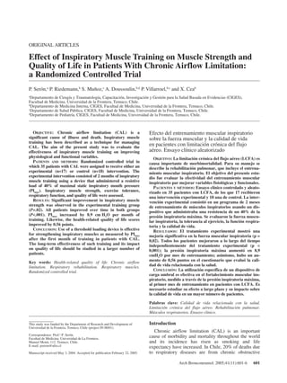
- 1. ORIGINAL ARTICLES Effect of Inspiratory Muscle Training on Muscle Strength and Quality of Life in Patients With Chronic Airflow Limitation: a Randomized Controlled Trial P. Serón,a P. Riedemann,b S. Muñoz,c A. Doussoulin,b,d P. Villarroel,b,c and X. Ceab aDepartamento de Cirugía y Traumatología, Capacitación, Investigación y Gestión para la Salud Basada en Evidencias (CIGES), Facultad de Medicina, Universidad de la Frontera, Temuco, Chile. bDepartamento de Medicina Interna, CIGES, Facultad de Medicina, Universidad de la Frontera, Temuco, Chile. cDepartamento de Salud Pública, CIGES, Facultad de Medicina, Universidad de la Frontera, Temuco, Chile. dDepartamento de Pediatría, CIGES, Facultad de Medicina, Universidad de la Frontera, Temuco, Chile. Introduction Chronic airflow limitation (CAL) is an important cause of morbidity and mortality throughout the world and its incidence has risen as smoking and life expectancy have increased. In Chile, 20% of deaths due to respiratory diseases are from chronic obstructive This study was funded by the Department of Research and Development of Universidad de la Frontera, Temuco, Chile (project IN 00/01). Correspondence: Prof.a P. Serón. Facultad de Medicina. Universidad de La Frontera. Manuel Montt, 112. Temuco. Chile. E-mail: pseron@ufro.cl Manuscript received May 3, 2004. Accepted for publication February 22, 2005. Arch Bronconeumol. 2005;41(11):601-6 601 OBJECTIVE: Chronic airflow limitation (CAL) is a significant cause of illness and death. Inspiratory muscle training has been described as a technique for managing CAL. The aim of the present study was to evaluate the effectiveness of inspiratory muscle training on improving physiological and functional variables. PATIENTS AND METHODS: Randomized controlled trial in which 35 patients with CAL were assigned to receive either an experimental (n=17) or control (n=18) intervention. The experimental intervention consisted of 2 months of inspiratory muscle training using a device that administered a resistive load of 40% of maximal static inspiratory mouth pressure (PImax). Inspiratory muscle strength, exercise tolerance, respiratory function, and quality of life were assessed. RESULTS: Significant improvement in inspiratory muscle strength was observed in the experimental training group (P=.02). All patients improved over time in both groups (P<.001). PImax increased by 8.9 cm H2O per month of training. Likewise, the health-related quality of life scores improved by 0.56 points. CONCLUSION: Use of a threshold loading device is effective for strengthening inspiratory muscles as measured by PImax after the first month of training in patients with CAL. The long-term effectiveness of such training and its impact on quality of life should be studied in a larger number of patients. Key words: Health-related quality of life. Chronic airflow limitation. Respiratory rehabilitation. Respiratory muscles. Randomized controlled trial. Efecto del entrenamiento muscular inspiratorio sobre la fuerza muscular y la calidad de vida en pacientes con limitación crónica del flujo aéreo. Ensayo clínico aleatorizado OBJETIVO: La limitación crónica del flujo aéreo (LCFA) es causa importante de morbimortalidad. Para su manejo se describe la rehabilitación pulmonar, que incluye el entrena-miento muscular inspiratorio. El objetivo del presente estu-dio fue evaluar la efectividad del entrenamiento muscular inspiratorio par mejorar variables fisiológicas y funcionales. PACIENTES Y MÉTODOS: Ensayo clínico controlado y aleato-rizado en 35 pacientes con LCFA, de los que 17 recibieron una intervención experimental y 18 una de control. La inter-vención experimental consistió en un programa de 2 meses de entrenamiento de músculos inspiratorios usando un dis-positivo que administraba una resistencia de un 40% de la presión inspiratoria máxima. Se evaluaron la fuerza muscu-lar inspiratoria, la tolerancia al ejercicio, la función respira-toria y la calidad de vida. RESULTADOS: El tratamiento experimental mostró una mejoría significativa en la fuerza muscular inspiratoria (p = 0,02). Todos los pacientes mejoraron a lo largo del tiempo independientemente del tratamiento experimental (p < 0,001); la presión inspiratoria máxima aumentó en 8,9 cmH2O por mes de entrenamiento; asimismo, hubo un au-mento de 0,56 puntos en el cuestionario que evaluó la cali-dad de vida relacionada con la salud. CONCLUSIÓN: La utilización específica de un dispositivo de carga umbral es efectiva en el fortalecimiento muscular ins-piratorio, medido a través de la presión inspiratoria máxima, al primer mes de entrenamiento en pacientes con LCFA. Es necesario estudiar su efecto a largo plazo y su impacto sobre la calidad de vida en un mayor número de pacientes. Palabras clave: Calidad de vida relacionada con la salud. Limitación crónica del flujo aéreo. Rehabilitación pulmonar. Músculos respiratorios. Ensayo clínico.
- 2. SERÓN P, ET AL. EFFECT OF INSPIRATORY MUSCLE TRAINING ON MUSCLE STRENGTH AND QUALITY OF LIFE IN PATIENTS WITH CHRONIC AIRFLOW LIMITATION: A RANDOMIZED CONTROLLED TRIAL pulmonary disease (COPD), the main cause of CAL; COPD causes an average of 1600 deaths every year in the country.1 CAL causes physical changes and apart from experiencing mechanical inconveniences, many patients are unable to enjoy life fully because of breathing difficulties and the inactivity imposed on them by symptoms.2 As a result, a primary objective in the management of these patients is to improve health-related quality of life (HRQL).3,4 Respiratory rehabilitation plays a fundamental role in the management of patients with CAL, contributing to social reinsertion and improved quality of life. Respiratory rehabilitation programs have various components, and it is difficult to distinguish the relative importance of each of them in the light of available evidence.5-8 Among the components are general health care, respiratory health care (bronchial drainage, assisted coughing, etc), exercise in general, functional training, education, psychosocial training,4 optional ventilatory assistance, noninvasive ventilation, and home health care.2 The potential role of respiratory muscle weakness and possible fatigue as a cause of CAL and respiratory failure in patients with chronic airway disease has led to the introduction of muscle training in rehabilitation programs. Several studies on inspiratory muscle training have demonstrated small differences in physiological parameters and symptoms.9-16 The results have been contradictory and have depended on the mode of resistance applied and intensity of exercise. Nevertheless, it has been asserted that training with a substantial inspiratory resistive load and a controlled breathing pattern can lead to evident improvements in strength, exercise tolerance, and dyspnea, with a resulting reduction in discomfort during activities of daily living, increased functional capacity, and improved quality of life.12,14,15 In this context, the present study aimed to determine the effect of inspiratory muscle training with a controlled flow rate on a) inspiratory muscle strength, b) exercise tolerance, c) respiratory function, and d) HRQL in patients with CAL. Patients and Methods Design In this controlled clinical trial patients were assigned to an experimental or control group by randomization from blocks of varying sizes. An independent person who did not otherwise participate in the study took charge of the randomization process. The patients were studied for a period of 2 months. Patients, supervisors, and persons charged with taking measurements were blinded to group assignment. Subjects A consecutive sample of patients was established for distribution. The sample included men and women, patients over the age of 20 years with CAL being monitored and treated at the Department of Pulmonary Medicine and Tuberculosis of the Diagnosis and Treatment Center of the city clinics of Temuco, Chile. The patients all had a forced expiratory volume in the first second (FEV1) less than 80% of the predicted value; they had all been informed of the nature of the study and had given their signed consent. Patients were excluded if they were clinically unstable as indicated by the need for emergency care or hospitalization in the month before enrollment in the study. Patients were also excluded if they were unable to perform the experimental maneuver or any maneuver necessary for the outcome measures, whether because of inability to understand the instructions or because of osteomuscular problems. Also excluded were patients with incapacitating heart disease (recent acute myocardial infarction, functional class IV heart failure, unstable angina). The research protocol was approved by the ethics committee of the Regional Hospital of Temuco. Outcome Measures Inspiratory muscle strength was assessed by maximal inspiratory pressure (PImax). This measurement, which is widely used for the purpose, has the advantage of being the simplest, oldest, and most useful way to measure respiratory muscle strength,17 and it is also noninvasive. Additionally, reference values are available for adults and children.18 To determine PImax, the patient was asked to take a normal breath and insert the mouthpiece at the moment he or she finished exhaling the tidal volume; then the patient made a maximal inspiratory effort lasting at least 2 seconds, against the closed valve. The highest value of PImax in 3 reproducible maneuvers was recorded.17,18 Exercise tolerance was evaluated during a 6-minute walk test carried out according to the protocol of Sciurva and Slivka.19 This measure, considered simple and useful as an index of general physical capacity, measures the distance the patient can walk in 6 minutes. The analysis was performed in a well-ventilated hallway 25 meters long that had been previously marked at 1-meter intervals. The supervisor gave the same instructions to each patient before and during the test. A training walk was carried out 30 minutes before the real test. Respiratory function was determined by spirometry with a VM PLUS portable spirometer (Clement Clarke, Harlow, UK). The theoretical reference values were calculated for each patient according to the method of Gutiérrez et al.20 HRQL was assessed with the Chronic Respiratory Questionnaire (CRQ), one of several instruments available to measure that parameter in patients with chronic respiratory diseases; developed in English and later translated to Spanish at the department of respiratory medicine of Hospital de la Santa Creu i de Sant Pau in Barcelona, Spain,21 the CRQ has been validated for use with Chilean patients and its psychometric properties and usefulness have been reported in various studies.21-27 The CRQ consists of 20 questions, distributed into 4 domains or dimensions: dyspnea, fatigue, emotional function, and mastery of disease. Each question is answered on a Likert-type scale of 7 points, where 1 indicates the most severe degree of dysfunction and 7 the best function. Five scores—total and domain-specific scores on a comparative scale from 1 to 7—can be obtained from each completed questionnaire. All measurements, by trained personnel who were blinded as to treatment group, were recorded at the start of training (baseline) and twice more (at the end of the first month and after 2 months). 602 Arch Bronconeumol. 2005;41(11):601-6
- 3. SERÓN P, ET AL. EFFECT OF INSPIRATORY MUSCLE TRAINING ON MUSCLE STRENGTH AND QUALITY OF LIFE Interventions IN PATIENTS WITH CHRONIC AIRFLOW LIMITATION: A RANDOMIZED CONTROLLED TRIAL Patients in both the treatment group and the control group took part in an exercise program that targeted the inspiratory muscles, supervised by a physical therapist. An adaptation period of 1 week included a phase of proprioceptive re-education, instruction on how to perform the exercises, and information on adherence. After this period, all patients were instructed to perform the exercises daily and to come to the clinic twice a week for 2 months. Strength training was carried out with the Threshold Inspiratory Muscle Trainer (Respironics HealthScan, Inc, Cedar Grove, New York, USA), a device that incorporates a 1-way flow valve that could be set to provide a load ranging from 7 to 41 cm H2O. The setting for the treatment group was individualized, such that a patient worked with a load that was 40% of inspiratory muscle strength measured by PImax at baseline. The load was reset after 4 weeks of training in accordance with the new level of inspiratory muscle strength at that time. The control group worked with the minimal loading offered by the exercise device (7 cm H2O). Inspiratory maneuvers were performed in a series of 5 repetitions with 1-minute periods between them. At the beginning of the program, patients had to complete 10 minutes of training; the duration was gradually increased until exercise sessions lasted 30 minutes a day in the last 3 weeks. Sample Size Calculation of sample size was based on finding a clinically significant difference of 1 point on the CRQ as a reflection of HRQL. Assuming a SD of 1, a statistical power of 80%, and a significance level of 5%, it was estimated that 17 patients needed to be assigned to each treatment group. Statistical Analysis Data was entered into the EpiInfo program and checked before being analyzed with the Stata statistical package, version 7.0. The hypothesis to be tested was that inspiratory muscle training has an effect on the strength of those muscles, on respiratory function, or on HRQL. Differences between groups at baseline and following treatment, and the differences in variables over time in both groups were analyzed by multiple linear regression. The treatments were assessed according to the following outcome measures: PImax, distance covered (in meters) on the 6-minute walk test, FEV1 expressed as a percentage of the theoretical value (FEV1, % of predicted), CRQ total score and domain scores (dyspnea, fatigue, emotional function, and mastery of disease). The variable used for between-group comparison was treatment (group 1: resistive loading of 40% of PImax; group 0: minimum loading). Longitudinal change was analyzed by comparing measurements at 3 times (0, baseline; 1, after 1 month; 2, after 2 months). Covariables were age, weight, height, and sex, as well as the previous measurement obtained for each study variable and adherence expressed as a percentage. Fisher’s F test was used to assess effect in each model. Relative bias was calculated to determine the influence of confounding factors.28 Results Figure 1 is a flow diagram showing the movement of patients through the study phases. The reason for loss of 41 Patients Assessed for Eligibility 6 Fail to Meet Inclusion Criteria 35 Patients Randomized 17 Assigned to the Experimental Group 2 Lost to Follow-up Data Analyzed for 15 Patients 18 Assigned to the Control Group 2 Lost to Follow-up Data Analyzed for 16 Patients Figure 1. Diagram showing the flow of patients through the phases of the study. patients in the second month was their self-declared inability to attend training sessions. Table 1 gives the baseline characteristics of patients by group. A between-group difference was detected for height, but there were no significant differences in age, weight, sex, CRQ domain scores, total CRQ score, PImax, 6-minute walk test, or FEV1 (% of predicted). Even though no statistically significant differences were detected between groups for most measurements or demographic variables, it was considered necessary to include those variables in regression models to assess their possible effect as confounders. Arch Bronconeumol. 2005;41(11):601-6 603 Analysis Follow-up Assignement Recruitment TABLE 1 Baseline Characteristics in the Treatment Group (40% Resistive Load) and the Control Group (Minimum Resistance)* Treatment Control Group (n=17) Group (n=18) P Age, years 55.1 (12.8) 59.2 (14.9) .19 Weight, kg 72.7 (14.9) 66.7 (12.6) .10 Height, cm 148.6 (10.4) 154.0 (6.7) .03 Sex: women 53% 46% .2 CRQ score Dyspnea 3.3 (1.2) 2.9 (0.8) .23 Fatigue 3.6 (1.4) 4.1 (1.2) .31 Emotional function 4.0 (1.6) 4.2 (1.3) .76 Mastery of disease 4.1 (1.5) 4.3 (1.6) .63 Total 3.8 (1.2) 3.9 (1.1) .70 PImax, cm H2O 56.5 (19.5) 53.2 (18.2) .60 6-minute walk test, m 448 (65.1) 451 (94.1) .90 FEV1, % pred 55.4 (19.2) 50.7 (19.6) .47 *Results are expressed as means (SD) or a percentage (sex). CRQ indicates Chronic Respiratory Questionnaire; PImax, maximal inspiratory pressure; FEV1, % pred, forced expiratory volume in the first second expressed as a percentage of the theoretical reference value.
- 4. SERÓN P, ET AL. EFFECT OF INSPIRATORY MUSCLE TRAINING ON MUSCLE STRENGTH AND QUALITY OF LIFE IN PATIENTS WITH CHRONIC AIRFLOW LIMITATION: A RANDOMIZED CONTROLLED TRIAL 7 6 5 4 3 2 1 Total CRQ Score Experimental Group Score 1 2 3 PImax Experimental Group At the end of the first month of training, an improvement was detected in inspiratory muscle strength between treatment groups. PImax was 11.5 cm H2O higher in the treatment group (P=.02, adjusted for age, weight, height, sex, baseline PImax, and adherence). No significant differences were found for other variables. Nor significant between-group differences were found after 2 months of training for any of the outcome measures. Change over time was also assessed. Statistically significant differences were observed for every CRQ domain, for the total CRQ score, and for PImax (P<.0001, adjusted for age, weight, height, sex, baseline value, and adherence). CRQ scores improved over the 2 months of treatment in both groups. The score for Control Group Score PImax Control Group dyspnea increased by 0.82 points, fatigue by 0.43 points, emotional function by 0.5, mastery by 0.5, and the total by 0.56. PImax increased 8.9 cm H2O per month of training over the 2-month study period. No differences were found for FEV1 (% of predicted) or for distance covered on the 6-minute walk test. Figure 2 shows how total CRQ score and PImax changed for both study groups. Table 2 shows the evolution of all variables during the study for both groups. Discussion A threshold inspiratory muscle resistive loading device was used in this study to provide the same level of resistance for each inhalation during training. The 604 Arch Bronconeumol. 2005;41(11):601-6 7 6 5 4 3 2 1 Total CRQ Score 1 2 3 7 6 5 4 3 2 1 PImax (cm H2O) 1 2 3 7 6 5 4 3 2 1 PImax (cm H2O) 1 2 3 Figure 2. Changes in outcome measures for the treatment group (40% resistive loading on inspiration) and control group (minimal resistance). CRQ indicates Chronic Respiratory Questionnaire; PImax, maximal inspiratory pressure. TABLE 2 Changes in Outcome Measures After the First and Second Months of Training in the Treatment Group (40% Resistive Load) and Control Group (Minimal Resistance)* Treatment Group Control Group Month 1 Month 2 Month 1 Month 2 CRQ score Dyspnea 4.6 (1.1) 5.1 (1.3) 3.9 (1) 4.5 (1.2) Fatigue 4.3 (1.5) 4.6 (1.6) 4.6 (1) 4.9 (0.9) Emotional function 5 (1.4) 5.3 (1.2) 4.5 (1.3) 4.9 (1.2) Mastery of disease 4.9 (1.5) 5.4 (1.4) 5.1 (1.4) 5.1 (1.2) Total 4.7 (1.2) 5.1 (1.2) 4.6 (1) 4.9 (1) PImax, cm H2O 72.4 (19.8) 77.7 (20.5) 64.1 (16.1) 67.6 (15.1) 6-minute walk test, m 458 (47.4) 452.6 (52.3) 457.2 (67.6) 458.5 (75.4) FEV1, % pred 60.9 (28.5) 60.6 (24) 53.5 (20.6) 54.6 (21.9) Percentage adherence 84 (14.2) 85 (13.8) 89 (13.3) 87.7 (12.1) *Results are expressed as means (SD). CRQ indicates Chronic Respiratory Questionnaire; PImax, maximal inspiratory pressure; FEV1, % pred, forced expiratory volume in the first second expressed as a percentage of the theoretical reference value.
- 5. SERÓN P, ET AL. EFFECT OF INSPIRATORY MUSCLE TRAINING ON MUSCLE STRENGTH AND QUALITY OF LIFE IN PATIENTS WITH CHRONIC AIRFLOW LIMITATION: A RANDOMIZED CONTROLLED TRIAL device was developed after it was demonstrated that strength training with uncontrolled flow rates was ineffective.29 Resistance to inhalation comes from a constant pressure loading that takes inspiratory flow into account. A valve allows the load to be set to the desired level.9 Patients in the treatment group, who trained with a load that was 40% of PImax, experienced a significant improvement in inspiratory muscle strength after 1 month. This finding is consistent with reports from other studies, such as that of Larson et al,10 who was the first to show a statistically significant increase in inspiratory muscle strength, respiratory muscle resistance, and tolerance to exercise in patients with moderate-to-severe COPD. Similarly, Belman and Shadmehr11 demonstrated significant improvement in inspiratory muscle strength in a group of COPD patients in whom high resistance training was compared to low resistance training applied 15 minutes every day over a period of 6 weeks. It is important to emphasize that when analyzing the results of the present study, we adjusted for the previous measurement of the outcome measure and for adherence, an aspect mentioned in previous studies as being important, although others have not made such adjustments. Furthermore, random assignment to groups and the fact that trainers and assessors were blinded to patient assignment, served to minimize bias in favor of the treatment group. These characteristics of the study strengthen the conclusion that the exercise treatment applied increases muscle strength. That finding is of clinical importance as it will mean that the respiratory system is better prepared mechanically to withstand a crisis if muscle fatigue reappears.10 Results after 2 months of training demonstrated no difference between groups. Similar findings were reported by Harver et al,12 who observed a statistically significant difference in inspiratory muscle strength between baseline and interim follow-up measurements in the treatment group but no difference between groups at the end of the study. Lisboa et al15 similarly found a significant difference in PImax that appeared after 2 weeks in a group that trained with a resistive load of 30%, whereas the significant increase in PImax appeared after the fifth week in the group training with a load of 10%. Dikhuijzen et al,14 on the other hand, found significant differences after 10 weeks of respiratory rehabilitation between an experimental group that received inspiratory muscle training and a control group that received only conventional respiratory rehabilitation. It is relevant that the control group in our study underwent training with a minimal resistive load of 7 cm H2O provided by the device. This was done in order to improve blinding. That minimal resistive load was between 21% and 25% of PImax for a quarter of the patients who were randomized to the control group. That would explain the control group’s improvement, which came somewhat more slowly but was significant over time (Figure 2). An additional point to consider is that our study, like others, evaluated muscle force by way of PImax, yet it seems necessary to use direct measures of diaphragmatic contractility, such as magnetic stimulation of the phrenic nerve30 or to use outcome measures based on clinical or functional aspects, as we have done in the present study by assessing HRQL. In this sense, we believe that the lack of a statistically significant difference in HRQL between groups is attributable to a real statistical power of 65% rather than the higher power of 80% assumed at the time of calculating the sample size. This would be the case because both the variation observed and the SD (1.2 vs the assumed value of 1) were greater than expected. The same pattern was seen with functional and physiological variables. Even so, other studies have reported findings similar to ours. No significant differences in subjective measurements such as respiratory symptoms or HRQL were seen in the studies by Dekhuijzen et al14 or Berman et al,31 for example. In the comparison of measurements over time, it is important to emphasize that there were significant improvements in all the quality of life domains, from a clinical standpoint as well as a statistical one. We were able to detect that over time the minimal clinically significant difference described for the CRQ was 0.5 points,32,33 the level that would allow respiratory rehabilitation interventions to be said to produce improvement in subjective health measures such as HRQL. In this case, the outcome can not be attributed directly to inspiratory muscle training, although it can be hypothesized that it is due to the overall set of measures studied (such as education of patients about their respiratory problem, correct use of inhalers, and postures that favor respiratory mechanics) and the respiratory exercises aside from the inspiratory muscle training intervention (such as diaphragmatic breathing and sustained maximal inhalation) which were used in every maneuver.34 The results of this study lead to the conclusion that patients with CAL can use a device specifically designed to provide a threshold inspiratory resistive load to effectively strengthen inspiratory muscles as reflected by PImax after 1 month of training. The long-term effect and impact on HRQL should be studied in a larger number of patients. REFERENCES 1. González P. Consenso Nacional en Enfermedad Pulmonar Obstructiva Crónica (EPOC). Rev Ch Enf Respir. 1998;14:77-82. 2. Celli B, Snider G, Heffner J, Tiep B, Ziment I, Make B, et al. ATS statement. Standards for the diagnosis and care of patients with chronic obstructive pulmonary disease. Am J Respir Crit Care Med. 1995;152 Supl 1:78-121. 3. Saroea H. Chronic obstructive pulmonary disease. Major objectives of management. Postgrad Med. 1993;94:113-22. 4. Barr R. Pulmonary rehabilitation. In: Hillegass H, Sadowsky S, editors. Essentials of cardiopulmonary physical therapy. Philadelphia: WB Saunders Company; 1994, p. 677-701. Arch Bronconeumol. 2005;41(11):601-6 605
- 6. SERÓN P, ET AL. EFFECT OF INSPIRATORY MUSCLE TRAINING ON MUSCLE STRENGTH AND QUALITY OF LIFE IN PATIENTS WITH CHRONIC AIRFLOW LIMITATION: A RANDOMIZED CONTROLLED TRIAL 5. Lacasse Y, Guyatt GH, Goldstein RS. The components of a respiratory rehabilitation program: a systematic overview. Chest. 1997; 111:1077-88. 6. Cambach W, Wagenaar RC, Koelman TW, van Keimpema AR, Kemper HC. The long-term effects of pulmonary rehabilitation in patients with asthma and chronic obstructive pulmonary disease: a research synthesis. Arch Phys Med Rehabil. 1999;80: 103-11. 7. Salman GF, Mosier MC, Beasley BW, Calkins DR. Rehabilitation for patients with chronic obstructive pulmonary disease: metaanalysis of randomized controlled trials. J Gen Intern Med. 2003; 18:213-21. 8. Lacasse Y, Brosseau L, Milne S, Martin S, Wong E, Guyatt GH, et al. Pulmonary rehabilitation for chronic obstructive pulmonary disease (Cochrane Review). In: The Cochrane Library, Issue 4, 2004. 9. Guyatt G, Keller J, Singer J, Halcrow S, Newhouse M. Controlled trial of respiratory muscle training in chronic airflow limitation. Thorax. 1992;47:598-602. 10. Larson J, Kim M, Sharp J, Larson D. Inspiratory muscle training with a pressure threshold breathing device in patients with chronic obstructive pulmonary disease. Am Rev Respir Dis. 1988;138: 689-96. 11. Belman MJ, Shadmehr R. Targeted resistive ventilatory muscle training in chronic obstructive pulmonary disease. J Appl Physiol. 1988;65:2726-35. 12. Harver A, Mahler D, Daubenspeck A. Targeted inspiratory muscle training improves respiratory muscle function and reduces dyspnea in patients with chronic obstructive pulmonary disease. Ann Intern Med. 1989;111:117-24. 13. Goldstein R, Derosie J, Long S. Applicability of a threshold loa-ding device for inspiratory muscle testing and training in patients with COPD. Chest. 1989;96:564-71. 14. Dekhuijzen PN, Folgerin HT, van Herwaarden CL. Target-flow inspiratory muscle training during pulmonary rehabilitation in patients with COPD. Chest. 1991;99:128-33. 15. Lisboa C, Villafranca C, Leiva A, Cruz E, Pertuzé J, Borzone G. Inspiratory muscle training in chronic airflow limitation: effect on exercise performance. Eur Respir J. 1997;10:537-42. 16. Lisboa C, Borzone G, Cruz E. Entrenamiento muscular inspiratorio en pacientes con enfermedad pulmonar obstructiva crónica. Rev Med Chil. 1998;126:563-8. 17. Celli B, Grassino A. Respiratory muscles: functional evaluation. Sem Respir Crit Care Med. 1998;19:367-81. 18. Syabbalo N. Assessment of respiratory muscle function and strength. Postgrad Med. 1998;74:208-15. 19. Sciurba F, Slivka W. Six-minute walk testing. Sem Respir Crit Care Med. 1998;19:383-92. 20. Gutiérrez M, Rioseco F, Rojas A, Casanova D. Determinación de valores espirométricos en una población chilena normal mayor de 5 años, a nivel del mar. Rev Med Chil. 1996;124:1295-306. 21. Güell R, Casan P, Sangenís M, Morante F, Belda J, Guyatt G. Quality of life in patients with chronic respiratory disease: the Spanish version of the Chronic Respiratory Questionnaire (CRQ). Eur Respir J. 1998;11:55-60. 22. Guyatt G, Berman L, Towsend M, Pugsley S, Chambers L. A measure of quality of life for clinical trials in chronic lung disease. Thorax. 1987;42:773-8. 23. Hajiro T, Nishimura K, Tsukino M, Ikeda A, Koyama H, Izumi T. Comparison of discriminative properties among Disease-specific Questionnaires for measuring health-related quality of life in patients with chronic obstructive pulmonary disease. Am J Respir Crit Care Med. 1998;157:785-90. 24. Wijstra P, Tenvergert E, Vanaltena R, Otten V, Postma D, Kraan J, et al. Reliability and validity of the chronic respiratory question-naire (CRQ). Thorax. 1994;49:465-7. 25. Sans-Torres J, Domingo C, Rue M, Durán-Tauleria E, Marín A. Valoración de la calidad de vida de los pacientes con EPOC e hipoxemia crónica mediante la versión española del Chronic Respiratory Disease Questionnaire. Arch Bronconeumol. 1999;35: 428-34. 26. Lisboa C, Villafranca C, Caiozzi G, Berrocal C, Leiva A, Pinochet R. Calidad de vida en pacientes con enfermedad pulmonar obstructiva crónica e impacto del entrenamiento físico. Rev Med Chil. 2001;129:359-66. 27. Serón P, Riedemann P, Sanhueza A, Doussoulin A, Villarroel P. Validación del Cuestionario de la Enfermedad Respiratoria Crónica en pacientes chilenos con limitación crónica del flujo aéreo. Rev Med Chil. 2003;131:1243-50. 28. Kleinbaum DG, Kupper LL, Muller KE, Nizam A. Applied regression analysis and multivariable methods. 3rd ed. California: Duxbury Press; 1998. 29. Smith K, Cook D, Guyatt G, Madhaven J, Oxman A. Respiratory muscle training in chronic airflow limitation: a meta-analysis. Am Rev Respir Dis. 1992;145:533-9. 30. Polkey MI, Moxham J. Improvement in volitional tests of muscle function alone may not be adequate evidence that inspiratory muscle training is effective. Eur Respir J. 2004;23:5-6. 31. Berman LB, Goldsmith CH, Jones NL, McIntosh JM. Resistive Inspiratory Breathing and Cycle Ergometer in Chronic Airflow Obstruction. Final report (DM545) Ontario Ministry of Health-and Ontario Thoracic Society, 1986-08, p. 37 + 9 appendices. 32. Jaeschke R, Singer J, Guyatt G. Measurement of health status. Ascertaining the minimal clinically important difference. Control Clin Trials. 1989;10:407-15. 33. Redelmeier DA, Guyatt GH, Goldstein RS. Assessing the minimal important difference in symptoms: a comparison of two techniques. J Clin Epidemiol. 1996;49:1215-9. 34. Hough A. Management of breathlessness and pulmonary rehabilitation. In: Physiotherapy in Respiratory Care. A problem-solving approach to respiratory and cardiac management. 2nd ed. Cheltenham: Stanley Thornes; 1996. p. 145-68. 606 Arch Bronconeumol. 2005;41(11):601-6