1. Cytoplasm
From Wikipedia, the free encyclopedia
Jump to: navigation, search
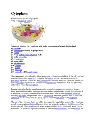
Schematic showing the cytoplasm, with major components of a typical animal cell
(organelles):
(3) ribosomes (indicated by purple dots)
(4) vesicle
(5) rough endoplasmic reticulum (ER)
(6) Golgi apparatus
(7) cytoskeleton
(8) smooth ER
(9) mitochondria
(10) vacuole
(11) cytosol
(12) lysosome
(13) centrioles within centrosome
The cytoplasm is a thick liquid residing between the cell membrane holding all the cell's internal
sub-structures (called organelles), except for the nucleus. All the contents of the cells of
prokaryote organisms (which lack a cell nucleus) are contained within the cytoplasm. Within the
cells of eukaryote organisms the contents of the cell nucleus are separated from the cytoplasm,
and are then called the nucleoplasm.
In eukaryotic cells also, the cytoplasm contains organelles, such as mitochondria, which are
filled with liquid that is kept separate from the rest of the cytoplasm by biological membranes. It
is within the cytoplasm that most cellular activities occur, such as many metabolic pathways
including glycolysis, and processes such as cell division. The inner, granular mass is called the
endoplasm and the outer, clear and glassy layer is called the cell cortex or the ectoplasm.
The part of the cytoplasm that is not held within organelles is called the cytosol. The cytosol is a
complex mixture of cytoskeleton filaments, dissolved molecules, and water that fills much of the
volume of a cell. The cytosol is a gel, with a network of fibers dispersed through water. Due to
this network of pores and high concentrations of dissolved macromolecules, such as proteins, an
2. effect called macromolecular crowding occurs and the cytosol does not act as an ideal solution.
This crowding effect alters how the components of the cytosol interact with each other.
Movement of calcium ions in and out of the cytoplasm is thought to be a signaling activity for
metabolic processes.[1]
NUCLEOULES++From Wikipedia, the free encyclopedia
Jump to: navigation, search
The nucleolus is contained within the cell nucleus.
Schematic of typical animal cell, showing subcellular components. Organelles:
(1) nucleolus
(2) nucleus
(3) Ribosomes (little dots)
(4) vesicle
(5) rough endoplasmic reticulum (ER)
(6) Golgi apparatus
(7) Cytoskeleton
(8) smooth endoplasmic reticulum (ER)
(9) mitochondria
(10) vacuole
3. (11) cytosol (not cytoplasm as that includes all the organelles)
(12) lysosome
(13) centrioles within centrosome
The nucleolus (also called nucleole) is a non-membrane bound structure[1] composed of proteins
and nucleic acids found within the nucleus. Ribosomal RNA (rRNA) is transcribed and
assembled within the nucleolus. The nucleolus ultrastructure can be visualized through an
electron microscope, while the organization and dynamics can be studied through fluorescent
protein tagging and fluorescent recovery after photobleaching (FRAP). Malfunction of nucleoli
can be the cause for several human diseases.
From Wikipedia, the free encyclopedia
Jump to: navigation, search
The nucleolus is contained within the cell nucleus.
Schematic of typical animal cell, showing subcellular components. Organelles:
(1) nucleolus
(2) nucleus
(3) Ribosomes (little dots)
(4) vesicle
(5) rough endoplasmic reticulum (ER)
(6) Golgi apparatus
4. (7) Cytoskeleton
(8) smooth endoplasmic reticulum (ER)
(9) mitochondria
(10) vacuole
(11) cytosol (not cytoplasm as that includes all the organelles)
(12) lysosome
(13) centrioles within centrosome
The nucleolus (also called nucleole) is a non-membrane bound structure[1] composed of proteins
and nucleic acids found within the nucleus. Ribosomal RNA (rRNA) is transcribed and
assembled within the nucleolus. The nucleolus ultrastructure can be visualized through an
electron microscope, while the organization and dynamics can be studied through fluorescent
protein tagging and fluorescent recovery after photobleaching (FRAP). Malfunction of nucleoli
can be the cause for several human diseases.
From Wikipedia, the free encyclopedia
Jump to: navigation, search
The nucleolus is contained within the cell nucleus.
Schematic of typical animal cell, showing subcellular components. Organelles:
(1) nucleolus
(2) nucleus
5. (3) Ribosomes (little dots)
(4) vesicle
(5) rough endoplasmic reticulum (ER)
(6) Golgi apparatus
(7) Cytoskeleton
(8) smooth endoplasmic reticulum (ER)
(9) mitochondria
(10) vacuole
(11) cytosol (not cytoplasm as that includes all the organelles)
(12) lysosome
(13) centrioles within centrosome
The nucleolus (also called nucleole) is a non-membrane bound structure[1] composed of proteins
and nucleic acids found within the nucleus. Ribosomal RNA (rRNA) is transcribed and
assembled within the nucleolus. The nucleolus ultrastructure can be visualized through an
electron microscope, while the organization and dynamics can be studied through fluorescent
protein tagging and fluorescent recovery after photobleaching (FRAP). Malfunction of nucleoli
can be the cause for several human diseases.
NUCLEOLUS=
From Wikipedia, the free encyclopedia
Jump to: navigation, search
The nucleolus is contained within the cell nucleus.
6. Schematic of typical animal cell, showing subcellular components. Organelles:
(1) nucleolus
(2) nucleus
(3) Ribosomes (little dots)
(4) vesicle
(5) rough endoplasmic reticulum (ER)
(6) Golgi apparatus
(7) Cytoskeleton
(8) smooth endoplasmic reticulum (ER)
(9) mitochondria
(10) vacuole
(11) cytosol (not cytoplasm as that includes all the organelles)
(12) lysosome
(13) centrioles within centrosome
The nucleolus (also called nucleole) is a non-membrane bound structure[1] composed of proteins
and nucleic acids found within the nucleus. Ribosomal RNA (rRNA) is transcribed and
assembled within the nucleolus. The nucleolus ultrastructure can be visualized through an
electron microscope, while the organization and dynamics can be studied through fluorescent
protein tagging and fluorescent recovery after photobleaching (FRAP). Malfunction of nucleoli
can be the cause for several human diseases.
CELL NECLUES=
In cell biology, the nucleus (pl. nuclei; from Latin nucleus or nuculeus, meaning kernel) is a
membrane-enclosed organelle found in eukaryotic cells. It contains most of the cell's genetic
material, organized as multiple long linear DNA molecules in complex with a large variety of
proteins, such as histones, to form chromosomes. The genes within these chromosomes are the
cell's nuclear genome. The function of the nucleus is to maintain the integrity of these genes and
to control the activities of the cell by regulating gene expression — the nucleus is, therefore, the
control center of the cell. The main structures making up the nucleus are the nuclear envelope, a
double membrane that encloses the entire organelle and separates its contents from the cellular
cytoplasm, and the nuclear lamina, a meshwork within the nucleus that adds mechanical support,
much like the cytoskeleton, which supports the cell as a whole. Because the nuclear membrane is
impermeable to most molecules, nuclear pores are required to allow movement of molecules
7. across the envelope. These pores cross both of the membranes, providing a channel that allows
free movement of small molecules and ions. The movement of larger molecules such as proteins
is carefully controlled, and requires active transport regulated by carrier proteins. Nuclear
transport is crucial to cell function, as movement through the pores is required for both gene
expression and chromosomal maintenance.
Although the interior of the nucleus does not contain any membrane-bound subcompartments, its
contents are not uniform, and a number of subnuclear bodies exist, made up of unique proteins,
RNA molecules, and particular parts of the chromosomes. The best-known of these is the
nucleolus, which is mainly involved in the assembly of ribosomes. After being produced in the
nucleolus, ribosomes are exported to the cytoplasm where they translate mRNA.
RIBOSOMES=
A ribosome is an an organelle (an internal component of a biological cell) the function of which
is to assemble the twenty specific amino acid molecules to form the particular protein molecule
determined by the nucleotide sequence of an RNA molecule.
One of the central tenets of biology, often referred to as the central dogma of molecular biology,
is that DNA is used to make RNA, which is used to make proteins. The DNA sequence in genes
is copied into a messenger RNA (mRNA). Ribosomes then read the information in this mRNA
and use it to create proteins. This process is known as translation; the ribosome translates the
genetic information from the RNA into proteins. Ribosomes do this by binding to an mRNA and
using it as a template for determining the correct sequence of amino acids in a particular protein.
The amino acids are attached to transfer RNA (tRNA) molecules, which enter one part of the
ribosome and bind to the messenger RNA sequence. The attached amino acids are then joined
together by another part of the ribosome. The ribosome moves along the mRNA, "reading" its
sequence and producing a corresponding chain of amino acids.
Ribosomes are made from complexes of RNAs and proteins. Ribosomes are divided into two
subunits. The smaller subunit binds to the mRNA, while the larger subunit binds to the tRNA
and the amino acids. When a ribosome finishes reading a mRNA, these two subunits split apart.
Ribosomes have been classified as ribozymes, because the ribosomal RNA seems to be most
important for the peptidyl transferase activity that links amino acids together.
Ribosomes from bacteria, archaea and eukaryotes (the three domains of life on Earth), have
significantly different structures and RNA sequences. These differences in structure allow some
antibiotics to kill bacteria by inhibiting their ribosomes, while leaving human ribosomes
unaffected. The ribosomes in the mitochondria of eukaryotic cells functionally resemble in many
features those in bacteria, reflecting the likely evolutionary origin of this organelle.[1] [2] The
word ribosome comes from ribonucleic acid and the Greek: soma (meaning body).
VESICLE=
A vesicle can be visualised as a bubble of liquid within another liquid, a supramolecular
assembly made up of many different molecules. More technically, a vesicle is a small
8. membrane-enclosed sack that can store or transport substances. Vesicles can form naturally
because of the properties of lipid membranes (see micelle), or they may be prepared. Artificially
prepared vesicles are known as liposomes. Most vesicles have specialized functions depending
on what materials they contain.
Because vesicles tend to look alike, it is very difficult to tell the difference between different
types.
The vesicle is separated from the cytosol by at least one phospholipid bilayer. If there is only one
phospholipid bilayer, they are called unilamellar vesicles; otherwise they are called
multilamellar.
Vesicles store, transport, or digest cellular products and waste. The membrane enclosing the
vesicle is similar to that of the plasma membrane, and vesicles can fuse with the plasma
membrane to release their contents outside of the cell. Vesicles can also fuse with other
organelles within the cell.
Because it is separated from the cytosol, the inside of the vesicle can be made to be different
from the cytosolic environment. For this reason, vesicles are a basic tool used by the cell for
organizing cellular substances. Vesicles are involved in metabolism, transport, buoyancy
control,[1] and enzyme storage. They can also act as chemical reaction chambers.
ENDOOLASMIC RETICULUM=
The endoplasmic reticulum (ER) is a eukaryotic organelle that forms an interconnected
network of tubules, vesicles, and cisternae within cells. Rough endoplasmic reticula synthesize
proteins, while smooth endoplasmic reticula synthesize lipids and steroids, metabolize
carbohydrates and steroids (but not lipids), and regulate calcium concentration, drug metabolism,
and attachment of receptors on cell membrane proteins. Sarcoplasmic reticula solely regulate
calcium levels.
The lacey membranes of the endoplasmic reticulum were first seen by Keith R. Porter, Albert
Claude, and Ernest F. Fullam in 1945.[1]
GOLGI APPERATUS=The Golgi apparatus (also Golgi body or the Golgi complex) is an
organelle found in most eukaryotic cells.[1] It was identified in 1897 by the Italian physician
Camillo Golgi, after whom the Golgi apparatus is named.[2]
It processes and packages macromolecules, such as proteins and lipids, after their synthesis and
before they make their way to their destination; it is particularly important in the processing of
proteins for secretion. The Golgi apparatus forms a part of the cellular endomembrane system.
CYTOSKELETON=
9. The cytoskeleton (also CSK) is a cellular "scaffolding" or "skeleton" contained within the
cytoplasm and is made out of protein. The cytoskeleton is present in all cells; it was once thought
to be unique to eukaryotes, but recent research has identified the prokaryotic cytoskeleton. It has
structures such as flagella, cilia and lamellipodia and plays important roles in both intracellular
transport (the movement of vesicles and organelles, for example) and cellular division. The
concept of a protein mosaic that dynamically coordinated cytoplasmic biochemistry was
proposed by Rudolph Peters in 1929 [1] while the term (cytosquelette, in French) was first
introduced by French embryologist Paul Wintrebert in 1931.[2]
MITOCHODRIAN=
In cell biology, a mitochondrion (plural mitochondria) is a membrane-enclosed organelle
found in most eukaryotic cells.[1] These organelles range from 0.5 to 10 micrometers (μm) in
diameter. Mitochondria are sometimes described as "cellular power plants" because they
generate most of the cell's supply of adenosine triphosphate (ATP), used as a source of chemical
energy.[2] In addition to supplying cellular energy, mitochondria are involved in a range of other
processes, such as signaling, cellular differentiation, cell death, as well as the control of the cell
cycle and cell growth.[3] Mitochondria have been implicated in several human diseases, including
mitochondrial disorders[4] and cardiac dysfunction,[5] and may play a role in the aging process.
The word mitochondrion comes from the Greek μίτος or mitos, thread + χονδρίον or chondrion,
granule.
Several characteristics make mitochondria unique. The number of mitochondria in a cell varies
widely by organism and tissue type. Many cells have only a single mitochondrion, whereas
others can contain several thousand mitochondria.[6][7] The organelle is composed of
compartments that carry out specialized functions. These compartments or regions include the
outer membrane, the intermembrane space, the inner membrane, and the cristae and matrix.
Mitochondrial proteins vary depending on the tissue and the species. In humans, 615 distinct
types of proteins have been identified from cardiac mitochondria,[8] whereas in Murinae (rats),
940 proteins encoded by distinct genes have been reported.[9] The mitochondrial proteome is
thought to be dynamically regulated.[10] Although most of a cell's DNA is contained in the cell
nucleus, the mitochondrion has its own independent genome. Further, its DNA shows substantial
similarity to bacterial genomes.[11]
CENTRIOLE=
A centriole is a barrel-shaped cell structure[1] found in most animal eukaryotic cells, though it is
absent in higher plants and most fungi.[2] The walls of each centriole are usually composed of
nine triplets of microtubules (protein of the cytoskeleton). Deviations from this structure include
Drosophila melanogaster embryos, with nine doublets, and Caenorhabditis elegans sperm cells
and early embryos, with nine singlets; [3][4]. Crabs may also exhibit nine doublets, (see picture).
An associated pair of centrioles, arranged perpendicularly and surrounded by an amorphous mass
of dense material (the pericentriolar material) constitutes the compound structure known as the
centrosome.[1]
10. VACOULE=
A vacuole is a membrane-bound organelle which is present in all plant and fungal cells and some
protist, animal[1] and bacterial cells.[2] Vacuoles are essentially enclosed compartments which are
filled with water containing inorganic and organic molecules including enzymes in solution,
though in certain cases they may contain solids which have been engulfed. Vacuoles are formed
by the fusion of multiple membrane vesicles and are effectively just larger forms of these.[3] The
organelle has no basic shape or size; its structure varies according to the needs of the cell.
The function and importance of vacuoles varies greatly according to the type of cell in which
they are present, having much greater prominence in the cells of plants, fungi and certain protists
than those of animals and bacteria. In general, the functions of the vacuole include:
Isolating materials that might be harmful or a threat to the cell
Containing waste products
Containing water in plant cells
Maintaining internal hydrostatic pressure or turgor within the cell
Maintaining an acidic internal pH
Containing small molecules
Exporting unwanted substances from the cell
Allows plants to support structures such as leaves and flowers due to the pressure of the central
vacuole
In seeds, stored proteins needed for germination are kept in 'protein bodies', which are modified
vacuoles.[4]
Vacuoles also play a major role in autophagy, maintaining a balance between biogenesis
(production) and degradation (or turnover), of many substances and cell structures in certain
organisms. They also aid in the lysis and recycling of misfolded proteins that have begun to build
up within the cell. Thomas Boller [4] and others proposed that the vacuole participates in the
destruction of invading bacteria and Robert B Mellor proposed organ-specific forms have a role
in 'housing' symbiotic bacteria. In protists, vacuoles have the additional function of storing food
which has been absorbed by the organism and assisting in the digestive and waste management
process for the cell.[5]
CYTOSOL=
The cytosol or intracellular fluid (or cytoplasmic matrix) is the liquid found inside cells. It is
separated into compartments by membranes. For example, the mitochondrial matrix separates the
mitochondrion into compartments.
The contents of a eukaryotic cell within the cell membrane (excluding the cell nucleus), is
referred to as the cytoplasm. In prokaryotes, most of the chemical reactions of metabolism take
place in the cytosol, while a few take place in membranes or in the periplasmic space. In
eukaryotes, while many metabolic pathways still occur in the cytosol, others are contained within
organelles.
11. The cytosol is a complex mixture of substances dissolved in water. Although water forms the
large majority of the cytosol, its structure and properties within cells is not well understood. The
concentrations of ions such as sodium and potassium are different in the cytosol than in the
extracellular fluid; these differences in ion levels are important in processes such as
osmoregulation and cell signaling. The cytosol also contains large amounts of macromolecules,
which can alter how molecules behave, through macromolecular crowding.
Although once thought to be a simple solution of molecules, multiple levels of organization exist
in the cytosol. These include concentration gradients of small molecules such as calcium, large
complexes of enzymes that act together to carry out metabolic pathways, and protein complexes
such as proteasomes and carboxysomes that enclose and separate parts of the cytosol.
LYSOSOME=
Lysosomes are cellular organelles that contain acid hydrolase enzymes to break down waste
materials and cellular debris. They are found in animal cells, while in yeast and plants the same
roles are performed by lytic vacuoles.[1] Lysosomes digest excess or worn-out organelles, food
particles, and engulfed viruses or bacteria. The membrane around a lysosome allows the
digestive enzymes to work at the 4.5 pH they require. Lysosomes fuse with vacuoles and
dispense their enzymes into the vacuoles, digesting their contents. They are created by the
addition of hydrolytic enzymes to early endosomes from the Golgi apparatus. The name
lysosome derives from the Greek words lysis, to separate, and soma, body. They are frequently
nicknamed "suicide-bags" or "suicide-sacs" by cell biologists due to their role in autolysis.
Lysosomes were discovered by the Belgian cytologist Christian de Duve in the 1950s.
The size of lysosomes varies from 0.1–1.2 μm.[2] At pH 4.8, the interior of the lysosomes is
acidic compared to the slightly alkaline cytosol (pH 7.2). The lysosome maintains this pH
differential by pumping protons (H+ ions) from the cytosol across the membrane via proton
pumps and chloride ion channels. The lysosomal membrane protects the cytosol, and therefore
the rest of the cell, from the degradative enzymes within the lysosome. The cell is additionally
protected from any lysosomal acid hydrolases that leak into the cytosol, as these enzymes are
pH-sensitive and do not function as well in the alkaline environment of the cytosol
CENTRIOLE=
A centriole is a barrel-shaped cell structure[1] found in most animal eukaryotic cells, though it is
absent in higher plants and most fungi.[2] The walls of each centriole are usually composed of
nine triplets of microtubules (protein of the cytoskeleton). Deviations from this structure include
Drosophila melanogaster embryos, with nine doublets, and Caenorhabditis elegans sperm cells
and early embryos, with nine singlets; [3][4]. Crabs may also exhibit nine doublets, (see picture).
An associated pair of centrioles, arranged perpendicularly and surrounded by an amorphous mass
of dense material (the pericentriolar material) constitutes the compound structure known as the
centrosome.[1]
![effect called macromolecular crowding occurs and the cytosol does not act as an ideal solution.
This crowding effect alters how the components of the cytosol interact with each other.
Movement of calcium ions in and out of the cytoplasm is thought to be a signaling activity for
metabolic processes.[1]
NUCLEOULES++From Wikipedia, the free encyclopedia
Jump to: navigation, search
The nucleolus is contained within the cell nucleus.
Schematic of typical animal cell, showing subcellular components. Organelles:
(1) nucleolus
(2) nucleus
(3) Ribosomes (little dots)
(4) vesicle
(5) rough endoplasmic reticulum (ER)
(6) Golgi apparatus
(7) Cytoskeleton
(8) smooth endoplasmic reticulum (ER)
(9) mitochondria
(10) vacuole](data:image/gif;base64,R0lGODlhAQABAIAAAAAAAP///yH5BAEAAAAALAAAAAABAAEAAAIBRAA7)