SlideShare una empresa de Scribd logo
1 de 32
Anatomy of Nose & Paranasal
Sinuses
Vestibule
 Examination :- By Lifting the tip of the nose.
 Dilated Passage way leading from external nose to
the nasal fossa.
 Demarcated by Limen Nasi
 Superiorly lined by skin, contains hairs, sebaceous
gland and sweat glands
Furunculosis of Nose
 Skin loosely attached at the dorsun
and lateral sides but firmly attached
at tip and the Alar cartilage – it
contains sebaceous gland –
Rhinophyma
Supporting framework of external
nose
 Lower lateral cartilage has horse shoe shape,
lateral cross is broad and strong forms alae,
medial portion is weak and extends partly along
the free caudal end of cartilaginous septum and
partly within the membranous columella.
Muscles of the external Nose
 Nasal cavity has – medial wall,
lateral wall, roof and floor.
Nasal Septum (Medial wall)
Septum has 3 parts
 Bony
 Cartilagenous
 Membranous (Columella)
Lateral Wall
 Most important for
FESS
 Osteomeatal complex
Roof
 Upper and lower lateral cartilage.
 Nasal bones
 Nasal process of frontal bones
 body of ethmoid
 body of sphenoid
 Cribiform plate – Major part of roof.
 Transmits the filament of the olfactory nerve, which
distributes in the mucous membrane covering the
upper part of the nose
 2-4 cm2 area
Floor
 Palatine process of maxilla
 Horizontal process of palatine bone
Turbinates
 Inferior turbinates – largest, separate bones,
bones of the inferior turbinate is deeply pitted,
cellular, surface of the turbinate is perforated by
blood vessels.
 (Turbinectomy - Turbinoplasty)
Inferior Meatus
– Nasolacrimal duct (Hasner's Valve)
Middle turbinate
 Smaller then inferior turbinate
 Stroma of the middle turbinate contains many
gland
 Key structure in FESS
Variations of middle turbinate
 Concha bullosa
 Paradoxical MT
 Double Middle turbinate
 Polypoidal change in MT
Middle Meatus
Osteomeatal complex
 All the anterior group of sinuses (Maxillary,
anterior ethmoid, frontal sinus) drains.
 Deep crescentric groove hidden by the MT is
infundibulum
 Fissure leading from MT into infundibulum is
hiatus semilunaris
 Ethmoidal bulla – bulge from anterior ethmoid
cells.
 Superior turbinate – not visualized by anterior
rhinoscopy or nasal endoscopy.
 Smallest of all the turbinates
 Landmark for the ostium of sphenoid sinus
 Superior Meatus
 Posterior ethmoid drains
 Supreme turbinate occasionally present
 Sphenoethmoidal recess – above and behind the
superior turbinate – sphenoid sinus opens
 Functional Anatomy
 Respiration
 Olfaction
 Paranasal sinuses
 Frontal Sinuses
 Ethmoid Sinuses
 Maxillary Sinuses
 Sphenoid Sinuses
Maxillary Sinus (Antrum of
Highmore)
 Largests of all the sinuses
 Presence since birth
 At birth floor of maxillary sinuses is above
the nasal floor, descends continuously until
it reaches the age of 8 yrs – Same level
 At the age of 18yrs floor of maxillary sinus
is below the floor of nasal cavity.
 Shape- Pyramidal, base towards the nasal
cavity, apex towards the zygoma.
 Natural osteum of maxillary sinus opens in
middle meatus.
 Anti-gravity drainage, cilliary movement is
towards the natural osteum in middle
meatus.
 Inferior meatal anstrostomy – non
physiological
Relation
 Roof – floor of orbit – infraorbital nerve
 Blow out # - entrapment of inferior rectus
muscles
 Medial wall – lateral wall of nose
 Anterolateral wall – bone undernith cheek
 Posterior wall – separates its from pterygo
palatine fosa.
 Floor – upper alveolus and hard palate
 2nd premolar and 1st & 2nd molar in relation
with maxillary antrum
Chronic Maxillary Sinusitis
Sphenoid Sinus
 Small before 3yrs but fully develop by 12 – 15
yrs.
 Situated within the sphenoid bone, variable in
size and shape
 Separated from each other by bony septum.
Communicates with superior meatus by small
aperture which opens into spheno ethmoidal
rescess. Size varies – 0.5 – 4 mm
 Anti-gravity drainage
Relation
 Anteriorly – posterior ethmoid cells
 Posteriorly – Posterior cranial fossa
 Superiorly – Anterior and middle cranial fossa
 Optic chiasma anteriorly and pituitary fossa
posteriorly.
 ICA, optic nerve and cavernous sinus laterally
 Pituitory fossa – transnasal / transsphenoidal
hypophysectomy
Ethmoid Sinus
 Ethmoid cells lies in either side just lateral to the
superior 1 half of the nasal cavity medial to bony
orbit. Ethmoid bones have horizontal plates and
vertical plates that are at right angle with each other.
 Vertical portion has superior part called crista galli and
inferior part called perpendicular plate of ethmoid.
 The horizontal plate is comprised of medial portion –
cribiform plate and lateral portion that forms the roof
of ethmoid.
Contd…..
Two groups of ethmoid air cells
 anterior group
 Posterior group
Separated by basal laella
 Anterior group drains to middle
meatus
 Posterior group drains to superior
meatus
Clinically – attachment of middle
turbinate to lateral nasal wall marks the
 Anterior group lies infront and posterior
group lies above and behind
 The two group differ in size, usually the
posterior ethmoidal cells are fewer in
number 3-7 and larger in size.
Contd…..
Mucoele in children
 It is relatively well developed at
birth.
 It has a close relation with orbit
(Lamina papyracea) and
anterior cranial fossa, infection
may spread easily to these
structure.
 During FESS in inexperience
hand there is a chance of injury
to these area.
Frontal Sinus
 Absent at birth, rudimentary till the age of 7
yrs.
 Rarely identical, varies greatly in size and
shape.
 Bony septa may divide sinus into one or
more compartment.
 Occasionally it may not phenumatize at all.
 It drains to middle meatus through fronto-
nasal duct.
 The duct is tortuous bony passage runs
through the anterior ethmoid sinus down to
Contd…..
 It is because of its tortuosity
and length the duct gets block
to form frontal mucoele.
 Frontal sinus has anterior
table, posterior table (which
also forms the roof) and floor
 Relation – posteriorly –frontal
lobe
 Inferiorly – roof of ethmoid
medially and roof of orbit
laterally.
 Inferiorly in the floor the
fronto-nasal duct, its anterior
Nasal Mucus membrane
 Nasal cavity, nasopharynx and sinuses are
lined by pseudo stratified columnar ciliated
epithelium (Respiratory type)
 The vestibula area of nasal cavity is lined
by cuboidal epithelium
 Area above the superior turbinate is lined
by olfactory epithelim – it is brownish in
color, confined to cribiform plate, it extends
medially to septum and laterally to superior
turbinate. Olfactory nerve may get damage
by head injury – loss of smell (anosmia)
Nerve supply
Nose and paranasal sinuses has
 Sensory
 autonomic
 specific sensory (olfactory)
Sensory: Through the ophthalmic and
maxillary division 5th nerve
Autonomic: the secretory gland of the nasal
mucosa are under the control of autonomic
nervous system via the nerve of pterygoid
canal (vidian nerve), which contains both
sympathetic fibres and para sympathetic
fibres.
Contd……
 Sympathetic stimulation constrict the mucosal
vessel, reduces the size of turbinate –
enlarging airway. Parasympathetic stimulation
reverse the condition.
 Olfactory nerve fibre
 The bipolar olfactory cells that lies in the
olfactory epithelium are the first order of
neurons. Axons from these combined to form
about 20 olfactory nerves.
 These nerves pass through the cribiform plate
to relay at the olfactory bulb.
 The neurons from the bulb run in the olfactory
tract to the 2nd olfactory centre in the frontal
Blood Supply of Nasal cavity
 Recurrent posterior epistaxis –
sphenopalatine artery ligation.
Venous drainage –
 Venous drainage of nose and paranasal
sinuses is to the ophthalmic vein, facial vein
and to the pterygoid and pharyngeal plexus.
 Cavernous sinus thrombophlebitis
Lymphatic drainage
 The anterior part of the nose and the sinuses
drains to submandibular lymphnode and to
deep cervical chain.
 Posterior part of nose and posterior sinuses
drain to retropharyngeal lymph node.
 Functions of paranasal sinuses
 air conditioning
 pressure damping
 reduction of skull weight
 heat insulation
 increase the olfactory area
 vocal resonance
 mucus production
Anatomy of nose & paranasal sinuses

Más contenido relacionado

La actualidad más candente

Nasal Cavity and Sphenoid Sinus Anatomy
Nasal Cavity and Sphenoid Sinus AnatomyNasal Cavity and Sphenoid Sinus Anatomy
Nasal Cavity and Sphenoid Sinus AnatomyFarrukh Javeed
 
Anatomy of Lateral wall of nose
Anatomy of Lateral wall of noseAnatomy of Lateral wall of nose
Anatomy of Lateral wall of noseJinu Iype
 
Anatomy of Paranasal Sinuses
Anatomy of Paranasal SinusesAnatomy of Paranasal Sinuses
Anatomy of Paranasal SinusesMeghna Rai
 
All about uncinate process of nose and paranasal sinuses
All about uncinate process of nose and paranasal sinusesAll about uncinate process of nose and paranasal sinuses
All about uncinate process of nose and paranasal sinusesBikash Shrestha
 
Development of nose by av sharma
Development of nose by av sharmaDevelopment of nose by av sharma
Development of nose by av sharmaajay sharma
 
Surgical anatomy of nose
Surgical anatomy of noseSurgical anatomy of nose
Surgical anatomy of noseAugustine raj
 
Physiology of nose and pns
Physiology of nose and pnsPhysiology of nose and pns
Physiology of nose and pnsManpreet Nanda
 
Middle ear anatomy
Middle ear anatomyMiddle ear anatomy
Middle ear anatomykbn2914
 
Endoscopic anatomy of ethmoid bone
Endoscopic anatomy of ethmoid boneEndoscopic anatomy of ethmoid bone
Endoscopic anatomy of ethmoid boneSaaketh Boddupalli
 
Temporal & Infratemporal Region -I
Temporal &  Infratemporal  Region -ITemporal &  Infratemporal  Region -I
Temporal & Infratemporal Region -IPrabhakar Yadav
 
anatomy of The nose
anatomy of  The noseanatomy of  The nose
anatomy of The noseddert
 
Anatomy of the external and middle ear
Anatomy of the external and middle earAnatomy of the external and middle ear
Anatomy of the external and middle earSalman Syed
 

La actualidad más candente (20)

Anatomy of pharynx
Anatomy of pharynxAnatomy of pharynx
Anatomy of pharynx
 
Embryology nose and paranasal sinuses
Embryology nose and paranasal sinusesEmbryology nose and paranasal sinuses
Embryology nose and paranasal sinuses
 
Nasal Cavity and Sphenoid Sinus Anatomy
Nasal Cavity and Sphenoid Sinus AnatomyNasal Cavity and Sphenoid Sinus Anatomy
Nasal Cavity and Sphenoid Sinus Anatomy
 
Physiology of Nose
Physiology of Nose Physiology of Nose
Physiology of Nose
 
Anatomy of Lateral wall of nose
Anatomy of Lateral wall of noseAnatomy of Lateral wall of nose
Anatomy of Lateral wall of nose
 
Anatomy of Paranasal Sinuses
Anatomy of Paranasal SinusesAnatomy of Paranasal Sinuses
Anatomy of Paranasal Sinuses
 
Anatomy of nose& PNS
Anatomy of nose& PNSAnatomy of nose& PNS
Anatomy of nose& PNS
 
All about uncinate process of nose and paranasal sinuses
All about uncinate process of nose and paranasal sinusesAll about uncinate process of nose and paranasal sinuses
All about uncinate process of nose and paranasal sinuses
 
Development of nose by av sharma
Development of nose by av sharmaDevelopment of nose by av sharma
Development of nose by av sharma
 
Surgical anatomy of nose
Surgical anatomy of noseSurgical anatomy of nose
Surgical anatomy of nose
 
Physiology of nose and pns
Physiology of nose and pnsPhysiology of nose and pns
Physiology of nose and pns
 
Anatomy of nose
Anatomy of noseAnatomy of nose
Anatomy of nose
 
Middle ear anatomy
Middle ear anatomyMiddle ear anatomy
Middle ear anatomy
 
Facial nerve done
Facial nerve doneFacial nerve done
Facial nerve done
 
Endoscopic anatomy of ethmoid bone
Endoscopic anatomy of ethmoid boneEndoscopic anatomy of ethmoid bone
Endoscopic anatomy of ethmoid bone
 
Palatine tonsil
Palatine tonsilPalatine tonsil
Palatine tonsil
 
Temporal & Infratemporal Region -I
Temporal &  Infratemporal  Region -ITemporal &  Infratemporal  Region -I
Temporal & Infratemporal Region -I
 
Pterygopalatine fossa
Pterygopalatine  fossaPterygopalatine  fossa
Pterygopalatine fossa
 
anatomy of The nose
anatomy of  The noseanatomy of  The nose
anatomy of The nose
 
Anatomy of the external and middle ear
Anatomy of the external and middle earAnatomy of the external and middle ear
Anatomy of the external and middle ear
 

Destacado (20)

Presentation the lymphatic system
Presentation the lymphatic systemPresentation the lymphatic system
Presentation the lymphatic system
 
Anatomy and physiology of the ear
Anatomy and physiology of the earAnatomy and physiology of the ear
Anatomy and physiology of the ear
 
Chest pain
Chest painChest pain
Chest pain
 
Diagnostic Investigations of Cardiovascular System
Diagnostic Investigations of Cardiovascular System Diagnostic Investigations of Cardiovascular System
Diagnostic Investigations of Cardiovascular System
 
Nose and paranasal sinuses
Nose and paranasal sinusesNose and paranasal sinuses
Nose and paranasal sinuses
 
ppt on Eye anatomy
ppt on Eye anatomy ppt on Eye anatomy
ppt on Eye anatomy
 
Dyspnea
DyspneaDyspnea
Dyspnea
 
Anatomy of the ear
Anatomy of the earAnatomy of the ear
Anatomy of the ear
 
The human ear
The human earThe human ear
The human ear
 
ABCDs of Chest Pain
ABCDs of Chest PainABCDs of Chest Pain
ABCDs of Chest Pain
 
Chest pain approach
Chest pain approachChest pain approach
Chest pain approach
 
Chest pain 2009 ppt
Chest pain 2009 pptChest pain 2009 ppt
Chest pain 2009 ppt
 
Tongue Anatomy
Tongue AnatomyTongue Anatomy
Tongue Anatomy
 
Anatomy of the tongue
Anatomy of the tongueAnatomy of the tongue
Anatomy of the tongue
 
Anatomy of nose and paranasal sinus
Anatomy of nose and paranasal sinusAnatomy of nose and paranasal sinus
Anatomy of nose and paranasal sinus
 
The human tongue
The human tongueThe human tongue
The human tongue
 
Shortness Of Breath
Shortness Of BreathShortness Of Breath
Shortness Of Breath
 
Dyspnea
DyspneaDyspnea
Dyspnea
 
PPT on Lymphatic system
PPT on Lymphatic systemPPT on Lymphatic system
PPT on Lymphatic system
 
Upper limb ppt
Upper limb pptUpper limb ppt
Upper limb ppt
 

Similar a Anatomy of nose & paranasal sinuses

Ct anatomy of paranasal sinuses( PNS) pk.pdf ppt
Ct anatomy of paranasal sinuses( PNS) pk.pdf pptCt anatomy of paranasal sinuses( PNS) pk.pdf ppt
Ct anatomy of paranasal sinuses( PNS) pk.pdf pptDr pradeep Kumar
 
CT ANATOMY OF PARA NASAL SINUSES
CT ANATOMY OF PARA NASAL SINUSESCT ANATOMY OF PARA NASAL SINUSES
CT ANATOMY OF PARA NASAL SINUSESPrasanna Kumaravel
 
Nose and pns 25.04.16 dr.sithandandhakumar
Nose and pns 25.04.16 dr.sithandandhakumarNose and pns 25.04.16 dr.sithandandhakumar
Nose and pns 25.04.16 dr.sithandandhakumarophthalmgmcri
 
Nose and paranasal sinuses
Nose and paranasal sinusesNose and paranasal sinuses
Nose and paranasal sinusesspecialclass
 
Endoscopic anatomy of nose ,paranasal sinus and anterior skull base
Endoscopic anatomy of nose ,paranasal sinus and anterior skull baseEndoscopic anatomy of nose ,paranasal sinus and anterior skull base
Endoscopic anatomy of nose ,paranasal sinus and anterior skull baseRajat Jain
 
nose and paranasal sinuses EXCELLENT SLIDES TO CULL FROM NOSE SINUSES [Autosa...
nose and paranasal sinuses EXCELLENT SLIDES TO CULL FROM NOSE SINUSES [Autosa...nose and paranasal sinuses EXCELLENT SLIDES TO CULL FROM NOSE SINUSES [Autosa...
nose and paranasal sinuses EXCELLENT SLIDES TO CULL FROM NOSE SINUSES [Autosa...TariqJamilFaridi
 
PNS- radiological anatomy (wecompress.com) (1).pptx
PNS- radiological anatomy (wecompress.com) (1).pptxPNS- radiological anatomy (wecompress.com) (1).pptx
PNS- radiological anatomy (wecompress.com) (1).pptxVishnuDutt40
 
Cross Sectional Anatomy of Paranasal sinus
Cross Sectional Anatomy of Paranasal sinus Cross Sectional Anatomy of Paranasal sinus
Cross Sectional Anatomy of Paranasal sinus Sarbesh Tiwari
 
Anatomy of Internal Nose
Anatomy of Internal Nose  Anatomy of Internal Nose
Anatomy of Internal Nose Arjun Graison
 
Anatomy of nose and pns
Anatomy of nose and pnsAnatomy of nose and pns
Anatomy of nose and pnsManpreet Nanda
 
Anatomy of Nose & Paranasal sinuses.pptx
Anatomy of Nose & Paranasal sinuses.pptxAnatomy of Nose & Paranasal sinuses.pptx
Anatomy of Nose & Paranasal sinuses.pptxHtet Ko
 
EXTERNAL ANATOMY OF NOS by dr gangaE .pptx
EXTERNAL ANATOMY OF NOS by dr gangaE .pptxEXTERNAL ANATOMY OF NOS by dr gangaE .pptx
EXTERNAL ANATOMY OF NOS by dr gangaE .pptxabhishektengli2
 
Nose and para nasal sinus meher
Nose and para nasal sinus meherNose and para nasal sinus meher
Nose and para nasal sinus mehermehermoinkhan
 
Anatomy Of Nose And Paranasal Sinuses - Copy.pptx
Anatomy Of Nose And Paranasal Sinuses - Copy.pptxAnatomy Of Nose And Paranasal Sinuses - Copy.pptx
Anatomy Of Nose And Paranasal Sinuses - Copy.pptxHtet Ko
 
Anatomy of Nose and Paranasal Sinuses
Anatomy of Nose and Paranasal SinusesAnatomy of Nose and Paranasal Sinuses
Anatomy of Nose and Paranasal SinusesAlkaKapil
 
9. Anatomy of the Nose and Paranasal sinuses, Dr. Maxprince.pptx
9. Anatomy of the Nose and Paranasal sinuses, Dr. Maxprince.pptx9. Anatomy of the Nose and Paranasal sinuses, Dr. Maxprince.pptx
9. Anatomy of the Nose and Paranasal sinuses, Dr. Maxprince.pptxIvanMorgan2
 

Similar a Anatomy of nose & paranasal sinuses (20)

Ct anatomy of paranasal sinuses( PNS) pk.pdf ppt
Ct anatomy of paranasal sinuses( PNS) pk.pdf pptCt anatomy of paranasal sinuses( PNS) pk.pdf ppt
Ct anatomy of paranasal sinuses( PNS) pk.pdf ppt
 
CT ANATOMY OF PARA NASAL SINUSES
CT ANATOMY OF PARA NASAL SINUSESCT ANATOMY OF PARA NASAL SINUSES
CT ANATOMY OF PARA NASAL SINUSES
 
Nose and pns 25.04.16 dr.sithandandhakumar
Nose and pns 25.04.16 dr.sithandandhakumarNose and pns 25.04.16 dr.sithandandhakumar
Nose and pns 25.04.16 dr.sithandandhakumar
 
Nose and paranasal sinuses
Nose and paranasal sinusesNose and paranasal sinuses
Nose and paranasal sinuses
 
Endoscopic anatomy of nose ,paranasal sinus and anterior skull base
Endoscopic anatomy of nose ,paranasal sinus and anterior skull baseEndoscopic anatomy of nose ,paranasal sinus and anterior skull base
Endoscopic anatomy of nose ,paranasal sinus and anterior skull base
 
nose and paranasal sinuses EXCELLENT SLIDES TO CULL FROM NOSE SINUSES [Autosa...
nose and paranasal sinuses EXCELLENT SLIDES TO CULL FROM NOSE SINUSES [Autosa...nose and paranasal sinuses EXCELLENT SLIDES TO CULL FROM NOSE SINUSES [Autosa...
nose and paranasal sinuses EXCELLENT SLIDES TO CULL FROM NOSE SINUSES [Autosa...
 
PNS- radiological anatomy (wecompress.com) (1).pptx
PNS- radiological anatomy (wecompress.com) (1).pptxPNS- radiological anatomy (wecompress.com) (1).pptx
PNS- radiological anatomy (wecompress.com) (1).pptx
 
Cross Sectional Anatomy of Paranasal sinus
Cross Sectional Anatomy of Paranasal sinus Cross Sectional Anatomy of Paranasal sinus
Cross Sectional Anatomy of Paranasal sinus
 
Anatomy of Internal Nose
Anatomy of Internal Nose  Anatomy of Internal Nose
Anatomy of Internal Nose
 
Anatomy of nose and pns
Anatomy of nose and pnsAnatomy of nose and pns
Anatomy of nose and pns
 
Anatomy of Nose & Paranasal sinuses.pptx
Anatomy of Nose & Paranasal sinuses.pptxAnatomy of Nose & Paranasal sinuses.pptx
Anatomy of Nose & Paranasal sinuses.pptx
 
EXTERNAL ANATOMY OF NOS by dr gangaE .pptx
EXTERNAL ANATOMY OF NOS by dr gangaE .pptxEXTERNAL ANATOMY OF NOS by dr gangaE .pptx
EXTERNAL ANATOMY OF NOS by dr gangaE .pptx
 
Nose and para nasal sinus meher
Nose and para nasal sinus meherNose and para nasal sinus meher
Nose and para nasal sinus meher
 
Anatomy Of Nose And Paranasal Sinuses - Copy.pptx
Anatomy Of Nose And Paranasal Sinuses - Copy.pptxAnatomy Of Nose And Paranasal Sinuses - Copy.pptx
Anatomy Of Nose And Paranasal Sinuses - Copy.pptx
 
Anatomy of Nose and Paranasal Sinuses
Anatomy of Nose and Paranasal SinusesAnatomy of Nose and Paranasal Sinuses
Anatomy of Nose and Paranasal Sinuses
 
9. Anatomy of the Nose and Paranasal sinuses, Dr. Maxprince.pptx
9. Anatomy of the Nose and Paranasal sinuses, Dr. Maxprince.pptx9. Anatomy of the Nose and Paranasal sinuses, Dr. Maxprince.pptx
9. Anatomy of the Nose and Paranasal sinuses, Dr. Maxprince.pptx
 
Paranasal air sinus 7.pptx
Paranasal air sinus 7.pptxParanasal air sinus 7.pptx
Paranasal air sinus 7.pptx
 
Anatomy of para nasal sinuses
Anatomy of para nasal sinusesAnatomy of para nasal sinuses
Anatomy of para nasal sinuses
 
Middle ear
Middle earMiddle ear
Middle ear
 
Nose and PNS Anatomy.pptx
Nose and PNS Anatomy.pptxNose and PNS Anatomy.pptx
Nose and PNS Anatomy.pptx
 

Más de Khem Chalise

Leishmanisis(kala azar)
Leishmanisis(kala azar)Leishmanisis(kala azar)
Leishmanisis(kala azar)Khem Chalise
 
Deep neck space infections
Deep neck space infectionsDeep neck space infections
Deep neck space infectionsKhem Chalise
 
Dd of peripheral vertigo mbbs 2010
Dd of peripheral vertigo mbbs 2010Dd of peripheral vertigo mbbs 2010
Dd of peripheral vertigo mbbs 2010Khem Chalise
 
Sensorineural hearing loss
Sensorineural hearing loss Sensorineural hearing loss
Sensorineural hearing loss Khem Chalise
 
Acute otitis media
Acute otitis mediaAcute otitis media
Acute otitis mediaKhem Chalise
 

Más de Khem Chalise (13)

Esophageal cancer
Esophageal cancerEsophageal cancer
Esophageal cancer
 
Leishmanisis(kala azar)
Leishmanisis(kala azar)Leishmanisis(kala azar)
Leishmanisis(kala azar)
 
Malaria
MalariaMalaria
Malaria
 
Salmonella
SalmonellaSalmonella
Salmonella
 
Uveitis
Uveitis Uveitis
Uveitis
 
Deep neck space infections
Deep neck space infectionsDeep neck space infections
Deep neck space infections
 
Dd of peripheral vertigo mbbs 2010
Dd of peripheral vertigo mbbs 2010Dd of peripheral vertigo mbbs 2010
Dd of peripheral vertigo mbbs 2010
 
Bppv & vertigo
Bppv & vertigoBppv & vertigo
Bppv & vertigo
 
Sensorineural hearing loss
Sensorineural hearing loss Sensorineural hearing loss
Sensorineural hearing loss
 
Atrophic rhinitis
Atrophic rhinitisAtrophic rhinitis
Atrophic rhinitis
 
Allergic rhinitis
Allergic rhinitisAllergic rhinitis
Allergic rhinitis
 
Acute pharyngitis
Acute pharyngitisAcute pharyngitis
Acute pharyngitis
 
Acute otitis media
Acute otitis mediaAcute otitis media
Acute otitis media
 

Anatomy of nose & paranasal sinuses

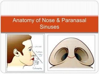
  • 1. Anatomy of Nose & Paranasal Sinuses
  • 2. Vestibule  Examination :- By Lifting the tip of the nose.  Dilated Passage way leading from external nose to the nasal fossa.  Demarcated by Limen Nasi  Superiorly lined by skin, contains hairs, sebaceous gland and sweat glands
  • 3. Furunculosis of Nose  Skin loosely attached at the dorsun and lateral sides but firmly attached at tip and the Alar cartilage – it contains sebaceous gland – Rhinophyma
  • 4. Supporting framework of external nose  Lower lateral cartilage has horse shoe shape, lateral cross is broad and strong forms alae, medial portion is weak and extends partly along the free caudal end of cartilaginous septum and partly within the membranous columella.
  • 5. Muscles of the external Nose  Nasal cavity has – medial wall, lateral wall, roof and floor. Nasal Septum (Medial wall) Septum has 3 parts  Bony  Cartilagenous  Membranous (Columella)
  • 6. Lateral Wall  Most important for FESS  Osteomeatal complex
  • 7. Roof  Upper and lower lateral cartilage.  Nasal bones  Nasal process of frontal bones  body of ethmoid  body of sphenoid  Cribiform plate – Major part of roof.  Transmits the filament of the olfactory nerve, which distributes in the mucous membrane covering the upper part of the nose  2-4 cm2 area
  • 8. Floor  Palatine process of maxilla  Horizontal process of palatine bone
  • 9. Turbinates  Inferior turbinates – largest, separate bones, bones of the inferior turbinate is deeply pitted, cellular, surface of the turbinate is perforated by blood vessels.  (Turbinectomy - Turbinoplasty)
  • 10. Inferior Meatus – Nasolacrimal duct (Hasner's Valve) Middle turbinate  Smaller then inferior turbinate  Stroma of the middle turbinate contains many gland  Key structure in FESS
  • 11. Variations of middle turbinate  Concha bullosa  Paradoxical MT  Double Middle turbinate  Polypoidal change in MT
  • 12. Middle Meatus Osteomeatal complex  All the anterior group of sinuses (Maxillary, anterior ethmoid, frontal sinus) drains.  Deep crescentric groove hidden by the MT is infundibulum  Fissure leading from MT into infundibulum is hiatus semilunaris  Ethmoidal bulla – bulge from anterior ethmoid cells.
  • 13.  Superior turbinate – not visualized by anterior rhinoscopy or nasal endoscopy.  Smallest of all the turbinates  Landmark for the ostium of sphenoid sinus
  • 14.  Superior Meatus  Posterior ethmoid drains  Supreme turbinate occasionally present  Sphenoethmoidal recess – above and behind the superior turbinate – sphenoid sinus opens
  • 15.  Functional Anatomy  Respiration  Olfaction  Paranasal sinuses  Frontal Sinuses  Ethmoid Sinuses  Maxillary Sinuses  Sphenoid Sinuses
  • 16. Maxillary Sinus (Antrum of Highmore)  Largests of all the sinuses  Presence since birth  At birth floor of maxillary sinuses is above the nasal floor, descends continuously until it reaches the age of 8 yrs – Same level
  • 17.  At the age of 18yrs floor of maxillary sinus is below the floor of nasal cavity.  Shape- Pyramidal, base towards the nasal cavity, apex towards the zygoma.  Natural osteum of maxillary sinus opens in middle meatus.  Anti-gravity drainage, cilliary movement is towards the natural osteum in middle meatus.  Inferior meatal anstrostomy – non physiological
  • 18. Relation  Roof – floor of orbit – infraorbital nerve  Blow out # - entrapment of inferior rectus muscles  Medial wall – lateral wall of nose  Anterolateral wall – bone undernith cheek  Posterior wall – separates its from pterygo palatine fosa.  Floor – upper alveolus and hard palate  2nd premolar and 1st & 2nd molar in relation with maxillary antrum Chronic Maxillary Sinusitis
  • 19. Sphenoid Sinus  Small before 3yrs but fully develop by 12 – 15 yrs.  Situated within the sphenoid bone, variable in size and shape  Separated from each other by bony septum. Communicates with superior meatus by small aperture which opens into spheno ethmoidal rescess. Size varies – 0.5 – 4 mm  Anti-gravity drainage
  • 20. Relation  Anteriorly – posterior ethmoid cells  Posteriorly – Posterior cranial fossa  Superiorly – Anterior and middle cranial fossa  Optic chiasma anteriorly and pituitary fossa posteriorly.  ICA, optic nerve and cavernous sinus laterally  Pituitory fossa – transnasal / transsphenoidal hypophysectomy
  • 21. Ethmoid Sinus  Ethmoid cells lies in either side just lateral to the superior 1 half of the nasal cavity medial to bony orbit. Ethmoid bones have horizontal plates and vertical plates that are at right angle with each other.  Vertical portion has superior part called crista galli and inferior part called perpendicular plate of ethmoid.  The horizontal plate is comprised of medial portion – cribiform plate and lateral portion that forms the roof of ethmoid.
  • 22. Contd….. Two groups of ethmoid air cells  anterior group  Posterior group Separated by basal laella  Anterior group drains to middle meatus  Posterior group drains to superior meatus Clinically – attachment of middle turbinate to lateral nasal wall marks the
  • 23.  Anterior group lies infront and posterior group lies above and behind  The two group differ in size, usually the posterior ethmoidal cells are fewer in number 3-7 and larger in size. Contd…..
  • 24. Mucoele in children  It is relatively well developed at birth.  It has a close relation with orbit (Lamina papyracea) and anterior cranial fossa, infection may spread easily to these structure.  During FESS in inexperience hand there is a chance of injury to these area.
  • 25. Frontal Sinus  Absent at birth, rudimentary till the age of 7 yrs.  Rarely identical, varies greatly in size and shape.  Bony septa may divide sinus into one or more compartment.  Occasionally it may not phenumatize at all.  It drains to middle meatus through fronto- nasal duct.  The duct is tortuous bony passage runs through the anterior ethmoid sinus down to
  • 26. Contd…..  It is because of its tortuosity and length the duct gets block to form frontal mucoele.  Frontal sinus has anterior table, posterior table (which also forms the roof) and floor  Relation – posteriorly –frontal lobe  Inferiorly – roof of ethmoid medially and roof of orbit laterally.  Inferiorly in the floor the fronto-nasal duct, its anterior
  • 27. Nasal Mucus membrane  Nasal cavity, nasopharynx and sinuses are lined by pseudo stratified columnar ciliated epithelium (Respiratory type)  The vestibula area of nasal cavity is lined by cuboidal epithelium  Area above the superior turbinate is lined by olfactory epithelim – it is brownish in color, confined to cribiform plate, it extends medially to septum and laterally to superior turbinate. Olfactory nerve may get damage by head injury – loss of smell (anosmia)
  • 28. Nerve supply Nose and paranasal sinuses has  Sensory  autonomic  specific sensory (olfactory) Sensory: Through the ophthalmic and maxillary division 5th nerve Autonomic: the secretory gland of the nasal mucosa are under the control of autonomic nervous system via the nerve of pterygoid canal (vidian nerve), which contains both sympathetic fibres and para sympathetic fibres.
  • 29. Contd……  Sympathetic stimulation constrict the mucosal vessel, reduces the size of turbinate – enlarging airway. Parasympathetic stimulation reverse the condition.  Olfactory nerve fibre  The bipolar olfactory cells that lies in the olfactory epithelium are the first order of neurons. Axons from these combined to form about 20 olfactory nerves.  These nerves pass through the cribiform plate to relay at the olfactory bulb.  The neurons from the bulb run in the olfactory tract to the 2nd olfactory centre in the frontal
  • 30. Blood Supply of Nasal cavity  Recurrent posterior epistaxis – sphenopalatine artery ligation. Venous drainage –  Venous drainage of nose and paranasal sinuses is to the ophthalmic vein, facial vein and to the pterygoid and pharyngeal plexus.  Cavernous sinus thrombophlebitis
  • 31. Lymphatic drainage  The anterior part of the nose and the sinuses drains to submandibular lymphnode and to deep cervical chain.  Posterior part of nose and posterior sinuses drain to retropharyngeal lymph node.  Functions of paranasal sinuses  air conditioning  pressure damping  reduction of skull weight  heat insulation  increase the olfactory area  vocal resonance  mucus production