SlideShare una empresa de Scribd logo
1 de 19
Descargar para leer sin conexión
Module 1 Microbiology
STAPHYLOCOCCI
1. To what family of bacteria do the Staphylococci belong?
• Staphylococcus belongs to the family Micrococcaceae
2. What tests may be performed to differentiate Micrococci from Staphylococci?
o Differentiation of staphylococci and micrococci:
o Oxidation fermentation
 Staphylococci ferment glucose
 Micrococci do not ferment glucose
o Oxidase test
 Rapidly differentiates staphylococci from micrococci
• Most staphylococci will be negative
• Micrococci will be positive
o Bacitracin susceptibility test
 Micrococcus are susceptible
 Staphylococcus spp. are resistant
Differentiation Between Staphylococci and Micrococci

3. Name two tests other than coagulase which may be used to differentiate S. aureus from S.
epidermidis.
• Hemolysis
o S. aureus produces B hemolytic zones and yellow pigment production with extended incubation
o S. epidermidis is nonhemolytic (may be weakly hemolytic
• PYR
o Used to distinguish S. aureus from other CNS
• Oxacillin Disks
o Used with S. aureus to detect the presence of B-lactamase

Page 1
Microbiology
4. List 4 disease processes involving Staphylococcus aureus and 2 involving S. epidermidis.
• S. aureus
o Folliculitis
 Mild inflammation of a hair follicle or oil gland; red and raised
o Furuncles (boils)
 Large, raised, superficial abscesses
o Carbuncles
 Larger abscess, involve multiple furuncles, more invasive, fever & chills, systemic
spreading of bacteria
o Bullous impetigo
 Highly contagious spread by direct contact, fomites or autoinoculation; large pustules
surrounded by small zone of erythema
o Toxic Shock Syndrome
• S. epidermidis
o UTIs
 Usually nosocomial infections (usually involve development of biofilm
o Endocarditis
5. Why are stools sometimes cultured for Staphylococci?
• For the detection of Staphylococcus aureus food poisoning which can lead to Staphylococcal enteritis
a stool culture may be required.
o A stool culture is used to detect the presence of disease-causing bacteria and help diagnose
an infection of the digestive tract.
o In the case of Staphylococcal enteritis it is conducted to see if the stool is positive for a
pathogenic bacterium
6. List types of toxins produced by Staphylococci and discuss their roles in disease processes.
• Produced by S. aureus
o Exfoliative toxin
 AKA epidermolytic toxin
 Causes the epidermal layer to slough off and known to cause SSS (scalded skin
syndrome)
 Scalded Skin Syndrome
• Also called Ritter disease
• Extensive exfoliative dermatitis primarily in newborns, young children and adults
with renal failure
• Caused by staphylococcal exfoliative toxin in a lesion located elsewhere on the
body
• Ranges from bullous impetigo (somewhat localized) to a more generalized
condition
o Cytolytic toxins
 Four types of hemolysins (affect RBCs)
 α, β, γ, δ hemolysins
o Protein A
 Identified in the cell wall of S. aureus
 Able to bind to Fc on IgG (neutralizes it) and blocks phagocytosis
o Enterotoxins A, D, or B
 Food poisoning
• Caused by ingestion of S. aureus enterotoxins A, D, or B
o Improperly handled food allowing bacteria and toxin production to
continue
Page 2
Microbiology
7. Discuss disease processes involving Staphylococcus saprophyticus. How is it differentiated from
other Staphylococci?
• S. saprophyticus disease
o Urinary tract infections in young women
 Can be found in low numbers but still significant
• Identification (S. saprophyticus)
o Colony Morphology
 Slightly larger colony
 Half strains produce yellow pigment on BAP
• Presumptive Identification of S. saprophyticus
o Novobiocin
 S. saprophyticus is resistant to novobiocin
8. Discuss the role of Protein A of S. aureus used as a reagent in rapid identification kits, such as
Staphylococcus typing kits.
• Protein A
o One of several cellular components that have been identified in the cell wall of S. aureus.
o Most significant role of protein A in infections caused by S. aureus is its ability to bind the Fc
portion of IgG
 Binding IgG in this manner neutralizes IgG and can block phagocytosis.
• Rapid identification kits
o These kits use plasma-coated carrier particles, such as latex.
o The plasma detects both clumping factor (with fibrinogen) and protein A in the cell wall of S.
aureus (with IgG).
o These kits often have a higher specificity and sensitivity than the traditional plasma slide test
and are commonly used in clinical laboratories.
o They are particularly useful for the identification of MRSA (methicillin-resistant Staphylococcus
aureus) that is often weakly or negative in the slide coagulase test
 Some strains of S. saprophyticus, S. sciuri, S. lugdunensis, and Micrococcus spp. may
produce positive tests with the latex-coated assays, but they would be tube coagulase
negative on further testing.
o Procedure
 Slide coagulase test = used to ID S. aureus
• A heavy suspension of organism is prepared on a glass slide in water or saline
and mixed with a drop of plasma (human, pig or rabbit)
o If clumping = positive, ID as S. aureus following additional testing
• Coagulase can also be performed in a tube method;
o Detects staphylocoagulase (free coagulase)
 Staphylocoagulase is an extracellular molecule that causes a clot
to form when bacteria are incubated with plasma

Page 3
Microbiology
9. What is the gram reaction and morphology of Staphylococci?
• Gram reaction
o Gram positive
• Morphology
o Cocci that can appear singly, in pairs, or in clusters
o Non-motile
o Non-spore-forming
o Aerobic or facultatively anaerobic4
 Except S. saccharolyticus
o Medium, cream or white colored colonies (buttery looking)
o Some species are β hemolytic
10. What two methods are used for coagulase testing? What does each measure?
• Coagulase test
o Staphylocoagulase is the active enzyme
o Presumptive identification for S. aureus
o CoNS means coagulase-negative staphylococci
• Two methods
o Slide coagulase test
 Detects:
• Clumping factor
 Because about 5% of S. aureus organisms do not produce clumping factor, any
negative slide coagulase test result must be confirmed with the tube method, which
detects staphylocoagulase, or free coagulase.
 Procedure
• Heavy suspension of the suspected organism is prepared on a glass slide in
water or saline and is mixed with a drop of plasma.
• If agglutination occurs, the isolate can be identified as S. aureus.
• Some strains of S. lugdunensis and S. schleiferi are also positive for clumping
factor.
o Tube coagulase test
 Detects:
• Staphylocoagulase (free coagulase
o Staphylocoagulase is an extracellular molecule that causes a clot to form
when bacterial cells are incubated with plasma
o Staphylocoagulase reacts with a thermostable, thrombin-like molecule
called coagulase-reacting factor (CRF) to form coagulase-CRF complex.
o The complex resembles thrombin and indirectly converts fibrinogen to
fibrin.
o The clot formed in the tube may have a tendency to undergo autolysis
(because of fibrinolysin), giving the appearance of a negative result.
o Clinical laboratorians should look for clot formation after 4 hours of
incubation at 37° C.
 If no clot appears, the tube should be left at room temperature
and checked the following day.
o False negative result
 Fibrinolysin activity is enhanced at 37° C which might result in a
false negative result.
 Will differentiate other coag positive staphylococci species from S. aureus
 Staphylococcus species that are capable of producing coagulase include S. aureus
(potentially pathogenic in humans and animals) and the animal isolates S. intermedius
Page 4
Microbiology
and S. hyicus
CNS = coagulase negative staphylococci
11. Identify two body sites where Staphylococcus may be found as part of the normal flora.
• Primary reservoir
o Nares
• Secondary
o Axillae
o Vagina
o Pharynx
o Other skin surfaces
12. Discuss beta lactamase production and its role in the development of penicillin resistant
Staphylococcus.
• Penicillin resistance due to β-lactamases
o S. aureus produce β-lactamases
 Production of β-lactamases break down the β-lactam ring of many penicillins
 Most S. aureus isolates are resistant to penicillin.
 Increasing resistance to alternative antimicrobial agents is a major concern.
13. What class of antibiotics is generally used to treat methicillin resistant Staphylococcus aureus
infections?
• Vancomycin is used to treat methicillin-resistant staphylococcus
14. Describe the term M.R.S.A. How are these organisms detected?
• Methicillin-Resistant Staphylococci
o Organisms resistant to oxacillin or nafcillin are termed MR
 Can be MRSA, MRSE
• CA-MRSA = community associated
o Seen in athletes, inmates, military recruits in close quarters, pediatric
patients, tattoo recipients
• HA-MRSA = hospital-associated
o Risk factors such as recent hospitalization, long-term care, dialysis or
indwelling devices

Page 5
Microbiology
STREPTOCOCCI
1. Describe the appearance of the three major types of hemolysis produced by Streptococci.
• Alpha α
o Partial lysis of RBCs resulting in greenish pigment of agar surrounding colone
• Beta β
o Complete lysis of RBCs clearing surrounding colony
•

Gamma γ
o Lack of lysis of RBCs, no change in agar

2. Discuss Streptolysin S and O and describe 2 methods used to demonstrate these hemolytic
properties.
• Streptolysin O or S
• Responsible for hemolysis of agar RBCs
o O = oxygen labile
 Occurs when incubated anaerobically
o S = oxygen stable
 Occurs when incubated aerobically
• Can test patients for the presence of antistreptolysin O (ASO) to check for recent strep infection
3. Name two diseases which may appear as sequelae to Group A Streptococcus infection.
• Rheumatic fever
o Typically follow pharyngitis
• Acute glomerulonephritis
o Sometimes occurs after a cutaneous or pharyngitis infection
4. Name two disease processes in which Streptococcus pneumoniae may be found as the etiological
agent.
• Otitis media
o Kids under 3
• Bacterial pneumonia
o Mainly elderly or immunocompromised
Page 6
Microbiology
5. What is the gram reaction and morphology of:
a. Streptococci agalactiae
• Colonies appear grayish white and mucoid on BAP and have small zone of β hemolysis
• Organisms appear as GPC in chains
• Capsule prevents phagocytosis but is ineffective after opsonization
b. Streptococcus pneumoniae
• Presence of capsule
• Gram positive
• Catalase negative
6. What is the best single test for differentiating Staphylococci from Streptococci? What interference
do RBCs have in this test?
• Catalase test
o Negative with streptococcus
o Positive with staphylococcus
7. Name 2 disease processes associated with each of the following groups of streptococci and a test
or tests used for routine biochemical identification:
Associated Diseases
a. Group A
(strep. pyogenes)

Biochemical
Identification

•
•
•
•
•

Scarlet fever
Bacterial pharyngitis
Pyodermal (skin) infections
Necrotizing fasciitis
Streptococcal Toxic shock
syndrome
Post-streptococcal sequelae
Acute glomerulonephritis

•
•

Bacitracin
susceptibility
PYR

Invasive disease of newborn
• Early onset <7d old
• Late onset > 7d old
Pregnant women should be
screened at 35 – 37 wks.
gestation

•
•

CAMP test
Hippurate hydrolysis

•
•
b. Group B
(strep agalactiae)

•
•

c. Group D
non-enterococcus

•

Wounds, eyes, ears (not big
deal)

•
•
•

Bile esculin
PYR
Salt Tolerance

d. Enterococcus

•

Nosocomial infections

•
•

Bile esculin
Salt tolerance

e. Streptococcus viridans

•
•
•
•
•
•

Bacterial endocarditis
Oral infections (gingivitis)
Meningitis/Septicemia
Abscesses
Bacterial pneumonia
Otitis media

•
•

Neg for optochin
Neg for Bile solubility

•
•

Optochin (P disk)
Bile solubility

f. Streptococcus
pneumoniae

Page 7
Microbiology
8. Name two tests used to differentiate pneumococci from other alpha hemolytic streptococci.
• Optochin (P disk)
o Presumptive ID os S. pneumoniae
o Zone of inhibition = susceptible
• Bile solubility
o Further identification of S. pneumoniae
9. What colonial characteristic helps to distinguish Strep pneumoniae on a blood agar plate?
•
•

α hemolysis – partial lysing of RBCs that results in green discoloration of the agar
Additionally shape of colony (concave red blood cells)

10. What is the drug of choice for treatment of most streptococcal infections?
• Penicillin can be used to treat most streptococci
o Penicillin-resistant S. pneumoniae and viridans
 Use erythromycin and narrow spectrum cephalosporins
• Vancomycin is effective against GPC
o Not currently a widespread resistance
11. Describe the PYR test and what organisms it is used to identify.
• Presumptive ID for Group A and Group D
o Group A = only strep that is positive
o Enterococcus = also PYR positive
o More specific for Group A than bacitracin
12. Describe the term V.R.E. How are these organisms detected?
• VRE = vancomycin resistant enterococci
• Molecular typing methods, such as pulsed-field gel electrophoresis, contour-clamped
homogeneous electric-field electrophoresis, ribotyping and PCR-based typing methods have
been used mainly to type enterococcal species in epidemiologic studies and investigations of
vancomycin-resistant enterococci (VRE).

Page 8
Microbiology
HAEMOPHILUS
1. (L) What is the satellite phenomenon?
• Growth of a fastidious organism around other bacteria that release necessary growth factors
• Hemolytic S. aureus, S. pneumoniae, Neisseria species release the factors or naturally produce V
factor
o H. ducreyi is exception
2. (L) Name a disease process from which each of the following may be isolated:
a. H. influenzae
• Haemophilus influenzae serotype b (Hib)
o Primarily causes disease in children
o Hib vaccine is useful in reducing incidence of disease
o Causes bacteremia and then spreads to other tissues
• Disease of Hib strains
o Meningitis
o Epiglottitis
o Bacterial tracheitis
o Cellulitis
o Acute pharyngitis and pneumonia
b. Haemophilus-aegyptius
• Conjunctivitis
o Pink eye
c. H. ducreyi
• Sexually transmitted infection that can cause chancroid
d. Gardnerella vaginalis
• Bacterial vaginosis
3. What is the gram stain and morphology of Haemophilus species?
• Gram negative
• Pleomorphic coccobacilli or rods
• Biochemical reactions
o Nonmotile
o Oxidase positive
o Catalase positive
o Nitrate reduction
4. What are the X and V factors?
• Haemophilus requires X and V factors
o X factor
 Hemin or hematin
o V factor
 Nicotinamide-adenine dinucleotide (NAD)
• Para – require only V factor
o Haemophilus parainfluenzae
 Produces X factor but requires V factor
• Sheep blood agar contains only X factor NOT V factor

Page 9
Microbiology
5. Explain the importance of doing beta-lactamase tests.
• Checking for resistance to beta-lactams
• Nitrocefin test
o Put on glass slide with drop of water
o Put disc on there
o Put loop of organism on there and changes to red
6. What tests and media are used for screening for and identifying G. vaginalis?
• Vaginal discharge collected from suspected BV cases is the most common specimen used for the
isolation of G. vaginalis.
• Because it is part of the urogenital microbiota, the organism can also be isolated from urine.
• Because G. vaginalis can be found as normal vaginal biota and its role in BV is questionable, cultures
for G. vaginalis are infrequently performed.
• It often takes longer than 24 hours to develop visible colonies, and G. vaginalis grows best in 5% to 7%
CO2 at a temperature of 35° to 37° C.
• G. vaginalis grows on SBA as pinpoint, nonhemolytic colonies.
• It will also grow on chocolate agar.
• The medium of choice for G. vaginalis, however, is human blood bilayer Tween (HBT) agar.
• When cultured on human blood, colonies are β-hemolytic, small, gray, and opaque.
• G. vaginalis will also produce β-hemolytic colonies on media made with rabbit blood.

Page 10
Microbiology
NEISSERIA / MORAXELLA
1. Name 2 pathogenic species of Neisseria and an important disease that each causes and 1 species
of Moraxella.
• Two pathogenic Neisseria spp.
o N. gonorrhoeae
 Always pathogenic when present
o N. meningitidis
 May be found as normal flora in respiratory tract of carriers
2. What is the gram reaction and morphology of Neisseria species?
• Aerobic, nonmotile, non-spore-forming, oxidase positive, catalase positive, GNDC
• Requires enriched media to grow
3. What is one simple test that aids in the identification of Neisseria species?
• Presumptive Identification
o Colonial Morphology
o Small, tan, translucent and raised after 24-48hrs
o Fresh subculture should be used for ID tests
• Definitive Identification
o Immunologic Assays
 Coagglutination tests using antibodies against N. gonorrhoeae fused to killed S.
aureus cells; agglutination is a positive reaction
o Nucleic Acid Assays
 See Table 17-4; able to detect organism directly from specimens
• Treatment
o Cephalosporins are currently the drug of choice
o Often used in combination with other drugs if Chlamydia is also present
4. Name several sources from which each of these species can be isolated.
• Neisseria meningitidis
o Normal flora in nasopharynx or oropharynx
• Neisseria gonorrhea
o Clinical Infections
o Acute, pyogenic infection, primarily acquired by sexual contact & infect urethra, endocervix,
anal canal, pharynx and conjunctiva
o Short incubation period of 2-7 days
o Men = acute urethritis with purulent discharge and painful urination
 Only 3-5% will be asymptomatic
o Women = cervical discharge, lower abdominal pain & dysuria
 As many as 50% may be asymptomatic carriers
 Pelvic Inflammatory Disease – caused by untreated gonococcal cervicitis (can cause
sterility & ectopic pregnancy)

Page 11
Microbiology
5. What factors have to do with the viability of Neisseria meningitidis?
• Humans are its only host
• Requires enriched media
• Virulence Factors (same for meningitidis and gonorrhoeae)
• Receptors for human transferrin
• Capsule
• Pili (fimbriae)
o Inhibit phagocytosis; aid in the initial attachment to host tissues
o Aid in the exchange of genetic material between cells
• Cell membrane proteins
o IgA protease
o Lipooligosaccharide (LOS) endotoxin
 Mediates damage to host tissues and induces inflammatory response
• Culture
o Requires the use of chocolate agar
o But chocolate will allow other organisms to grow
o Media should be allowed to come to room temp due to Neisseria’s sensitivity to temperature
changes
• Incubation
o Plates should be incubated at 35C in 5% CO2; also prefer increased humidity
6. When is M. catarrhalis implicated in disease? What screening test differentiates it from Neisseria?
• Clinical Infections
o An opportunistic pathogen that causes URT infections in children and elderly
o The third most common cause of acute otitis media and sinusitis in children
• Intracellular GNDC, most produce β-lactamase (makes them resistant to ampicillin & amoxicillin)

Page 12
Microbiology
MISCELLANEOUS GRAM NEGATIVE RODS
1. Identify the source from which most infections with Pasteurella multocida can be attributed and
name clinical specimens where it may be found.
• Colonize the URT and GI tract of mammals and birds
o Exposure from cat and dog bites or scratches
2.

Name the causative organism of whooping cough and the medium used for primary isolation.
• Bordetella pertussis and Bordetella parapertussis
• Specimens are best from the nasopharynx
o Aspirates of fluid
o Calcium alginate or Dacron swabs up each nasal passage as deep as possible
• Culture
o BAP and MAC agar
o Chocolate if you need to rule out Haemophilus
• Incubate plates
o 35C in aerobic conditions
o 7 days
o Adequate moisture to prevent plate drying

3. Name the causative organism of tularemia and discuss the primary reason for not culturing it in the
laboratory.
• Francisella spp
• Diagnosis is from serology because culture is less safe
4. Describe the gram stain morphology of Pasteurella.
• Gram negative pleomorphic coccobacilli with bipolar staining
o Oval shaped, short rods, longer filaments
o Grow on BAP and chocolate agar
 Nonhemolytic mucoid colonies with a narrow green or brown halo
5. Discuss the fluorescent antibody techniques used for diagnosis of whooping cough (pertussis)
and name 2 suitable specimens.
• Nasopharyngeal swab with booger juice
• Put this on glass slide
• Adding fluorescent monoclonal tag to this
• If there is binding we know the organism is there due to the presence of fluorescent

Page 13
Microbiology
ENTEROBACTERIACEAE
1. What is the definition of the family Enterobacteriaceae?
• Enterics: Members of the family Eterobacteriaceae consisting of organisms that all ferment glucose,
nearly all fail to produce the cytochrome oxidase, and nearly all reduce nitrates to nitrites. Most are
resident flora of the gastrointestinal tract.
•
•

•
•
•
•
•
•
•
•

Includes many genera and species
Common clinical isolates include:
o E. coli
o Klebsiella pneumoniae
o Proteus mirabilis
A wide variety of differential and selective media, such as MacConkey (MAC) agar, and highly
selective media, such as Hektoen enteric (HE) agar and xylose-lysine-desoxycholate (XLD) agar, are
available for the presumptive identification of enteric pathogens.
These media contain one or more carbohydrates, such as lactose and sucrose, which show the ability
of the species to ferment specific carbohydrates.
Fermentation is indicated by a color change on the medium, which results from a drop in pH detected
by a pH indicator incorporated into the medium.
Nonfermenting species are differentiated by lack of color change, and colonies retain the original color
of the medium.
Species that produce hydrogen sulfide (H2S) may be readily distinguished when placed on HE or XLD
agar.
HE and XLD agars contain sodium thiosulfate and ferric ammonium citrate, which produce blackening
of H2S-producing colonies.
These features have been used to initially differentiate and characterize certain genera.
Definitive identification depends on the biochemical reactions and serologic antigenic structures
demonstrated by the particular species.

Page 14
Microbiology
2. Why must a TSI slant be read within 18-24 hours?
• Enterics: Members of the family Eterobacteriaceae consisting of organisms that all ferment glucose,
nearly all fail to produce the cytochrome oxidase, and nearly all reduce nitrates to nitrites. Most are
resident flora of the gastrointestinal tract.
o
o

Reading the results after fewer than 12 hours of incubation gives the false appearance of an organism
capable of fermenting glucose and lactose (or sucrose in the case of TSI agar).
For this reason, TSI agar or KIA must be incubated for 18 to 24 hours.

3.

What is meant by K antigens? somatic antigens? flagellar antigens?
• K antigen, or capsular antigen—is a heat-labile polysacharide found only in certain encapsulated
species.
o Examples
 K1 antigen of E. coli
 Vi antigen of Salmonella enterica
• O antigen, or somatic antigen—is a heat-stable antigen located on the cell wall
• H antigen, or flagellar antigen—is a heat-labile antigen found on the surface of flagella, structures
responsible for motility.

4.

What is the Vi antigen? How do you destroy this antigen? What is the purpose of destroying the Vi
antigen?.
• Capsular antigen of Salmonella typhi
o Heat labile

5. Describe disease processes caused by enterotoxigenic E. coli (ETEC), enteropathogenic E. coli
(EPEC) and enterohemorrhagic E. coli.
•

ETEC- Enterotoxigenic E. coli (ETEC) Diarrhea (Traveler’s Diarrhea). This disease consists of a watery
diarrhea with or without mucus or blood, vomiting, and abdominal cramping. Dehydration and lowgrade fever may occur. Enterotoxigenic strains of E. coli are the most common cause of traveler’s
diarrhea worldwide and a common cause of diarrheal disease in children in developing countries.

•

EPEC

•

EHEC- Enterohemorrhagic E. coli (EHEC) Diarrhea. This disease consists of a hemorrhagic, watery
diarrhea with abdominal cramping. Usually, patients have no fever or only a slight fever. About 5% of
infected people (especially children younger than age 5 and the elderly) develop hemolytic- uremic
syndrome (HUS), with anemia, low platelet count, and kidney failure. The first recognized outbreak of
diarrhea caused by enterohemorrhagic E. coli (O157:H7) occurred in 1982, involving contaminated
hamburger meat—hamburger meat contaminated with cattle feces. Since then, several well-publicized
epidemics involving the same serotype have occurred. Not all of the outbreaks involved meat; some
resulted from ingestion of or unpasteurized milk or apple juice, lettuce, or other raw vegetables.

Page 15
Microbiology
6. Name the causative organism of plague. Identify the vector of this disease and three clinical forms
recognized in man..
• Yersinia pestis
o Transmitted by fleas from infected rodent
o Pneumonic plague through respiratory droplets
o Bubonic
o Glandular
7. Describe how lactose fermenters, nonlactose fermenters and H2S (+) organisms appear on the
following media: MacConkey, Hektoen, XLD (xylose-lysine-desoxycholate), and SS (SalmonellaShigella).

Media

Enteric organism Reactions
Lactose Fermenters
Non-lactose
H2S postive
Fermenters

MacConkey

Red/Pink

No change

Does not indicate

Hektoen

Yellow or Orange

No change (green is
color of agar)

Produces a black
precipitate

XLD

Yellow

Colorless or red

Black precipitate

SS

Does not indicate
lactose fermenters

Salmonella – colorless

Black

Shigella – colorless

8. Triple sugar iron agar (TSI) is used as a screening medium for the pathogenic enterics. What do
the following reactions in a TSI slant indicate with regards to fermentation or utilization of
ingredients in the agar:
TSI reactions

Indication of Fermentation or Utilization

a. Acid Butt

(yellow) ferments lactose, glucose, and/or sucrose

b. Alkaline slant

(red) does not ferment either lactose or sucrose

c. Gas bubbles in butt

Gas has been formed

d. Blackening of butt

H2S has been formed

Page 16
Microbiology
9. List possible organisms giving the following reactions on TSI agar:
Slant
Acid

Butt
Acid

H2S
Gas None

Alkaline

Acid

Gas +

Alkaline

Acid

None

Acid

Acid

Possible Organisms
E. coli,
Klebsiella spp,
Enterobactor spp.,
Serratia spp

Gas +

10. What is the IMViC profile? What are the two most common organisms which are used as positive
and negative controls for IMViC and what reactions do they give?

Indole
Methyl Red
VP
Citrate

Organism 1:
E. coli
E. coli
Enterobacter aerogenes
Klebsiella pnumoniae

Organism 2:
Klebsiella pneumoniae
Enterobacter aerogenes
E. coli
E. coli

11. Which group of enterics may produce a red pigment and at what temperature?
• Often produce a characteristic pink to red pigment, prodigiosin, especially when the cultures are
incubated at room temperature
o S. marcescens,
o S. rubidaea
o S. plymuthica
12. What is characteristic of Proteus species with regards to colony growth?
• Swarming
13. How do the colonies of E. coli generally appear on MacConkey agar?
• Pink due to lactose fermentation
Page 17
Microbiology
14. Name three Enterobacteriaceae genera considered pathogenic when isolated from stool, describe
pathogenesis of infection, and outline the procedure for their identification.
• Salmonella- non lactose fermenting, h2s producing, negative urease.
• Shigella- non lactose fermenting, non h2s producing,
• Yersinia- non lactose fermenting, positive urease
15. List 4 genera which produce H2S in TSI
• Salmonella
• Proteus
• Citrobacter
• Edwardsiella
16. Yersinia enterocolitica is generally isolated from what clinical specimen? Describe incubation
temperatures, and urease reactions of this organism.
•
•
•
•

Routinely isolated from stool,
Can survive cold temperatures
Incubation temperatures recommended to be below 37C.
A positive urease test is presumptive to identifying Yersinia

Page 18
Microbiology
AEROMONAS, VIBRIO,
CAMPYLOBACTER,
1. List 3 sources from which Aeromonas may be isolated.
• Retail produce
• Animal meat products
• Aquatic wound infections
2. Identify the mechanism by which Vibrio cholera produces disease.
• Cholera toxin also known as choleragen, once ingested the bacteria colonize the small intestine where
they multiply and produce choleragen.
• Read further on how this mechanism works on (text page 465)
3. Name the Vibrio species which are associated with the consumption of contaminated seafood. How
are they identified?
•
•
•

Vibrio parahaemolyticus
Vibrio vulnificus
Page 467-468

4. Identify the disease processes associated with Campylobacter jejuni.
• Patients with C.jejuni present with a diarrheal disease that begins with mild abdominal pain within 2-10
days after ingesting the organism.
• Cramps and bloody diarrhea often follow the initial signs.
• Illness is usually self-limiting and resolves in 2-6 days.
5. How do the Campylobacter appear on gram stain? What is their unique atmosphere requirement?
• Curved non-spore forming gram negative rods
• Enteric campylobacters may appear as long spirals, S shapes, or seagull wings
• Organisms stain poorly
• Page 478
• Anerobic, some microanerobic
6. What organism in this group is characterized by "rice water" stools?
• Vibrio cholera
7. What disease is associated with Helicobacter? Why is this organism difficult to isolate? ULCERS
•
•
•
•
•
•

Not thought to be a cause of diarrheal illness, H. pylori nonetheless is a significant cause of disease.
Living in the stomach and duodenum, H. pylori has been associated with ulcer disease.
This infection is common, in that many patients have detectable antibody against this organism.
When the organism is recovered from a gastric or duodenal ulcer, there is evidence that patients have
faster, more durable ulcer recovery with antibiotic therapy.
Active infection can be diagnosed by detecting the organism on an endoscopically derived biopsy
specimen or through breath testing
This testing is based on the fact that the organism shows high urease activity; urea labeled with either
13C or 14C will, when ingested, rapidly produce labeled CO2 in the patient's breath if the organism is
present.

Page 19

Más contenido relacionado

La actualidad más candente

Pathological examination of body fluids
Pathological examination of body fluidsPathological examination of body fluids
Pathological examination of body fluidsUtkarsh Sharma
 
Entamaoeba Histolytica (Exam Point of View)
Entamaoeba Histolytica (Exam Point of View)Entamaoeba Histolytica (Exam Point of View)
Entamaoeba Histolytica (Exam Point of View)Sijo A
 
AUTOMATED STAINING DEVICE.pdf
AUTOMATED STAINING DEVICE.pdfAUTOMATED STAINING DEVICE.pdf
AUTOMATED STAINING DEVICE.pdfFranklinEmitati
 
Sample Collection
Sample CollectionSample Collection
Sample CollectionYESANNA
 
Thrombocytopenia
ThrombocytopeniaThrombocytopenia
ThrombocytopeniaPriya
 
Donor selection ppt
Donor selection pptDonor selection ppt
Donor selection pptdas nelaturi
 
Identification of bacteria
Identification of bacteriaIdentification of bacteria
Identification of bacteriavinaya warad
 
Stool occult blood test
Stool occult blood testStool occult blood test
Stool occult blood testAniah Marcelo
 
Safety and Infection control in hematology laboratory
Safety and Infection control in hematology laboratorySafety and Infection control in hematology laboratory
Safety and Infection control in hematology laboratoryNegash Alamin
 
L13 medical microbiology laboratory (staphylococcus spp.)
L13   medical microbiology laboratory (staphylococcus spp.)L13   medical microbiology laboratory (staphylococcus spp.)
L13 medical microbiology laboratory (staphylococcus spp.)Hussein Al-tameemi
 

La actualidad más candente (20)

Pathological examination of body fluids
Pathological examination of body fluidsPathological examination of body fluids
Pathological examination of body fluids
 
Museum
MuseumMuseum
Museum
 
culture media
culture mediaculture media
culture media
 
Salmonella
SalmonellaSalmonella
Salmonella
 
Entamaoeba Histolytica (Exam Point of View)
Entamaoeba Histolytica (Exam Point of View)Entamaoeba Histolytica (Exam Point of View)
Entamaoeba Histolytica (Exam Point of View)
 
biology behind malaria
biology behind malaria biology behind malaria
biology behind malaria
 
SCHISTONOMA MANSONI
SCHISTONOMA MANSONISCHISTONOMA MANSONI
SCHISTONOMA MANSONI
 
Stool Examinations
Stool ExaminationsStool Examinations
Stool Examinations
 
AUTOMATED STAINING DEVICE.pdf
AUTOMATED STAINING DEVICE.pdfAUTOMATED STAINING DEVICE.pdf
AUTOMATED STAINING DEVICE.pdf
 
Sample Collection
Sample CollectionSample Collection
Sample Collection
 
Thrombocytopenia
ThrombocytopeniaThrombocytopenia
Thrombocytopenia
 
Donor selection ppt
Donor selection pptDonor selection ppt
Donor selection ppt
 
Identification of bacteria
Identification of bacteriaIdentification of bacteria
Identification of bacteria
 
Stool occult blood test
Stool occult blood testStool occult blood test
Stool occult blood test
 
APTT.pptx
APTT.pptxAPTT.pptx
APTT.pptx
 
Klebsiella spp
Klebsiella sppKlebsiella spp
Klebsiella spp
 
Streptococci.ppt
Streptococci.pptStreptococci.ppt
Streptococci.ppt
 
Hypogammaglobulinemia
HypogammaglobulinemiaHypogammaglobulinemia
Hypogammaglobulinemia
 
Safety and Infection control in hematology laboratory
Safety and Infection control in hematology laboratorySafety and Infection control in hematology laboratory
Safety and Infection control in hematology laboratory
 
L13 medical microbiology laboratory (staphylococcus spp.)
L13   medical microbiology laboratory (staphylococcus spp.)L13   medical microbiology laboratory (staphylococcus spp.)
L13 medical microbiology laboratory (staphylococcus spp.)
 

Destacado

1. must to know in clinical chemistry 001
1. must to know in clinical chemistry 0011. must to know in clinical chemistry 001
1. must to know in clinical chemistry 001Nhil Abaya
 
Clinical chemistry review sheet for mlt certification and ascp
Clinical chemistry review sheet for mlt certification and ascpClinical chemistry review sheet for mlt certification and ascp
Clinical chemistry review sheet for mlt certification and ascpDonna Kim
 
Compilation of previous board examination questions
Compilation of previous board examination questionsCompilation of previous board examination questions
Compilation of previous board examination questionsRhem Rick Corpuz
 
Medical Laboratory Scientist-microbiology summary
Medical Laboratory Scientist-microbiology summaryMedical Laboratory Scientist-microbiology summary
Medical Laboratory Scientist-microbiology summaryProtegeNithi
 
clinical chemistry
clinical chemistry clinical chemistry
clinical chemistry Moha Haji
 
Introduction to clinical chemistry
Introduction to clinical chemistryIntroduction to clinical chemistry
Introduction to clinical chemistrygoogle
 
Clinical chemistry lecture slide show
Clinical chemistry lecture slide showClinical chemistry lecture slide show
Clinical chemistry lecture slide showGhie Santos
 
Quiz maker - plant and animal cell
Quiz maker - plant and animal cellQuiz maker - plant and animal cell
Quiz maker - plant and animal cellsandhyaprabha
 
laboratory diagnosis of staphylococcus
laboratory diagnosis of staphylococcuslaboratory diagnosis of staphylococcus
laboratory diagnosis of staphylococcusSHALINI BISHT
 
Ln hematology mlt_final
Ln hematology mlt_finalLn hematology mlt_final
Ln hematology mlt_finalRohit Kumar
 
Morphology and general and general properties of fungi
Morphology and general and general properties of fungiMorphology and general and general properties of fungi
Morphology and general and general properties of fungiMicrobiology
 
TURKISH POWER SYSTEM AND RENEWABLE ENERGY IN TURKEY
TURKISH POWER SYSTEM AND RENEWABLE ENERGY IN TURKEYTURKISH POWER SYSTEM AND RENEWABLE ENERGY IN TURKEY
TURKISH POWER SYSTEM AND RENEWABLE ENERGY IN TURKEYSuat Furkan ISIK
 
Microbiology - Agents of Bioterrorism
Microbiology - Agents of BioterrorismMicrobiology - Agents of Bioterrorism
Microbiology - Agents of BioterrorismDonna Kim
 
Yersinia pasteurella fransicella
Yersinia pasteurella fransicellaYersinia pasteurella fransicella
Yersinia pasteurella fransicellaRiyaz Sheriff
 
Culture media & culture methods copy
Culture media & culture methods   copyCulture media & culture methods   copy
Culture media & culture methods copyNagapavan Edara
 
4immunohematologylab
4immunohematologylab4immunohematologylab
4immunohematologylabMiami Dade
 

Destacado (19)

1. must to know in clinical chemistry 001
1. must to know in clinical chemistry 0011. must to know in clinical chemistry 001
1. must to know in clinical chemistry 001
 
Clinical chemistry review sheet for mlt certification and ascp
Clinical chemistry review sheet for mlt certification and ascpClinical chemistry review sheet for mlt certification and ascp
Clinical chemistry review sheet for mlt certification and ascp
 
Compilation of previous board examination questions
Compilation of previous board examination questionsCompilation of previous board examination questions
Compilation of previous board examination questions
 
Microbiology Q & A for exam (short note)
Microbiology Q & A  for exam (short note)Microbiology Q & A  for exam (short note)
Microbiology Q & A for exam (short note)
 
Medical Laboratory Scientist-microbiology summary
Medical Laboratory Scientist-microbiology summaryMedical Laboratory Scientist-microbiology summary
Medical Laboratory Scientist-microbiology summary
 
clinical chemistry
clinical chemistry clinical chemistry
clinical chemistry
 
Introduction to clinical chemistry
Introduction to clinical chemistryIntroduction to clinical chemistry
Introduction to clinical chemistry
 
Clinical chemistry lecture slide show
Clinical chemistry lecture slide showClinical chemistry lecture slide show
Clinical chemistry lecture slide show
 
Quiz maker - plant and animal cell
Quiz maker - plant and animal cellQuiz maker - plant and animal cell
Quiz maker - plant and animal cell
 
Quiz by yp-with answers
Quiz by yp-with answersQuiz by yp-with answers
Quiz by yp-with answers
 
laboratory diagnosis of staphylococcus
laboratory diagnosis of staphylococcuslaboratory diagnosis of staphylococcus
laboratory diagnosis of staphylococcus
 
MLT Resume4
MLT Resume4MLT Resume4
MLT Resume4
 
Ln hematology mlt_final
Ln hematology mlt_finalLn hematology mlt_final
Ln hematology mlt_final
 
Morphology and general and general properties of fungi
Morphology and general and general properties of fungiMorphology and general and general properties of fungi
Morphology and general and general properties of fungi
 
TURKISH POWER SYSTEM AND RENEWABLE ENERGY IN TURKEY
TURKISH POWER SYSTEM AND RENEWABLE ENERGY IN TURKEYTURKISH POWER SYSTEM AND RENEWABLE ENERGY IN TURKEY
TURKISH POWER SYSTEM AND RENEWABLE ENERGY IN TURKEY
 
Microbiology - Agents of Bioterrorism
Microbiology - Agents of BioterrorismMicrobiology - Agents of Bioterrorism
Microbiology - Agents of Bioterrorism
 
Yersinia pasteurella fransicella
Yersinia pasteurella fransicellaYersinia pasteurella fransicella
Yersinia pasteurella fransicella
 
Culture media & culture methods copy
Culture media & culture methods   copyCulture media & culture methods   copy
Culture media & culture methods copy
 
4immunohematologylab
4immunohematologylab4immunohematologylab
4immunohematologylab
 

Similar a Micro part1 study guide

Mdl 237 Staphylococci
Mdl 237 StaphylococciMdl 237 Staphylococci
Mdl 237 Staphylococciraj kumar
 
Staphylococcus Aureus
Staphylococcus AureusStaphylococcus Aureus
Staphylococcus AureusSajjad Fadhil
 
Staphylococci and Streptococci organisms.ppt
Staphylococci and Streptococci organisms.pptStaphylococci and Streptococci organisms.ppt
Staphylococci and Streptococci organisms.pptvinuthdp
 
Pathogenic Cocci. Staphylococci, Streptococci. Laboratory diagnostics of dise...
Pathogenic Cocci. Staphylococci, Streptococci. Laboratory diagnostics of dise...Pathogenic Cocci. Staphylococci, Streptococci. Laboratory diagnostics of dise...
Pathogenic Cocci. Staphylococci, Streptococci. Laboratory diagnostics of dise...Eneutron
 
Diagnosis of Staphylococcus aureus (Updated )
Diagnosis of Staphylococcus aureus (Updated )Diagnosis of Staphylococcus aureus (Updated )
Diagnosis of Staphylococcus aureus (Updated )Dr. Samira Fattah
 
Identification of gram positive and gram negative bacteria
Identification of gram positive and gram negative bacteriaIdentification of gram positive and gram negative bacteria
Identification of gram positive and gram negative bacteriaMicrobiology
 
Identification of gram positive and gram negative bacteria
Identification of gram positive and gram negative bacteriaIdentification of gram positive and gram negative bacteria
Identification of gram positive and gram negative bacteriaMicrobiology
 
Identification of gram positive and gram negative bacteria
Identification of gram positive and gram negative bacteriaIdentification of gram positive and gram negative bacteria
Identification of gram positive and gram negative bacteriaMicrobiology
 
streptococci dr . ihsan alsaimary
 streptococci  dr . ihsan alsaimary streptococci  dr . ihsan alsaimary
streptococci dr . ihsan alsaimarydr.Ihsan alsaimary
 
. staphylococcus dr. ihsan alsaimary
.  staphylococcus  dr. ihsan alsaimary.  staphylococcus  dr. ihsan alsaimary
. staphylococcus dr. ihsan alsaimarydr.Ihsan alsaimary
 
Sero and phage typing bls 209
Sero and phage typing bls 209Sero and phage typing bls 209
Sero and phage typing bls 209Bruno Mmassy
 
Gram positive and gram negitive bacteria
Gram positive and gram negitive bacteriaGram positive and gram negitive bacteria
Gram positive and gram negitive bacteriaCollege of Agriculture
 
Chapter_4_Pathogenic_Gram_positive_cocci.pptx
Chapter_4_Pathogenic_Gram_positive_cocci.pptxChapter_4_Pathogenic_Gram_positive_cocci.pptx
Chapter_4_Pathogenic_Gram_positive_cocci.pptxAshenafiKochare
 
Practical 40 biochemical test microbiology.pdf
Practical 40 biochemical test microbiology.pdfPractical 40 biochemical test microbiology.pdf
Practical 40 biochemical test microbiology.pdfssuser668f10
 
Lec (1) - staphylococus.pptx
Lec (1) - staphylococus.pptxLec (1) - staphylococus.pptx
Lec (1) - staphylococus.pptxOsamaJafar1
 
Staphylococcus aureus - Jahnvi arora (11228108) , mmdu, mullana .pptx
Staphylococcus aureus - Jahnvi arora (11228108) , mmdu, mullana .pptxStaphylococcus aureus - Jahnvi arora (11228108) , mmdu, mullana .pptx
Staphylococcus aureus - Jahnvi arora (11228108) , mmdu, mullana .pptxjahnviarora989
 
Medical Microbiology Laboratory (streptococcus spp.)
Medical Microbiology Laboratory (streptococcus spp.)Medical Microbiology Laboratory (streptococcus spp.)
Medical Microbiology Laboratory (streptococcus spp.)Hussein Al-tameemi
 

Similar a Micro part1 study guide (20)

Staphylococcus Spp
Staphylococcus SppStaphylococcus Spp
Staphylococcus Spp
 
Mdl 237 Staphylococci
Mdl 237 StaphylococciMdl 237 Staphylococci
Mdl 237 Staphylococci
 
Staphylococcus Aureus
Staphylococcus AureusStaphylococcus Aureus
Staphylococcus Aureus
 
Staphylococci and Streptococci organisms.ppt
Staphylococci and Streptococci organisms.pptStaphylococci and Streptococci organisms.ppt
Staphylococci and Streptococci organisms.ppt
 
Pathogenic Cocci. Staphylococci, Streptococci. Laboratory diagnostics of dise...
Pathogenic Cocci. Staphylococci, Streptococci. Laboratory diagnostics of dise...Pathogenic Cocci. Staphylococci, Streptococci. Laboratory diagnostics of dise...
Pathogenic Cocci. Staphylococci, Streptococci. Laboratory diagnostics of dise...
 
4.streptococcus
4.streptococcus4.streptococcus
4.streptococcus
 
Diagnosis of Staphylococcus aureus (Updated )
Diagnosis of Staphylococcus aureus (Updated )Diagnosis of Staphylococcus aureus (Updated )
Diagnosis of Staphylococcus aureus (Updated )
 
Staphylococcus Aureus
Staphylococcus AureusStaphylococcus Aureus
Staphylococcus Aureus
 
Identification of gram positive and gram negative bacteria
Identification of gram positive and gram negative bacteriaIdentification of gram positive and gram negative bacteria
Identification of gram positive and gram negative bacteria
 
Identification of gram positive and gram negative bacteria
Identification of gram positive and gram negative bacteriaIdentification of gram positive and gram negative bacteria
Identification of gram positive and gram negative bacteria
 
Identification of gram positive and gram negative bacteria
Identification of gram positive and gram negative bacteriaIdentification of gram positive and gram negative bacteria
Identification of gram positive and gram negative bacteria
 
streptococci dr . ihsan alsaimary
 streptococci  dr . ihsan alsaimary streptococci  dr . ihsan alsaimary
streptococci dr . ihsan alsaimary
 
. staphylococcus dr. ihsan alsaimary
.  staphylococcus  dr. ihsan alsaimary.  staphylococcus  dr. ihsan alsaimary
. staphylococcus dr. ihsan alsaimary
 
Sero and phage typing bls 209
Sero and phage typing bls 209Sero and phage typing bls 209
Sero and phage typing bls 209
 
Gram positive and gram negitive bacteria
Gram positive and gram negitive bacteriaGram positive and gram negitive bacteria
Gram positive and gram negitive bacteria
 
Chapter_4_Pathogenic_Gram_positive_cocci.pptx
Chapter_4_Pathogenic_Gram_positive_cocci.pptxChapter_4_Pathogenic_Gram_positive_cocci.pptx
Chapter_4_Pathogenic_Gram_positive_cocci.pptx
 
Practical 40 biochemical test microbiology.pdf
Practical 40 biochemical test microbiology.pdfPractical 40 biochemical test microbiology.pdf
Practical 40 biochemical test microbiology.pdf
 
Lec (1) - staphylococus.pptx
Lec (1) - staphylococus.pptxLec (1) - staphylococus.pptx
Lec (1) - staphylococus.pptx
 
Staphylococcus aureus - Jahnvi arora (11228108) , mmdu, mullana .pptx
Staphylococcus aureus - Jahnvi arora (11228108) , mmdu, mullana .pptxStaphylococcus aureus - Jahnvi arora (11228108) , mmdu, mullana .pptx
Staphylococcus aureus - Jahnvi arora (11228108) , mmdu, mullana .pptx
 
Medical Microbiology Laboratory (streptococcus spp.)
Medical Microbiology Laboratory (streptococcus spp.)Medical Microbiology Laboratory (streptococcus spp.)
Medical Microbiology Laboratory (streptococcus spp.)
 

Último

Biomechanics- Shoulder Joint!!!!!!!!!!!!
Biomechanics- Shoulder Joint!!!!!!!!!!!!Biomechanics- Shoulder Joint!!!!!!!!!!!!
Biomechanics- Shoulder Joint!!!!!!!!!!!!ibtesaam huma
 
METHODS OF ACQUIRING KNOWLEDGE IN NURSING.pptx by navdeep kaur
METHODS OF ACQUIRING KNOWLEDGE IN NURSING.pptx by navdeep kaurMETHODS OF ACQUIRING KNOWLEDGE IN NURSING.pptx by navdeep kaur
METHODS OF ACQUIRING KNOWLEDGE IN NURSING.pptx by navdeep kaurNavdeep Kaur
 
Music Therapy's Impact in Palliative Care| IAPCON2024| Dr. Tara Rajendran
Music Therapy's Impact in Palliative Care| IAPCON2024| Dr. Tara RajendranMusic Therapy's Impact in Palliative Care| IAPCON2024| Dr. Tara Rajendran
Music Therapy's Impact in Palliative Care| IAPCON2024| Dr. Tara RajendranTara Rajendran
 
Nutrition of OCD for my Nutritional Neuroscience Class
Nutrition of OCD for my Nutritional Neuroscience ClassNutrition of OCD for my Nutritional Neuroscience Class
Nutrition of OCD for my Nutritional Neuroscience Classmanuelazg2001
 
Presentació "Real-Life VR Integration for Mild Cognitive Impairment Rehabilit...
Presentació "Real-Life VR Integration for Mild Cognitive Impairment Rehabilit...Presentació "Real-Life VR Integration for Mild Cognitive Impairment Rehabilit...
Presentació "Real-Life VR Integration for Mild Cognitive Impairment Rehabilit...Badalona Serveis Assistencials
 
LUNG TUMORS AND ITS CLASSIFICATIONS.pdf
LUNG TUMORS AND ITS  CLASSIFICATIONS.pdfLUNG TUMORS AND ITS  CLASSIFICATIONS.pdf
LUNG TUMORS AND ITS CLASSIFICATIONS.pdfDolisha Warbi
 
History and Development of Pharmacovigilence.pdf
History and Development of Pharmacovigilence.pdfHistory and Development of Pharmacovigilence.pdf
History and Development of Pharmacovigilence.pdfSasikiranMarri
 
Clinical Pharmacotherapy of Scabies Disease
Clinical Pharmacotherapy of Scabies DiseaseClinical Pharmacotherapy of Scabies Disease
Clinical Pharmacotherapy of Scabies DiseaseSreenivasa Reddy Thalla
 
Informed Consent Empowering Healthcare Decision-Making.pptx
Informed Consent Empowering Healthcare Decision-Making.pptxInformed Consent Empowering Healthcare Decision-Making.pptx
Informed Consent Empowering Healthcare Decision-Making.pptxSasikiranMarri
 
April 2024 ONCOLOGY CARTOON by DR KANHU CHARAN PATRO
April 2024 ONCOLOGY CARTOON by  DR KANHU CHARAN PATROApril 2024 ONCOLOGY CARTOON by  DR KANHU CHARAN PATRO
April 2024 ONCOLOGY CARTOON by DR KANHU CHARAN PATROKanhu Charan
 
Measurement of Radiation and Dosimetric Procedure.pptx
Measurement of Radiation and Dosimetric Procedure.pptxMeasurement of Radiation and Dosimetric Procedure.pptx
Measurement of Radiation and Dosimetric Procedure.pptxDr. Dheeraj Kumar
 
97111 47426 Call Girls In Delhi MUNIRKAA
97111 47426 Call Girls In Delhi MUNIRKAA97111 47426 Call Girls In Delhi MUNIRKAA
97111 47426 Call Girls In Delhi MUNIRKAAjennyeacort
 
Study on the Impact of FOCUS-PDCA Management Model on the Disinfection Qualit...
Study on the Impact of FOCUS-PDCA Management Model on the Disinfection Qualit...Study on the Impact of FOCUS-PDCA Management Model on the Disinfection Qualit...
Study on the Impact of FOCUS-PDCA Management Model on the Disinfection Qualit...MehranMouzam
 
The next social challenge to public health: the information environment.pptx
The next social challenge to public health:  the information environment.pptxThe next social challenge to public health:  the information environment.pptx
The next social challenge to public health: the information environment.pptxTina Purnat
 
Tans femoral Amputee : Prosthetics Knee Joints.pptx
Tans femoral Amputee : Prosthetics Knee Joints.pptxTans femoral Amputee : Prosthetics Knee Joints.pptx
Tans femoral Amputee : Prosthetics Knee Joints.pptxKezaiah S
 
VarSeq 2.6.0: Advancing Pharmacogenomics and Genomic Analysis
VarSeq 2.6.0: Advancing Pharmacogenomics and Genomic AnalysisVarSeq 2.6.0: Advancing Pharmacogenomics and Genomic Analysis
VarSeq 2.6.0: Advancing Pharmacogenomics and Genomic AnalysisGolden Helix
 
Apiculture Chapter 1. Introduction 2.ppt
Apiculture Chapter 1. Introduction 2.pptApiculture Chapter 1. Introduction 2.ppt
Apiculture Chapter 1. Introduction 2.pptkedirjemalharun
 
PNEUMOTHORAX AND ITS MANAGEMENTS.pdf
PNEUMOTHORAX   AND  ITS  MANAGEMENTS.pdfPNEUMOTHORAX   AND  ITS  MANAGEMENTS.pdf
PNEUMOTHORAX AND ITS MANAGEMENTS.pdfDolisha Warbi
 
Presentation for Bella Mahl 2024-03-28-24-MW-Overview-Bella.pptx
Presentation for Bella Mahl 2024-03-28-24-MW-Overview-Bella.pptxPresentation for Bella Mahl 2024-03-28-24-MW-Overview-Bella.pptx
Presentation for Bella Mahl 2024-03-28-24-MW-Overview-Bella.pptxpdamico1
 

Último (20)

Biomechanics- Shoulder Joint!!!!!!!!!!!!
Biomechanics- Shoulder Joint!!!!!!!!!!!!Biomechanics- Shoulder Joint!!!!!!!!!!!!
Biomechanics- Shoulder Joint!!!!!!!!!!!!
 
METHODS OF ACQUIRING KNOWLEDGE IN NURSING.pptx by navdeep kaur
METHODS OF ACQUIRING KNOWLEDGE IN NURSING.pptx by navdeep kaurMETHODS OF ACQUIRING KNOWLEDGE IN NURSING.pptx by navdeep kaur
METHODS OF ACQUIRING KNOWLEDGE IN NURSING.pptx by navdeep kaur
 
Music Therapy's Impact in Palliative Care| IAPCON2024| Dr. Tara Rajendran
Music Therapy's Impact in Palliative Care| IAPCON2024| Dr. Tara RajendranMusic Therapy's Impact in Palliative Care| IAPCON2024| Dr. Tara Rajendran
Music Therapy's Impact in Palliative Care| IAPCON2024| Dr. Tara Rajendran
 
Nutrition of OCD for my Nutritional Neuroscience Class
Nutrition of OCD for my Nutritional Neuroscience ClassNutrition of OCD for my Nutritional Neuroscience Class
Nutrition of OCD for my Nutritional Neuroscience Class
 
Epilepsy
EpilepsyEpilepsy
Epilepsy
 
Presentació "Real-Life VR Integration for Mild Cognitive Impairment Rehabilit...
Presentació "Real-Life VR Integration for Mild Cognitive Impairment Rehabilit...Presentació "Real-Life VR Integration for Mild Cognitive Impairment Rehabilit...
Presentació "Real-Life VR Integration for Mild Cognitive Impairment Rehabilit...
 
LUNG TUMORS AND ITS CLASSIFICATIONS.pdf
LUNG TUMORS AND ITS  CLASSIFICATIONS.pdfLUNG TUMORS AND ITS  CLASSIFICATIONS.pdf
LUNG TUMORS AND ITS CLASSIFICATIONS.pdf
 
History and Development of Pharmacovigilence.pdf
History and Development of Pharmacovigilence.pdfHistory and Development of Pharmacovigilence.pdf
History and Development of Pharmacovigilence.pdf
 
Clinical Pharmacotherapy of Scabies Disease
Clinical Pharmacotherapy of Scabies DiseaseClinical Pharmacotherapy of Scabies Disease
Clinical Pharmacotherapy of Scabies Disease
 
Informed Consent Empowering Healthcare Decision-Making.pptx
Informed Consent Empowering Healthcare Decision-Making.pptxInformed Consent Empowering Healthcare Decision-Making.pptx
Informed Consent Empowering Healthcare Decision-Making.pptx
 
April 2024 ONCOLOGY CARTOON by DR KANHU CHARAN PATRO
April 2024 ONCOLOGY CARTOON by  DR KANHU CHARAN PATROApril 2024 ONCOLOGY CARTOON by  DR KANHU CHARAN PATRO
April 2024 ONCOLOGY CARTOON by DR KANHU CHARAN PATRO
 
Measurement of Radiation and Dosimetric Procedure.pptx
Measurement of Radiation and Dosimetric Procedure.pptxMeasurement of Radiation and Dosimetric Procedure.pptx
Measurement of Radiation and Dosimetric Procedure.pptx
 
97111 47426 Call Girls In Delhi MUNIRKAA
97111 47426 Call Girls In Delhi MUNIRKAA97111 47426 Call Girls In Delhi MUNIRKAA
97111 47426 Call Girls In Delhi MUNIRKAA
 
Study on the Impact of FOCUS-PDCA Management Model on the Disinfection Qualit...
Study on the Impact of FOCUS-PDCA Management Model on the Disinfection Qualit...Study on the Impact of FOCUS-PDCA Management Model on the Disinfection Qualit...
Study on the Impact of FOCUS-PDCA Management Model on the Disinfection Qualit...
 
The next social challenge to public health: the information environment.pptx
The next social challenge to public health:  the information environment.pptxThe next social challenge to public health:  the information environment.pptx
The next social challenge to public health: the information environment.pptx
 
Tans femoral Amputee : Prosthetics Knee Joints.pptx
Tans femoral Amputee : Prosthetics Knee Joints.pptxTans femoral Amputee : Prosthetics Knee Joints.pptx
Tans femoral Amputee : Prosthetics Knee Joints.pptx
 
VarSeq 2.6.0: Advancing Pharmacogenomics and Genomic Analysis
VarSeq 2.6.0: Advancing Pharmacogenomics and Genomic AnalysisVarSeq 2.6.0: Advancing Pharmacogenomics and Genomic Analysis
VarSeq 2.6.0: Advancing Pharmacogenomics and Genomic Analysis
 
Apiculture Chapter 1. Introduction 2.ppt
Apiculture Chapter 1. Introduction 2.pptApiculture Chapter 1. Introduction 2.ppt
Apiculture Chapter 1. Introduction 2.ppt
 
PNEUMOTHORAX AND ITS MANAGEMENTS.pdf
PNEUMOTHORAX   AND  ITS  MANAGEMENTS.pdfPNEUMOTHORAX   AND  ITS  MANAGEMENTS.pdf
PNEUMOTHORAX AND ITS MANAGEMENTS.pdf
 
Presentation for Bella Mahl 2024-03-28-24-MW-Overview-Bella.pptx
Presentation for Bella Mahl 2024-03-28-24-MW-Overview-Bella.pptxPresentation for Bella Mahl 2024-03-28-24-MW-Overview-Bella.pptx
Presentation for Bella Mahl 2024-03-28-24-MW-Overview-Bella.pptx
 

Micro part1 study guide

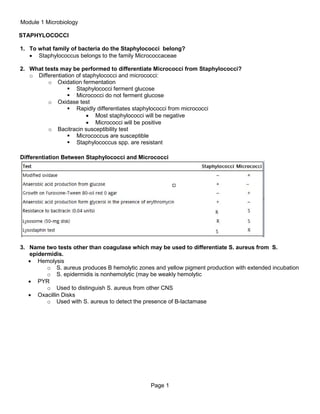
  • 1. Module 1 Microbiology STAPHYLOCOCCI 1. To what family of bacteria do the Staphylococci belong? • Staphylococcus belongs to the family Micrococcaceae 2. What tests may be performed to differentiate Micrococci from Staphylococci? o Differentiation of staphylococci and micrococci: o Oxidation fermentation  Staphylococci ferment glucose  Micrococci do not ferment glucose o Oxidase test  Rapidly differentiates staphylococci from micrococci • Most staphylococci will be negative • Micrococci will be positive o Bacitracin susceptibility test  Micrococcus are susceptible  Staphylococcus spp. are resistant Differentiation Between Staphylococci and Micrococci 3. Name two tests other than coagulase which may be used to differentiate S. aureus from S. epidermidis. • Hemolysis o S. aureus produces B hemolytic zones and yellow pigment production with extended incubation o S. epidermidis is nonhemolytic (may be weakly hemolytic • PYR o Used to distinguish S. aureus from other CNS • Oxacillin Disks o Used with S. aureus to detect the presence of B-lactamase Page 1
  • 2. Microbiology 4. List 4 disease processes involving Staphylococcus aureus and 2 involving S. epidermidis. • S. aureus o Folliculitis  Mild inflammation of a hair follicle or oil gland; red and raised o Furuncles (boils)  Large, raised, superficial abscesses o Carbuncles  Larger abscess, involve multiple furuncles, more invasive, fever & chills, systemic spreading of bacteria o Bullous impetigo  Highly contagious spread by direct contact, fomites or autoinoculation; large pustules surrounded by small zone of erythema o Toxic Shock Syndrome • S. epidermidis o UTIs  Usually nosocomial infections (usually involve development of biofilm o Endocarditis 5. Why are stools sometimes cultured for Staphylococci? • For the detection of Staphylococcus aureus food poisoning which can lead to Staphylococcal enteritis a stool culture may be required. o A stool culture is used to detect the presence of disease-causing bacteria and help diagnose an infection of the digestive tract. o In the case of Staphylococcal enteritis it is conducted to see if the stool is positive for a pathogenic bacterium 6. List types of toxins produced by Staphylococci and discuss their roles in disease processes. • Produced by S. aureus o Exfoliative toxin  AKA epidermolytic toxin  Causes the epidermal layer to slough off and known to cause SSS (scalded skin syndrome)  Scalded Skin Syndrome • Also called Ritter disease • Extensive exfoliative dermatitis primarily in newborns, young children and adults with renal failure • Caused by staphylococcal exfoliative toxin in a lesion located elsewhere on the body • Ranges from bullous impetigo (somewhat localized) to a more generalized condition o Cytolytic toxins  Four types of hemolysins (affect RBCs)  α, β, γ, δ hemolysins o Protein A  Identified in the cell wall of S. aureus  Able to bind to Fc on IgG (neutralizes it) and blocks phagocytosis o Enterotoxins A, D, or B  Food poisoning • Caused by ingestion of S. aureus enterotoxins A, D, or B o Improperly handled food allowing bacteria and toxin production to continue Page 2
  • 3. Microbiology 7. Discuss disease processes involving Staphylococcus saprophyticus. How is it differentiated from other Staphylococci? • S. saprophyticus disease o Urinary tract infections in young women  Can be found in low numbers but still significant • Identification (S. saprophyticus) o Colony Morphology  Slightly larger colony  Half strains produce yellow pigment on BAP • Presumptive Identification of S. saprophyticus o Novobiocin  S. saprophyticus is resistant to novobiocin 8. Discuss the role of Protein A of S. aureus used as a reagent in rapid identification kits, such as Staphylococcus typing kits. • Protein A o One of several cellular components that have been identified in the cell wall of S. aureus. o Most significant role of protein A in infections caused by S. aureus is its ability to bind the Fc portion of IgG  Binding IgG in this manner neutralizes IgG and can block phagocytosis. • Rapid identification kits o These kits use plasma-coated carrier particles, such as latex. o The plasma detects both clumping factor (with fibrinogen) and protein A in the cell wall of S. aureus (with IgG). o These kits often have a higher specificity and sensitivity than the traditional plasma slide test and are commonly used in clinical laboratories. o They are particularly useful for the identification of MRSA (methicillin-resistant Staphylococcus aureus) that is often weakly or negative in the slide coagulase test  Some strains of S. saprophyticus, S. sciuri, S. lugdunensis, and Micrococcus spp. may produce positive tests with the latex-coated assays, but they would be tube coagulase negative on further testing. o Procedure  Slide coagulase test = used to ID S. aureus • A heavy suspension of organism is prepared on a glass slide in water or saline and mixed with a drop of plasma (human, pig or rabbit) o If clumping = positive, ID as S. aureus following additional testing • Coagulase can also be performed in a tube method; o Detects staphylocoagulase (free coagulase)  Staphylocoagulase is an extracellular molecule that causes a clot to form when bacteria are incubated with plasma Page 3
  • 4. Microbiology 9. What is the gram reaction and morphology of Staphylococci? • Gram reaction o Gram positive • Morphology o Cocci that can appear singly, in pairs, or in clusters o Non-motile o Non-spore-forming o Aerobic or facultatively anaerobic4  Except S. saccharolyticus o Medium, cream or white colored colonies (buttery looking) o Some species are β hemolytic 10. What two methods are used for coagulase testing? What does each measure? • Coagulase test o Staphylocoagulase is the active enzyme o Presumptive identification for S. aureus o CoNS means coagulase-negative staphylococci • Two methods o Slide coagulase test  Detects: • Clumping factor  Because about 5% of S. aureus organisms do not produce clumping factor, any negative slide coagulase test result must be confirmed with the tube method, which detects staphylocoagulase, or free coagulase.  Procedure • Heavy suspension of the suspected organism is prepared on a glass slide in water or saline and is mixed with a drop of plasma. • If agglutination occurs, the isolate can be identified as S. aureus. • Some strains of S. lugdunensis and S. schleiferi are also positive for clumping factor. o Tube coagulase test  Detects: • Staphylocoagulase (free coagulase o Staphylocoagulase is an extracellular molecule that causes a clot to form when bacterial cells are incubated with plasma o Staphylocoagulase reacts with a thermostable, thrombin-like molecule called coagulase-reacting factor (CRF) to form coagulase-CRF complex. o The complex resembles thrombin and indirectly converts fibrinogen to fibrin. o The clot formed in the tube may have a tendency to undergo autolysis (because of fibrinolysin), giving the appearance of a negative result. o Clinical laboratorians should look for clot formation after 4 hours of incubation at 37° C.  If no clot appears, the tube should be left at room temperature and checked the following day. o False negative result  Fibrinolysin activity is enhanced at 37° C which might result in a false negative result.  Will differentiate other coag positive staphylococci species from S. aureus  Staphylococcus species that are capable of producing coagulase include S. aureus (potentially pathogenic in humans and animals) and the animal isolates S. intermedius Page 4
  • 5. Microbiology and S. hyicus CNS = coagulase negative staphylococci 11. Identify two body sites where Staphylococcus may be found as part of the normal flora. • Primary reservoir o Nares • Secondary o Axillae o Vagina o Pharynx o Other skin surfaces 12. Discuss beta lactamase production and its role in the development of penicillin resistant Staphylococcus. • Penicillin resistance due to β-lactamases o S. aureus produce β-lactamases  Production of β-lactamases break down the β-lactam ring of many penicillins  Most S. aureus isolates are resistant to penicillin.  Increasing resistance to alternative antimicrobial agents is a major concern. 13. What class of antibiotics is generally used to treat methicillin resistant Staphylococcus aureus infections? • Vancomycin is used to treat methicillin-resistant staphylococcus 14. Describe the term M.R.S.A. How are these organisms detected? • Methicillin-Resistant Staphylococci o Organisms resistant to oxacillin or nafcillin are termed MR  Can be MRSA, MRSE • CA-MRSA = community associated o Seen in athletes, inmates, military recruits in close quarters, pediatric patients, tattoo recipients • HA-MRSA = hospital-associated o Risk factors such as recent hospitalization, long-term care, dialysis or indwelling devices Page 5
  • 6. Microbiology STREPTOCOCCI 1. Describe the appearance of the three major types of hemolysis produced by Streptococci. • Alpha α o Partial lysis of RBCs resulting in greenish pigment of agar surrounding colone • Beta β o Complete lysis of RBCs clearing surrounding colony • Gamma γ o Lack of lysis of RBCs, no change in agar 2. Discuss Streptolysin S and O and describe 2 methods used to demonstrate these hemolytic properties. • Streptolysin O or S • Responsible for hemolysis of agar RBCs o O = oxygen labile  Occurs when incubated anaerobically o S = oxygen stable  Occurs when incubated aerobically • Can test patients for the presence of antistreptolysin O (ASO) to check for recent strep infection 3. Name two diseases which may appear as sequelae to Group A Streptococcus infection. • Rheumatic fever o Typically follow pharyngitis • Acute glomerulonephritis o Sometimes occurs after a cutaneous or pharyngitis infection 4. Name two disease processes in which Streptococcus pneumoniae may be found as the etiological agent. • Otitis media o Kids under 3 • Bacterial pneumonia o Mainly elderly or immunocompromised Page 6
  • 7. Microbiology 5. What is the gram reaction and morphology of: a. Streptococci agalactiae • Colonies appear grayish white and mucoid on BAP and have small zone of β hemolysis • Organisms appear as GPC in chains • Capsule prevents phagocytosis but is ineffective after opsonization b. Streptococcus pneumoniae • Presence of capsule • Gram positive • Catalase negative 6. What is the best single test for differentiating Staphylococci from Streptococci? What interference do RBCs have in this test? • Catalase test o Negative with streptococcus o Positive with staphylococcus 7. Name 2 disease processes associated with each of the following groups of streptococci and a test or tests used for routine biochemical identification: Associated Diseases a. Group A (strep. pyogenes) Biochemical Identification • • • • • Scarlet fever Bacterial pharyngitis Pyodermal (skin) infections Necrotizing fasciitis Streptococcal Toxic shock syndrome Post-streptococcal sequelae Acute glomerulonephritis • • Bacitracin susceptibility PYR Invasive disease of newborn • Early onset <7d old • Late onset > 7d old Pregnant women should be screened at 35 – 37 wks. gestation • • CAMP test Hippurate hydrolysis • • b. Group B (strep agalactiae) • • c. Group D non-enterococcus • Wounds, eyes, ears (not big deal) • • • Bile esculin PYR Salt Tolerance d. Enterococcus • Nosocomial infections • • Bile esculin Salt tolerance e. Streptococcus viridans • • • • • • Bacterial endocarditis Oral infections (gingivitis) Meningitis/Septicemia Abscesses Bacterial pneumonia Otitis media • • Neg for optochin Neg for Bile solubility • • Optochin (P disk) Bile solubility f. Streptococcus pneumoniae Page 7
  • 8. Microbiology 8. Name two tests used to differentiate pneumococci from other alpha hemolytic streptococci. • Optochin (P disk) o Presumptive ID os S. pneumoniae o Zone of inhibition = susceptible • Bile solubility o Further identification of S. pneumoniae 9. What colonial characteristic helps to distinguish Strep pneumoniae on a blood agar plate? • • α hemolysis – partial lysing of RBCs that results in green discoloration of the agar Additionally shape of colony (concave red blood cells) 10. What is the drug of choice for treatment of most streptococcal infections? • Penicillin can be used to treat most streptococci o Penicillin-resistant S. pneumoniae and viridans  Use erythromycin and narrow spectrum cephalosporins • Vancomycin is effective against GPC o Not currently a widespread resistance 11. Describe the PYR test and what organisms it is used to identify. • Presumptive ID for Group A and Group D o Group A = only strep that is positive o Enterococcus = also PYR positive o More specific for Group A than bacitracin 12. Describe the term V.R.E. How are these organisms detected? • VRE = vancomycin resistant enterococci • Molecular typing methods, such as pulsed-field gel electrophoresis, contour-clamped homogeneous electric-field electrophoresis, ribotyping and PCR-based typing methods have been used mainly to type enterococcal species in epidemiologic studies and investigations of vancomycin-resistant enterococci (VRE). Page 8
  • 9. Microbiology HAEMOPHILUS 1. (L) What is the satellite phenomenon? • Growth of a fastidious organism around other bacteria that release necessary growth factors • Hemolytic S. aureus, S. pneumoniae, Neisseria species release the factors or naturally produce V factor o H. ducreyi is exception 2. (L) Name a disease process from which each of the following may be isolated: a. H. influenzae • Haemophilus influenzae serotype b (Hib) o Primarily causes disease in children o Hib vaccine is useful in reducing incidence of disease o Causes bacteremia and then spreads to other tissues • Disease of Hib strains o Meningitis o Epiglottitis o Bacterial tracheitis o Cellulitis o Acute pharyngitis and pneumonia b. Haemophilus-aegyptius • Conjunctivitis o Pink eye c. H. ducreyi • Sexually transmitted infection that can cause chancroid d. Gardnerella vaginalis • Bacterial vaginosis 3. What is the gram stain and morphology of Haemophilus species? • Gram negative • Pleomorphic coccobacilli or rods • Biochemical reactions o Nonmotile o Oxidase positive o Catalase positive o Nitrate reduction 4. What are the X and V factors? • Haemophilus requires X and V factors o X factor  Hemin or hematin o V factor  Nicotinamide-adenine dinucleotide (NAD) • Para – require only V factor o Haemophilus parainfluenzae  Produces X factor but requires V factor • Sheep blood agar contains only X factor NOT V factor Page 9
  • 10. Microbiology 5. Explain the importance of doing beta-lactamase tests. • Checking for resistance to beta-lactams • Nitrocefin test o Put on glass slide with drop of water o Put disc on there o Put loop of organism on there and changes to red 6. What tests and media are used for screening for and identifying G. vaginalis? • Vaginal discharge collected from suspected BV cases is the most common specimen used for the isolation of G. vaginalis. • Because it is part of the urogenital microbiota, the organism can also be isolated from urine. • Because G. vaginalis can be found as normal vaginal biota and its role in BV is questionable, cultures for G. vaginalis are infrequently performed. • It often takes longer than 24 hours to develop visible colonies, and G. vaginalis grows best in 5% to 7% CO2 at a temperature of 35° to 37° C. • G. vaginalis grows on SBA as pinpoint, nonhemolytic colonies. • It will also grow on chocolate agar. • The medium of choice for G. vaginalis, however, is human blood bilayer Tween (HBT) agar. • When cultured on human blood, colonies are β-hemolytic, small, gray, and opaque. • G. vaginalis will also produce β-hemolytic colonies on media made with rabbit blood. Page 10
  • 11. Microbiology NEISSERIA / MORAXELLA 1. Name 2 pathogenic species of Neisseria and an important disease that each causes and 1 species of Moraxella. • Two pathogenic Neisseria spp. o N. gonorrhoeae  Always pathogenic when present o N. meningitidis  May be found as normal flora in respiratory tract of carriers 2. What is the gram reaction and morphology of Neisseria species? • Aerobic, nonmotile, non-spore-forming, oxidase positive, catalase positive, GNDC • Requires enriched media to grow 3. What is one simple test that aids in the identification of Neisseria species? • Presumptive Identification o Colonial Morphology o Small, tan, translucent and raised after 24-48hrs o Fresh subculture should be used for ID tests • Definitive Identification o Immunologic Assays  Coagglutination tests using antibodies against N. gonorrhoeae fused to killed S. aureus cells; agglutination is a positive reaction o Nucleic Acid Assays  See Table 17-4; able to detect organism directly from specimens • Treatment o Cephalosporins are currently the drug of choice o Often used in combination with other drugs if Chlamydia is also present 4. Name several sources from which each of these species can be isolated. • Neisseria meningitidis o Normal flora in nasopharynx or oropharynx • Neisseria gonorrhea o Clinical Infections o Acute, pyogenic infection, primarily acquired by sexual contact & infect urethra, endocervix, anal canal, pharynx and conjunctiva o Short incubation period of 2-7 days o Men = acute urethritis with purulent discharge and painful urination  Only 3-5% will be asymptomatic o Women = cervical discharge, lower abdominal pain & dysuria  As many as 50% may be asymptomatic carriers  Pelvic Inflammatory Disease – caused by untreated gonococcal cervicitis (can cause sterility & ectopic pregnancy) Page 11
  • 12. Microbiology 5. What factors have to do with the viability of Neisseria meningitidis? • Humans are its only host • Requires enriched media • Virulence Factors (same for meningitidis and gonorrhoeae) • Receptors for human transferrin • Capsule • Pili (fimbriae) o Inhibit phagocytosis; aid in the initial attachment to host tissues o Aid in the exchange of genetic material between cells • Cell membrane proteins o IgA protease o Lipooligosaccharide (LOS) endotoxin  Mediates damage to host tissues and induces inflammatory response • Culture o Requires the use of chocolate agar o But chocolate will allow other organisms to grow o Media should be allowed to come to room temp due to Neisseria’s sensitivity to temperature changes • Incubation o Plates should be incubated at 35C in 5% CO2; also prefer increased humidity 6. When is M. catarrhalis implicated in disease? What screening test differentiates it from Neisseria? • Clinical Infections o An opportunistic pathogen that causes URT infections in children and elderly o The third most common cause of acute otitis media and sinusitis in children • Intracellular GNDC, most produce β-lactamase (makes them resistant to ampicillin & amoxicillin) Page 12
  • 13. Microbiology MISCELLANEOUS GRAM NEGATIVE RODS 1. Identify the source from which most infections with Pasteurella multocida can be attributed and name clinical specimens where it may be found. • Colonize the URT and GI tract of mammals and birds o Exposure from cat and dog bites or scratches 2. Name the causative organism of whooping cough and the medium used for primary isolation. • Bordetella pertussis and Bordetella parapertussis • Specimens are best from the nasopharynx o Aspirates of fluid o Calcium alginate or Dacron swabs up each nasal passage as deep as possible • Culture o BAP and MAC agar o Chocolate if you need to rule out Haemophilus • Incubate plates o 35C in aerobic conditions o 7 days o Adequate moisture to prevent plate drying 3. Name the causative organism of tularemia and discuss the primary reason for not culturing it in the laboratory. • Francisella spp • Diagnosis is from serology because culture is less safe 4. Describe the gram stain morphology of Pasteurella. • Gram negative pleomorphic coccobacilli with bipolar staining o Oval shaped, short rods, longer filaments o Grow on BAP and chocolate agar  Nonhemolytic mucoid colonies with a narrow green or brown halo 5. Discuss the fluorescent antibody techniques used for diagnosis of whooping cough (pertussis) and name 2 suitable specimens. • Nasopharyngeal swab with booger juice • Put this on glass slide • Adding fluorescent monoclonal tag to this • If there is binding we know the organism is there due to the presence of fluorescent Page 13
  • 14. Microbiology ENTEROBACTERIACEAE 1. What is the definition of the family Enterobacteriaceae? • Enterics: Members of the family Eterobacteriaceae consisting of organisms that all ferment glucose, nearly all fail to produce the cytochrome oxidase, and nearly all reduce nitrates to nitrites. Most are resident flora of the gastrointestinal tract. • • • • • • • • • • Includes many genera and species Common clinical isolates include: o E. coli o Klebsiella pneumoniae o Proteus mirabilis A wide variety of differential and selective media, such as MacConkey (MAC) agar, and highly selective media, such as Hektoen enteric (HE) agar and xylose-lysine-desoxycholate (XLD) agar, are available for the presumptive identification of enteric pathogens. These media contain one or more carbohydrates, such as lactose and sucrose, which show the ability of the species to ferment specific carbohydrates. Fermentation is indicated by a color change on the medium, which results from a drop in pH detected by a pH indicator incorporated into the medium. Nonfermenting species are differentiated by lack of color change, and colonies retain the original color of the medium. Species that produce hydrogen sulfide (H2S) may be readily distinguished when placed on HE or XLD agar. HE and XLD agars contain sodium thiosulfate and ferric ammonium citrate, which produce blackening of H2S-producing colonies. These features have been used to initially differentiate and characterize certain genera. Definitive identification depends on the biochemical reactions and serologic antigenic structures demonstrated by the particular species. Page 14
  • 15. Microbiology 2. Why must a TSI slant be read within 18-24 hours? • Enterics: Members of the family Eterobacteriaceae consisting of organisms that all ferment glucose, nearly all fail to produce the cytochrome oxidase, and nearly all reduce nitrates to nitrites. Most are resident flora of the gastrointestinal tract. o o Reading the results after fewer than 12 hours of incubation gives the false appearance of an organism capable of fermenting glucose and lactose (or sucrose in the case of TSI agar). For this reason, TSI agar or KIA must be incubated for 18 to 24 hours. 3. What is meant by K antigens? somatic antigens? flagellar antigens? • K antigen, or capsular antigen—is a heat-labile polysacharide found only in certain encapsulated species. o Examples  K1 antigen of E. coli  Vi antigen of Salmonella enterica • O antigen, or somatic antigen—is a heat-stable antigen located on the cell wall • H antigen, or flagellar antigen—is a heat-labile antigen found on the surface of flagella, structures responsible for motility. 4. What is the Vi antigen? How do you destroy this antigen? What is the purpose of destroying the Vi antigen?. • Capsular antigen of Salmonella typhi o Heat labile 5. Describe disease processes caused by enterotoxigenic E. coli (ETEC), enteropathogenic E. coli (EPEC) and enterohemorrhagic E. coli. • ETEC- Enterotoxigenic E. coli (ETEC) Diarrhea (Traveler’s Diarrhea). This disease consists of a watery diarrhea with or without mucus or blood, vomiting, and abdominal cramping. Dehydration and lowgrade fever may occur. Enterotoxigenic strains of E. coli are the most common cause of traveler’s diarrhea worldwide and a common cause of diarrheal disease in children in developing countries. • EPEC • EHEC- Enterohemorrhagic E. coli (EHEC) Diarrhea. This disease consists of a hemorrhagic, watery diarrhea with abdominal cramping. Usually, patients have no fever or only a slight fever. About 5% of infected people (especially children younger than age 5 and the elderly) develop hemolytic- uremic syndrome (HUS), with anemia, low platelet count, and kidney failure. The first recognized outbreak of diarrhea caused by enterohemorrhagic E. coli (O157:H7) occurred in 1982, involving contaminated hamburger meat—hamburger meat contaminated with cattle feces. Since then, several well-publicized epidemics involving the same serotype have occurred. Not all of the outbreaks involved meat; some resulted from ingestion of or unpasteurized milk or apple juice, lettuce, or other raw vegetables. Page 15
  • 16. Microbiology 6. Name the causative organism of plague. Identify the vector of this disease and three clinical forms recognized in man.. • Yersinia pestis o Transmitted by fleas from infected rodent o Pneumonic plague through respiratory droplets o Bubonic o Glandular 7. Describe how lactose fermenters, nonlactose fermenters and H2S (+) organisms appear on the following media: MacConkey, Hektoen, XLD (xylose-lysine-desoxycholate), and SS (SalmonellaShigella). Media Enteric organism Reactions Lactose Fermenters Non-lactose H2S postive Fermenters MacConkey Red/Pink No change Does not indicate Hektoen Yellow or Orange No change (green is color of agar) Produces a black precipitate XLD Yellow Colorless or red Black precipitate SS Does not indicate lactose fermenters Salmonella – colorless Black Shigella – colorless 8. Triple sugar iron agar (TSI) is used as a screening medium for the pathogenic enterics. What do the following reactions in a TSI slant indicate with regards to fermentation or utilization of ingredients in the agar: TSI reactions Indication of Fermentation or Utilization a. Acid Butt (yellow) ferments lactose, glucose, and/or sucrose b. Alkaline slant (red) does not ferment either lactose or sucrose c. Gas bubbles in butt Gas has been formed d. Blackening of butt H2S has been formed Page 16
  • 17. Microbiology 9. List possible organisms giving the following reactions on TSI agar: Slant Acid Butt Acid H2S Gas None Alkaline Acid Gas + Alkaline Acid None Acid Acid Possible Organisms E. coli, Klebsiella spp, Enterobactor spp., Serratia spp Gas + 10. What is the IMViC profile? What are the two most common organisms which are used as positive and negative controls for IMViC and what reactions do they give? Indole Methyl Red VP Citrate Organism 1: E. coli E. coli Enterobacter aerogenes Klebsiella pnumoniae Organism 2: Klebsiella pneumoniae Enterobacter aerogenes E. coli E. coli 11. Which group of enterics may produce a red pigment and at what temperature? • Often produce a characteristic pink to red pigment, prodigiosin, especially when the cultures are incubated at room temperature o S. marcescens, o S. rubidaea o S. plymuthica 12. What is characteristic of Proteus species with regards to colony growth? • Swarming 13. How do the colonies of E. coli generally appear on MacConkey agar? • Pink due to lactose fermentation Page 17
  • 18. Microbiology 14. Name three Enterobacteriaceae genera considered pathogenic when isolated from stool, describe pathogenesis of infection, and outline the procedure for their identification. • Salmonella- non lactose fermenting, h2s producing, negative urease. • Shigella- non lactose fermenting, non h2s producing, • Yersinia- non lactose fermenting, positive urease 15. List 4 genera which produce H2S in TSI • Salmonella • Proteus • Citrobacter • Edwardsiella 16. Yersinia enterocolitica is generally isolated from what clinical specimen? Describe incubation temperatures, and urease reactions of this organism. • • • • Routinely isolated from stool, Can survive cold temperatures Incubation temperatures recommended to be below 37C. A positive urease test is presumptive to identifying Yersinia Page 18
  • 19. Microbiology AEROMONAS, VIBRIO, CAMPYLOBACTER, 1. List 3 sources from which Aeromonas may be isolated. • Retail produce • Animal meat products • Aquatic wound infections 2. Identify the mechanism by which Vibrio cholera produces disease. • Cholera toxin also known as choleragen, once ingested the bacteria colonize the small intestine where they multiply and produce choleragen. • Read further on how this mechanism works on (text page 465) 3. Name the Vibrio species which are associated with the consumption of contaminated seafood. How are they identified? • • • Vibrio parahaemolyticus Vibrio vulnificus Page 467-468 4. Identify the disease processes associated with Campylobacter jejuni. • Patients with C.jejuni present with a diarrheal disease that begins with mild abdominal pain within 2-10 days after ingesting the organism. • Cramps and bloody diarrhea often follow the initial signs. • Illness is usually self-limiting and resolves in 2-6 days. 5. How do the Campylobacter appear on gram stain? What is their unique atmosphere requirement? • Curved non-spore forming gram negative rods • Enteric campylobacters may appear as long spirals, S shapes, or seagull wings • Organisms stain poorly • Page 478 • Anerobic, some microanerobic 6. What organism in this group is characterized by "rice water" stools? • Vibrio cholera 7. What disease is associated with Helicobacter? Why is this organism difficult to isolate? ULCERS • • • • • • Not thought to be a cause of diarrheal illness, H. pylori nonetheless is a significant cause of disease. Living in the stomach and duodenum, H. pylori has been associated with ulcer disease. This infection is common, in that many patients have detectable antibody against this organism. When the organism is recovered from a gastric or duodenal ulcer, there is evidence that patients have faster, more durable ulcer recovery with antibiotic therapy. Active infection can be diagnosed by detecting the organism on an endoscopically derived biopsy specimen or through breath testing This testing is based on the fact that the organism shows high urease activity; urea labeled with either 13C or 14C will, when ingested, rapidly produce labeled CO2 in the patient's breath if the organism is present. Page 19