About us
•Descargar como DOCX, PDF•
0 recomendaciones•641 vistas

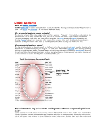
Dental sealants are plastic coatings placed on the chewing surfaces of back teeth to protect the deep grooves from decay. They are applied after the permanent molars and premolars have erupted, and can last for many years, helping to prevent cavities. Dental sealants are clear or white and are applied using acid etching and light curing to bond to the tooth surface.

Recomendados
Recomendados
Más contenido relacionado
La actualidad más candente
La actualidad más candente (20)
Similar a About us
Similar a About us (20)
Más de Dentist Imus Cavite
Más de Dentist Imus Cavite (20)
About us
- 1. Dental Sealants What are dental sealants? Dental sealants are plastic coatings that are usually placed on the chewing (occlusal) surface of the permanent ba teeth — the molars and premolars — to help protect them from decay. Why are dental sealants placed on teeth? The chewing surfaces of the molar and premolar teeth have grooves — "fissures" — that make them vulnerable to dec These fissures can be deep, are difficult to clean, and can be narrower than even a single bristle of a toothbrush. Plaqueaccumulates in these areas, and the acid from bacteria in the plaque attacks the enamel and cavities can develop. Fluoride helps prevent decay and helps protect all the surfaces of the teeth, dental sealants provide extra protection for the grooved and pitted areas by providing a smooth surface covering over the fissured area. When are dental sealants placed? The first dental sealant to be placed is usually on the fissure of the first permanent molartooth, once the chewing surfac the tooth has erupted completely beyond the gum. This tooth grows in behind the baby teeth. If the chewing (occlusal) surfaces of these teeth are sealed, the dental sealant will help protect the tooth. Except for the wisdom teeth, which com through much later, the molars and premolars continue to erupt until eleven-thirteen years of age and the chewing surfa of these teeth can be sealed after they have erupted beyond the gum. Are dental sealants only placed on the chewing surface of molar and premolar permanent teeth? Dental sealants are usually placed on the chewing surfaces of these teeth because these are the areas and teeth that typically have deep fissures. Dental sealants are sometimes also used on other permanent teeth if they have grooves o pits, to help protect these surfaces. In some children, the molars in the primary dentition (baby teeth) also have grooves
- 2. could benefit from dental sealants and in this situation your dentist orhygienist may recommend dental sealants on the chewing surfaces of these primary teeth. Can dental sealants be place on the teeth of adults? Yes — while less common, dental sealants are sometimes placed in adults at risk for caries, on deep grooves and fissu that do not already have fillings or dental sealants. What do dental sealants look like? Dental sealants can be clear, white or have a slight tint depending upon the dental sealant used. How are dental sealants placed? Firstly the tooth surface is thoroughly cleaned with a paste and rotating brush by your dentist or hygienist. Next the toot washed with water and dried. Then a solution that is acidic is placed on the fissured area of the tooth’s chewing surface a number of seconds before being rinsed off. This creates small microscopic areas and a fine rougher surface than the surrounding tooth enamel, that can be seen with a microscope. The rough surface and microscopic areas enable the de sealant to attach to the tooth. After the tooth is dried again, the liquid dental sealant is placed on the tooth and hardene Dental sealants are hardened by using a light that hardens the dental sealant, or sometimes by using a two-componen dental sealant that sets without using a light. Once the dental sealant has hardened it becomes a hard plastic varnish coating, and you can chew on the tooth again. How long does a dental sealant last? Dental sealants have been used and have been proven to be effective since the 1970s. Many studies have shown that are effective in helping to prevent decay on chewing (occlusal) surfaces. Dental sealants can last many years. If necess it is also possible to place a new dental sealant on the tooth. Do I still need to use fluoride if I have dental sealants? Yes. Dental sealants only protect the surface area that they are placed on. Fluoride helps protect all the surfaces of the tooth from decay and cavities. What is flexite denture? A flexite denture is a partial denture made of elastic nylon resin, more flexible than plastic of which a regular denture is made. Because the flexite denture base is fixed to the gum and can be thin, no clasp is needed and more comfortable to wear. Flexite Denture Diastema (Gap Between Teeth) What Is It? ~A diastema is a space or gap between two teeth. It appears most often between the two upper front teeth. However, gaps can occur between any two teeth. ~A diastema also can be caused by an oversized labial frenum. The labial frenum is the piece of tissue that normally extends from the inside of your upper lip to the gum just above your two upper front teeth. In some situations, the labial frenum continues to grow and passes between
- 3. the two front teeth. If this happens, it blocks the natural closing of the space between these teeth. ~Habits can also lead to gaps between the teeth. Thumb sucking tends to pull the front teeth forward, creating gaps. Spaces can develop from an incorrect swallowing reflex. For most people, the tongue presses against the roof of the mouth (palate) during swallowing. Some people develop a different reflex known as a tongue thrust. When they swallow, the tongue presses against the front teeth. Over time the pressure will push the front teeth forward. This can cause spaces to develop. Symptoms A diastema that occurs because of a mismatch between the teeth and the jaw does not have symptoms. However, spaces caused by a tongue thrust habit or periodontal diseasewill tend to expand or grow with time. The teeth may become loose, and discomfort or pain may occur, particularly during biting or chewing. Diagnosis You may notice a space when brushing or flossing. Your dentist can see spaces during an examination. Treatment ~Sometimes, a diastema is part of a set of problems that require orthodontic treatment. In other cases, a diastema is the only problem. However, some people may seek treatment for reasons of appearance. ~Some people get braces, which move the teeth together. Often, no matter where the diastema is, you must wear a full set of braces — on both your upper and lower teeth. That's because moving any teeth affects your entire mouth. If your lateral incisors are too small, your dentist may suggest widening them usingcrowns, veneers or bonding. ~If a large labial frenum is causing the gap, the frenum can be reduced through surgery called a frenectomy. If a frenectomy is done in a younger child, the space may close on its own. If it is done in an older child or an adult, the space may need to be closed with braces. Prognosis ~If a diastema is closed through orthodontics or dental repair, the space will tend to stay closed. However, to help prevent the space from coming back, wear your retainers as directed by your orthodontist. Your orthodontist may also splint (attach) the backs of the teeth to other teeth with composite (plastic) and a wire to prevent them from moving. Visit your dentist regularly to make sure your dental work is in good repair. Dental Caries (Cavities) What Is It? ~Dental caries is the medical term for tooth decay or cavities. It is caused by specific types of bacteria. They produce acid that destroys the tooth's enamel and the layer under it, the dentin. Many different types of bacteria normally live in the human mouth. They build up on the teeth in a sticky film called plaque. This plaque also contains saliva, bits of food and other natural substances. It forms most easily in certain places. Symptoms ~Early caries may not have any symptoms. Later, when the decay has eaten through the enamel, the teeth may be sensitive to sweet, hot or cold foods or drinks. Diagnosis ~A dentist will look for caries at each office visit. This will be part of the exam, whether it is a routine visit or an appointment made because of pain. The dentist will look at the teeth and may
- 4. probe them with a tool called an explorer to look for pits or areas of damage. The problem with these methods is that they often do not catch cavities when they are just forming. Occasionally, if too much force is used, an explorer can puncture the enamel. This could allow the cavitycausing bacteria to spread to healthy teeth. Newer devices also have been developed to detect tooth decay. They are useful in some situations, and they do not spread decay. The one most commonly used in dental offices is a liquid dye or stain. Your dentist brushes the nontoxic dye over your teeth, then rinses it off with water. It rinses away cleanly from healthy areas but sticks to the decayed areas. Some dentists also use high-tech devices such as lasers to detect cavities. Under many conditions, these devices can detect very early tooth decay, which can actually be reversed. Lastly, more advanced caries can be seen on X-rays. They are taken on a set schedule, or to find out the cause of symptoms such as pain. Treatment ~Caries is a process. In its early stages, tooth decay can be stopped. It can even be reversed. Fluorides and other prevention methods also help a tooth in early stages of decay to repair itself (remineralize). White spots are the last stage of early caries. Once caries gets worse and there is a break in the enamel, only the dentist can repair the tooth. ~Then the standard treatment for a cavity is to fill the tooth. If a drill is used, the dentist will numb the area. If a laser is used, a numbing shot is not usually required. The decayed material in the cavity is removed and the cavity is filled. ~Many fillings are made of dental amalgam or composite resin. Amalgam is a silver-gray material made from silver, mercury, copper or other metals. Composite resin offers a better appearance because it is tooth-colored. Newer resins are very durable. ~Amalgams are used in molars and premolars because the metal is not seen in the back of the mouth. Composite and ceramic materials are used for all teeth. If a cavity is large, the remaining tooth may not be able to support enough filling material to repair it. In this case, the dentist will remove the decay and cover the tooth with a ceramic inlay, onlay or artificial crown. These may be made in the office or in a lab. Sometimes the part of the tooth you can see remains relatively intact, but there is decay in the pulp inside the tooth. In this case, the tooth will need root canal treatment. A general dentist or an endodontist will be able to remove the tooth's pulp and replace it with an inert material. In most cases, the tooth will need a crown. Prognosis I~f caries is not treated, it likely will cause the tooth to decay significantly. Eventually, uncontrolled decay may destroy the tooth. Having caries increases your risk of more caries for several reasons: Caries is caused by bacteria. The more decay you have, the more bacteria exist in your mouth. The same oral care and dietary habits that led to the decay of your teeth will cause more decay. Bacteria tend to stick to fillings and other restorations more than to smooth teeth, so those areas will be more likely to have new caries. Cracks or gaps in the fillings may allow bacteria and food to enter the tooth, leading to decay from beneath the filling. Many of those fortunate to have dental benefits are allowed preventive dental care twice a year because for the most part dental disease is preventable. There is still time to take advantage of your full benefits this year. Hurry and call us today at (XXX) XXX-XX or visit yourwebsite.com to request your first appointment before July 1. Health card logo
- 5. Retainer dentist healthway medical Can asia San Miguel yamamuraasia About Us Our Vision To contribute to the health and wellness of every individual by providing quality dental services. Our Mission To provide dental services in a timely and friendly manner. To provide dental services and product with professional quality that will meet the standard of Philippine Dental Association. To be responsive to the suggestions and concern of every patient. To provide quality service and address to the needs of every patient. Our Company Profile Established in 2006 at Barcelona Ph.2, BuhaynaTubig, Imus, Cavite. In 2009, LE Dental Clinic moved to B5 L22 Blanca street,Primarosa Subdivision, Phase 5, BuhaynaTubig, Imus, Cavite. The practice is owned by Dr. Genelyn L. Estrada. LE Dental Clinic offers quality dental care, preventive, prosthodontic, aesthetic and orthodontic dentistry. LE Dental Clinic is accepting different health cards such as: Maxicare Intellicare Medocare Dental Network Affinity Health and Wellness Fortune Care Valucare OMNI HMI Asian Life Philcare Health Partners Dental Access Medaccess and other health cards.
- 6. LE Dental Clinic will be a comfortable, relaxing dental clinic that provides a waiting room with a television, music, wifi and a playing area for kids to help relieve anxiety and nervousness of patients. The two examination rooms will be comfortable and decorated in a professional manner, but with a home-style touch. Health card holders will visit LE Dental Clinic for their dentistry needs and return because of the excellent customer service provided by the staff. What the clinic will provide you? The Clinic will provide information and education on preventive care. Quality dental care with a comfortable and professional environment. Will build community trust and word of mouth referrals for future patients. Core Values Compassion Provide the best care, treating patients and family members with sensitivity and empathy. Quality We provide quality care by evaluating technology, learning new techniques and utilizing the best equipment and materials for our patients. Integrity We speak the truth and honor our word. Honesty Doing the right thing under all circumstances.