Early Diagnosis and Treatment for Alzheimer's
•Descargar como PPT, PDF•
4 recomendaciones•2,681 vistas

A Novel Multi-phased Project that investigates the Glutathione Pathway by Creating Alzheimer’s model Drosophila with Pan-neuronal Over-expression of the GCLc Gene, Inducing Redox Stress through Sleep Deprivation, and Analyzing Mitochondrial Electron Transport Chain using Colorimetric Enzymatic Assays.

Recomendados
Recomendados
Más contenido relacionado
La actualidad más candente
La actualidad más candente (20)
Destacado
Destacado (20)
Similar a Early Diagnosis and Treatment for Alzheimer's
Similar a Early Diagnosis and Treatment for Alzheimer's (20)
Último
Último (20)
Early Diagnosis and Treatment for Alzheimer's
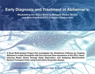
- 1. Early Diagnosis and Treatment of Alzheimer’s: Modulating the GCLc Gene to Mitigate Redox Stress and Mitochondrial ETC Complex Dysfunction A Novel Multi-phased Project that investigates the Glutathione Pathway by Creating Alzheimer’s model Drosophila with Pan-neuronal Over-expression of the GCLc Gene, Inducing Redox Stress through Sleep Deprivation, and Analyzing Mitochondrial Electron Transport Chain using Colorimetric Enzymatic Assays. Lisa Michaels • Plano West Senior High School • Plano, TX •
- 2. ALZHEIMER’S BRAIN Source: Alzheimer’s.org, National Center for health statistics Brain scans done with Positron Emission Tomography (PET) show how Alzheimer’s affects brain activity. The top images show a normal brain, while the right images are from a person with Alzheimer’s. The red and yellow areas in the bottom scans show Amyloid plaques, made visible with Mathis and Klunk’s traceable dye. As Alzheimer’s disease progresses, brain tissue shrinks. However, the ventricles, chambers within the brain that contain cerebrospinal fluid, are noticeably enlarged. In the early stages of Alzheimer's disease, short-term memory begins to decline when the cells in the hippocampus degenerate. 2
- 3. NATIONAL SIGNIFICANCE MAJOR CAUSES OF DEATH - % Change 2000-08 66% Increase in Alzheimer’s during 2000-2008 as a Cause of Death! Source: Alzheimer’s.org, National Center for health statistics AD has emerged as the sixth largest killer disease in the US, rapidly growing while deaths from other diseases are declining. If we make no progress in diagnosis and treatment of AD, the associated cost will rise to over a trillion dollars a year in 2050. The President recently signed into law the National Alzheimer’s Project to overcome the disease before 2025. This project contributes to this national public health goal. 3
- 4. PREVALENT THEORY: AMYLOID CASCADE Source: National Institutes of Health website 4
- 5. PREVALENT THEORY II: MITOCHONDRIAL CASCADE Source: Alzheimer’s.org, National Center for health statistics 5
- 6. PREVALENT THEORY III: OXIDATIVE STRESS OXIDATIVE STRESS O2 H 2 O2 OH – NO – ONOO– GSSG Amyloid Plaques Tau Tangles NEURO-DEGENERATION NEURO-PROTECTION CAT SOD GSH peroxidase GSH-S-transferase GSH ANTIOXIDANT DEFENSE Balance of ROS generation and antioxidantive systems is critical for neuronal survival. An imbalance of both systems due to either excessive production of ROS (left) or reduced antioxidant defense (right) results in oxidative stress. In AD, increases in oxidative stress, mainly promoted by the interplay between the reactive species and transition metals, lead to oxidative damage that induces neuronal damage or death. Antioxidant therapies that increase endogenous antioxidant defenses or reduce oxidative sources should reduce oxidative damage, and thereby prevent or delay disease symptoms.
- 7. PROPOSED THEORY AMYLOID TOXICITY ROS ALZHEIMER’S MITOCHONDRIAL DYSFUNCTION PROGRRESSION MITOCHONDRIAL PROTECTION Source: Thinkquest.org ANTIOXIDANT CAPACITY GCLc GENE GLUTATHIONE GENE EXPRESSION The research thesis is that in vivo synthesis of Glutathione through over-expression of the GCLc gene mitigates Oxidative stress, Mitochondrial dysfunction and Amyloid toxicity, thereby slowing down the progression of AD. The Oxidative stress caused by Amyloid toxicity results in ROS, impairment of the Electron Transport Chain, further ROS generation and Mitochondrial dysfunction. Switching on the GCLc gene may protect Mitochondria from damage by improving the Redox balance (GSH/GSSG) which mitigates Mitochondrial dysfunction and the progression of Alzheimer’s.
- 8. SUGGESTED SOLUTION: GCLc_GLUTATHIONE PATHWAY C10H17N3O6S http://max-one-cure.org/max-one/max-one-qa Glutathione is the primary endogenous antioxidant. It is the body's most effective detoxifier. Stress depletes the Glutathione stores. It modulates the immune response. It assists in the regulation of the cell's vital functions, such as the synthesis and repair of DNA, the synthesis of proteins, and the activation and regulation of enzymes. As Glutathione is a large molecule the supplements are not absorbed into the body. Glutathione-rich foods include asparagus, grapefruit and peaches, garlic, whey protein. We need 250 milligrams of Glutathione a day; the average American consumes less than 35 mg! REACTION 1: REACTION 2: Glutamate ϒ-glutamylcysteine ϒ-glutamylcysteinylglycine GSH Cysteine Glycine GSH GCL ADP Synthase ATP ATP ADP Glutathione is synthesized de novo by the action of two enzymes: glutamate-cysteine ligase-catalytic subunit (GCLc) and by GSH synthase. The determinants of Glutathione synthesis are the availability of cysteine, activity of 8 GCLc gene and feedback regulation.
- 9. RESEARCH DESIGN Home Lab GENETIC INCREASED H1: MODULATION Aβ42-GCLc GLUTATHIONE LIFESPAN PRODUCTION Home Lab INCREASED H2: RESISTANCE TO Stressed RESISTANCE TO LIFESPAN REDOX STRESS Aβ42-GCLc REDOX STRESS Genetics Lab INCREASED H3: MITOCHONDRIAL BIOENERGETICS Aβ42-GCLc MITOCHONDRIAL ETC BIOENERGETICS ACTIVITY
- 10. HYPOTHESES Hypothesis 1: IF lifespans of Alzheimer’s model Aβ42 Drosophila and Aβ42-GCLc are studied, THEN the Aβ42-GCLc group will outlive the Aβ42 group of flies; all groups will exhibit shorter lifespans compared to the control group of yellow-white Drosophila melanogaster. Hypothesis 2: IF Aβ42 and Aβ42-GCLc Drosophila are subjected to continuous oxidative stress, THEN the Aβ42-GCLc flies will be able to better resist stress, as exhibited by longer lifespan; all groups will exhibit shorter lifespans compared to the control group of yellow-white Drosophila melanogaster. Hypothesis 3: IF the Mitochondrial Electron Transport Chain of Aβ42 and Aβ42-GCLc groups of Drosophila are analyzed, THEN Aβ42-GCLc will exhibit more enzymatic activity in the mitochondrial complexes compared to Aβ42; all groups will exhibit lower enzyme activity compared to the control group of yellow-white Drosophila melanogaster. 10
- 11. MATERIALS 11
- 12. Home Lab: Lifespan and Oxidative stress Experiments Source: All Charts and Photographs by the Researcher Oscillator For inducing Oxidative Stress University Lab: ETC studies 12
- 13. TRANSGENIC DROSOPHILA http://sanpatricio.co.uk/Innexins/pg2%20dev.php Neuronal network of Natural (A) and Transgenic (B) Drosophila visualized by immuno-staining technique . • Drosophila melanogaster is an ideal animal model widely used in human longevity and disease studies for several reasons. About 75% of the known human disease genes have a recognizable match in the genetic code of fruit flies and 50% of fly protein sequences have human analogues (Culliton, 2000; Reiter et al, 2001; UCSD, 2001). It has a relatively short life cycle, is easily reared in the laboratory, and the fecund females produce large populations that make statistical studies easy and reliable. • Drosophila melanogaster is ideal for genetic intervention studies in neurodegenerative diseases. Several genes have been identified that are preferentially expressed inneurons, and have been used to drive transgene expression in tissues. In this research, elav-GAL4 technique for pan-neuronal expression was used in the A β42- GCLc Drosophila. • A yeast transgene for the transcription factor GAL4 (elav) is inserted in an arbitrary location in the Drosophila genome. There, the expression of GAL4 is controlled by flanking Drosophila enhancers (GCLc) and suppressors that normally regulate a Drosophila gene in the neighborhood. The resulting expression pattern of GAL4 might include certain cells and tissues. As GAL4 is itself a transcription factor, it can drive the expression of other genes that are placed downstream of a DNA sequence that binds GAL4. Hence the combination of GAL4 driver and effector gene is a way to target the effector to tissues that happen to express GAL4 in a particular driver line. 13
- 14. METHODS 1 SUBJECT PREPARATION 1. Breeding: Male and female natural yellow-white Drosophila melanogaster and the transgenic GAL4-GCLc flies were obtained from the Southern Methodist University Biology lab in Dallas. Alzheimer model Aβ42 (elav-GAL4 ; UAS Aβ42/cyo) Drosophila melanogaster flies were obtained from Thomas Jefferson Universities in Philadelphia to be used as the parent population. 2. The Alzheimer’s/GCLc crosses were made by cross breeding virgin female Azheimer elav- Aβ42 flies with male GAL4-GCLc flies. Multiple culture bottles were maintained till a large sample subject population was obtained http://www.nature.com/nrg/journal/v3/n3/box/nrg751_BX2.html 14
- 15. ALZHEIMER’S MODEL DROSOPHILA The dominating role of the mouse in modeling Alzheimer's disease has been challenged by Drosophila melanogaster. Its well organized brain permits the study of complex behaviors such as learning and memory. Transgenic flies have been established that express human Aβ42 in the nervous system using the UAS-GAL4 system. These flies developed age- dependent short-term memory impairment and neurodegeneration. Transgenic flies with overexpression of Abeta42 peptides in the nervous system results in phenotypes associated with neuronal degeneration in a dose- and age-dependent manner. It is these peptides that accumulate in human disease and are thought to be the initiating factor in Alzheimer's disease. The flies exhibit a clear phenotype from a few days of age, including reduced locomotor function, impaired olfactory memory and shortened lifespan. Therapeutic agents that interfere with the generation of toxic aggregates of beta-amyloid peptides have been shown to rescue the flies. Spatial targeting of transgene expression in Drosophila. GAL4/UAS system. Driver lines expressing the transcriptional activator GAL4 in a tissue-specific fashion are crossed to UAS-lines with genomic inserts of a target gene fused to five GAL4-binding sites arrayed in tandem (5 × UAS) (shown here as UAS-Aβ42). Source: Adapted from Development, Brand and Perrimon, 118: 401-415, 1993 15
- 16. METHODS – Phase 2 EXPERIMENTS PROCEDURE 1) Breeding: Male and female yellow-white Drosophila, Alzheimer model “c155-elav/elav GAL4; UAS- Aβ42/cyo” Drosophila and GCLc-GAL4 Drosophila melanogaster parent flies were obtained from the SMU biology Department and Thomas Jefferson Universities. A total of about 1000 subject Drosophila were used for this research study. 2) Mating: They were bred at the home lab in Drosophila glass culture bottles with Carolina blue medium 424 formula, activated yeast and water. The Alzheimer’s/GCLc transgenic fly crosses were made in the home lab by breeding virgin female Alzheimer’s elav-GAL4;UAS-Aβ42 flies with male GCLc-GAL4 flies. 3) Anesthetizing: Once the larvae hatched, they were immediately isolated, anesthetized by a CO2 anesthetizer, and divided on an ice pack by gender. Only male flies were used as the subject population. 4) Grouping: The research subject flies were divided into 100 flies each in four vials of about 25 each. There were 3 types of flies consisting of Male Yellow White, Male Aβ42 Alzheimer’s and Male Aβ42/GCLc flies. There were three study groups in each type of fly, one group was used for natural lifespan, one for redox stress lifespan and one for the mitochondrial ETC assays. 5) Redox Stress: The 24 hr Redox stress was achieved by constant oscillation, noise and light in each group. The non stressed flies were in an insect culture incubator with no movement, natural noise and light. 1) Life Span: The 40 vials of 25 flies each were changed every other day. This consists of making the right food for every vial (Carolina 424 blue formula with dry activated yeast and water) and transferring the flies from one vial to the other and the number of flies that died each time was recorded. This was continued until the last fly died in each vial. The average 50%survival life spans for each group of flies in each group (in each of the 4 vials) were recorded, tabulated and graphed. 7) The ETC subject group of flies were collected and frozen, 15 each time, at four weeks for further testing at the genetics laboratory 16
- 17. METHODS – Phase 3 The Mitochondrial Electron Transport Chain (ETC) colorimetric enzymatic assays and analysis was The Mitochondrial Electron Transport Chain (ETC) colorimetric enzymatic assays and analysis was performed at a Human Genetics Lab in a Medical College. performed at a Human Genetics Lab in a Medical College. HOMOGENIZING: HOMOGENIZING: For each group of Drosophila studied, (YW, Aβ42, and Aβ42-GCLc) 15 flies each were frozen at four weeks of their lifespan. They were For each group of Drosophila studied, (YW, Aβ42, and Aβ42-GCLc) 15 flies each were frozen at four weeks of their lifespan. They were taken on dry ice to the Baylor Medical Genetics Lab to do the Electron Transport Chain Complex Enzymatic Assays using the taken on dry ice to the Baylor Medical Genetics Lab to do the Electron Transport Chain Complex Enzymatic Assays using the Spectrophotometer. Spectrophotometer. Flies were homogenized and Drosophila lysate transferred to eppendorph tubes. Flies were homogenized and Drosophila lysate transferred to eppendorph tubes. Whole fly lysate was spun at 2500rpms for 10 minutes and supernatant containing mitochondria was separately placed into new tubes. Whole fly lysate was spun at 2500rpms for 10 minutes and supernatant containing mitochondria was separately placed into new tubes. STANDARDIZING: STANDARDIZING: Bradford protein reagent was used to measure diluted concentrations of Drosophila lysate described above. Bradford protein reagent was used to measure diluted concentrations of Drosophila lysate described above. BSA was used to create a standard curve, to which each sample was measured against. BSA was used to create a standard curve, to which each sample was measured against. Each Drosophila lysate was standardized to 1ug/ul. Each Drosophila lysate was standardized to 1ug/ul. SET-UP: SET-UP: The activities of complex II (succinate dehydrogenase), total and rotenone sensitive complex I+III (NADH: Cytochrome C Oxido reductase), The activities of complex II (succinate dehydrogenase), total and rotenone sensitive complex I+III (NADH: Cytochrome C Oxido reductase), and complex IV (cytochrome c oxidase), and CS (Citrate Synthase) was measured using appropriate electron acceptors/donors according to and complex IV (cytochrome c oxidase), and CS (Citrate Synthase) was measured using appropriate electron acceptors/donors according to published procedures. Each assay was performed in duplicate. published procedures. Each assay was performed in duplicate. COLORIMETRIC ASSAY: COLORIMETRIC ASSAY: ETC enzymes will be assayed at 30 °C using a temperature-controlled spectrophotometer; Ultraspec 6300 pro, Biochrom Ltd., ETC enzymes will be assayed at 30 °C using a temperature-controlled spectrophotometer; Ultraspec 6300 pro, Biochrom Ltd., (Cambridge, England). (Cambridge, England). The increase or decrease in the absorbance of cytochrome C at 550 nm will be measured for complexes I+III, II+III, and complex IV. For The increase or decrease in the absorbance of cytochrome C at 550 nm will be measured for complexes I+III, II+III, and complex IV. For complex II, the reduction of 2,6-dichloroindophenol (DCIP) at 600 nm was measured. complex II, the reduction of 2,6-dichloroindophenol (DCIP) at 600 nm was measured. Citrate Synthase (CS) was used as a marker for mitochondrial content and was measured by reduction of Ellman's reagent at 412 nm. Citrate Synthase (CS) was used as a marker for mitochondrial content and was measured by reduction of Ellman's reagent at 412 nm. Enzyme activities are expressed as nmol/min/mg protein. Enzyme activities are expressed as nmol/min/mg protein. Data was recorded, graphed, tested for statistical significance using the two tailed T test, analyzed and conclusions drawn. Data was recorded, graphed, tested for statistical significance using the two tailed T test, analyzed and conclusions drawn. (Complete protocol in research folder) (Complete protocol in research folder) Sources: Kirby M. Biochemical assays of respiratory chain complex activity. Methods in Cell Biology. 2007;80:93-119. Sources: Kirby M. Biochemical assays of respiratory chain complex activity. Methods in Cell Biology. 2007;80:93-119. Medja F. Development and implementation of standardized spectrophotometric assays for clinical diagnosis. Mitochondrion. 2009 Medja F. Development and implementation of standardized spectrophotometric assays for clinical diagnosis. Mitochondrion. 2009 Sept; 9(5); 331-339. Sept; 9(5); 331-339. 17
- 18. RESULTS: HYPOTHESIS 1 Results: There was a dramatic decrease in lifespan as hypothesized. The increase was 40% in the Aβ42 Alzheimer model flies compared to the natural yellow whites. GCLc overexpression in the Aβ42-GCLc flies increased lifespan by 28%! * +28% * -40% * T * Statistically Significant at 99% level; using two-tailed t-tests; SE shown on the bars. Charts conceived and created by researcher 18
- 19. RESULTS: HYPOTHESIS 2 Results: As hypothesized, stress was extremely significant in the Alzheimer model flies. Stress decreased average lifespan from 25 days to 11 days! But the lifespan of the Aβ42-GCLc flies was 26 days (136% increase compared to Aβ42) showing that increased Glutathione can mitigate redox stress especially in Alzheimer’s. * +136% -62% ** * T * Statistically Significant at 99% level; using two-tailed t-tests; SE shown on the bars. Charts conceived and created by researcher 19
- 20. RESULTS: HYPOTHESIS 3 Hypothesis 3: As hypothesized Aβ42-GCLc exhibited more activity in mitochondrial complexes I+III, and IV compared to Aβ42. As hypothesized Aβ42-GCLc exhibited dramatically more activity in mitochondrial complex IV compared to Aβ42. The GCLc activity in the Alzheimer flies restored and even increased complex IV activity compared to the control group of yellow-white. +63% -34% * Two trials; Each trial done in duplicate; SD shown on bars Original ETC Assay readings in nmol/min/mg protein; converted to percentages to adjust for Citrate Synthase variations +116% -50% Charts conceived and created by researcher
- 21. RESULTS: HYPOTHESIS 3 Hypothesis 3: As hypothesized Aβ42-GCLc exhibited more activity in mitochondrial complexes II and II+III compared to Aβ42. Complex II and II+III Aβ42-GCLc exhibited lower enzyme activity compared to the yellow-white Drosophila melanogaster. T T +141% -68% T * Two trials; Each trial done in duplicate; SD shown on bars Original ETC Assay readings in nmol/min/mg protein; converted to percentages to adjust for Citrate Synthase variations T +151% T -69% Charts conceived and created by researcher T
- 22. CONCLUSIONS Hypothesis 1: STRONGLY SUPPORTED. The Alzheimer flies had a 40% decrease in lifespan compared to the Yellow Whites. But it was found that the Aβ42-GCLc group had a 28% increased lifespan compared to the Aβ42 group! The increased average lifespan was found significant at the 99% confidence level. Hypothesis 2: STRONGLY SUPPORTED. The Alzheimer flies had a 62% decrease in lifespan when stressed. But the Aβ42-GCLc group under stress had an increase of 136% in lifespan compared to the stressed Alzheimer’s group! The increased average lifespan was found significant at the 99% confidence level. Hypothesis 3: SUPPORTED. The Mitochondrial activities in all four complexes tested were significantly lower in the Alzheimer flies compared to the yw, reduced by 34-69%. But in each complex tested, the Aβ42-GCLc flies enzyme activity increased 63-151% compared to the Alzheimer’s flies! In complex I+III and IV, the enzyme activities in Aβ42-GCLc flies were even higher than the YW flies, suggesting increased protection for mtDNA. The research findings underscore the importance of an integrated model on Alzheimer’s and the key role of antioxidant genes, like GCLc in redox stress management. The dramatic change in lifespan and mitochondrial function when Glutathione was increased in the Alzheimer’s flies (Aβ42-GCLc), opens new research avenues in the field of antioxidants in Alzheimer’s as preventive and therapeutic agents. AD therapies designed to reduce Aβ42 thus far have had very limited clinical benefits. This research identifies alternative therapeutic targets. Since it is increasingly accepted that mitochondria play an important role in the late-onset forms Alzheimer’s, this could potentially advance our understanding especially of sporadic, late-onset AD, making mitochondria an ideal diagnostic and therapeutic target. This is consistent with the Mitochondrial Cascade Hypothesis. This study also shows that novel animal models such as Aβ42-GCLc are valuable tools in investigating AD diagnostics and therapies. 22
- 23. SCIENTIFIC INSIGHTS This study offers support for an integrated approach toward Alzheimer’s. An improper mitochondrial complex function leads to a decreased mitochondrial membrane potential of the organelle impairing ATP formation. Increased ROS levels act at multiple levels to impair mitochondrial function, and increase Amyloid accumulation. Also the critical role of mitochondria in the early pathogenesis of AD may make them attractive as a preferential target for treatment strategies. In view of the increasing interest in mitochondrial protection as a treatment strategy in dementia, besides strategies with regard to the treatment and/or removal of both Aβ and tau pathology, the findings of a substantial protection of mitochondria against Aβ-induced dysfunction deserve further attention. This project underscores efforts to develop therapies aimed at modulating GSH levels so as to modulate disease risk and progression. Increasing the neuronal GSH level, whether endogenously or exogenously, would prevent the progression of Alzheimer’s by protecting against oxidative stress. It is unclear whether exogenous GSH supplements are clinically effective, whereas genetic pathways inducing GSH synthesis might be an alternative strategy against neuro- degenerative diseases. 23
- 24. APPLICATIONS This research has medical implications for diagnosis, treatment and prognosis of Alzheimer’s. Diagnostic tools can be developed to map mitochondrial function and test enzymatic assay levels as a biomarker for susceptibility to disease. Biotechnology research may be developed and targeted at the 15 million Americans who are at risk of Alzheimer’s. a.This research confirmed the importance of redox stress on health and longevity. Antioxidant genes like GCLc may be able to mitigate some of the negative effects of stress on health and longevity, especially in people with neurodegenerative diseases of aging. b.People with Alzheimer’s with decreased Glutathione levels may have a decreased innate capacity to withstand stress. Over-expressing the GCLc genes through epigenetic intervention may be a potential Biotechnology application for the future. c.Physicians may want to test serum Glutathione levels in patients who suffer from stress, sleep deprivation and Alzheimer’s. As Glutathione oxidizes quickly outside the body, blood tests may not reveal the antioxidant deficiency; this research suggests that genetic testing may produce more reliable results. d.We need 250 milligrams of Glutathione a day; the average American consumes less than 35 mg! Over the counter, Glutathione supplements could substitute the 60 million prescriptions every year for sleep disorders and may be a valuable supplement in delaying Alzheimer’s disease. e.As Glutathione is not absorbed well into the body, Glutathione precursors such as N-acetyl-cysteine (NAC), Selenium and Vitamin C may be taken. Glutathione-rich foods such as asparagus, mangoes, eggs, garlic and whey protein would also help. f.Future studies may explore genetic intervention in the brain to treat Alzheimer’s. Increasing Glutathione (GSH) levels in the brain by exploring new molecular targets and transcription factors may be a focus area for such research. 24
- 25. AD DIAGNOSIS: CURRENT VS. PROPOSED BIOMARKERS FOR AD > CURRENT - PROPOSED Plasma Biomarkers: ApoE MILD COGNITIVE Age, Genetics, genotype status; Plasma Abeta 1 IM PAIRMENT: metabolic, traumatic Risk Factors Biomarkers Beta Amyloid Cleaving Mitochondrial Enzyme (BACE1) Cerebrospinal Fluid Biomarkers Dysfunction using ETC MILD AD: Early Enzymatic Assays 2 Molecular activity; Brain Abeta; such as T-tau, P-tau and Abeta42 Mechanisms Tau Complex I+III: NADH- Cytochrome C Reductase Complex II: Succinate- CoQ Reductase Complex II+III: Ubiquinol - Cytochrome Cerebrospinal Fluid Biomarkers C Reductase MODERATE AD: Tau, Neurofibrillary Complex IV: such as T-tau, P-tau and Abeta42 3 Neuro- degeneration Tangles, Volume loss Cytochrome C Oxidase Biomarkers SEVERE AD: Tau, Neurofibrillary Cerebrospinal Fluid Biomarkers 4 Neuro- degeneration Tangles, Volume loss such as T-tau, P-tau and Abeta42 Biomarkers Source: Adapted from Hampel, H et al. (2011), Biomarkers for Alzheimer’s disease therapeutic trials, Progress in Neurobiology 95 (2011) 579–593.
- 26. AD TREATMENT: CURRENT VS. PROPOSED THERAPEUTIC TARGETS FOR AD > CURRENT - PROPOSED MITOCHONDRIAL 1 DY SFUNCTION MitoQ; CoQ10; Creatine; Idebenone, SS31 2 OXIDATIVE Vitamin E; Vitamin C; Melatonin; Pycnogenol; STRESS SOD2 GCLc-Glutathione Pathway targeted on Mitochondria: Anti-Abeta – Rifampicin; 8-hidroxy-3R-methyl-2R, • Gene Regulation AMYLOID 3 TOXICITY Type IV collagen; Danuomycin; Fullerine; Tannic acid; Savianolic acid • Epigenetics • Gene Therapy Hormone therapy using Estrogen; Gene therapy 4 OTHER using Lentiviral vectors; Stem cell transplantation; Neurosteroids; NMDA receptor antagonists – Memantine; Prednisone. Source: Tarawneh R and Galvin JE (2010), Potential Future Neuroprotective Therapies For Neurodegnerative Disorders, Clinical Geriatric Medicine, February; 26 (1): 125-147. Bolognesi ML, Matera R, Minarini A et al. (2009) Alzheimer’s Disease: New Approaches to Drug Discovery. Current Opinions in Chemical Biology 2009; 13:303.
- 27. REFERENCES & ACKNOWLEDGEMENTS James Michael Luchak, This research was made possible with the Baylor Genetics Laboratory. access to the Genetics Labs at three research institutions. The project idea, research design and Professor Lee-Jun Wong, methodology are original and independent, and Dept of Molecular Medicine. not a part of any ongoing projects at any of these institutions. Professor Koichi Iijima, A patent application has been filed by the Dept of Molecular Biology. researcher with USPTO on the novel animal model created for the project, and the diagnostic tool proposed for Alzheimer's Professor. William Orr, Disease based on the findings from this Chairman of the Dept of Biology. research. I thankfully acknowledge the guidance of the
Notas del editor
- MITOCHONDRIA & ALZHEIMER’S: Modulating GCLc Genes to Mitigate Redox Stress and ETC Complex Dysfunction A novel multi-phased research project · investigating the effects of modifying the GCLc-Glutathione pathway by creating Alzheimer’s model Drosophila with pan-neuronal over-expression of GCLc, using the UAS-GAL system ( A β 42-GCLc) · inducing Redox Stress through sleep deprivation and · analyzing Mitochondrial Electron Transport Chain activity using Colorimetric Enzymatic Assays.
- While most cases of Alzheimer’s Disease are not directly caused by a gene, there are some identified genetic links. For a fuller discussion of the genetics of Alzheimer’s disease, see the Cause page. There are genetic tests for these genes, but they are typically only necessary in cases where there is a family history of younger onset dementia. Younger onset Alzheimer’s disease (onset before age 65) is known to be caused by at least three genes: APP (amyloid precursor protein) gene Presenilin 1 gene Presenilin 2 gene There are genetic tests for these genes, but they are typically only necessary in cases where there is a family history of younger onset dementia. The E4 sub-type of the apolipoprotein E (ApoE) gene increases the risk of developing late onset Alzheimer’s disease, but does not cause the disease. It is important to emphasize that while the ApoE4 variant may increase risk of developing late onset Alzheimer’s disease, having the ApoE4 gene does not mean that a person will develop the disease. Therefore, while ApoE4 gene tests do exist, they are usually not recommended. One of the hallmarks of Alzheimer's disease is the accumulation ofa myloid plaques between nerve cells (neurons) in the brain. Amyloid is a general term for protein fragments that the body produces normally. Beta amyloid is a fragment of a protein snipped from another protein called amyloid precursor protein (APP). In a healthy brain, these protein fragments would break down and be eliminated. In Alzheimer's disease, the fragments accumulate to form hard, insoluble plaques. Neurofibrillary tangles are insoluble twisted fibers found inside the brain's nerve cells. They primarily consist of a protein called tau, which forms part of a structure called a microtubule. The microtubule helps transport nutrients and other important substances from one part of the nerve cell to another. Axons are long threadlike extensions that conduct nerve impulses away from the nerve cell; dendrites are short branched threadlike extensions that conduct nerve impulses towards the nerve cell body. In Alzheimer's disease the tau protein is abnormal and the microtubule structures collapse. As Alzheimer’s disease progresses, brain tissue shrinks. However, the ventricles , chambers within the brain that contain cerebrospinal fluid, are noticeably enlarged. In the early stages of Alzheimer's disease, short-term memory begins to decline when the cells in the hippocampus degenerate. Those with the disease lose the ability to perform routine tasks. As Alzheimer's disease spreads through the cerebral cortex (the outer layer of the brain), judgment worsens, emotional outbursts may occur and language is impaired. Advancement of the disease leads to the death of more nerve cells and subsequent changes in behavior, such as wandering and agitation. In the final stages, people may lose the ability to feed themselves, speak, recognize people and control bodily functions. Memory worsens and may become almost non-existent. Constant care is typically necessary. On average, those with Alzheimer’s live for 8 to 10 years after diagnosis, but this terminal disease can last for as long as 20 years.
- Alzheimer’s has emerged as the sixth largest killer disease in the US, growing fast while deaths from other diseases are declining. President signed into law the National Alzheimer’s Project in 2011 to overcome the disease before 2025. We spend $5.6 billion a year funding cancer studies, $1 billion a year for heart disease ... and $500 million to study Alzheimer's," says Dr. Ronald Petersen, director of the Mayo Clinic Alzheimer's Disease Research Center. "Yet what is going to get most of us in the next few years is Alzheimer's."
- AD Progression: Overproduction, decreased clearance of Abeta42 – Abeta42 aggregation as plaques – Abeta affects synapses – synaptic and neuritic injury – oxidative injury – breakdown of tau/Tangles – dysfunction and neuronal death – dementia. In AD, many of these clumps form, disrupting the work of neurons. This affects the hippocampus and other areas of the cerebral cortex. A dominant theory of Alzheimer’s Disease is the Amyloid Cascade Hypothesis which states that accumulation of the amyloid precursor protein, beta amyloid is the cause of neurodegeneration.
- Oxidative stress is a fundamental process contributing to the neuronal degeneration and death observed in AD, and many studies using markers of oxidative damage have provided evidence supporting this hypothesis (for review, see Moreira et al., 2004, 2006). While the clinical value of antioxi- dants for the prevention of AD is often ambiguous, they seem to be the most promising weapons against AD evolution owing to their low toxicity, low cost, and ability to target the earliest sources of oxidative stress in AD. However, the broad occurrence of the disease, the limited regener- ative nature of the central nervous system, and the fact that diagnosis often does not occur until late in disease progression suggest that the ideal antioxidant should be used as a prophylactic treatment. Therefore, the prevention of degenerative diseases through antioxidant intervention either by the rec- ommendation of specific diets or by the use of dietary supplements has great potential for improving human health. Indeed, it has been recently reported hat people who carefully followed the Mediterranean diet (heavy on fish, fruits, and vegetables, monounsaturated fats such as those found in olive oil, and low on meat and dairy products) had a 40% lower risk of developing AD than those who ate the conventional American diet (Scarmeas, Stern, Tang, Mayeux, & Luchsinger, 2006). In conclusion, it is important to edu- cate the public about the crucial importance of healthy diets and the potential benefits of something as simple and affordable as a daily multivitamin/mineral supplement. Source: The Potential Application of Antioxidant Agents in Alzheimer Disease Therapeutics Paula I. Moreira, Mark A. Smith, Xiongwei Zhu, Akihiko Nunomura, and George Perry
- ROS effects on mitochondria. ROS can increase biogenesis and antioxidative capacity of mitochondria through different signalling pathways. With a high burden of ROS, however, the direct damage to mitochondria will prevail and cause AD. Proposed mechanisms through which overexpression of the GCLc gene prevents oxidative stress, mitochondrial dysfunction and amyloid toxicity. In Abeta42 flies, the oxidative stress caused by Amyloid toxicity results in ROS production and impairment of the Electron Transport Chain, further ROS generation and mitochondrial dysfunction. GCLc overexpression may protect mitochondria from damage by improving the redox balance (GSH/GSSG) which ameliorates mitochondrial dysfunction and progression of Alzheimer’s. A gene is a stretch of DNA that contains the instructions for making or regulating a specific protein. Some genes make proteins that are important for the early development and growth of the infant brain. Certain genes make proteins that in turn make neurotransmitters that transmit information from one neuron to the next. Other proteins are important for establishing physical connections that link various neurons together. Still other genes make proteins that act as housekeepers in the brain, keeping neurons and their networks in good working order. For example, the SOD1 gene makes a protein that fights DNA damage in neurons. Perhaps GCLc gene serves a similar function. Each cell turns on only a fraction of its genes, while it silences the rest. Some genes are turned on during the early months of human development and then are silenced later. What determines these unique patterns of gene expression? Like people, cells have a unique lineage, and they tend to inherit traits from their parents. So, a cell ’s origins influence the genes it turns on to make proteins. The cell’s environment – its exposure to surrounding cells and to hormones and other signals – also helps determine which proteins the cell makes.
- This diagram summarizes the project, and its conclusions.
- Drosophila melanogaster is an animal model widely used in human longevity and disease studies for several reasons. About 75% of the known human disease genes have a recognizable match in the genetic code of fruit flies and 50% of fly protein sequences have human analogues (UCSD, 2001). It has a relatively short life cycle, is easily reared in the laboratory, and the fecund females produce large populations that make statistical studies easy and reliable. The male fruit flies are chosen for the study as the results are not affected by their rate of reproduction. Drosophila sleep approximately 12 hours per day (University of Pennsylvania, 2008). Sleep patterns in Drosophila have been shown to mimic human sleep brain waves.
- Drosophila melanogaster is an animal model widely used in human longevity and disease studies for several reasons. About 75% of the known human disease genes have a recognizable match in the genetic code of fruit flies and 50% of fly protein sequences have human analogues (UCSD, 2001). It has a relatively short life cycle, is easily reared in the laboratory, and the fecund females produce large populations that make statistical studies easy and reliable. The male fruit flies are chosen for the study as the results are not affected by their rate of reproduction. Drosophila sleep approximately 12 hours per day (University of Pennsylvania, 2008). Sleep patterns in Drosophila have been shown to mimic human sleep brain waves.
- Drosophila melanogaster is an animal model widely used in human longevity and disease studies for several reasons. About 75% of the known human disease genes have a recognizable match in the genetic code of fruit flies and 50% of fly protein sequences have human analogues (UCSD, 2001). It has a relatively short life cycle, is easily reared in the laboratory, and the fecund females produce large populations that make statistical studies easy and reliable. The male fruit flies are chosen for the study as the results are not affected by their rate of reproduction. Drosophila sleep approximately 12 hours per day (University of Pennsylvania, 2008). Sleep patterns in Drosophila have been shown to mimic human sleep brain waves.
- Drosophila melanogaster is an animal model widely used in human longevity and disease studies for several reasons. About 75% of the known human disease genes have a recognizable match in the genetic code of fruit flies and 50% of fly protein sequences have human analogues (UCSD, 2001). It has a relatively short life cycle, is easily reared in the laboratory, and the fecund females produce large populations that make statistical studies easy and reliable. The male fruit flies are chosen for the study as the results are not affected by their rate of reproduction. Drosophila sleep approximately 12 hours per day (University of Pennsylvania, 2008). Sleep patterns in Drosophila have been shown to mimic human sleep brain waves.
- Drosophila melanogaster is an animal model widely used in human longevity and disease studies for several reasons. About 75% of the known human disease genes have a recognizable match in the genetic code of fruit flies and 50% of fly protein sequences have human analogues (UCSD, 2001). It has a relatively short life cycle, is easily reared in the laboratory, and the fecund females produce large populations that make statistical studies easy and reliable. The male fruit flies are chosen for the study as the results are not affected by their rate of reproduction. Drosophila sleep approximately 12 hours per day (University of Pennsylvania, 2008). Sleep patterns in Drosophila have been shown to mimic human sleep brain waves.
- Drosophila melanogaster is an animal model widely used in human longevity and disease studies for several reasons. About 75% of the known human disease genes have a recognizable match in the genetic code of fruit flies and 50% of fly protein sequences have human analogues (UCSD, 2001). It has a relatively short life cycle, is easily reared in the laboratory, and the fecund females produce large populations that make statistical studies easy and reliable. The male fruit flies are chosen for the study as the results are not affected by their rate of reproduction. Drosophila sleep approximately 12 hours per day (University of Pennsylvania, 2008). Sleep patterns in Drosophila have been shown to mimic human sleep brain waves.
- Drosophila melanogaster is an animal model widely used in human longevity and disease studies for several reasons. About 75% of the known human disease genes have a recognizable match in the genetic code of fruit flies and 50% of fly protein sequences have human analogues (UCSD, 2001). It has a relatively short life cycle, is easily reared in the laboratory, and the fecund females produce large populations that make statistical studies easy and reliable. The male fruit flies are chosen for the study as the results are not affected by their rate of reproduction. Drosophila sleep approximately 12 hours per day (University of Pennsylvania, 2008). Sleep patterns in Drosophila have been shown to mimic human sleep brain waves.
- Kirby DM, Thornburn DR, Turnbull DM, Taylor RW. Biochemical assays of respiratory chain complex activity. Methods in Cell Biology. 2007;80:93-119. Medja F, Allouche S, Frachon P, Jardel C, Malgat M, de Camaret BM, Slama A, Lunardi J, Mazat JP, Lombès A. Development and implementation of standardized respiratory chain spectrophotometric assays for clinical diagnosis. Mitochondrion. 2009 Sept; 9(5); 331-339.
- -40%; +28% Standard error is abut 10% here. Standard Deviation divided by square root of n (=100) Drosophila melanogaster is an animal model widely used in human longevity and disease studies for several reasons. About 75% of the known human disease genes have a recognizable match in the genetic code of fruit flies and 50% of fly protein sequences have human analogues (UCSD, 2001). It has a relatively short life cycle, is easily reared in the laboratory, and the fecund females produce large populations that make statistical studies easy and reliable. The male fruit flies are chosen for the study as the results are not affected by their rate of reproduction. Drosophila sleep approximately 12 hours per day (University of Pennsylvania, 2008). Sleep patterns in Drosophila have been shown to mimic human sleep brain waves.
- -62%; +136%
- -34%; +63%; -50%; 116% Drosophila melanogaster is an animal model widely used in human longevity and disease studies for several reasons. About 75% of the known human disease genes have a recognizable match in the genetic code of fruit flies and 50% of fly protein sequences have human analogues (UCSD, 2001). It has a relatively short life cycle, is easily reared in the laboratory, and the fecund females produce large populations that make statistical studies easy and reliable. The male fruit flies are chosen for the study as the results are not affected by their rate of reproduction. Drosophila sleep approximately 12 hours per day (University of Pennsylvania, 2008). Sleep patterns in Drosophila have been shown to mimic human sleep brain waves.
- -68%; 141%; -69%; 151%
- In AD, Aβ peptides enter mitochondria and interact with mitochondrial proteins, induce free radicals, decrease cytochrome oxidase activity, and inhibit ATP generation. In AD brains, APP is transported to outer mitochondrial membranes, blocks the import of nuclear cytochrome oxidase proteins to mitochondria, and may be responsible for decreased cytochrome oxidase activity. In HD neurons, mutant Htt binds to the outer mitochondrial membrane and induces free radical production. Free radicals may interrupt with calcium uptake. In PD neurons, mutant proteins of α-synuclein, parkin, PINK1, and DJ1 are associated with mitochondria and cause mitochondrial dysfunction. Complex I activity is inhibited in PD neurons. Beyond Beta Amyloid and Tau The key question we still need to resolve," muses neurogeneticist John Hardy of the Mayo Clinic in Jacksonville, Fla., "is, What is the relationship between beta amyloid and tau?" That is why Hardy and others are so excited by the new strain of transgenic mice that scientists are breeding. By crossing mice that develop tangles with mice that develop plaques, they should finally be able to provide scientists with a research tool they've sorely lacked: lab animals that closely approximate the disease in humans. Cholesterol-lowering drugs, nerve-growth factors, antioxidants, estrogen replacement in postmenopausal women--the verdict on the capacity of such substances to protect against Alzheimer's is not yet in, but it is coming. lready the program has isolated a few dozen intriguing protein markers in blood and spinal fluid that may herald Alzheimer's disease and could help researchers identify high-risk individuals before symptoms set in. Also, newer, better brain scans are helping detect the amyloid patterns that previously could be verified only by autopsy. Being able to say, "This patient appears to have Alzheimer's" — as opposed to, "This deceased patient had Alzheimer's" — is no small thing. Still, as with the blood and spinal-fluid tests, the challenge remains to understand the link between the plaques and the actual symptoms. What is the threshold between normal and diseased states? Read more: http://www.time.com/time/magazine/article/0,9171,2025572,00.html#ixzz1pOcCVKOv
- Complex V: Citrate Synthase used to standardize Neurochemical and imaging candidate biomarkers generally have to undergo certain stages of development before they can be established as biomarkers that can be used for clinical trials. One approach to exploring neurochemical candidate biomarkers is scanning of large arrays of various proteins and molecules (proteomics) and then testing a small number of most promising candidates in dedicated confirmatory studies. Currently, manual hippocampus volumetry, 18F-FDG-PET, amyloid PET ligands as well as core AD CSF biomarkers (T-tau, P-tau, Ab1–40, Ab1–42) have reached the most advanced stage of development (stage III) and are being used in large multicenter controlled clinical trials. Molecular Imaging of Amyloid plaques using PET with F-18 binding ligands; Structural Imaging of cerebral atrophy in hippocampus using MRI With the ever-increasing population of aged individuals at risk of developing Alzheimer’s disease (AD), there is an urgent need for a sensitive, specific, non-invasive, and diagnostic standard. The majority of efforts have focused on auto-anti- bodies against amyloid-b (Ab) protein, both as a potential treatment, and a reliable biomarker of AD pathology. Naturally occurring antibodies against Ab are found in the CSF and plasma of patients with AD as well as healthy control subjects. To date, differences between diseased and control subjects have been highly variable. Various risk factors may initiate an initial adaptive ‘‘functional’’ neuroplasticity stage of disease—a proposed non-linear, dynamic, fully reversible (preventive) state of altered and/or abnormally increased network function (as observed, e.g. in young healthy subjects at increased genetic risk for LOAD). This chronic aberrant functional state may be a self-propagating factor of subsequent pathophysiologically relevant molecular mechanisms (e.g. dysbalance and deterioration of Ab homeostasis leading to early oxidative and synaptotoxic neural network effects), whereas pathological Ab may further negatively drive coordinated neural network function, leading to progressive functional ‘‘compensation’’ and ‘‘disconnection’’ and subsequently to functional breakdown of relevant large-scale cognitive interconnectivity networks (maladaptive and compensatory state with altered molecular mechanisms). The self-propagating functional disconnection syndrome feeds compensatory network processes and is hypothesized to be a prerequisite for impaired cognitive capacity and leads – after transition into a structural neurodegenerative stage (irreversible decompensation) of the disease – ultimately to cognitive failure and functional disability (dementia syndrome). CVD: cardiovascular disease.
- Alterations in mitochondrial size, structure and function have ben extensively reported in AD. These include deficiency in enzymes involved in oxidative metabolism (alpha ketoglutarate dehydrogenase complex), calcium homeostasis and sporadic mtDNA rearrangements. APP accumulates in mitochondria and levels of mitochondrial APP seem to directly correlate with mitochondrial dysfunction and severity of the disease. AD is also associated with abnormal distribution of mitochondria as they accumulate in the soma and are reduced in the neuronal processes of AD pyramidal neurons.Furthermore synaptic dysfunction is one of the early and most robust correlates of AD-associated cognitice deficits suggesting a role for Abeta-induced mitochondrial dysfunction in the pathogenesis of AD. Su B, Wang, Zheng Gene therapies utilize a viral vector to deliver a protein of interest to specific brain regions and may offer utility as a mean for delivery of tropic factors
- I thankfully acknowledge the guidance of the following in this research project: * James Michael Luchak, at Baylor College of Medicine Genetic laboratory, for supervising my Drosophila Home lab set up, advising me and guiding me through the ETC assays and inspiring me with his passion for research. * Dr. Lee-Jun Wong, Ph.D of the Baylor Genetics Laboratory for allowing me to use her laboratory under supervision to perform the Mitochondrial ETC enzymatic assay studies. * Dr. Koichi Iijima, Ph.D., Dept of Molecular Biology at Thomas Jefferson University for providing me with the Alzheimer’s model Aβ42 flies and educating me on Alzheimer fly genetics. Dr. William Orr, Ph.D, Chairman of the Department of Biology, SMU, for introducing me to Drosophila research four years ago and providing me with the yellow white and GCLc flies.