SlideShare una empresa de Scribd logo
1 de 6
Descargar para leer sin conexión
BRIEF REPORTS


Fine Needle Aspiration Findings
in Malignant Ameloblastoma:
A Case Report and Differential Diagnosis
Michelle Reid-Nicholson, M.B.B.S.,1* Daniel Teague, M.D.,1 Barry White,                      M.D.,
                                                                                                     1

Preetha Ramalingam, M.B.B.S.,1 and Rafik Abdelsayed, D.D.S., M.S.2


We present a case of malignant ameloblastoma presenting in the           been linked to faulty regulation of the genes involved in
posterior mandible and cervical lymph nodes of an African                tooth development.3 On histology, AB contains epithelial
American child. This case is somewhat unusual in that the
patient was an adolescent and presented with metastatic disease.         islands that resemble the enamel organ. These islands
This partly clinical as well as cytologic diagnosis was facilitated      consist of two principal, but distinct cell types; (1) the
by the presence of typical ameloblastoma cytology in multiple            centrally located stellate-shaped cells, which resemble the
cervical lymph nodes adjacent to the histologically confirmed             stellate reticulum of the enamel organ in a developing
intraosseous ameloblastoma. Although cytology is helpful in              tooth, and (2) the peripherally located ameloblast-like co-
diagnosing ameloblastoma, its features are by no means defini-
tive as there are several cytologic mimics. A high index of suspi-       lumnar cells, which exhibit peripheral palisading and
cion is therefore necessary to confirm or exclude ameloblastoma           reversed polarization of nuclei. Cellular pleomorphism,
when evaluating any jaw lesion and/or adjacent enlarged lymph            necrosis, and abnormal mitoses are not typical features of
nodes by cytologic examination. Adequate sampling is para-               conventional AB.
mount to accurate diagnosis, and is especially important when               Malignant or metastasizing AB (MAB) is extremely
attempting to distinguish ameloblastoma from ameloblastic car-
cinoma. Diagn. Cytopathol. 2009;37:586–591. ' 2009 Wiley-Liss, Inc.      rare and accounts for fewer than 1% of ABs.1 These
                                                                         tumors are histologically identical to conventional AB but
Key Words:     malignant ameloblastoma; fine needle aspiration            are associated with metastasis.3 The diagnosis of MAB is
                                                                         therefore a clinical one and is often made in hindsight, af-
Ameloblastoma (AB) is defined as a benign, but locally                    ter the tumor has metastasized.4 MAB should be distin-
aggressive epithelial odontogenic neoplasm arising in the                guished from ameloblastic carcinoma (ABC) which is a
jaw and having close resemblance to the enamel organ                     true malignant neoplasm. ABC typically shows frankly
epithelium.1 Although the most common odontogenic                        malignant histologic features including nuclear pleomor-
neoplasm, it accounts for only 1% of all jaw tumors. AB                  phism, prominent nucleoli, abnormal mitoses, necrosis,
is slow growing and typically behaves in a benign fashion                and/or perineural invasion.1,4
however, it is capable of local invasion and destruction.                   We herein describe a case of MAB that presented in
Tumors removed by curettage have a high tendency for                     the posterior mandible of an African American child. This
recurrence.2 The etiology of AB is still unknown but has                 case is somewhat unusual in that the patient presented
                                                                         with metastatic disease involving adjacent submandibular
                                                                         lymph nodes, and had developed in an adolescent rather
   1
    Department of Pathology, Medical College of Georgia, Augusta,        than an adult in whom it is more frequent. The diagnosis
Georgia                                                                  of malignant AB was based on histologic findings in the
   2
    Department of Oral and Maxillofacial Pathology, Medical College of
Georgia, Augusta, Georgia                                                biopsy of the mandibular lesion and fine needle aspiration
   *Correspondence to: Michelle Reid-Nicholson, M.B.B.S., Department     (FNA) findings in multiple enlarged cervical lymph
of Pathology, Medical College of Georgia, BAE-2575, 1120 15th Street,    nodes, both of which showed features consistent with AB.
Augusta, GA 30912.
E-mail: mrnicholson@mcg.edu or mreidnicholson@yahoo.com
   Received 29 October 2008; Accepted 11 February 2009                   Case Reports
   DOI 10.1002/dc.21083
   Published online 16 April 2009 in Wiley InterScience (www.            A 15-year-old African American male presented to an
interscience.wiley.com).                                                 outside institution with a 1 month history of a non-tender,

586      Diagnostic Cytopathology, Vol 37, No 8                                                                    '   2009 WILEY-LISS, INC.
Diagnostic Cytopathology DOI 10.1002/dc
                                                                                                 MALIGNANT AMELOBLASTOMA; FNA

                                                                               examination of the surgical (resection) specimen, a well
                                                                               circumscribed partially cystic tumor was identified in the
                                                                               posterior region of the mandible. The tumor measured
                                                                               7 3 7 3 3 cm and on sectioning showed a dense white
                                                                               cut surface with a large central cyst. Tissue sections were
                                                                               cut, fixed in 10% formalin, embedded in paraffin and cut
                                                                               3–4 microns thick then stained with H&E.

                                                                               Cytologic Findings
                                                                               The aspirates and cell blocks from both lymph nodes
                                                                               showed similar findings. Smears were hypercellular and
                                                                               composed of sheets, clusters, and singly disposed basaloid
                                                                               cells (Figs. C-1A and B). The basaloid cell clusters con-
                                                                               tained central loose spindled and stellate-shaped cells as
                                                                               well as peripherally arranged columnar cells with pali-
Fig. 1. CT scan showing a 7 cm circumscribed unicystic lytic lesion involv-    saded deeply-chromatic nuclei and clear cytoplasm (Fig.
ing the body, angle, and condyle of the mandible (see arrow). This was asso-   C-1C). These were morphologically consistent with ame-
ciated with enlarged submandibular cervical lymph nodes (arrow head).          loblast-like cells. Another interesting and striking low
                                                                               power finding was the arrangement of tumor cells in lin-
slowly increasing, left facial swelling. On examination, a                     ear streams (Fig. C-1D). Cells had high nuclear to cyto-
fixed, non-tender, expansile mass was identified in the left                     plasmic ratios with oval to round, or spindled nuclei and
submandibular area. Oral examination revealed granular,                        scant pale cytoplasm (Fig. C-2A). Nuclear molding was
erythematous mandibular gingival mucosa, in the region                         also a prominent feature. Nuclei had fine powdery chro-
of the posterior mandibular molar teeth overlying the                          matin, small peripheral nucleoli (Fig. C-2B) and focal nu-
mass. The mandibular buccal and lingual plates were                            clear ‘‘holes’’. There was minimal nuclear pleomorphism
expanded, and the mandible was deviated to the right but                       however, and rare mitotic figures were seen (Fig. C-2A).
there was no associated dysfunction or malocclusion. A                         Occasional benign squamous cells, rosettes, and stromal
computerized tomography (CT) scan revealed a 7 3 7 3                           fragments were also identified (Fig. C-2C). In addition,
3 cm circumscribed, lytic lesion involving the mandibular                      mature lymphocytes with standard maturation sequence
body, angle, and condyle and coronoid processes (Fig. 1).                      and occasional lymphoglandular bodies were seen (Fig.
This was associated with multiple enlarged left-sided lev-                     C-2D). On low power, the lymphocytes were almost
els I, II, and III cervical lymph nodes, ranging in size                       indistinguishable from the basaloid tumor cells. The cell
from 15 mm to 4.5 cm. The radiologic findings were felt                         block sections showed similar basaloid cells with high nu-
to be consistent with an AB with likely metastatic disease                     clear to cytoplasmic ratios, embedded in dense eosino-
to multiple cervical lymph nodes. An incisional biopsy of                      philic stroma (Fig. C-3A).
the mandibular mass was performed at an outside oral
surgery clinic and was microscopically confirmed to be                          Histologic Findings
AB. The patient was then referred to the Medical College                       The H and E-stained sections from the mandibular resec-
of Georgia for management. FNA was performed on two                            tion and biopsy specimens were composed of anastomoz-
enlarged levels I and II cervical lymph nodes, and showed                      ing islands and sheets of stellate and basaloid cells with a
cytologic features consistent with metastatic AB. On the                       follicular and plexiform arrangement (Fig. C-3B). These
basis of the latter finding, a diagnosis of malignant AB                        were surrounded by dense fibrous stroma. The basaloid
was made. A left hemi-mandibulectomy and left cervical                         cells had high nuclear to cytoplasmic ratios with minimal
lymph node dissection were then performed.                                     atypia, while the more central stellate-shaped cells
                                                                               resembled the stellate reticulum in a developing tooth
Materials and Methods                                                          enamel organ (Fig. C-3C). Focal central squamous differ-
FNA was performed using 25-gauge needles and 10-ml                             entiation was also present. The peripheral ameloblast-like
syringes and material was collected as needle and syringe                      columnar cells had clear cytoplasm and elongated pali-
washes. Air-dried and alcohol-fixed smears were prepared                        saded nuclei that were positioned away from the basement
and stained with the Diff-Quik (DQ), Papanicolaou (Pap),                       membrane (Fig. C-3C). The follicular arrangement of tu-
and hematoxylin and eosin (H&E) stains, respectively.                          mor cells closely resembled cellular aggregates within the
Material was also submitted in phosphate buffered saline                       cytologic smears (Fig. C-3D). There was no overt evi-
for cell block preparation, placed in 10% formalin, em-                        dence of carcinoma. Left neck dissection revealed 2 of 45
bedded in paraffin and stained with the H&E stain. On                           positive lymph nodes.

                                                                                                 Diagnostic Cytopathology, Vol 37, No 8   587
Diagnostic Cytopathology DOI 10.1002/dc
REID-NICHOLSON ET AL.


Fig. C-1. A. Hypercellular smear with clusters and singly disposed basaloid cells (DQ stain, 340). B. Cluster of basaloid cells with central loose
spindled stellate reticulum-like cells and focal peripherally palisading tumor cells (DQ stain, 3100). C. The elongated tumor cells which are present on
the periphery of branching clusters (arrow) are consistent with ameloblasts (DQ stain, 3200). D. Smear showing linear streaming arrangement of single
tumor cells (DQ stain, 3200).


Discussion                                                                    as the follicular and plexiform subtypes) have little clini-
AB is divided into two main types, the multicystic (MC                        cal significance. The acanthomatous subtype of AB
AB) variant, which accounts for 80% of tumors and                             deserves special mention as in addition to its conventional
occurs in the mandible of adults5 and the unicystic (UC                       AB histology it also exhibits extensive squamous differen-
AB) variant, which accounts for 10–15% and is more fre-                       tiation and keratin formation, which may be mistaken for
quently observed in the posterior mandible of children                        squamous cell carcinoma (SQCA).
and adolescents.1 On X-Ray, MC AB has a multilocular                             Malignant or metastasizing AB is histologically iden-
‘‘soap bubble’’ appearance,1 whereas UC AB appears as a                       tical to conventional AB but is associated with metasta-
circumscribed unilocular ‘‘cyst-like’’ lucency, which often                   sis. The most common sites of metastasis include the
surrounds an unerupted tooth. The two most common his-                        lung and cervical lymph nodes.1 In contrast, ABC has
tologic subtypes of AB are the follicular and plexiform                       frankly malignant histology, and may also show areas of
variants. The follicular variant exhibits islands and sheets                  conventional AB.1,4 ABC may arise de novo or as a
of stellate-shaped cells with central cyst formation. The                     transformation of conventional AB.5 Most cases of
plexiform subtype consists of anastomozing cords of                           MAB and ABC are diagnosed in adults and are aggres-
stellate-shaped tumor cells with two to three layers of                       sive tumors, with poor clinical outcome and 50% of
ameloblast-like cells peripherally. Various less frequent                     patients documented in the literature have died of their
histologic subtypes are also described but these (as well                     disease.1

588       Diagnostic Cytopathology, Vol 37, No 8
Diagnostic Cytopathology DOI 10.1002/dc
                                                                                                  MALIGNANT AMELOBLASTOMA; FNA


Fig. C-2. A. Smear showing tumor cells with high nuclear/cytoplasmic ratios, nuclear grooves, and focal nuclear molding. Columnar cells are present
in the lower right and a single central mitotic figure is also present (Papanicolaou stain, 3400). B. Nuclei have fine powdery chromatin and small
nucleoli. Note central benign squamous cell (Papanicolaou stain, 3600). C. Note centrally located benign squamous cell (C-1) (DQ stain, 3200) and
rosette (C-2) (Hematoxylin and Eosin stain, 3600). D. DQ stain showing admixture of basaloid tumor cells, mature lymphocytes, and central lympho-
glandular body (arrow) (DQ stain, 3400).


   The cytologic diagnosis of conventional AB has been                      stromal fragments, and granular cells with abundant cyto-
well documented in the literature.6–15 In fact, Ucok et al.                 plasm. The latter is seen in the granular cell variant of AB,
in their report of over 40 cases of AB found that preoper-                  which has also been definitively diagnosed on cytology.7
ative FNA diagnosis of AB was not only possible but                            Although the cytology of conventional AB has been
also an invaluable non-invasive tool for determining the                    previously reported, only a handful of reports have
need for, and type of, surgical management of these                         described the cytologic features of malignant AB.10,15–17
patients.14 Despite its intraosseous location, AB is amena-                 The first such report was by Levine et al. in 1981.10 On
ble to FNA, because the tumor often causes marked thin-                     cytology MAB is indistinguishable from conventional AB
ning of the overlying cortex. On cytology AB is charac-                     and unless identified in locations distant from the primary
terized by (1) small basaloid cells with high nuclear to                    tumor cannot be classified as malignant on cytology
cytoplasmic ratios, minimal atypia, fine chromatin, vari-                    alone. Because ABC shows frank malignant cytologic fea-
able nuclear molding, and spindling, (2) peripheral colum-                  tures, in addition to areas of conventional AB,4,5,18–20 cy-
nar-type cells with palisaded basophilic nuclei, and (3)                    tology alone can be used to distinguish this tumor from
benign squamous cells.11–13 These squamous cells are                        conventional AB and MAB. However, there are only iso-
more abundant in the acanthomatous variant of AB. Other                     lated reports in the literature that describe the cytologic
less frequently reported cytologic findings include rosettes,                features of ABC.20,21

                                                                                                  Diagnostic Cytopathology, Vol 37, No 8      589
Diagnostic Cytopathology DOI 10.1002/dc
REID-NICHOLSON ET AL.


Fig. C-3. A. Cell block section showing basaloid tumor cells with surrounding dense stroma (Hematoxylin and Eosin stain, 3400). B. Tissue section
from needle core biopsy of intraosseous tumor showing islands of tumor cells with both follicular and plexiform arrangement of cells (Hematoxylin
and Eosin stain, 3200). C. Tissue section showing a cluster of tumor cells with central loosely arranged stellate reticulum and peripheral columnar
ameloblasts (Hematoxylin and Eosin stain, 3400). D. Smear showing cluster of central spindled stellate reticulum and peripheral columnar ameloblasts
(Papanicolaou stain, 3200).

   Despite the obvious benefit of the preoperative FNA diag-                 illary lesion are seen. These include small cell carcinoma,
nosis of AB, MAB, and ABC there are several limitations                     lymphoma, adenoid cystic carcinoma (ACC), poorly dif-
to FNA. These include inadequate sampling due to exten-                     ferentiated SQCA, and ameloblastic fibroma (AF). The
sive cyst formation within the tumor. This may lead to                      small basaloid cells of AB may show prominent nuclear
false-negative results which can be especially dangerous                    molding similar to that seen in small cell carcinoma how-
in ABC. Failure to identify frankly malignant tumor cells                   ever unlike small cell carcinoma the nuclei of AB have
can lead to misclassification of ABC as conventional AB.                     finely dispersed chromatin and not the typical ‘‘salt and
In fact, there is one such report in the literature in which                pepper’’ chromatin pattern of small cell carcinoma. Malig-
an FNA of ABC was read as negative because of ‘‘a geo-                      nant lymphoma may also resemble the basaloid cells of
graphic miss’’ of frankly malignant tumor cells.22 The                      AB however unlike AB background smears of lymphoma
accurate preoperative diagnosis of AB is very important                     show prominent lymphoglandular bodies, which would
because it helps prevent unnecessary or suboptimal surgi-                   not be seen in AB. In our case, an interesting finding in
cal management. Another limitation of cytology is its                       the FNA of the submandibular lymph nodes was the pres-
inability to distinguish conventional AB from MAB, with-                    ence of background lymphoglandular bodies. We attribute
out prior knowledge of metastatic disease.                                  this finding to partial nodal involvement by AB with sur-
   Several differentials should always be excluded when-                    rounding mature lymphocytes and their associated cyto-
ever basaloid cells of an intraosseous mandibular or max-                   plasmic fragments. This phenomenon could be a potential

590      Diagnostic Cytopathology, Vol 37, No 8
Diagnostic Cytopathology DOI 10.1002/dc
                                                                                           MALIGNANT AMELOBLASTOMA; FNA

confounding factor in the FNA diagnosis of AB. ACC is                 2. Gardner DG, Shear M, Philipsen HP, Coleman. Ameloblastomas.
                                                                         In: Barnes L, Eveson JW, Reichart P, Sidransky D, editors. World
an aggressive salivary gland tumor that may also metastasize             heath organization pathology and genetics. Head and neck tumors.
to bone. The solid variant of ACC where one sees fewer                   1st ed. Lyon, France: IARC Press; 2005. p 296–300.
extracellular hyaline globules of basement membrane and               3. Heikinheimo K, Jee KJ, Niini T, et al. Gene expression profiling of
abundant basaloid cells may look similar to AB. The                      ameloblastoma and human tooth germ by means of a cDNA micro-
                                                                         array. J Dent Res 2002;81:525–530.
identification of these basement membrane globules is
                                                                      4. Slootweg PJ, Muller H. Malignant ameloblastoma or ameloblastic
probably the most helpful cytologic feature in distinguish-              carcinoma. Oral Surg Oral Med Oral Pathol 1984;57:168–176.
ing the two entities. Metastatic poorly differentiated                5. Mendenhall WM, Werning JW, Fernandes R, Malyapa RS, Menden-
SQCA with a predominant basaloid pattern and few kera-                   hall NP. Ameloblastoma. Am J Clin Oncol 2007;30:645–648.
tinized cells may resemble the acanthomatous variant of               6. Choudhury M, Dhar S, Bajaj P. Primary diagnosis of ameloblas-
                                                                         toma by fine-needle aspiration: A report of two cases. Diagn Cyto-
AB. However, SQCA would have frankly malignant fea-                      pathol 2000;23:414–416.
tures, which would not be expected in conventional AB                 7. Deshpande A, Umap P, Munshi M. Granular cell ameloblastoma of
or MAB. Two additional differentials that are more spe-                  the jaw. A report of two cases with fine needle aspiration cytology.
cifically related to our case are metastatic lobular carci-               Acta Cytol 2000;44:81–85.
                                                                      8. Gunhan O. Fine needle aspiration cytology of ameloblastoma. A
noma of the breast and polymorphous low grade adeno-                     report of 10 cases. Acta Cytol 1996;40:967–969.
carcinoma of salivary gland. These two differentials were             9. Gunhan O, Finci R, Celasun B, Demiriz M. A case of ameloblas-
considered because of the unique and striking linear                     toma diagnosed by fine-needle aspiration cytology. J Nihon Univ
arrangement of tumor cells in our patient’s lymph node                   Sch Dent 1989;31:565–569.
FNAs, a finding that has not been previously described in             10. Levine SE, Mossler JA, Johnston WW. The cytologic appearance of
                                                                         metastatic ameloblastoma. Acta Cytol 1981;25:295–298.
the cytology literature. The presence of spindled basaloid           11. Okada H, Matsumoto T, Yamamoto H. Imprint cytology of amelo-
cells and squamous cells would not be expected in either                 blastoma. J Oral Sci 2002;44:97–101.
of the latter two tumors and so distinction from AB is               12. Parate SN, Anshu, Helwatkar SB, Munshi MM. Cytology of recur-
possible. AF is a primary intraosseous tumor that should                 rent ameloblastoma with malignant change. A case report. Acta
                                                                         Cytol 1999;43:1105–1107.
be distinguished from AB. Both tumors show a predomi-
                                                                     13. Radhika S, Nijhawan R, Das A, Dey P. Ameloblastoma of the man-
nance of basaloid cells with peripheral tumor cell palisad-              dible: Diagnosis by fine-needle aspiration cytology. Diagn Cytopa-
ing.23 AF is also common in children and adolescents, as                 thol 1993;9:310–313.
is UC AB. AF, however, has more stromal fragments than               14. Ucok O, Dogan N, Ucok C, Gunhan O. Role of fine needle aspira-
                                                                         tion cytology in the preoperative presumptive diagnosis of amelo-
AB and this perhaps is the most helpful cytologic feature                blastoma. Acta Cytol 2005;49:38–42.
that may distinguish the two entities.                               15. Weir MM, Centeno BA, Szyfelbein WM. Cytological features of
   In summary, we present a case of malignant AB, which                  malignant metastatic ameloblastoma: A case report and differential
was diagnosed as such on FNA. This partly clinical as                    diagnosis. Diagn Cytopathol 1998;18:125–130.
                                                                     16. Sharma S, Misra K, Dev G. Malignant ameloblastoma. A case
well as cytologic diagnosis was facilitated by the presence              report. Acta Cytol 1993;37:543–546.
of typical AB cytology in enlarged cervical lymph nodes              17. Campbell D, Jeffrey RR, Wallis F, Hulks G, Kerr KM. Metastatic
adjacent to a histologically confirmed mandibular AB.                     pulmonary ameloblastoma. An unusual case. Br J Oral Maxillofac
Although cytology is helpful in diagnosing AB it does not                Surg 2003;41:194–196.
distinguish between conventional AB and malignant AB.                18. Corio RL, Goldblatt LI, Edwards PA, Hartman KS. Ameloblastic
                                                                         carcinoma: A clinicopathologic study and assessment of eight cases.
Adequate sampling is also important when attempting to                   Oral Surg Oral Med Oral Pathol 1987;64:570–576.
distinguish conventional AB from ABC. Because there                  19. Akrish S, Buchner A, Shoshani Y, Vered M, Dayan D. Ameloblas-
are several cytologic mimics of AB, a high index of sus-                 tic carcinoma: Report of a new case, literature review, and compari-
                                                                         son to ameloblastoma. J Oral Maxillofac Surg 2007;65:777–783.
picion is always necessary when evaluating FNA material
                                                                     20. Khalbuss WE, Loya A, Bazooband A. Fine-needle aspiration cytol-
from jaw lesions and/or regional lymph nodes. The need                   ogy of pulmonary ameloblastic carcinoma of mandibular origin.
for correlation of cytologic findings with clinical and                   Diagn Cytopathol 2006;34:208–209.
radiologic information cannot be overstated.                         21. Ingram EA, Evans ML, Zitsch RP, III. Fine-needle aspiration cytol-
                                                                         ogy of ameloblastic carcinoma of the maxilla: A rare tumor. Diagn
                                                                         Cytopathol 1996;14:249–252.
                                                                     22. Verneuil A, Sapp P, Huang C, Abemayor E. Malignant ameloblas-
References                                                               toma: Classification, diagnostic, and therapeutic challenges. Am J
 1. Waldron CA. Odontogenic cysts and tumors. In: Neville BW,            Otolaryngol 2002;23:44–48.
    Damm DD, Allen CM, Bouquot JE, editors. Oral and maxillofacial   23. Kumar N, Jain S. Aspiration cytology of ameloblastic fibroma: A
    pathology. 2nd ed. Pennsylvania: Saunders; 2002. p 589–642.          diagnostic challenge. Diagn Cytopathol 2003;29:101–104.


                                                                                           Diagnostic Cytopathology, Vol 37, No 8       591

Más contenido relacionado

La actualidad más candente

Radiological and pathological correlation of bone tumours Dr.Argha Baruah
Radiological and pathological correlation of bone tumours  Dr.Argha BaruahRadiological and pathological correlation of bone tumours  Dr.Argha Baruah
Radiological and pathological correlation of bone tumours Dr.Argha BaruahArgha Baruah
 
Atypical ameloblastoma – an enigma in diagnosis review of literature and rep...
Atypical ameloblastoma – an enigma in diagnosis  review of literature and rep...Atypical ameloblastoma – an enigma in diagnosis  review of literature and rep...
Atypical ameloblastoma – an enigma in diagnosis review of literature and rep...Quách Bảo Toàn
 
Introduction to bone tumours and GCT
Introduction to bone tumours and GCTIntroduction to bone tumours and GCT
Introduction to bone tumours and GCTlokesh277
 
Fibro-osseous lesions of the jaws
Fibro-osseous lesions of the jawsFibro-osseous lesions of the jaws
Fibro-osseous lesions of the jawssachidanand giri
 
Bone tumour , enchondroma , osteochondroma
Bone tumour , enchondroma , osteochondromaBone tumour , enchondroma , osteochondroma
Bone tumour , enchondroma , osteochondromaSagar Savsani
 
Approach to a case of musculoskeletal tumour
Approach to a case of musculoskeletal tumourApproach to a case of musculoskeletal tumour
Approach to a case of musculoskeletal tumourorthoprince
 
General principles of tumour management.
General principles of tumour management.General principles of tumour management.
General principles of tumour management.Sudhan Subramaniam
 
GCT of bone presentation by prof.Ahmad shaheen,M.D. prof.of orthopedic surger...
GCT of bone presentation by prof.Ahmad shaheen,M.D. prof.of orthopedic surger...GCT of bone presentation by prof.Ahmad shaheen,M.D. prof.of orthopedic surger...
GCT of bone presentation by prof.Ahmad shaheen,M.D. prof.of orthopedic surger...ahmad shaheen
 
METASTATIC TUMORS OF THE JAW
METASTATIC  TUMORS OF THE JAWMETASTATIC  TUMORS OF THE JAW
METASTATIC TUMORS OF THE JAWUpama Sishan
 
Giant cell tumour of bone
Giant cell tumour of boneGiant cell tumour of bone
Giant cell tumour of bonePonnilavan Ponz
 
Tumors Musculoskeletal.. VAPMS college of physiotherapy, vskp.
Tumors Musculoskeletal.. VAPMS college of physiotherapy, vskp.Tumors Musculoskeletal.. VAPMS college of physiotherapy, vskp.
Tumors Musculoskeletal.. VAPMS college of physiotherapy, vskp.Rajani Cartor
 
Giant Cell Tumors of Bones: Management & Single Author Experience
Giant Cell Tumors of Bones: Management & Single Author ExperienceGiant Cell Tumors of Bones: Management & Single Author Experience
Giant Cell Tumors of Bones: Management & Single Author ExperienceMohamed Abdulla
 

La actualidad más candente (20)

Radiological and pathological correlation of bone tumours Dr.Argha Baruah
Radiological and pathological correlation of bone tumours  Dr.Argha BaruahRadiological and pathological correlation of bone tumours  Dr.Argha Baruah
Radiological and pathological correlation of bone tumours Dr.Argha Baruah
 
tumour -benign tumours
tumour -benign tumourstumour -benign tumours
tumour -benign tumours
 
Atypical ameloblastoma – an enigma in diagnosis review of literature and rep...
Atypical ameloblastoma – an enigma in diagnosis  review of literature and rep...Atypical ameloblastoma – an enigma in diagnosis  review of literature and rep...
Atypical ameloblastoma – an enigma in diagnosis review of literature and rep...
 
Introduction to bone tumours and GCT
Introduction to bone tumours and GCTIntroduction to bone tumours and GCT
Introduction to bone tumours and GCT
 
Giant cell tumor
Giant cell tumorGiant cell tumor
Giant cell tumor
 
Tumors
TumorsTumors
Tumors
 
Fibro-osseous lesions of the jaws
Fibro-osseous lesions of the jawsFibro-osseous lesions of the jaws
Fibro-osseous lesions of the jaws
 
osteosarcoma
 osteosarcoma osteosarcoma
osteosarcoma
 
Bone tumour , enchondroma , osteochondroma
Bone tumour , enchondroma , osteochondromaBone tumour , enchondroma , osteochondroma
Bone tumour , enchondroma , osteochondroma
 
Approach to a case of musculoskeletal tumour
Approach to a case of musculoskeletal tumourApproach to a case of musculoskeletal tumour
Approach to a case of musculoskeletal tumour
 
General principles of tumour management.
General principles of tumour management.General principles of tumour management.
General principles of tumour management.
 
Giant Cell Tumour
Giant Cell TumourGiant Cell Tumour
Giant Cell Tumour
 
GCT of bone presentation by prof.Ahmad shaheen,M.D. prof.of orthopedic surger...
GCT of bone presentation by prof.Ahmad shaheen,M.D. prof.of orthopedic surger...GCT of bone presentation by prof.Ahmad shaheen,M.D. prof.of orthopedic surger...
GCT of bone presentation by prof.Ahmad shaheen,M.D. prof.of orthopedic surger...
 
METASTATIC TUMORS OF THE JAW
METASTATIC  TUMORS OF THE JAWMETASTATIC  TUMORS OF THE JAW
METASTATIC TUMORS OF THE JAW
 
Giant cell tumour of bone
Giant cell tumour of boneGiant cell tumour of bone
Giant cell tumour of bone
 
Soft tissue tumor
Soft tissue tumorSoft tissue tumor
Soft tissue tumor
 
Approach to bone tumor
Approach to bone tumorApproach to bone tumor
Approach to bone tumor
 
Osteosarcoma ppt
Osteosarcoma pptOsteosarcoma ppt
Osteosarcoma ppt
 
Tumors Musculoskeletal.. VAPMS college of physiotherapy, vskp.
Tumors Musculoskeletal.. VAPMS college of physiotherapy, vskp.Tumors Musculoskeletal.. VAPMS college of physiotherapy, vskp.
Tumors Musculoskeletal.. VAPMS college of physiotherapy, vskp.
 
Giant Cell Tumors of Bones: Management & Single Author Experience
Giant Cell Tumors of Bones: Management & Single Author ExperienceGiant Cell Tumors of Bones: Management & Single Author Experience
Giant Cell Tumors of Bones: Management & Single Author Experience
 

Destacado

Dentigerous cyst over maxillary sinus a case report and literature review
Dentigerous cyst over maxillary sinus  a case report and literature reviewDentigerous cyst over maxillary sinus  a case report and literature review
Dentigerous cyst over maxillary sinus a case report and literature reviewQuách Bảo Toàn
 
Ameloblastoma of the jaws a critical reappraisal based on a 40 years single i...
Ameloblastoma of the jaws a critical reappraisal based on a 40 years single i...Ameloblastoma of the jaws a critical reappraisal based on a 40 years single i...
Ameloblastoma of the jaws a critical reappraisal based on a 40 years single i...Quách Bảo Toàn
 

Destacado (7)

Search operator
Search operatorSearch operator
Search operator
 
Programa EF 1974 IV, p.392
Programa EF 1974 IV, p.392Programa EF 1974 IV, p.392
Programa EF 1974 IV, p.392
 
Dentigerous cyst over maxillary sinus a case report and literature review
Dentigerous cyst over maxillary sinus  a case report and literature reviewDentigerous cyst over maxillary sinus  a case report and literature review
Dentigerous cyst over maxillary sinus a case report and literature review
 
Ameloblastoma of the jaws a critical reappraisal based on a 40 years single i...
Ameloblastoma of the jaws a critical reappraisal based on a 40 years single i...Ameloblastoma of the jaws a critical reappraisal based on a 40 years single i...
Ameloblastoma of the jaws a critical reappraisal based on a 40 years single i...
 
1354
13541354
1354
 
Fichero de actividades EF, 2° ciclo
Fichero de actividades EF, 2° cicloFichero de actividades EF, 2° ciclo
Fichero de actividades EF, 2° ciclo
 
Aquasil ultra
Aquasil ultraAquasil ultra
Aquasil ultra
 

Similar a Fine needle aspiration findings in malignant ameloblastoma a case report and differential diagnosis

Basal cell ameloblastoma a rare case report and review of literature
Basal cell ameloblastoma a rare case report and review of literatureBasal cell ameloblastoma a rare case report and review of literature
Basal cell ameloblastoma a rare case report and review of literatureQuách Bảo Toàn
 
Fine needle aspiration biopsy of ameloblastic carcinoma of the mandible a cas...
Fine needle aspiration biopsy of ameloblastic carcinoma of the mandible a cas...Fine needle aspiration biopsy of ameloblastic carcinoma of the mandible a cas...
Fine needle aspiration biopsy of ameloblastic carcinoma of the mandible a cas...Quách Bảo Toàn
 
Fine needle aspiration cytology of neuroendocrine carcinoma of the breast
Fine needle aspiration cytology of neuroendocrine carcinoma of the breastFine needle aspiration cytology of neuroendocrine carcinoma of the breast
Fine needle aspiration cytology of neuroendocrine carcinoma of the breastAbKadir Rifaei Rashid Khairi
 
Basal cell Adenoma and Canalicular Adenoma Doctor Faris Alabeedi MSc, MMedSc,...
Basal cell Adenoma and Canalicular Adenoma Doctor Faris Alabeedi MSc, MMedSc,...Basal cell Adenoma and Canalicular Adenoma Doctor Faris Alabeedi MSc, MMedSc,...
Basal cell Adenoma and Canalicular Adenoma Doctor Faris Alabeedi MSc, MMedSc,...Doctor Faris Alabeedi
 
Melanoma Lipoblastic Cell Type
Melanoma Lipoblastic Cell TypeMelanoma Lipoblastic Cell Type
Melanoma Lipoblastic Cell TypeCelso Silva
 
Huge ameloblastoma of jaw a case report
Huge ameloblastoma of jaw a case reportHuge ameloblastoma of jaw a case report
Huge ameloblastoma of jaw a case reportQuách Bảo Toàn
 
Mammary-Type Myofibroblastoma of the Elbow: A Case of a Rare Upper Extremity ...
Mammary-Type Myofibroblastoma of the Elbow: A Case of a Rare Upper Extremity ...Mammary-Type Myofibroblastoma of the Elbow: A Case of a Rare Upper Extremity ...
Mammary-Type Myofibroblastoma of the Elbow: A Case of a Rare Upper Extremity ...CrimsonPublishersOPROJ
 
Rhabdomyosarcoma ; rare case report
Rhabdomyosarcoma ; rare case reportRhabdomyosarcoma ; rare case report
Rhabdomyosarcoma ; rare case reportnirmal rajan
 
Cytological diagnosis of mesothelioma:Recent Advances
Cytological diagnosis of mesothelioma:Recent AdvancesCytological diagnosis of mesothelioma:Recent Advances
Cytological diagnosis of mesothelioma:Recent AdvancesnehaSingh1543
 
Ameloblastoma of gingiva a case report
Ameloblastoma  of gingiva    a case reportAmeloblastoma  of gingiva    a case report
Ameloblastoma of gingiva a case reportQuách Bảo Toàn
 
Basal Cell Adenoma
Basal Cell AdenomaBasal Cell Adenoma
Basal Cell AdenomaAbhayBrar2
 
The Pathology Of Endometriosis
The Pathology Of EndometriosisThe Pathology Of Endometriosis
The Pathology Of EndometriosisCelso Silva
 
Clinical and radiologic behaviour of ameloblastoma in 4 cases
Clinical and radiologic behaviour of ameloblastoma in 4 casesClinical and radiologic behaviour of ameloblastoma in 4 cases
Clinical and radiologic behaviour of ameloblastoma in 4 casesQuách Bảo Toàn
 

Similar a Fine needle aspiration findings in malignant ameloblastoma a case report and differential diagnosis (20)

Basal cell ameloblastoma a rare case report and review of literature
Basal cell ameloblastoma a rare case report and review of literatureBasal cell ameloblastoma a rare case report and review of literature
Basal cell ameloblastoma a rare case report and review of literature
 
Fine needle aspiration biopsy of ameloblastic carcinoma of the mandible a cas...
Fine needle aspiration biopsy of ameloblastic carcinoma of the mandible a cas...Fine needle aspiration biopsy of ameloblastic carcinoma of the mandible a cas...
Fine needle aspiration biopsy of ameloblastic carcinoma of the mandible a cas...
 
Fine needle aspiration cytology of neuroendocrine carcinoma of the breast
Fine needle aspiration cytology of neuroendocrine carcinoma of the breastFine needle aspiration cytology of neuroendocrine carcinoma of the breast
Fine needle aspiration cytology of neuroendocrine carcinoma of the breast
 
Basal cell Adenoma and Canalicular Adenoma Doctor Faris Alabeedi MSc, MMedSc,...
Basal cell Adenoma and Canalicular Adenoma Doctor Faris Alabeedi MSc, MMedSc,...Basal cell Adenoma and Canalicular Adenoma Doctor Faris Alabeedi MSc, MMedSc,...
Basal cell Adenoma and Canalicular Adenoma Doctor Faris Alabeedi MSc, MMedSc,...
 
Melanoma Lipoblastic Cell Type
Melanoma Lipoblastic Cell TypeMelanoma Lipoblastic Cell Type
Melanoma Lipoblastic Cell Type
 
Phyllodes Tumour
Phyllodes TumourPhyllodes Tumour
Phyllodes Tumour
 
Fibrous Capsule Essay
Fibrous Capsule EssayFibrous Capsule Essay
Fibrous Capsule Essay
 
Huge ameloblastoma of jaw a case report
Huge ameloblastoma of jaw a case reportHuge ameloblastoma of jaw a case report
Huge ameloblastoma of jaw a case report
 
Ameloblastoma
AmeloblastomaAmeloblastoma
Ameloblastoma
 
Mammary-Type Myofibroblastoma of the Elbow: A Case of a Rare Upper Extremity ...
Mammary-Type Myofibroblastoma of the Elbow: A Case of a Rare Upper Extremity ...Mammary-Type Myofibroblastoma of the Elbow: A Case of a Rare Upper Extremity ...
Mammary-Type Myofibroblastoma of the Elbow: A Case of a Rare Upper Extremity ...
 
Rhabdomyosarcoma ; rare case report
Rhabdomyosarcoma ; rare case reportRhabdomyosarcoma ; rare case report
Rhabdomyosarcoma ; rare case report
 
Cytological diagnosis of mesothelioma:Recent Advances
Cytological diagnosis of mesothelioma:Recent AdvancesCytological diagnosis of mesothelioma:Recent Advances
Cytological diagnosis of mesothelioma:Recent Advances
 
Case record...Parasagittal meningioma
Case record...Parasagittal meningiomaCase record...Parasagittal meningioma
Case record...Parasagittal meningioma
 
Basal Cell Adenoma
Basal Cell AdenomaBasal Cell Adenoma
Basal Cell Adenoma
 
Ameloblastoma
AmeloblastomaAmeloblastoma
Ameloblastoma
 
Ameloblastoma of gingiva a case report
Ameloblastoma  of gingiva    a case reportAmeloblastoma  of gingiva    a case report
Ameloblastoma of gingiva a case report
 
Basal Cell Adenoma
Basal Cell AdenomaBasal Cell Adenoma
Basal Cell Adenoma
 
OSTEOSARCOMA.pptx
OSTEOSARCOMA.pptxOSTEOSARCOMA.pptx
OSTEOSARCOMA.pptx
 
The Pathology Of Endometriosis
The Pathology Of EndometriosisThe Pathology Of Endometriosis
The Pathology Of Endometriosis
 
Clinical and radiologic behaviour of ameloblastoma in 4 cases
Clinical and radiologic behaviour of ameloblastoma in 4 casesClinical and radiologic behaviour of ameloblastoma in 4 cases
Clinical and radiologic behaviour of ameloblastoma in 4 cases
 

Más de Quách Bảo Toàn

Profitable dental (impressions)
Profitable dental (impressions)Profitable dental (impressions)
Profitable dental (impressions)Quách Bảo Toàn
 
An alternative way to use gingival retraction paste
An alternative way to use gingival retraction pasteAn alternative way to use gingival retraction paste
An alternative way to use gingival retraction pasteQuách Bảo Toàn
 
A comparison of gingival inflammation related to retraction cords using bioch...
A comparison of gingival inflammation related to retraction cords using bioch...A comparison of gingival inflammation related to retraction cords using bioch...
A comparison of gingival inflammation related to retraction cords using bioch...Quách Bảo Toàn
 
Diagnostic ability of differential diagnosis in ameloblastoma and odontogenic...
Diagnostic ability of differential diagnosis in ameloblastoma and odontogenic...Diagnostic ability of differential diagnosis in ameloblastoma and odontogenic...
Diagnostic ability of differential diagnosis in ameloblastoma and odontogenic...Quách Bảo Toàn
 
Desmoplastic ameloblastoma of maxilla a case report
Desmoplastic ameloblastoma of maxilla  a case reportDesmoplastic ameloblastoma of maxilla  a case report
Desmoplastic ameloblastoma of maxilla a case reportQuách Bảo Toàn
 
Case reports treatment of ameloblastoma of the jaws in children
Case reports treatment of ameloblastoma of the jaws in childrenCase reports treatment of ameloblastoma of the jaws in children
Case reports treatment of ameloblastoma of the jaws in childrenQuách Bảo Toàn
 
Application of stereolithography in mandibular reconstruction following rese...
Application of stereolithography in mandibular reconstruction following  rese...Application of stereolithography in mandibular reconstruction following  rese...
Application of stereolithography in mandibular reconstruction following rese...Quách Bảo Toàn
 

Más de Quách Bảo Toàn (17)

Retraction2009handout
Retraction2009handoutRetraction2009handout
Retraction2009handout
 
Profitable dental (impressions)
Profitable dental (impressions)Profitable dental (impressions)
Profitable dental (impressions)
 
Jpd
JpdJpd
Jpd
 
Expasyltriedtrue dpr-2.08
Expasyltriedtrue dpr-2.08Expasyltriedtrue dpr-2.08
Expasyltriedtrue dpr-2.08
 
Expasyl nazarian-ce
Expasyl nazarian-ceExpasyl nazarian-ce
Expasyl nazarian-ce
 
Complex
ComplexComplex
Complex
 
An alternative way to use gingival retraction paste
An alternative way to use gingival retraction pasteAn alternative way to use gingival retraction paste
An alternative way to use gingival retraction paste
 
A comparison of gingival inflammation related to retraction cords using bioch...
A comparison of gingival inflammation related to retraction cords using bioch...A comparison of gingival inflammation related to retraction cords using bioch...
A comparison of gingival inflammation related to retraction cords using bioch...
 
200651850439300
200651850439300200651850439300
200651850439300
 
1447.full
1447.full1447.full
1447.full
 
335 339
335 339335 339
335 339
 
Us4871311
Us4871311Us4871311
Us4871311
 
Diagnostic ability of differential diagnosis in ameloblastoma and odontogenic...
Diagnostic ability of differential diagnosis in ameloblastoma and odontogenic...Diagnostic ability of differential diagnosis in ameloblastoma and odontogenic...
Diagnostic ability of differential diagnosis in ameloblastoma and odontogenic...
 
Desmoplastic ameloblastoma of maxilla a case report
Desmoplastic ameloblastoma of maxilla  a case reportDesmoplastic ameloblastoma of maxilla  a case report
Desmoplastic ameloblastoma of maxilla a case report
 
Case reports treatment of ameloblastoma of the jaws in children
Case reports treatment of ameloblastoma of the jaws in childrenCase reports treatment of ameloblastoma of the jaws in children
Case reports treatment of ameloblastoma of the jaws in children
 
Application of stereolithography in mandibular reconstruction following rese...
Application of stereolithography in mandibular reconstruction following  rese...Application of stereolithography in mandibular reconstruction following  rese...
Application of stereolithography in mandibular reconstruction following rese...
 
Application of stereolithography in mandibular reconstruction following rese...
Application of stereolithography in mandibular reconstruction following  rese...Application of stereolithography in mandibular reconstruction following  rese...
Application of stereolithography in mandibular reconstruction following rese...
 

Último

Call Girls Jp Nagar Just Call 7001305949 Top Class Call Girl Service Available
Call Girls Jp Nagar Just Call 7001305949 Top Class Call Girl Service AvailableCall Girls Jp Nagar Just Call 7001305949 Top Class Call Girl Service Available
Call Girls Jp Nagar Just Call 7001305949 Top Class Call Girl Service Availablenarwatsonia7
 
Russian Call Girl Brookfield - 7001305949 Escorts Service 50% Off with Cash O...
Russian Call Girl Brookfield - 7001305949 Escorts Service 50% Off with Cash O...Russian Call Girl Brookfield - 7001305949 Escorts Service 50% Off with Cash O...
Russian Call Girl Brookfield - 7001305949 Escorts Service 50% Off with Cash O...narwatsonia7
 
Call Girls Service in Bommanahalli - 7001305949 with real photos and phone nu...
Call Girls Service in Bommanahalli - 7001305949 with real photos and phone nu...Call Girls Service in Bommanahalli - 7001305949 with real photos and phone nu...
Call Girls Service in Bommanahalli - 7001305949 with real photos and phone nu...narwatsonia7
 
Artifacts in Nuclear Medicine with Identifying and resolving artifacts.
Artifacts in Nuclear Medicine with Identifying and resolving artifacts.Artifacts in Nuclear Medicine with Identifying and resolving artifacts.
Artifacts in Nuclear Medicine with Identifying and resolving artifacts.MiadAlsulami
 
Call Girls Hosur Just Call 7001305949 Top Class Call Girl Service Available
Call Girls Hosur Just Call 7001305949 Top Class Call Girl Service AvailableCall Girls Hosur Just Call 7001305949 Top Class Call Girl Service Available
Call Girls Hosur Just Call 7001305949 Top Class Call Girl Service Availablenarwatsonia7
 
College Call Girls Pune Mira 9907093804 Short 1500 Night 6000 Best call girls...
College Call Girls Pune Mira 9907093804 Short 1500 Night 6000 Best call girls...College Call Girls Pune Mira 9907093804 Short 1500 Night 6000 Best call girls...
College Call Girls Pune Mira 9907093804 Short 1500 Night 6000 Best call girls...Miss joya
 
See the 2,456 pharmacies on the National E-Pharmacy Platform
See the 2,456 pharmacies on the National E-Pharmacy PlatformSee the 2,456 pharmacies on the National E-Pharmacy Platform
See the 2,456 pharmacies on the National E-Pharmacy PlatformKweku Zurek
 
Russian Call Girls in Pune Riya 9907093804 Short 1500 Night 6000 Best call gi...
Russian Call Girls in Pune Riya 9907093804 Short 1500 Night 6000 Best call gi...Russian Call Girls in Pune Riya 9907093804 Short 1500 Night 6000 Best call gi...
Russian Call Girls in Pune Riya 9907093804 Short 1500 Night 6000 Best call gi...Miss joya
 
Call Girls Kanakapura Road Just Call 7001305949 Top Class Call Girl Service A...
Call Girls Kanakapura Road Just Call 7001305949 Top Class Call Girl Service A...Call Girls Kanakapura Road Just Call 7001305949 Top Class Call Girl Service A...
Call Girls Kanakapura Road Just Call 7001305949 Top Class Call Girl Service A...narwatsonia7
 
Sonagachi Call Girls Services 9907093804 @24x7 High Class Babes Here Call Now
Sonagachi Call Girls Services 9907093804 @24x7 High Class Babes Here Call NowSonagachi Call Girls Services 9907093804 @24x7 High Class Babes Here Call Now
Sonagachi Call Girls Services 9907093804 @24x7 High Class Babes Here Call NowRiya Pathan
 
Ahmedabad Call Girls CG Road 🔝9907093804 Short 1500 💋 Night 6000
Ahmedabad Call Girls CG Road 🔝9907093804  Short 1500  💋 Night 6000Ahmedabad Call Girls CG Road 🔝9907093804  Short 1500  💋 Night 6000
Ahmedabad Call Girls CG Road 🔝9907093804 Short 1500 💋 Night 6000aliya bhat
 
Bangalore Call Girls Marathahalli 📞 9907093804 High Profile Service 100% Safe
Bangalore Call Girls Marathahalli 📞 9907093804 High Profile Service 100% SafeBangalore Call Girls Marathahalli 📞 9907093804 High Profile Service 100% Safe
Bangalore Call Girls Marathahalli 📞 9907093804 High Profile Service 100% Safenarwatsonia7
 
Call Girls Frazer Town Just Call 7001305949 Top Class Call Girl Service Avail...
Call Girls Frazer Town Just Call 7001305949 Top Class Call Girl Service Avail...Call Girls Frazer Town Just Call 7001305949 Top Class Call Girl Service Avail...
Call Girls Frazer Town Just Call 7001305949 Top Class Call Girl Service Avail...narwatsonia7
 
Aspirin presentation slides by Dr. Rewas Ali
Aspirin presentation slides by Dr. Rewas AliAspirin presentation slides by Dr. Rewas Ali
Aspirin presentation slides by Dr. Rewas AliRewAs ALI
 
Low Rate Call Girls Mumbai Suman 9910780858 Independent Escort Service Mumbai
Low Rate Call Girls Mumbai Suman 9910780858 Independent Escort Service MumbaiLow Rate Call Girls Mumbai Suman 9910780858 Independent Escort Service Mumbai
Low Rate Call Girls Mumbai Suman 9910780858 Independent Escort Service Mumbaisonalikaur4
 
Call Girls Hebbal Just Call 7001305949 Top Class Call Girl Service Available
Call Girls Hebbal Just Call 7001305949 Top Class Call Girl Service AvailableCall Girls Hebbal Just Call 7001305949 Top Class Call Girl Service Available
Call Girls Hebbal Just Call 7001305949 Top Class Call Girl Service Availablenarwatsonia7
 
Call Girls Whitefield Just Call 7001305949 Top Class Call Girl Service Available
Call Girls Whitefield Just Call 7001305949 Top Class Call Girl Service AvailableCall Girls Whitefield Just Call 7001305949 Top Class Call Girl Service Available
Call Girls Whitefield Just Call 7001305949 Top Class Call Girl Service Availablenarwatsonia7
 
Housewife Call Girls Hoskote | 7001305949 At Low Cost Cash Payment Booking
Housewife Call Girls Hoskote | 7001305949 At Low Cost Cash Payment BookingHousewife Call Girls Hoskote | 7001305949 At Low Cost Cash Payment Booking
Housewife Call Girls Hoskote | 7001305949 At Low Cost Cash Payment Bookingnarwatsonia7
 
Book Call Girls in Yelahanka - For 7001305949 Cheap & Best with original Photos
Book Call Girls in Yelahanka - For 7001305949 Cheap & Best with original PhotosBook Call Girls in Yelahanka - For 7001305949 Cheap & Best with original Photos
Book Call Girls in Yelahanka - For 7001305949 Cheap & Best with original Photosnarwatsonia7
 
Call Girls Service In Shyam Nagar Whatsapp 8445551418 Independent Escort Service
Call Girls Service In Shyam Nagar Whatsapp 8445551418 Independent Escort ServiceCall Girls Service In Shyam Nagar Whatsapp 8445551418 Independent Escort Service
Call Girls Service In Shyam Nagar Whatsapp 8445551418 Independent Escort Serviceparulsinha
 

Último (20)

Call Girls Jp Nagar Just Call 7001305949 Top Class Call Girl Service Available
Call Girls Jp Nagar Just Call 7001305949 Top Class Call Girl Service AvailableCall Girls Jp Nagar Just Call 7001305949 Top Class Call Girl Service Available
Call Girls Jp Nagar Just Call 7001305949 Top Class Call Girl Service Available
 
Russian Call Girl Brookfield - 7001305949 Escorts Service 50% Off with Cash O...
Russian Call Girl Brookfield - 7001305949 Escorts Service 50% Off with Cash O...Russian Call Girl Brookfield - 7001305949 Escorts Service 50% Off with Cash O...
Russian Call Girl Brookfield - 7001305949 Escorts Service 50% Off with Cash O...
 
Call Girls Service in Bommanahalli - 7001305949 with real photos and phone nu...
Call Girls Service in Bommanahalli - 7001305949 with real photos and phone nu...Call Girls Service in Bommanahalli - 7001305949 with real photos and phone nu...
Call Girls Service in Bommanahalli - 7001305949 with real photos and phone nu...
 
Artifacts in Nuclear Medicine with Identifying and resolving artifacts.
Artifacts in Nuclear Medicine with Identifying and resolving artifacts.Artifacts in Nuclear Medicine with Identifying and resolving artifacts.
Artifacts in Nuclear Medicine with Identifying and resolving artifacts.
 
Call Girls Hosur Just Call 7001305949 Top Class Call Girl Service Available
Call Girls Hosur Just Call 7001305949 Top Class Call Girl Service AvailableCall Girls Hosur Just Call 7001305949 Top Class Call Girl Service Available
Call Girls Hosur Just Call 7001305949 Top Class Call Girl Service Available
 
College Call Girls Pune Mira 9907093804 Short 1500 Night 6000 Best call girls...
College Call Girls Pune Mira 9907093804 Short 1500 Night 6000 Best call girls...College Call Girls Pune Mira 9907093804 Short 1500 Night 6000 Best call girls...
College Call Girls Pune Mira 9907093804 Short 1500 Night 6000 Best call girls...
 
See the 2,456 pharmacies on the National E-Pharmacy Platform
See the 2,456 pharmacies on the National E-Pharmacy PlatformSee the 2,456 pharmacies on the National E-Pharmacy Platform
See the 2,456 pharmacies on the National E-Pharmacy Platform
 
Russian Call Girls in Pune Riya 9907093804 Short 1500 Night 6000 Best call gi...
Russian Call Girls in Pune Riya 9907093804 Short 1500 Night 6000 Best call gi...Russian Call Girls in Pune Riya 9907093804 Short 1500 Night 6000 Best call gi...
Russian Call Girls in Pune Riya 9907093804 Short 1500 Night 6000 Best call gi...
 
Call Girls Kanakapura Road Just Call 7001305949 Top Class Call Girl Service A...
Call Girls Kanakapura Road Just Call 7001305949 Top Class Call Girl Service A...Call Girls Kanakapura Road Just Call 7001305949 Top Class Call Girl Service A...
Call Girls Kanakapura Road Just Call 7001305949 Top Class Call Girl Service A...
 
Sonagachi Call Girls Services 9907093804 @24x7 High Class Babes Here Call Now
Sonagachi Call Girls Services 9907093804 @24x7 High Class Babes Here Call NowSonagachi Call Girls Services 9907093804 @24x7 High Class Babes Here Call Now
Sonagachi Call Girls Services 9907093804 @24x7 High Class Babes Here Call Now
 
Ahmedabad Call Girls CG Road 🔝9907093804 Short 1500 💋 Night 6000
Ahmedabad Call Girls CG Road 🔝9907093804  Short 1500  💋 Night 6000Ahmedabad Call Girls CG Road 🔝9907093804  Short 1500  💋 Night 6000
Ahmedabad Call Girls CG Road 🔝9907093804 Short 1500 💋 Night 6000
 
Bangalore Call Girls Marathahalli 📞 9907093804 High Profile Service 100% Safe
Bangalore Call Girls Marathahalli 📞 9907093804 High Profile Service 100% SafeBangalore Call Girls Marathahalli 📞 9907093804 High Profile Service 100% Safe
Bangalore Call Girls Marathahalli 📞 9907093804 High Profile Service 100% Safe
 
Call Girls Frazer Town Just Call 7001305949 Top Class Call Girl Service Avail...
Call Girls Frazer Town Just Call 7001305949 Top Class Call Girl Service Avail...Call Girls Frazer Town Just Call 7001305949 Top Class Call Girl Service Avail...
Call Girls Frazer Town Just Call 7001305949 Top Class Call Girl Service Avail...
 
Aspirin presentation slides by Dr. Rewas Ali
Aspirin presentation slides by Dr. Rewas AliAspirin presentation slides by Dr. Rewas Ali
Aspirin presentation slides by Dr. Rewas Ali
 
Low Rate Call Girls Mumbai Suman 9910780858 Independent Escort Service Mumbai
Low Rate Call Girls Mumbai Suman 9910780858 Independent Escort Service MumbaiLow Rate Call Girls Mumbai Suman 9910780858 Independent Escort Service Mumbai
Low Rate Call Girls Mumbai Suman 9910780858 Independent Escort Service Mumbai
 
Call Girls Hebbal Just Call 7001305949 Top Class Call Girl Service Available
Call Girls Hebbal Just Call 7001305949 Top Class Call Girl Service AvailableCall Girls Hebbal Just Call 7001305949 Top Class Call Girl Service Available
Call Girls Hebbal Just Call 7001305949 Top Class Call Girl Service Available
 
Call Girls Whitefield Just Call 7001305949 Top Class Call Girl Service Available
Call Girls Whitefield Just Call 7001305949 Top Class Call Girl Service AvailableCall Girls Whitefield Just Call 7001305949 Top Class Call Girl Service Available
Call Girls Whitefield Just Call 7001305949 Top Class Call Girl Service Available
 
Housewife Call Girls Hoskote | 7001305949 At Low Cost Cash Payment Booking
Housewife Call Girls Hoskote | 7001305949 At Low Cost Cash Payment BookingHousewife Call Girls Hoskote | 7001305949 At Low Cost Cash Payment Booking
Housewife Call Girls Hoskote | 7001305949 At Low Cost Cash Payment Booking
 
Book Call Girls in Yelahanka - For 7001305949 Cheap & Best with original Photos
Book Call Girls in Yelahanka - For 7001305949 Cheap & Best with original PhotosBook Call Girls in Yelahanka - For 7001305949 Cheap & Best with original Photos
Book Call Girls in Yelahanka - For 7001305949 Cheap & Best with original Photos
 
Call Girls Service In Shyam Nagar Whatsapp 8445551418 Independent Escort Service
Call Girls Service In Shyam Nagar Whatsapp 8445551418 Independent Escort ServiceCall Girls Service In Shyam Nagar Whatsapp 8445551418 Independent Escort Service
Call Girls Service In Shyam Nagar Whatsapp 8445551418 Independent Escort Service
 

Fine needle aspiration findings in malignant ameloblastoma a case report and differential diagnosis

  • 1. BRIEF REPORTS Fine Needle Aspiration Findings in Malignant Ameloblastoma: A Case Report and Differential Diagnosis Michelle Reid-Nicholson, M.B.B.S.,1* Daniel Teague, M.D.,1 Barry White, M.D., 1 Preetha Ramalingam, M.B.B.S.,1 and Rafik Abdelsayed, D.D.S., M.S.2 We present a case of malignant ameloblastoma presenting in the been linked to faulty regulation of the genes involved in posterior mandible and cervical lymph nodes of an African tooth development.3 On histology, AB contains epithelial American child. This case is somewhat unusual in that the patient was an adolescent and presented with metastatic disease. islands that resemble the enamel organ. These islands This partly clinical as well as cytologic diagnosis was facilitated consist of two principal, but distinct cell types; (1) the by the presence of typical ameloblastoma cytology in multiple centrally located stellate-shaped cells, which resemble the cervical lymph nodes adjacent to the histologically confirmed stellate reticulum of the enamel organ in a developing intraosseous ameloblastoma. Although cytology is helpful in tooth, and (2) the peripherally located ameloblast-like co- diagnosing ameloblastoma, its features are by no means defini- tive as there are several cytologic mimics. A high index of suspi- lumnar cells, which exhibit peripheral palisading and cion is therefore necessary to confirm or exclude ameloblastoma reversed polarization of nuclei. Cellular pleomorphism, when evaluating any jaw lesion and/or adjacent enlarged lymph necrosis, and abnormal mitoses are not typical features of nodes by cytologic examination. Adequate sampling is para- conventional AB. mount to accurate diagnosis, and is especially important when Malignant or metastasizing AB (MAB) is extremely attempting to distinguish ameloblastoma from ameloblastic car- cinoma. Diagn. Cytopathol. 2009;37:586–591. ' 2009 Wiley-Liss, Inc. rare and accounts for fewer than 1% of ABs.1 These tumors are histologically identical to conventional AB but Key Words: malignant ameloblastoma; fine needle aspiration are associated with metastasis.3 The diagnosis of MAB is therefore a clinical one and is often made in hindsight, af- Ameloblastoma (AB) is defined as a benign, but locally ter the tumor has metastasized.4 MAB should be distin- aggressive epithelial odontogenic neoplasm arising in the guished from ameloblastic carcinoma (ABC) which is a jaw and having close resemblance to the enamel organ true malignant neoplasm. ABC typically shows frankly epithelium.1 Although the most common odontogenic malignant histologic features including nuclear pleomor- neoplasm, it accounts for only 1% of all jaw tumors. AB phism, prominent nucleoli, abnormal mitoses, necrosis, is slow growing and typically behaves in a benign fashion and/or perineural invasion.1,4 however, it is capable of local invasion and destruction. We herein describe a case of MAB that presented in Tumors removed by curettage have a high tendency for the posterior mandible of an African American child. This recurrence.2 The etiology of AB is still unknown but has case is somewhat unusual in that the patient presented with metastatic disease involving adjacent submandibular lymph nodes, and had developed in an adolescent rather 1 Department of Pathology, Medical College of Georgia, Augusta, than an adult in whom it is more frequent. The diagnosis Georgia of malignant AB was based on histologic findings in the 2 Department of Oral and Maxillofacial Pathology, Medical College of Georgia, Augusta, Georgia biopsy of the mandibular lesion and fine needle aspiration *Correspondence to: Michelle Reid-Nicholson, M.B.B.S., Department (FNA) findings in multiple enlarged cervical lymph of Pathology, Medical College of Georgia, BAE-2575, 1120 15th Street, nodes, both of which showed features consistent with AB. Augusta, GA 30912. E-mail: mrnicholson@mcg.edu or mreidnicholson@yahoo.com Received 29 October 2008; Accepted 11 February 2009 Case Reports DOI 10.1002/dc.21083 Published online 16 April 2009 in Wiley InterScience (www. A 15-year-old African American male presented to an interscience.wiley.com). outside institution with a 1 month history of a non-tender, 586 Diagnostic Cytopathology, Vol 37, No 8 ' 2009 WILEY-LISS, INC.
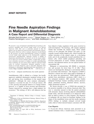
  • 2. Diagnostic Cytopathology DOI 10.1002/dc MALIGNANT AMELOBLASTOMA; FNA examination of the surgical (resection) specimen, a well circumscribed partially cystic tumor was identified in the posterior region of the mandible. The tumor measured 7 3 7 3 3 cm and on sectioning showed a dense white cut surface with a large central cyst. Tissue sections were cut, fixed in 10% formalin, embedded in paraffin and cut 3–4 microns thick then stained with H&E. Cytologic Findings The aspirates and cell blocks from both lymph nodes showed similar findings. Smears were hypercellular and composed of sheets, clusters, and singly disposed basaloid cells (Figs. C-1A and B). The basaloid cell clusters con- tained central loose spindled and stellate-shaped cells as well as peripherally arranged columnar cells with pali- Fig. 1. CT scan showing a 7 cm circumscribed unicystic lytic lesion involv- saded deeply-chromatic nuclei and clear cytoplasm (Fig. ing the body, angle, and condyle of the mandible (see arrow). This was asso- C-1C). These were morphologically consistent with ame- ciated with enlarged submandibular cervical lymph nodes (arrow head). loblast-like cells. Another interesting and striking low power finding was the arrangement of tumor cells in lin- slowly increasing, left facial swelling. On examination, a ear streams (Fig. C-1D). Cells had high nuclear to cyto- fixed, non-tender, expansile mass was identified in the left plasmic ratios with oval to round, or spindled nuclei and submandibular area. Oral examination revealed granular, scant pale cytoplasm (Fig. C-2A). Nuclear molding was erythematous mandibular gingival mucosa, in the region also a prominent feature. Nuclei had fine powdery chro- of the posterior mandibular molar teeth overlying the matin, small peripheral nucleoli (Fig. C-2B) and focal nu- mass. The mandibular buccal and lingual plates were clear ‘‘holes’’. There was minimal nuclear pleomorphism expanded, and the mandible was deviated to the right but however, and rare mitotic figures were seen (Fig. C-2A). there was no associated dysfunction or malocclusion. A Occasional benign squamous cells, rosettes, and stromal computerized tomography (CT) scan revealed a 7 3 7 3 fragments were also identified (Fig. C-2C). In addition, 3 cm circumscribed, lytic lesion involving the mandibular mature lymphocytes with standard maturation sequence body, angle, and condyle and coronoid processes (Fig. 1). and occasional lymphoglandular bodies were seen (Fig. This was associated with multiple enlarged left-sided lev- C-2D). On low power, the lymphocytes were almost els I, II, and III cervical lymph nodes, ranging in size indistinguishable from the basaloid tumor cells. The cell from 15 mm to 4.5 cm. The radiologic findings were felt block sections showed similar basaloid cells with high nu- to be consistent with an AB with likely metastatic disease clear to cytoplasmic ratios, embedded in dense eosino- to multiple cervical lymph nodes. An incisional biopsy of philic stroma (Fig. C-3A). the mandibular mass was performed at an outside oral surgery clinic and was microscopically confirmed to be Histologic Findings AB. The patient was then referred to the Medical College The H and E-stained sections from the mandibular resec- of Georgia for management. FNA was performed on two tion and biopsy specimens were composed of anastomoz- enlarged levels I and II cervical lymph nodes, and showed ing islands and sheets of stellate and basaloid cells with a cytologic features consistent with metastatic AB. On the follicular and plexiform arrangement (Fig. C-3B). These basis of the latter finding, a diagnosis of malignant AB were surrounded by dense fibrous stroma. The basaloid was made. A left hemi-mandibulectomy and left cervical cells had high nuclear to cytoplasmic ratios with minimal lymph node dissection were then performed. atypia, while the more central stellate-shaped cells resembled the stellate reticulum in a developing tooth Materials and Methods enamel organ (Fig. C-3C). Focal central squamous differ- FNA was performed using 25-gauge needles and 10-ml entiation was also present. The peripheral ameloblast-like syringes and material was collected as needle and syringe columnar cells had clear cytoplasm and elongated pali- washes. Air-dried and alcohol-fixed smears were prepared saded nuclei that were positioned away from the basement and stained with the Diff-Quik (DQ), Papanicolaou (Pap), membrane (Fig. C-3C). The follicular arrangement of tu- and hematoxylin and eosin (H&E) stains, respectively. mor cells closely resembled cellular aggregates within the Material was also submitted in phosphate buffered saline cytologic smears (Fig. C-3D). There was no overt evi- for cell block preparation, placed in 10% formalin, em- dence of carcinoma. Left neck dissection revealed 2 of 45 bedded in paraffin and stained with the H&E stain. On positive lymph nodes. Diagnostic Cytopathology, Vol 37, No 8 587
  • 3. Diagnostic Cytopathology DOI 10.1002/dc REID-NICHOLSON ET AL. Fig. C-1. A. Hypercellular smear with clusters and singly disposed basaloid cells (DQ stain, 340). B. Cluster of basaloid cells with central loose spindled stellate reticulum-like cells and focal peripherally palisading tumor cells (DQ stain, 3100). C. The elongated tumor cells which are present on the periphery of branching clusters (arrow) are consistent with ameloblasts (DQ stain, 3200). D. Smear showing linear streaming arrangement of single tumor cells (DQ stain, 3200). Discussion as the follicular and plexiform subtypes) have little clini- AB is divided into two main types, the multicystic (MC cal significance. The acanthomatous subtype of AB AB) variant, which accounts for 80% of tumors and deserves special mention as in addition to its conventional occurs in the mandible of adults5 and the unicystic (UC AB histology it also exhibits extensive squamous differen- AB) variant, which accounts for 10–15% and is more fre- tiation and keratin formation, which may be mistaken for quently observed in the posterior mandible of children squamous cell carcinoma (SQCA). and adolescents.1 On X-Ray, MC AB has a multilocular Malignant or metastasizing AB is histologically iden- ‘‘soap bubble’’ appearance,1 whereas UC AB appears as a tical to conventional AB but is associated with metasta- circumscribed unilocular ‘‘cyst-like’’ lucency, which often sis. The most common sites of metastasis include the surrounds an unerupted tooth. The two most common his- lung and cervical lymph nodes.1 In contrast, ABC has tologic subtypes of AB are the follicular and plexiform frankly malignant histology, and may also show areas of variants. The follicular variant exhibits islands and sheets conventional AB.1,4 ABC may arise de novo or as a of stellate-shaped cells with central cyst formation. The transformation of conventional AB.5 Most cases of plexiform subtype consists of anastomozing cords of MAB and ABC are diagnosed in adults and are aggres- stellate-shaped tumor cells with two to three layers of sive tumors, with poor clinical outcome and 50% of ameloblast-like cells peripherally. Various less frequent patients documented in the literature have died of their histologic subtypes are also described but these (as well disease.1 588 Diagnostic Cytopathology, Vol 37, No 8
  • 4. Diagnostic Cytopathology DOI 10.1002/dc MALIGNANT AMELOBLASTOMA; FNA Fig. C-2. A. Smear showing tumor cells with high nuclear/cytoplasmic ratios, nuclear grooves, and focal nuclear molding. Columnar cells are present in the lower right and a single central mitotic figure is also present (Papanicolaou stain, 3400). B. Nuclei have fine powdery chromatin and small nucleoli. Note central benign squamous cell (Papanicolaou stain, 3600). C. Note centrally located benign squamous cell (C-1) (DQ stain, 3200) and rosette (C-2) (Hematoxylin and Eosin stain, 3600). D. DQ stain showing admixture of basaloid tumor cells, mature lymphocytes, and central lympho- glandular body (arrow) (DQ stain, 3400). The cytologic diagnosis of conventional AB has been stromal fragments, and granular cells with abundant cyto- well documented in the literature.6–15 In fact, Ucok et al. plasm. The latter is seen in the granular cell variant of AB, in their report of over 40 cases of AB found that preoper- which has also been definitively diagnosed on cytology.7 ative FNA diagnosis of AB was not only possible but Although the cytology of conventional AB has been also an invaluable non-invasive tool for determining the previously reported, only a handful of reports have need for, and type of, surgical management of these described the cytologic features of malignant AB.10,15–17 patients.14 Despite its intraosseous location, AB is amena- The first such report was by Levine et al. in 1981.10 On ble to FNA, because the tumor often causes marked thin- cytology MAB is indistinguishable from conventional AB ning of the overlying cortex. On cytology AB is charac- and unless identified in locations distant from the primary terized by (1) small basaloid cells with high nuclear to tumor cannot be classified as malignant on cytology cytoplasmic ratios, minimal atypia, fine chromatin, vari- alone. Because ABC shows frank malignant cytologic fea- able nuclear molding, and spindling, (2) peripheral colum- tures, in addition to areas of conventional AB,4,5,18–20 cy- nar-type cells with palisaded basophilic nuclei, and (3) tology alone can be used to distinguish this tumor from benign squamous cells.11–13 These squamous cells are conventional AB and MAB. However, there are only iso- more abundant in the acanthomatous variant of AB. Other lated reports in the literature that describe the cytologic less frequently reported cytologic findings include rosettes, features of ABC.20,21 Diagnostic Cytopathology, Vol 37, No 8 589
  • 5. Diagnostic Cytopathology DOI 10.1002/dc REID-NICHOLSON ET AL. Fig. C-3. A. Cell block section showing basaloid tumor cells with surrounding dense stroma (Hematoxylin and Eosin stain, 3400). B. Tissue section from needle core biopsy of intraosseous tumor showing islands of tumor cells with both follicular and plexiform arrangement of cells (Hematoxylin and Eosin stain, 3200). C. Tissue section showing a cluster of tumor cells with central loosely arranged stellate reticulum and peripheral columnar ameloblasts (Hematoxylin and Eosin stain, 3400). D. Smear showing cluster of central spindled stellate reticulum and peripheral columnar ameloblasts (Papanicolaou stain, 3200). Despite the obvious benefit of the preoperative FNA diag- illary lesion are seen. These include small cell carcinoma, nosis of AB, MAB, and ABC there are several limitations lymphoma, adenoid cystic carcinoma (ACC), poorly dif- to FNA. These include inadequate sampling due to exten- ferentiated SQCA, and ameloblastic fibroma (AF). The sive cyst formation within the tumor. This may lead to small basaloid cells of AB may show prominent nuclear false-negative results which can be especially dangerous molding similar to that seen in small cell carcinoma how- in ABC. Failure to identify frankly malignant tumor cells ever unlike small cell carcinoma the nuclei of AB have can lead to misclassification of ABC as conventional AB. finely dispersed chromatin and not the typical ‘‘salt and In fact, there is one such report in the literature in which pepper’’ chromatin pattern of small cell carcinoma. Malig- an FNA of ABC was read as negative because of ‘‘a geo- nant lymphoma may also resemble the basaloid cells of graphic miss’’ of frankly malignant tumor cells.22 The AB however unlike AB background smears of lymphoma accurate preoperative diagnosis of AB is very important show prominent lymphoglandular bodies, which would because it helps prevent unnecessary or suboptimal surgi- not be seen in AB. In our case, an interesting finding in cal management. Another limitation of cytology is its the FNA of the submandibular lymph nodes was the pres- inability to distinguish conventional AB from MAB, with- ence of background lymphoglandular bodies. We attribute out prior knowledge of metastatic disease. this finding to partial nodal involvement by AB with sur- Several differentials should always be excluded when- rounding mature lymphocytes and their associated cyto- ever basaloid cells of an intraosseous mandibular or max- plasmic fragments. This phenomenon could be a potential 590 Diagnostic Cytopathology, Vol 37, No 8
  • 6. Diagnostic Cytopathology DOI 10.1002/dc MALIGNANT AMELOBLASTOMA; FNA confounding factor in the FNA diagnosis of AB. ACC is 2. Gardner DG, Shear M, Philipsen HP, Coleman. Ameloblastomas. In: Barnes L, Eveson JW, Reichart P, Sidransky D, editors. World an aggressive salivary gland tumor that may also metastasize heath organization pathology and genetics. Head and neck tumors. to bone. The solid variant of ACC where one sees fewer 1st ed. Lyon, France: IARC Press; 2005. p 296–300. extracellular hyaline globules of basement membrane and 3. Heikinheimo K, Jee KJ, Niini T, et al. Gene expression profiling of abundant basaloid cells may look similar to AB. The ameloblastoma and human tooth germ by means of a cDNA micro- array. J Dent Res 2002;81:525–530. identification of these basement membrane globules is 4. Slootweg PJ, Muller H. Malignant ameloblastoma or ameloblastic probably the most helpful cytologic feature in distinguish- carcinoma. Oral Surg Oral Med Oral Pathol 1984;57:168–176. ing the two entities. Metastatic poorly differentiated 5. Mendenhall WM, Werning JW, Fernandes R, Malyapa RS, Menden- SQCA with a predominant basaloid pattern and few kera- hall NP. Ameloblastoma. Am J Clin Oncol 2007;30:645–648. tinized cells may resemble the acanthomatous variant of 6. Choudhury M, Dhar S, Bajaj P. Primary diagnosis of ameloblas- toma by fine-needle aspiration: A report of two cases. Diagn Cyto- AB. However, SQCA would have frankly malignant fea- pathol 2000;23:414–416. tures, which would not be expected in conventional AB 7. Deshpande A, Umap P, Munshi M. Granular cell ameloblastoma of or MAB. Two additional differentials that are more spe- the jaw. A report of two cases with fine needle aspiration cytology. cifically related to our case are metastatic lobular carci- Acta Cytol 2000;44:81–85. 8. Gunhan O. Fine needle aspiration cytology of ameloblastoma. A noma of the breast and polymorphous low grade adeno- report of 10 cases. Acta Cytol 1996;40:967–969. carcinoma of salivary gland. These two differentials were 9. Gunhan O, Finci R, Celasun B, Demiriz M. A case of ameloblas- considered because of the unique and striking linear toma diagnosed by fine-needle aspiration cytology. J Nihon Univ arrangement of tumor cells in our patient’s lymph node Sch Dent 1989;31:565–569. FNAs, a finding that has not been previously described in 10. Levine SE, Mossler JA, Johnston WW. The cytologic appearance of metastatic ameloblastoma. Acta Cytol 1981;25:295–298. the cytology literature. The presence of spindled basaloid 11. Okada H, Matsumoto T, Yamamoto H. Imprint cytology of amelo- cells and squamous cells would not be expected in either blastoma. J Oral Sci 2002;44:97–101. of the latter two tumors and so distinction from AB is 12. Parate SN, Anshu, Helwatkar SB, Munshi MM. Cytology of recur- possible. AF is a primary intraosseous tumor that should rent ameloblastoma with malignant change. A case report. Acta Cytol 1999;43:1105–1107. be distinguished from AB. Both tumors show a predomi- 13. Radhika S, Nijhawan R, Das A, Dey P. Ameloblastoma of the man- nance of basaloid cells with peripheral tumor cell palisad- dible: Diagnosis by fine-needle aspiration cytology. Diagn Cytopa- ing.23 AF is also common in children and adolescents, as thol 1993;9:310–313. is UC AB. AF, however, has more stromal fragments than 14. Ucok O, Dogan N, Ucok C, Gunhan O. Role of fine needle aspira- tion cytology in the preoperative presumptive diagnosis of amelo- AB and this perhaps is the most helpful cytologic feature blastoma. Acta Cytol 2005;49:38–42. that may distinguish the two entities. 15. Weir MM, Centeno BA, Szyfelbein WM. Cytological features of In summary, we present a case of malignant AB, which malignant metastatic ameloblastoma: A case report and differential was diagnosed as such on FNA. This partly clinical as diagnosis. Diagn Cytopathol 1998;18:125–130. 16. Sharma S, Misra K, Dev G. Malignant ameloblastoma. A case well as cytologic diagnosis was facilitated by the presence report. Acta Cytol 1993;37:543–546. of typical AB cytology in enlarged cervical lymph nodes 17. Campbell D, Jeffrey RR, Wallis F, Hulks G, Kerr KM. Metastatic adjacent to a histologically confirmed mandibular AB. pulmonary ameloblastoma. An unusual case. Br J Oral Maxillofac Although cytology is helpful in diagnosing AB it does not Surg 2003;41:194–196. distinguish between conventional AB and malignant AB. 18. Corio RL, Goldblatt LI, Edwards PA, Hartman KS. Ameloblastic carcinoma: A clinicopathologic study and assessment of eight cases. Adequate sampling is also important when attempting to Oral Surg Oral Med Oral Pathol 1987;64:570–576. distinguish conventional AB from ABC. Because there 19. Akrish S, Buchner A, Shoshani Y, Vered M, Dayan D. Ameloblas- are several cytologic mimics of AB, a high index of sus- tic carcinoma: Report of a new case, literature review, and compari- son to ameloblastoma. J Oral Maxillofac Surg 2007;65:777–783. picion is always necessary when evaluating FNA material 20. Khalbuss WE, Loya A, Bazooband A. Fine-needle aspiration cytol- from jaw lesions and/or regional lymph nodes. The need ogy of pulmonary ameloblastic carcinoma of mandibular origin. for correlation of cytologic findings with clinical and Diagn Cytopathol 2006;34:208–209. radiologic information cannot be overstated. 21. Ingram EA, Evans ML, Zitsch RP, III. Fine-needle aspiration cytol- ogy of ameloblastic carcinoma of the maxilla: A rare tumor. Diagn Cytopathol 1996;14:249–252. 22. Verneuil A, Sapp P, Huang C, Abemayor E. Malignant ameloblas- References toma: Classification, diagnostic, and therapeutic challenges. Am J 1. Waldron CA. Odontogenic cysts and tumors. In: Neville BW, Otolaryngol 2002;23:44–48. Damm DD, Allen CM, Bouquot JE, editors. Oral and maxillofacial 23. Kumar N, Jain S. Aspiration cytology of ameloblastic fibroma: A pathology. 2nd ed. Pennsylvania: Saunders; 2002. p 589–642. diagnostic challenge. Diagn Cytopathol 2003;29:101–104. Diagnostic Cytopathology, Vol 37, No 8 591