Aortic Dissection: Causes, Symptoms, and Classification
•Descargar como DOCX, PDF•
1 recomendación•1,134 vistas

TO WHOM I CONCERN MY FAMILY

Recomendados
Más contenido relacionado
La actualidad más candente
La actualidad más candente (20)
Destacado
Destacado (20)
Similar a Aortic Dissection: Causes, Symptoms, and Classification
Similar a Aortic Dissection: Causes, Symptoms, and Classification (20)
Más de cardilogy
Más de cardilogy (20)
Último
Último (20)
Aortic Dissection: Causes, Symptoms, and Classification
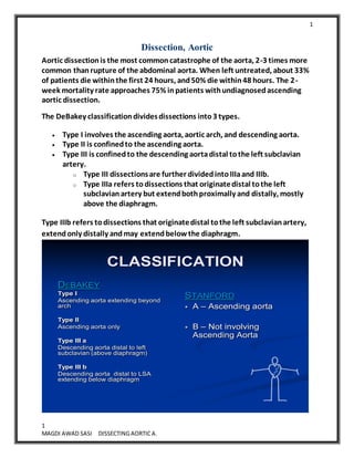
- 1. 1 1 MAGDI AWAD SASI DISSECTINGAORTICA. Dissection, Aortic Aortic dissectionis the most commoncatastrophe of the aorta, 2-3 times more common thanrupture of the abdominal aorta. When left untreated, about 33% of patients die withinthe first 24 hours, and50% die within48 hours. The 2- week mortality rate approaches 75% inpatients withundiagnosedascending aortic dissection. The DeBakey classificationdividesdissections into3 types. Type I involves the ascending aorta, aortic arch, and descending aorta. Type II is confinedto the ascending aorta. Type III is confinedto the descending aortadistal tothe left subclavian artery. o Type III dissectionsare further dividedintoIIIaand IIIb. o Type IIIa refers todissections that originatedistal tothe left subclavianartery but extendbothproximally and distally, mostly above the diaphragm. Type IIIb refers todissections that originatedistal tothe left subclavianartery, extendonly distally andmay extendbelowthe diaphragm. CLASSIFICATION DEBAKEY Type I Ascending aorta extending beyond arch Type II Ascending aorta only Type III a Descending aorta distal to left subclavian (above diaphragm) Type III b Descending aorta distal to LSA extending below diaphragm STANFORD A – Ascending aorta B – Not involving Ascending Aorta
- 2. 2 2 MAGDI AWAD SASI DISSECTINGAORTICA. Thoracic aortic dissections shouldbe distinguishedfromaneurysms (ie, localized abnormal dilationof the aorta) and transections, whichare causedmost commonly by high-energy trauma. Race Aortic dissectionis more commonin blacks than inwhites and less common in Asians thanin whites. Sex The male-to-female ratiois 3:1. Age 75% of dissections occur inthose aged40-70 years, withapeak in 50-65 years
- 3. 3 3 MAGDI AWAD SASI DISSECTINGAORTICA. Pathophysiology: The essential feature of aortic dissectionis atear in the intimal layer, followedby formationand propagationof a subintimal hematoma. The dissecting hematoma commonly occupies about half and occasionally the entire circumference of the aorta. This produces a false lumenor double-barreledaorta, which can reduce bloodflow to the major arteries arising fromthe aorta. If the dissectioninvolves the pericardial space, cardiac tamponade may result. Cystic medial necrosis The normal aorta contains collagen, elastin, andsmoothmuscle cells that contribute the intima, media, and adventitia, whichare the layers of the aorta. Withaging, degenerativechanges leadtobreakdownof the collagen, elastin, and smooth muscle and an increase in basophilic groundsubstance. This conditionis termedcystic medial necrosis. Atherosclerosis that causes occlusion
- 4. 4 4 MAGDI AWAD SASI DISSECTINGAORTICA. of the vasa vasorum alsoproduces this disorder. Cystic medial necrosis is the hallmark histologic change associatedwithdissectioninthose with Marfan syndrome. Researchershave usedthe termcystic medial degeneration inelder patients. Early on, cystic medial necrosis describedanaccumulationof basophilic ground substance inthe media withthe formationof cystlike pools. The mediain these focal areas may show loss of cells (ie, necrosis). This termstill is usedcommonly to describe the histopathologic changes that occur. Dissectionsites The most common site of dissectionis the first fewcentimetersof the ascending aorta, with90% occurring within10 centimetersof the aortic valve. The second most common site is just distal tothe left subclavianartery. Between5% and 10% of dissectionsdonot have an obvious intimal tear. These oftenare attributedtorupture of the aortic vasa vasorum as first describedby Krukenberg in1920.
- 5. 5 5 MAGDI AWAD SASI DISSECTINGAORTICA. Mortality/Morbidity From 1-2% of patients withaortic dissectiondie per hour for the first 24- 48 hours. Aortopathy may be present inheritable diseases suchas Marfan syndrome, Ehlers-Danlos syndrome, annuloaortic ectasia, familial aortic dissections, adult polycystic kidney disease, Turner syndrome, Noonan syndrome, osteogenesisimperfecta, bicuspidaortic valve, coarctationof the aorta, and connective tissuedisorders. It is alsoseeninheritable metabolic disorders suchas homocystinuriaandfamilial hypercholesterolemia. Incidence is increasedinpregnancy and syphilis. Thoracic aortic dissection also is associatedwithcrack cocaine use and iatrogenic causes, suchas cardiac catheterization. SYMPTOMS: No one signor symptom can positively identify acute aortic dissection(AAD). An estimated 38% of acute aortic dissections are missedoninitial evaluation There are no validatedclinical decisionrulesto helpidentify acute aortic dissection(ADD). The diagnosis is best made whenthere is highclinical suspiciongiventhe overall evaluationof the patient, including the history, physical examination, and supporting testsincluding ECG, laboratory studies, and radiology. ". . . Spontaneous tear of the arterial coats is associatedwithatrocious pain, with symptoms, indeed, inthe case of the aortaof angina pectoris andmany instances have beenmistakenfor it"WilliamOsler, 1910. Chest pain is the most common presenting symptominpatients withan aortic dissection. Consider thoracic aortic dissectioninthe differential diagnosis of all patients presenting with sudden chest pain. o The pain usually is describedas ripping or tearing. o The suddenonset of chest painhas beenshown to have a sensitivity of 84%. o This descriptionis not universal, andsome patients present with only mildpain, oftenmistakenfor musculoskeletal conditions, locatedin the thorax, groin, or back.
- 6. 6 6 MAGDI AWAD SASI DISSECTINGAORTICA. o The pain of aortic dissectiontypically is abrupt onset. o Aortic dissectionshouldbe consideredstrongly inall patients reporting acute, sudden, andsevere chest painthat is maximal at onset. o The nervi vascularis, bundles of nerve fibers foundinthe aortic adventitia, are involvedinthe productionof pain. The descriptionof the pain may indicate where the dissectionarises. o Anterior chest painandchest pain that mimics AMI usually are associatedwithanterior archor aortic root dissection. This is causedby the dissectioninterrupting flowtothe coronary arteries, resulting inmyocardial ischemia. o Pain that is describedinthe neck or jaw indicates that the dissectioninvolves the aortic archand extends intothe great vessels of the arch. o Tearing or ripping pain that is felt inthe intrascapular areamay indicate that the dissectioninvolves the descending aorta. The pain typically changes as the dissectionevolves. Aortic dissectionis painlessinabout 10% of patients. Painless dissectionis more common in those withneurologic complications fromthe dissection and those withMarfan syndrome. Presenting signs andsymptoms inacute thoracic aortic dissectioninclude: o Anterior chest pain - Ascending aortic dissection o Neck or jaw pain - Aortic archdissection o Interscapular tearing or ripping pain - Descending aortic dissection o Chest pain o Myocardial infarction o Neurologic symptoms Syncope Stroke symptoms Alteredmental status Limb paresthesias, pain, or weakness Hemiparesis or hemiplegia Horner syndrome o Dyspnea o Dysphagia
- 7. 7 7 MAGDI AWAD SASI DISSECTINGAORTICA. o Orthopnea o Anxiety andpremonitions of death o Flank pain if renal artery is involved o Dyspneaand hemoptysis if dissectionruptures intothe pleura SUMMARY=AAA is characterizedby: 1. Acute chest painwithacute inferior MI 2. Acute LVF/PULMONARY ODEMAdue toacute AR 3. Acute CVA AND wide mediastinum (aortic shadow widened) ABDOMINAL AORTA ANEURYSM Nonspecific abdominal symptoms (constipation,pain) Pulsating abdominal mass/backage Lower limbs intermittent cludication Physical Bloodpressure may increase or decrease. o Hypertensionmay result froma catecholamine surge or underlying essential hypertension. o Hypotensionis an ominous finding and may be the result of excessivevagal tone, cardiac tamponade, or hypovolemiafrom rupture of the dissection. o A bloodpressure differential of greater than20 mmHg was an independent predictorof aortic dissection. A pressuredifferential of greater than20 mm Hg shouldincrease the suspicionof aortic dissection, but it does not rule it in. Significant interarmblood pressure differentials may be found in 20% of people without aortic dissection. Neurologic deficits are a presenting sign in up to 20% of cases. o The most common neurologic findings are syncope and altered mental status. o Syncope is part of the early course of aortic dissection in about 5% of patients and may be the result of increased vagal tone, hypovolemia, or dysrhythmia. o Other causes of syncope or altered mental status include strokes from compromised blood flow to the brain or spinal cord and ischemia from interruption of blood flow to the spinal arteries. o Peripheral nerve ischemia can manifest with numbness and tingling in the extremities.
- 8. 8 8 MAGDI AWAD SASI DISSECTINGAORTICA. o Hoarseness from recurrent laryngeal nerve compression also has been described from AAA (ASCENDING AORTA). o Horner syndrome is caused by interruption in the cervical sympathetic ganglia and presents with ptosis, miosis, and anhidrosis. o Superior vena cava syndrome, caused by compression of the superior vena cava from a large distorted aorta, may occur. Dyspnea may be causedby congestive heart failure or tracheal or bronchial compression. Dysphagia from compressionof the esophagus may be present. Findings suggestive of cardiac tamponade, such as muffledheart sounds, hypotension, pulsus paradoxus, jugular venous distension, andKussmaul sign, must be recognizedquickly. Other diagnostic clues include anewdiastolic murmur, asymmetrical pulses, andasymmetrical bloodpressure measurements. Pay careful attentiontocarotid, brachial, and femoral pulses on initial examination and look for progressionof bruits or development of bruits on reexamination. Physical findings of a hemothorax may be found if the dissectionruptures intothe pleura. Causes Aortic dissection is more common in patients with hypertension, connective tissue disorders, congenital aortic stenosis or bicuspid aortic valve, and in those with first-degree relatives with history of thoracic dissections. These diseases affect the media of the aorta and predispose it to dissection. Aortopathy may be due to the following heritable diseases: A . Hereditary Connective Tissue Diseases Marfan Syndrome Ehler Danlos Syndrome B. Chromosomal Aberrations Turners Syndrome Noonans Syndrome C. Aortic Diseases
- 9. 9 9 MAGDI AWAD SASI DISSECTINGAORTICA. Aortic Dilatation Aortic Aneurysm Anuloaortic ectasia Aortic Arteritis Bicuspid Aortic Valve Hypertension or pulsatile blood flow can propagate the dissection. An estimated 50% of all cases of aortic dissection that occur in women younger than 40 years are associated with pregnancy. Syphilis may cause aortic dissection. Crack cocaine use may precipitate aortic dissection. Iatrogenic causes of aortic dissection include cardiac catheterization. Differential Diagnoses Aortic Regurgitation Pericarditis and Cardiac Tamponade Back Pain, Mechanical Pleural Effusion Gastroenteritis Pulmonary Embolism HypertensiveEmergencies Shock, Cardiogenic Myocardial Infarction Shock, Hypovolemic Thoracic Outlet Syndrome Pancreatitis Laboratory Studies Blood studies o Usually, the diagnosis is made before the blood work is returned;however, leukocytosis may be present. o BUN and creatinine are elevated if the dissection involves the renal arteries. o Troponin and creatine kinase (CK) can be elevated if the dissection has caused myocardial ischemia. o Decreases in the hemoglobin and hematocrit are ominous findings suggesting the dissection either is leaking or has ruptured. o Some studies suggest that D-dimer should be a part of the initial workup if aortic dissection is suspected.Anegative result makes the presence of the disease less likely. o Hematuria, oliguria, and even anuria (<50 mL/d) may occur if the dissection involves the renal arteries. o Routine bloods – non diagnostic o D-dimer < 500ng/ml unlikely to be dissection
- 10. 10 10 MAGDI AWAD SASI DISSECTINGAORTICA. ECG All patients with suspected thoracic aortic dissection should have an ECG. In acute thoracic dissection, ECG can mimic the changes seen in acute cardiac ischemia. In the presence of chest pain, these signs can make distinguishing dissection from AMI very difficult . Keep this in mind when administering thrombolytics to patients with chest pain. ST elevation can be seen in Stanford type A dissections because the dissection interrupts blood flow to the coronary arteries. The incidence of abnormal ECG findings is greater in Stanford type A dissections than in other types of dissections. In one study, 8% of patients with type A dissections had ST elevation, while no patients with type B dissections had ST elevation. More commonly, the ECG abnormality is ST depression. If the dissection involves the coronary ostia, it is the right coronary artery that is most commonly involved, leading to inferior ST-segment elevation pattern. DIAGNOSTIC EVALUATION Chest radiograph Tran thoracic echocardiogram Tran esophageal echocardiogram* Computed tomography* Magnetic resonance imaging* Aortography *Choice based on rapid availability and quality of performance
- 11. 11 11 MAGDI AWAD SASI DISSECTINGAORTICA. Chest radiography Findings are abnormal in 80% of patients and are abnormal in ascending aorta D. A widened mediastinum is sometimes difficult to identify on a portable anteroposterior (AP) radiograph. If the patient is hemodynamically stable and cooperative, an AP radiograph can be obtained at bedside. Look for a mediastinal width greater than 8 cm on AP chest radiograph. A tortuous aorta, common in hypertensive patients, may be hard to distinguish from a widened mediastinum(44-80%). If in doubt, a good posterior-anterior radiograph is recommended. The differential diagnosis of a widened mediastinum includes tumor, adenopathy, lymphoma, and enlarged thyroid. Abnormal (ie, blunted) aortic knob was observed in 66% of patients in one study. Ring sign (displacement of the aorta >5 mm past the calcified aortic intima) is considered a specific radiographic sign. Calcium sign -Displaced intimal calcification (>10mm) from outer aortic wall– useful in older patients. Other radiologic abnormalities seen on chest radiography include the following: Left apical cap Tracheal deviation Depression of left main stem bronchus Esophageal deviation Loss of the paratracheal stripe Over 12% of the chest radiographs of patients with aortic dissection were read as normal. Several studies concluded that the overall diagnosis of aortic dissection is not determined by any one sign, rather a combination of all findings leads to suspicion of dissection.
- 12. 12 12 MAGDI AWAD SASI DISSECTINGAORTICA. TTE Indicated as an initial test if patient is veryunwell and other imaging not available. Can be performed bedside ,Can detect intimal flap and AR Limitation: No information beyond aortic root and early part of proximal aorta. TRANSESOPHAGEAL ECHO Procedure of first choice for dissection, if readily available Portability of equipment facilities in emergency to ER or ICU High sensitivity (98%) and specificity(97%) Limitations :Unable to visualize distal part of ascending Aorta(beginning of aortic arch) and descending Aortabelow stomach .
- 13. 13 13 MAGDI AWAD SASI DISSECTINGAORTICA. Angiography Still considered by some as the diagnostic standard test for aortic dissection, it is being replaced by newer imaging modalities. Angiography leads to accurate diagnosis of aortic dissection in over 95% of patients and aids the surgeon in planning the repair operation because blood vessels of the arch can be assessed easily. Benefits include visualization of the true and false lumens, intimal flap, aortic regurgitation, and coronary arteries. Drawbacks include the following: The procedure is invasive. The patient must be transported to the radiology department . The use of contrast media may be harmful to patients who have renal insufficiency or an allergy to iodine. isdiagnoses can occur if the false channel is thrombosed. In this instance, the false lumen and intimal flap may not be visualized. Possible simultaneous opacification of the true and false lumens may make discerning the presence of a dissection difficult. Computedtomography (CT) scanning: The accepteddiagnostic criterionstandard, angiography, is being challengedby state-of-the-art CTangiography. Withthe advent of helical CT withmultiplanar and 3D reconstructionandCT angiography, CT scanning is quickly replacing angiography as the diagnostic test of choice in many institutions Spiral CT scanning is associated with a higher rate of detection and better resolution than incremental CT scanning. High-quality 2D and 3D reconstructions are possible with spiral CT scanning, which greatly adds to the usefulness of this imaging modality. More importantly, imaging information, including the type of lesion, location of the pathologic lesion, extent of the disease, and evaluation of the true and false lumen can be assessed quickly and help the surgeon plan the operation. This information helps determine if hypothermic circulatory arrest is necessary for surgery; this procedure increases the complexity, length, morbidity, and mortality associated with surgery.
- 14. 14 14 MAGDI AWAD SASI DISSECTINGAORTICA. Faster scanners have decreased the acquisition time to the range of a breath hold, resulting in less motion artifact from breathing. Drawbacks include the following: Transportation of a patient in potentially unstable condition from the ED, places the patient at risk. Requires the injection of iodinated contrast. The use of contrast material may harm a patient who has impaired renal function or an allergy to contrast media. No information on aortic regurgitation MRI MRI has over 90% sensitivity andgreater than95% specificity. It is the most sensitive methodfor diagnosing aortic dissectionandhas similar specificity toCTscanning. MRI shows the site of intimal tear, type and extent of dissection, andpresenceof aortic insufficiency, as well as the surrounding mediastinal structures. Other benefits are that MRI requires
- 15. 15 15 MAGDI AWAD SASI DISSECTINGAORTICA. no contrast mediumand no ionizing radiation. It is the preferredmodality for patients withrenal failure andthose withan allergy toiodine. Contrast-enhanced magnetic resonance angiography (CE-MRA) is a principle technique for evaluating the thoracic aorta. MRI is the preferred tool for imaging chronic dissections and postsurgical follow-up. Contrast 3D MRA is an accurate noninvasive imaging modality. It has the advantage of being able to evaluate the aortic valve more effectively than CT angiography. Drawbacks include the following: MRI is not readily available at most institutions, requiring transportation of patients in unstable condition away from the ED. MRI requires much more time to acquire images than CT scanning. Patients with permanent pacemakers cannot undergo MRI. Most patients with prosthetic heart valves or coronary stents can safely have an MRI. MRA • Good alternative toTEE or CT, if readily available • Highsensitivity (98%) and specificity (98%) • Provides three dimensional reconstruction • Can detect site of intimal tear and involvement of branch vessels • Non-invasive;neither x-rays nor contrast needed • Limitation: claustrophobic, more costly, not readily available
- 16. 16 16 MAGDI AWAD SASI DISSECTINGAORTICA. Immediate management: Initial therapy should begin when the diagnosis is suspected. This includes 2 large-bore intravenous lines (IVs), oxygen, respiratory monitoring, and monitoring of cardiac rhythm, blood pressure, and urine output.. Clinically, the patient must be assessed frequently for hemodynamic compromise, mental status changes, neurologic or peripheral vascular changes, and development or progression of carotid, brachial, and femoral bruits. Aggressive management of heart rate and blood pressure should be initiated. Beta-blockers should be given initially to reduce the rate of change of blood pressure and the shear forces on the aortic wall. The target heart rate should be 60-80 beats per minute. The target systolic blood pressure should be 100-120 mm Hg. Treat hypotension/ hypertension –aim for MAP 60-70 Betablockers eg esmalol, propranolol, labetalol Vasodilators Nanitroprusside Calcium channel blockers eg verapamil, diltiazem Indications for Definitive Surgical and Medical Therapy in AD
- 17. 17 17 MAGDI AWAD SASI DISSECTINGAORTICA. Urgent surgical intervention is required in type A dissections. The area of the aorta with the intimal tear usually is resected and replaced with a Dacron graft. The operative mortality rate is usually less than 10%. Dissections involving the arch are more complicated that those involving only the ascending aorta because the innominate, carotid, and subclavian vessels branch from the arch. Deep hypothermic arrest usually is required. If the arrest time is less than 45 minutes, the incidence of central nervous system complications is less than 10%. The definitive treatment for type B dissections is less clear(distal D.). Control blood pressure. Mortality rate - the same medically treated or surgically. Surgery is reserved for distal dissections that are leaking, ruptured, or compromising blood flow to a vital organ. Acute distal dissections in patients with Marfan syndrome usually are treated surgically. Inability to control hypertension with medication is also an indication for surgery. Patients with a distal dissection are usually hypertensive, emphysematous, or older. Long-term medical therapy involves a beta-adrenergic blocker combined with other antihypertensive medications. Avoid antihypertensives (eg, hydralazine, minoxidil) that produce a hyperdynamic response that would increase dP/dt (ie, alter the duration of P or T waves). Survivors of surgical therapy also should receive beta-adrenergic blockers. Definitive treatment involves segmental resection of the dissection with interposition of a synthetic graft. When thoracic dissections are associated with aortic valvular disease, replace the defective valve. With combined reconstruction–valve replacement, the operative mortality rate is approximately 5% with a late mortality rate of less than 10%. Operative repair of the transverse aortic arch is technically difficult, with an operative mortality rate of 10% despite induction of hypothermic cardiocirculatory arrest. Repair of the descending aorta is associated with a higher incidence of paraplegia than repair of other types of dissections because of interruption of segmental blood supply to the spinal cord. The operative mortality rate is approximately 5%
- 18. 18 18 MAGDI AWAD SASI DISSECTINGAORTICA. Sodium Nitroprusside Causes peripheral vasodilationby direct actionon venous and arteriolar smoothmuscle, thus reducing peripheral resistance. Commonly usedIV because of rapid onset and short durationof action. Easily titratable toreachdesiredeffect. Light sensitive; bottle andtubing shouldbe wrappedin aluminum foil. Prior to initiating nitroprusside, administer beta-blocker tocounteract physiologic response of reflex tachycardiathat occurs whennitroprusside usedalone. This physiologic response increases shear forcesagainst aortic wall, thus increasing dP/dt. Objectiveis tokeepheart rate at 60-80 bpm Sodium Nitroprusside for acute reduction starting 10 – 20 mcg/min and titrated upward Adding IV BB prior until desired effect such as HR 60 – 80s (propranolol 1 mg Q 3-5 minutes max 10 mg) Then Q 4-6 hrs at a dose of 2 – 6 mg 0.5-3 mcg/kg/min IV; rates >4 mcg/kg/min may lead to cyanide toxicity Labetolol Effectively lowersdP/dTas well as reducing arterial pressure Blocks alpha-, beta1-, and beta2-adrenergic receptor sites, decreasing BP. Initial dose is 20mg followedby 40 to 80 mg Q 10 – 15 minutes (max 300mg IV) Once BP controlledmaintenance by continuous infusion Infusionat 2mg/min titrating upto5 –10 mg/min Esmolol Ultrashort acting BB for those withlabile blood pressure or those that are surgical candidates. (Long acting medications may affect intraoperative bp management) Load with 500 mcg/kg bolus Infusionstarts @ 50mcg/kg/mintitrate to200 mcg/kg/minfor control Can be usedin patients withuncertainrisk for bronchospasm
- 19. 19 19 MAGDI AWAD SASI DISSECTINGAORTICA.