SlideShare una empresa de Scribd logo
1 de 21
Descargar para leer sin conexión
1 
ACUTE MYELOBLASTIC LEUKEMIA DR MAGDI AWAD SASI 2014 DETAILED APPROACH WITH SLIDES 
LEUKEMIA((LEUKAEMIA)) DEFINITION: Leukemia is a cancer of the blood cells, most generally white blood cells (WBCs). In leukemia, the WBCs have mutated and may divide too quickly, eventually crowding out normal functioning red and white blood cells. TYPES OF LEUKEMIA: There are several different types of leukemia. In general, leukemia is grouped by how fast it gets worse and what kind of white blood cell it affects. A. It may be acute or chronic. Acute leukemia gets worse very fast and may make patient feel sick right away. Chronic leukemia gets worse slowly and may not cause symptoms for years. B. It may be lymphocytic or myelogenous. Lymphocytic (or lymphoblastic) leukemia affects white blood cells called lymphocytes. Myelogenous leukemia affects the other type of cells that normally become granulocytes, red blood cells, or platelets. There are four main types of leukemia:  Acute myeloblastic leukemia (AML)  Chronic myelocytic leukemia (CML)  Acute lymphoblasti leukemia (ALL)  Chronic lymphocytic leukemia (CLL) Acute myeloid leukemia( AML ): Also known as acute myelogenous leukemia, acute myeloblastic leukemia, acute granulocytic leukemia or acute nonlymphocytic leukemia, is a fast-growing form of cancer of the blood and bone marrow. AML is the most common type of acute leukemia. It occurs when the bone marrow begins to make blasts, cells that have not yet completely matured. These blasts normally develop into white blood cells. However, in AML, these cells do not develop and are unable to ward off infections. In AML, the bone marrow may also make abnormal red blood cells and platelets. The number of these abnormal cells increases rapidly, and the abnormal (leukemia) cells begin to crowd out the normal white blood cells, red blood cells and platelets that the body needs. Due to their immaturity, they are unable to function properly to prevent or fight infection. Inadequate numbers of red cells and platelets being made by the marrow cause anaemia, and easy bleeding and/or bruising One of the main things that differentiates AML from the other main forms of leukemia is that it has eight different subtypes, which are based on the cell that the leukemia developed from. The types of acute myelogenous leukemia include:  Myeloblastic (M0) - on special analysis  Myeloblastic (M1) - without maturation  Myeloblastic (M2) - with maturation  Promyeloctic (M3)  Myelomonocytic (M4)
2 
ACUTE MYELOBLASTIC LEUKEMIA DR MAGDI AWAD SASI 2014 DETAILED APPROACH WITH SLIDES 
 Monocytic (M5)  Erythroleukemia (M6)  Megakaryocytic (M7) CHARACTERISTC FEATURES OF ACUTE LEUKEMIA: -The condition progresses rapidly and aggressively, requiring immediate treatment. -The site of the disease is bone marrow where all the cell lines may be depressed . -Bone marrow aspiration is mandatory for diagnosis (˃ 20% blasts ). -There is no enough time for the organs to be hugely enlarged (( may be palpable )). -The blood is circulating and malignant cells may invade the organs. -There is no surgical treatment for it. INCIDENCE: Acute leukaemia is an uncommon type of cancer. About 7,600 people are diagnosed with leukaemia each year in the UK. AML is an uncommon type of cancer. Around 2,600 people are diagnosed with the condition each year in the UK. AML can develop at any age, but it's more common in people over the age of 60. SYMPTOMS:  Many people often do not experience symptoms in the early stages of certain types of leukemia, or the symptoms develop slowly. Acute myeloid leukemia (AML) and acute lymphocytic leukemia (ALL) progress much faster and symptoms may worsen more quickly than with the chronic leukemias (CML and CLL).  Some leukemia symptoms, like night sweats, fever, fatigue and achiness, often resemble flu-like symptoms. If you have the flu, symptoms will likely subside as you get better. Make an appointment to see your doctor if the symptoms persist longer than expected.  Some general symptoms of leukemia include: 
Fever, chills 
Fatigue, weakness 
Loss of appetite, weight loss 
Night sweats 
Bone/joint pain 
Abdominal discomfort 
Headaches 
Shortness of breath 
Frequent infections 
Easy bruising or bleeding 
Petechiae (small red spots under the skin)
3 
ACUTE MYELOBLASTIC LEUKEMIA DR MAGDI AWAD SASI 2014 DETAILED APPROACH WITH SLIDES 
AML symptoms: 
Bone marrow depression—anemia , bleeding , infection 
Bony pain with sternal tenderness 
DIC , gum hyperatrophy 
S&S: 1. ANEMIA : 
Pallor 
Easy fatigability 
Dyspnea on mild exertion 
Lassitude 
Syncopal attacks 
Headache 
Palpitation 
Anorexia 
Taste disturbance 
Tinnitus 
Dizziness 
Dimension of vision 
Insomnia 
Angina 
Parasthesia in fingers and toes 
2. BLEEDING (( THROMBOCYTOPENIA)): 
Petechiae 
Bruisability 
Epistaxis 
Gingival bleeding (spongy bleeding) 
Melena 
Hematuria 
Purpura 
Spontaneous involving ---CNS , LUNG , GIT MUCOUS MEMBRANE 
Haemorrhage increased ˂ 20 x 10000/ml 
3. INFECTION: 
Is inversely related to number of circulating neutrophils 
Major risk in patient with granulocyte ˂ 0.5 x 10000/ml 
Neutrophil may function abnormally and comprises host defenses. 
The leukemia and its treatment cause a breakdown of mucosal barrier & systemic infections usually develop from skin, throat, GIT.
4 
ACUTE MYELOBLASTIC LEUKEMIA DR MAGDI AWAD SASI 2014 DETAILED APPROACH WITH SLIDES 
In leukocytosis, increase in blast cells of ˃ 100 x 10 000 /ml leads to leukostasis due to occlusion of the microcirculation by blast cells leading to hypoperfusion of lung and brain. 
Common sites of infections: 
Skin; Gingival; Ulcers in mouth and pharynx ;Perirectal tissues ;Lung;Urinary tract; 
Sore throat 
Septicemia usually occurs without an apparent cause. 
4. HEPATOMEGALLY AND SPLENOMEGALLY: 
Rare in AML. 
The visceral involvement can produce symptoms of nausea , abdominal fullness and satiety. 
Lymphoadenopathy –AML 50% 
Practical point—extramedullary leukemia can precede detectable involvement of B.M. 
5. EXTRAMEDULLARY TISSUES : 
Acute leukemia may infiltrate –skin, lung, eye, kidney ,nasopharynx 
CHLOROMAS----soft tissues masses of leukemic cells. 
6. SYMPTOMS RELATED TO THE EXPANDING MALIGNANT CELL MASS: 
Bony pain and sternal tenderness 
This occur in half of patients 
Osteolytic lesions are rare 
Renal abnormalities due to : 
A. Leukemic infiltration 
B. Ureteral obstruction by urate nephropathy—enlarged LN, uric acid stone, infections ,haemorrhage. 
GIT----bleeding, distention ,satiety ,constipation((due to organomegally or leukemic infiltrate)) . 
7. METABOLIC DISTURBANCE : 
Hypokalemia and hyponatremia due to renal involvement . 
LDH—lactic dehydrogenase may be increased. 
CLASSIFICATION OF ACUTE LEUKEMIA: 
Classification of acute leukemias used to rely entirely on morphology and histochemical changes, but now information regarding cytogenetic patterns and correlations with immunophenotype, oncogene expression, and gene mutation has changed the biology of this disease 
1. FAB classification(French-American-British) :1970 
A classification of acute leukemia produced by a three-nation joint collaboration. 
A schema that divides acute leukemias into lymphoid–ALL or myeloid–AML cell lines 
This is based on the study of microscopic features and cytochemistry of blast cells. 
Acute myelogenous leukemia is subdivided into eight types(M0-M7) based on the type of cell from which the leukemia developed and how mature the cells are.
5 
ACUTE MYELOBLASTIC LEUKEMIA DR MAGDI AWAD SASI 2014 DETAILED APPROACH WITH SLIDES 
Acute myeloid leukemia (AML) Subtypes M0 through M5 all start in precursors of white blood cells. M6 AML starts in very early forms of red blood cells, while M7 AML starts in early forms of cells that make platelets. M1 Myeloblasts without maturation M2 Myeloblasts with maturation (best AML prognosis) M3 Hypergranular promyelocytic leukemia (faggot cells) M3V Variant, microgranular promyelocytic leukemia M4 Myelomonocytic leukocytes M5 Monocytic, subtype a. Poorly differentiated monocytic leukemia b. Well differentiated monocytic leukemia M6 Erythroleukemia/DiGuglielmo syndrome M7 Megakaryocytic leukemia Pleomorphic undifferentiated cells with cytoplasmic blebs; myelofibrosis or ↑ BM reticulin; positive for platelet peroxidase antifactor VIII 
2. World Health Organization (WHO) classification of AML The FAB classification system is useful and is still commonly used to group AML into subtypes. But it doesn’t take into account many of the factors that are known to impact prognosis (outlook). In 2001, the World Health Organization (WHO) published a newer system that includes some of these factors to try to help better classify cases of AML based on a patient’s outlook. The WHO classification system divides AML into several broad groups: 1. AML with certain genetic abnormalities  AML with a translocation between chromosomes 8 and 21  AML with a translocation or inversion in chromosome 16  AML with changes in chromosome 11  APL (M3), which usually has translocation between chromosomes 15 and 17 2. AML with multilineage dysplasia (more than one abnormal myeloid cell type is involved) 3. AML related to previous chemotherapy or radiation 
4. AML not otherwise specified (includes cases of AML that don’t fall into one of the above groups; similar to the FAB classification)  Undifferentiated AML (M0)  AML with minimal maturation (M1)  AML with maturation (M2)  Acute myelomonocytic leukemia (M4)  Acute monocytic leukemia (M5)  Acute erythroid leukemia (M6)
6 
ACUTE MYELOBLASTIC LEUKEMIA DR MAGDI AWAD SASI 2014 DETAILED APPROACH WITH SLIDES 
 Acute megakaryoblastic leukemia (M7)  Acute basophilic leukemia  Acute panmyelosis with fibrosis  Myeloid sarcoma (also known as granulocytic sarcoma or chloroma) 5. Undifferentiated or biphenotypic acute leukemias (leukemias that have both lymphocytic and myeloid features). Sometimes called ALL with myeloid markers, AML with lymphoid markers, or mixed lineage leukemias. 
Morphological subtypes of AML: AML occurs more often in men and is usually diagnosed in individuals over 50, although it can occur at any age. M1 
 ((AML without maturation)) 
 20% of AML 
 Myeloblast predominant 
 Distinct nucleoli 
 Few Auer rods , Azurophilic granules 
 ( > 90% type I and II blasts ) of marrow nonerythroid cells is present. Most of the blasts are 
agranular. Auer rods are infrequent. 
 Staining: Relatively few blasts (5-10%) are MPO 
(myeloperoxidase) positive. A minimum of 3% MPO 
positive blasts are required for diagnosis. NSE and 
PAS are generally negative. 
 Immunophenotype: Variably positive for CD13, 
CD14, CD11b, CD33, and HLA-DR. 
 Chromosome Abnormalities: t(9;22) Philadelphia 
chromosome, 8+, -5, and -7.
7 
ACUTE MYELOBLASTIC LEUKEMIA DR MAGDI AWAD SASI 2014 DETAILED APPROACH WITH SLIDES 
M2 
AML with maturation ((acute myeloblastic with maturation)) 
The most common (20-40%) type of AML Maturation: Type II blasts ((Myeloblast)) are common and Auer rods are frequent (promyelocytes- myelocytes). Positive to peroxidase sudan black Staining: The blasts are largely MPO peroxidase sudan black positive. NSE and PAS are generally negative. 
 Immunophenotype: Variable positivity for 
CD13, CD33, and HLA-DR, but are -ve for 
CD14 and CD11b. 
 Chromosome Abnormalities: t(8;21), 8+, -5, 
and -7. 
 Cell Morphology: In some cases the 
immature cells have abundant, frequently 
basophilic cytoplasm, with variable 
numbers of often indistinct, sometimes coalescent granules. If such immature cells are < 10% the diagnosis is M1, but if > 10% the diagnosis becomes M2. M3 
o Acute Promyelocytic Leukemia (APL) 
 Maturation: the majority of promyelocytes are 
hypergranular. Auer rods are frequent and often multiple. 
 Staining: The cells are MPO and chloroacetate esterase 
(CAE) positive, but generally negative for NSE (NSE 
positive in 25%). 
 Immunophenotype: Positivity for CD13 and CD33, but are 
usually negative for HLA-DR. 
 Chromosome Abnormalities: The t(15;17) is unique to 
promyelocytic leukemia. 
 DIC is seen in M3 by thromboplastic material released by 
leukemic cells & exacerbated by chemotherapy. 
 Treatment Options: Why is it critical to distinguish M3 from other myeloid leukemias?  The t(15;17) splits the retinoic acid receptor (RAR) alpha-gene on 17. This is replaced by the myl gene from 15, producing a novel protein: myl/RAR-a. Myl/RAR-alpha may suppress the normal RAR- aalpha allele thus blocking expression of retinoic acid genes needed for myeloid differentiation.  Recognition of M3 leukemias is critical because M3s can be initially treated with retinoic acid rather than toxic chemotherapy. 
 Microgranular Variant: 
 In the microgranular variant, M3v, the leukemic cells have a 
monocytic appearance with clefted angel-wing nuclei and
8 
ACUTE MYELOBLASTIC LEUKEMIA DR MAGDI AWAD SASI 2014 DETAILED APPROACH WITH SLIDES 
abundant cytoplasm having at best indistinct cytoplasmic granulation. 
 The cytochemical, immunophenotypic and chromosomal features are indentical to the classic M3.  Both forms of acute promyelocytic leukemia are associated with a high incidence of disseminated intravascular coagulation (DIC) and hemorrhage. M4 
 Acute Myelomonocytic Leukemia (AMML) – ((30%)) 
 Maturation: Differentiation along both myeloid and monocytic 
lines. Monocytes and promonocytes represent > 20%, but < 
80% of the marrow differential. 
 Staining: More than 20% of the blasts should be MPO + and 
more than 20% should be NSE + . 
 Diagnosis Aides: 
I. High serum lysozyme (3x normal) 
II. A peripheral monocytosis of > 5x10/L in an otherwise M2 
marrow and increased lysozyme 
III. A peripheral monocytosis of >5 x10/L in M2 marrow and 
>20% NSE + marrow blasts. 
 Chromosome Abnormalities: t(4;11), t(9;11), 8+ 
and -7. 
 Variant: M4e variant in which eosinophils (> 5%) 
are increased in number and abnormal associated 
with abnormalities of chromosome 16. 
 Staining of Variant: CAE, usually negative in 
eosinophils, is frequently positive in the abnormal 
eosinophils of M4e. 
M5 
o Acute Monocytic Leukemia (AMoL) – ((10%)) 
o Differentiatiation: Monocytic------Two subtypes: 
1. M5a is the poorly differentiated form 
2. M5b is the well-differentiated form 
o Chromosome Abnormalities: t(9;11), 8+, -5, and -7. 
Chromosome abnormalities of 11q are closely associated 
with M5a 
o Cell Morphology: Note the nuclear folds and the relatively 
large nucleoli typical of monoblasts at right. 
Monoblast 
Myeloblasts 
fewer granules 
more granules 
occasionally will see slight nuclear fold 
typically round nucleus 
few, large nucleoli 
more nucleoli
9 
ACUTE MYELOBLASTIC LEUKEMIA DR MAGDI AWAD SASI 2014 DETAILED APPROACH WITH SLIDES 
M5a 
 This is the M5a or poorly differentiated form of acute monoblastic leukemia. >80% of cells are monoblasts. 
 Staining: The blasts in M5a are generally MPO negative, but strongly positive for the nonspecific esterase (NSE) and inhibited by Fluoride. 
M5b 
This is the M5b or differentiated form of acute monoblastic leukemia in which > 80% of the leukemic cells are monoblasts, promonocytes, and monocytes. Note the large nucleoli so typical of monoblasts 
Staining: The blasts of M5b usually contain some peroxidase activity and, of course, are positive for nonspecific esterase (NSE) and inhibited by Fluoride. 
M4 & M5 have extramedullary involvement of skin ,gingival and CNS. M6 
o Acute Erythroblastic Leukemia 
 Maturation: Erythroleukemia is rare and difficult to diagnose. More than 50%* of the nucleated marrow cells are abnormal nucleated red blood cells. 
 Morphology: The leukemic red cells are frequently bizarre with extreme dysplastic features including: giant 
forms, multinucleation, cytoplasmic vacuolization, cytoplasmic buds, and megaloblastoid changes. 
 30% considered adequate by some 
 Staining: The blasts are MPO negative, but often positive 
for NSE. The malignant red cells are PAS positive, 
(forming PAS positive lakes or containing coarse chunks 
of PAS positive material). 
 Immunophenotype: Positive for glycophorin A. 
 Chromosome Abnormalities: 8+, -5, del(5q), and -7. 
 Differential Diagnosis:
10 
ACUTE MYELOBLASTIC LEUKEMIA DR MAGDI AWAD SASI 2014 DETAILED APPROACH WITH SLIDES 
1. Congenital dyserythropoietic anemia 
2. Myelodysplastic syndrome 
3. Sideroblastic anemia 
4. Megaloblastic anemia. M7 
 Acute Megakaryocytic Leukemia (AMKL) – ((5%)) 
 M7 blasts are often resemble lymphoblasts, although M7 leukemias may be accompanied by atypical 
megakaryocytes. The marrow is often fibrotic. 
 Morphology: M7 blasts may have granular cytoplasm and shed 'platelets'. 
M7 blasts often clump together 
Immunoperoxidase staining (brown) for Factor VIII related protein identifies the blasts as being of megakaryocyte lineage 
 Staining: M7 blasts are MPO negative and variably positive for 
PAS and NSE. 
 Diagnosis: Determination of an M7 subtype is dependent on 
immunologic evidence or electron microscopic ultracytochemical 
identification of platelet peroxidase (PPO). 
 Immunophenotypic studies of M7 are positive for glycoproteins 
GP Ib andGP IIb/IIIa. 
 Factor VIII related protein is usually found in the megakaryoblast cytoplasm. 
 Chromosome Abnormalities: t(1;22), have been associated with M7 in infants. 
Myeloid Leukemia with Minimal Differentiation - FAB M0: 
 M0 is a recently defined FAB nonlymphoid leukemia 
dependent on the immunologic identification of myeloid 
antigens. Morphologic and cytochemical studies show no 
distinguishing features. 
 Staining: M0 blasts are nondescript (no Auer rods) and are 
MPO, PAS and NSE negative (<3%).
11 
ACUTE MYELOBLASTIC LEUKEMIA DR MAGDI AWAD SASI 2014 DETAILED APPROACH WITH SLIDES 
 Electron microscopic ultra cytochemical studies for platelet peroxidase are negative. 
 Immunophenotype: M0 blasts are negative for B and T lymphoid antigens, platelet glycoproteins GP Ib and GP IIb/IIIa, and erythroid glycophorin A. Myeloid antigens such as CD13, CD33 and CD11b are variably positive. CD34 and HLA-DR are generally positive. 
 Chromosome Abnormalities: No particular association. 
ADDITIONAL INFORMATION REGARDING LEUKEMIA: 
Eosinophilic leukemia is a rare variant of acute myeloid leukemia in which blasts and immature eosinophils proliferate. CNS involvement appears to be common. Should be distinguished from CML with large numbers of eosinophils. 
Basophilic leukemia is a rare subset of AML associated with t(6;9) and abnormal 12p. Ultrastructural studies of the immature basophilic granules may be necessary. Philadelphia chromosome positive cases may be related to the blast crisis of CML 
Blast crisis of chronic leukemia refers to a phase of a chronic leukemia resembling an acute leukemia ( in which blasts are >30%). In chronic myelogenous leukemia (CML), blast crisis signals the terminal phase of the disease for which there is no effective therapy. 
Secondary (therapy-induced) acute leukemia is nearly always myeloid and is usually preceeded by a myelodysplastic syndrome involving all three hematopoietic cell lines (panmyeloisis). 
Abnormalities of chromosomes 5, 7, and 11 are common in therapy-induced leukemias. 
This is most common after alkylating chemotherapy or radiation therapy, with an average latent period of 5 years (range1- 15 years). 
Hypocellular acute leukemia is a situation in which the marrow cellularity (< 30%) often resembles aplastic anemia, but with numerous (>30%) blasts in the interstitium of the marrow. About 5-10% of acute myeloid leukemias present in this form. 
Extramedullary myeloid cell tumor (EMCT) or granulocytic sarcoma or chloroma is an extramedullary tissue mass of blasts and immature myeloid cells. The surface of a freshly cut EMCT turns light green (hence the name chloroma) upon exposure to air as large amounts of peroxidase are oxidized. EMCT may represent the initial manifestation of acute leukemia or signal relapse. The blast crisis of chronic myeloid leukemia may present as EMCT. Masses of monoblasts as may be seen in M5 leukemias are also sometimes referred to as EMCT. 
The surface of this subcutaneous chloroma turned pale green after exposure to air. The patient had been in remission following Rx for AML. 
Acute biphenotypic leukemia – 
Cells contain both markers (lymphoid and myeloid) 
It is a leukemia of primitive pleuripotent stem cells. 
To differentiate ALL from AML ( M1 from L2 ): 
1. Cytochemical stains 
2. Analysis of myeloid and lymphoid antigen 
3. Electron microscopy to detect promyelocytic granules 
Leukemia in BM can’t be detected when cell mass ˂1000/ml
12 
ACUTE MYELOBLASTIC LEUKEMIA DR MAGDI AWAD SASI 2014 DETAILED APPROACH WITH SLIDES 
Clinical criteria of complete remission: 
1. ˂5% blast cells in the BM with absence of leukemic cells in peripheral blood 
2. The restoration of normal peripheral blood count 
3. The absence of physical findings attributable to extramedullary involvement leukemia 
THE SUMMARY SHEET OF AML FAB: Morphological Features of AML TYPE(FAB) CHARACTERISTIC MORPHOLOGY Acute myelocytic leukemia (M1) Cells very undifferentiated, occasionally cytoplasmic granules, some promyelocytes seen. Acute myelocytic leukemia (M2) Granulated blasts predominate, small number of monocytoid cells may be present, differentiation beyond promyelocytic stage evident, +/- Aeur bodies Acute promyelocytic leukemia (M3) Typically, hypergranular promyelocytes predominate, cells have large basophilic and esoinophilic granules, +/- Auder bodies Acute myelomonocytic leukemia (M4) Monocytic and granulocytic precursors seen, serum lysozyme elevated, +/- Auer bodies Acute monoblastic leukemia (M5) Large monoblasts with abundant, agranular cytoplasm that may be vacuolated and basophilic Erythroleukemia (M6) Megaloblastoid red cell precursors predominate, myeloid blasts also seen, multinucleated red cell precursors common Megakaryocytic leukemia (M7) Variable morphology, megakaryocytic features may not be seen with light microscopy  Identification of AML subtypes is important because several new drugs have more activity against some varieties than against others.  Prognosis and some clinical features may differ considerably among the various AML subtypes. Histochemical Features of AML FAB HISTOCHEMISTRY M1 Occasional peroxidate+ granules, PAS- M2 Strongly peroxidase+, PAS- M3 Strongly peroxidase+, PAS- M4 Strongly peroxidase+, some cells may be PAS+ M5 Many be peroxidase+ and PAS+, nonspecific esterase stains are strongly + and inhibited by NAF M6 Red cell precursors are PAS+, ringed sideroblasts are seen with iron stains M7 Variable, platelet peroxidase can be demonstrated by electron microscopy  Distinction among the various subgroups of AML is now possible with greater precision because of new histochemical studies and immunologic techniques.  Immunophenotyping and terminal deoxytransferase (TDT) determination may help distinguish between AML and ALL, and between the various subtypes of AML.
13 
ACUTE MYELOBLASTIC LEUKEMIA DR MAGDI AWAD SASI 2014 DETAILED APPROACH WITH SLIDES 
AML MARKERS  Morphologic ----------Auer bodies AND "malignant" primary granules  Cytoplasmic organelles o chloracetate esterase o Sudan B o myeloperoxidase  Cytogenetic o t(8;21) o t(15;17) o -5,-7  Immunologic/biochemical o lysozyme o cell surface markers 
QUESTIONS FOR TRAINING: 
1. A 12-year-old girl is noted to be pale and lethragic by her parents. You note some petechiae on her legs. 
A chest roentgenogram is normal. There is no lymphadenopathy. A CBC revealed a WBC of 25.0 x10 /L with 15% blasts.The bone marrow aspirate differential count included 32% type I blasts; 13% type II blasts; 10% type III blasts; 10% promyelocytes; 25% myelocytes; 3% PMNs and 7% monocytes. Many of the blasts contained Auer rods. There were fewer than 50% erythroblasts. 
With the results of this information you make the following diagnosis: 
A. Acute myelocytic leukemia, M1 
B. Acute myelocytic leukemia, M2 
C. Acute promyelocytic leukemia, M3 
D. Acute myelocytic leukemia, M4 
E. Acute myelocytic leukemia, M5 
ANSWER In this case >30% blasts and >10% cells with maturation ( promyelocytes - polymorphonuclear leukocytes) are seen. Type II blasts are common and Auer rods are frequently, all features of acute myelocytic leukemia with maturation - M2. Too much maturation (too many Type III blasts, promyelocytes and myelocytes) for an M1 Fewer than 20% were monocytic cells, thus not an M4 or M5. 
2. A 25-year-old woman comes to you because of a severe headache and epistaxis. She is pale and has petechiae on her arms and chest. 
There is no lymphadenopathy or hepatosplenomegaly. A chest roentgenogram is normal. The WBC was 18.0 x10 /L with 10% blasts. On the bone marrow aspirate it was difficult to distinguish between Type II and III blasts and promyelocytes. There were an estimated15% type I blasts and 75% immature cells (type II and type III blasts and promyelocytes). The remainder were dysplastic myelocytes. There were fewer than 50% erythroblasts. Cytogenetic studies showed a t(15;17) chromosome abnormality. 
With the results of this information you make the following diagnosis: 
A. Acute myelocytic leukemia, M1 
B. Acute myelocytic leukemia, M2 
C. Acute promyelocytic leukemia, M3 
D. Acute myelocytic leukemia, M4 
E. Acute myelocytic leukemia, M5 
ANSWER Because of the difficulty of distinguishing between Type II and III blasts and promyelocytes (75% type II and type III blasts and promyelocytes) the differential is between an FAB M2 and an FAB M3 myeloid leukemia. The key is the cytogenetic study showing a t(15;17) chromosome abnormality which is diagnositic of promyelocytic leukemia (FAB-M3).
14 
ACUTE MYELOBLASTIC LEUKEMIA DR MAGDI AWAD SASI 2014 DETAILED APPROACH WITH SLIDES 
3. A 45-year-old woman is referred to you from her dentist because of swollen and bleeding gums. Her conjunctiva were pale and she had been feeling exceptionally wornout. 
There is mild splenomegaly. A chest roentgenogram is normal. The WBC was 23.0 x10 /L with 44% blasts. The bone marrow aspirate showed > 55% large blasts with folded nuclei and large nucleoli. The remainder were promonocytes, monocytes and a few dysplastic myelocytes. There were fewer than 50% erythroblasts. Cytogenetic studies showed a t(9;11) chromosome abnormality. 
With the results of this information you make the following diagnosis: 
A. Acute myelocytic leukemia, M1 
B. Acute myelocytic leukemia, M2 
C. Acute promyelocytic leukemia, M3 
D. Acute myelomonocytic leukemia, M4 
E. Acute monocytic leukemia, M5 
ANSWER The finding of > 55% large blasts with folded nuclei and large nucleoli accompanied by numerous promonocytes, monocytes and a few dysplastic myelocytes indicates a monoblastic leukemia. An M4 might also be considered as monocytes are part of the M4 milieu. Key is the cytogenetic study showing a t(9;11) chromosome abnormality which is closely associated with acute monocytic leukemia (FAB M5). 
4. A 37-year-old man comes to you complaining of fatigue and increasing SOB, and now a fever (the fever is what got him to see you). He has had to stop jogging. 
He is pale and diaphoretic. There is no lymphadenopathy or hepatosplenomegaly. A chest roentgenogram is shows dense bilateral infiltrates c/w pneumonia. The WBC is 78.0 x10 /L with 20% blasts. On the bone marrow aspirate there were an estimated 25% type I blasts and 55% immature cells (type II and type III blasts and promyelocytes). The remainder were dysplastic myelocytes.Cytogenetic studies showed a t(8;21) chromosome abnormality. 
With the results of this information you make the following diagnosis: 
A. Acute myelocytic leukemia, M1 
B. Acute myelocytic leukemia, M2 
C. Acute promyelocytic leukemia, M3 
D. Acute myelocytic leukemia, M4 
E. Acute lymphoblastic leukemia ANSWER The difficulty of distinguishing among type II blasts, type III blasts and promyelocytes can make the morphologic distinction between an M2 and an M3 (and possibly an M4) leukemia difficult. The finding of a t(8;21) chromosome abnormality is of great help in classifying this leukemia, a t(8;21) abnormality is associated with acute myelocytic leukemia with differentiation. Other chromosome abnormalities commonly associated with M2 AMLs are 8+, -5, and -7. 
5. A 32-year-old man comes to you because of a persistent cough x3 weeks and noted some shortness of breath. He is pale and has several ecchymoses and petechiae on his back and legs. 
He has several small axillary nodes and an ill-defined mediastinal mass. The WBC is 114.0 x10 /L with 90% blasts. The marrow aspirate showed 54% blasts; 8% promyelocytes; 15% myelocytes; 3% bands;12% segmented neutrophils; 5% eosinophils, and 3% basophils. The blasts are negative for myeloperoxidase; nonspecific esterase and PAS. Chromosome studies showed a t(10;14)(q24;q11) abnormality. Immunologic marker studies are pending. 
With the results of this information you make the following diagnosis: 
A. Acute myelocytic leukemia, M1 
B. Acute myelocytic leukemia, M2
15 
ACUTE MYELOBLASTIC LEUKEMIA DR MAGDI AWAD SASI 2014 DETAILED APPROACH WITH SLIDES 
C. Acute promyelocytic leukemia, M3 
D. Acute myelocytic leukemia, M4 
E. Acute lymphoblastic leukemia ANSWER 
A 32-year-old man comes to you because of a persistent cough x3 weeks and noted some shortness of breath. He is pale and has several ecchymoses and petechiae on his back and legs. 
The ill-defined mediastinal mass and the high peripheral blood blast count > 100 x10 /L is typical of acute lymphoblastic leukemia of T-cell type. The nonmyeloid nature of the blasts is indicated by the negative myeloperoxidase, nonspecific esterase and PAS. The chromosome studies showing a t(10;14)(q24;q11) abnormality is further evidence of the T-lymphoblastic nature of this lesion. AML ---- 
THE PRESENCE OF AUER RODS DIAGNOSTIC-----BY Romanorsky dye and cytochemical stains 
MORE COMMON IN ADULT 
POLYCLONAL 
SEVER BLEEDING MAY OCCUR Cytogenatic studies -----the most powerful prognostic factor Favorable in AML---t(8 , 21) , t(15 , 17) , inv 169 Favorable in ALL --- Hyperlipoid state Risk Factors for Acute Myeloid Leukemia If something increases your risk of getting a disease, it's called a risk factor. Risk factors don't tell the whole story. For example, you can have few risk factors and still get a disease or have several and not get it. Acute myeloid leukemia risk factors include: 1. Smoking 2. Exposure to certain chemicals such as benzene , certain cleaning products, detergents, and paint strippers. 3. Treatment with certain chemotherapy drugs used to treat other cancers, such as mechlorethamine, procarbazine, and chlorambucil -- especially when combined with radiation therapy 4. Exposure to high doses of radiation 5. Certain blood disorders, such as myeloproliferative disorders (for example, chronic myelogenous leukemia) 6. Certain congenital syndromes, such as Down syndrome 7. Being male 8. For most people, the cause of AML is unknown. There is not a way to prevent it, but you may reduce your risk by quitting smoking and avoiding exposure to chemicals. 
Diagnostically Important AML Markers  Morphologic o Auer bodies o "malignant" primary granules  Cytoplasmic organelles o chloracetate esterase o Sudan B
16 
ACUTE MYELOBLASTIC LEUKEMIA DR MAGDI AWAD SASI 2014 DETAILED APPROACH WITH SLIDES 
o myeloperoxidase  Cytogenetic o t(8;21) o t(15;17) o -5,-7  Immunologic/biochemical o lysozyme o cell surface markers  A variety of morphologic, cytoplasmic, cytochemical, and biochemical features are associated with AML that can aid in diagnosis.  No available marker is either totally specific or sensitive enough to be used in all cases.  Cytogenetic analyses should be performed at diagnosis in all patients with AML. Approximately 65% of patients will have abnormal karyotypes.  Cytogenetic abnormalities may help determine prognosis 
Work-Up for Patient with AML:  Complete history: o family, work, and medical history ---radiation and chemical exposure history  Complete physical examination, with special attention to: o Temperature, Iymph node-bearing areas and splenic area, optic fundi and cranial nerves o Potential sites of infection:  Skin , including axillae  Oropharynx , including gingivae  Lungs  Perianal area o Peripheral blood studies:  Hematocrit , leukocyte count, platelet count  Leukocyte differential count o Bone marrow examination:  Perform biopsy to determine cellularity  Aspirate smears stained with Wright's, Sudan black, and esterase stains; periodic acid-Schiff reagent; iron stain; and immunofluorescent stain for terminal transferase  Aspirate for karyotyping and immunophenotyping  Blood chemistries and other studies:  Serum electrolytes, uric acid, blood urea nitrogen, and muramidase (lysozyme)  Coagulation profile, including fibrinogen level, prothrombin time, and partial thromboplastin time  Liver function analysis  Chest radiographs:  chest posteroanterior and lateral radiographic films  Transfusion work-up:  determine blood type and human leulocyte antigen type if patient has circulating lymphocytes  do same for family members who are willing to serve as platelet, granulocyte, or potential marrow donors 
Peripheral Blood Features of AML
17 
ACUTE MYELOBLASTIC LEUKEMIA DR MAGDI AWAD SASI 2014 DETAILED APPROACH WITH SLIDES 
 The peripheral white blood cell count may be increased, decreased, or normal with approximately equal frequency.  Granulocytopenia is very common. Approximately 1/2 of all patients will have granulocyte counts < 1,000/uL.  Thrombocytopenia is frequently observed and platelet counts <20,000/uL are common.  The hematocrit is generally low but severe anemia is uncommon.  Circulating blast cells are absent from the peripheral blood in approximately 15% of AML patients initially, and in 1/2 of patients presenting with leukopenia Certain factors affect prognosis (chance of recovery) and treatment options: The prognosis (chance of recovery) and treatment options depend on:  The age of the patient.  The subtype of AML.  Whether the patient received chemotherapy in the past to treat a different cancer.  Whether there is a history of a blood disorder such as myelodysplastic syndrome.  Whether the cancer has spread to the central nervous system.  Whether the cancer has been treated before or recurred (come back). There is no standard staging system for adult AML The disease is described as untreated, in remission, or recurrent. A. Untreated adult AML In untreated adult AML, the disease is newly diagnosed. It has not been treated except to relieve signs and symptoms such as fever, bleeding, or pain, and the following are true:  The complete blood count is abnormal.  At least 20% of the cells in the bone marrow are blasts (leukemia cells).  There are signs or symptoms of leukemia. B. Adult AML in remission In adult AML in remission, the disease has been treated and the following are true:  The complete blood count is normal.  Less than 5% of the cells in the bone marrow are blasts (leukemia cells).  There are no signs or symptoms of leukemia in the brain and spinal cord or elsewhere in the body. C. Recurrent Adult AML Recurrent AML is cancer that has recurred (come back) after it has been treated. The AML may come back in the blood or bone marrow.
18 
ACUTE MYELOBLASTIC LEUKEMIA DR MAGDI AWAD SASI 2014 DETAILED APPROACH WITH SLIDES 
Treatment for Acute Myeloid Leukemia 
This is a disease which need to be treated by haematologist in specialized center. If the patient had pancytopenia , supportive measures are used to correct them by packed red blood cells for anemia , granulocyte colony stimulating factor for leucopenia , and platelet concentrate for thrombocytopenia. The patient should be admitted to haematology department and quick screening for external orifices should be carried out to look for infections. Because AML is a group of related diseases, treatment depends upon each subtype as well as on other factors. It is important to start treatment as soon as possible after diagnosis. The 2 treatment phases of adult AML are:  Remission induction therapy: This is the first phase of treatment. The goal is to kill the leukemic cells in the blood and bone marrow. This puts the leukemia into remission.  Post-remission therapy: This is the second phase of treatment. It begins after the leukemia is in remission. The goal of post-remission therapy is to kill any remaining leukemia cells that may not be active but could begin to regrow and cause a relapse. This phase is also called remission continuation therapy. You may have more than one type of treatment, including: Chemotherapy 
Chemotherapy is a cancer treatment that uses drugs to stop the growth of cancer cells, either by killing the cells or by stopping them from dividing. 
When chemotherapy is taken by mouth or injected into a vein or muscle, the drugs enter the bloodstream and can reach cancer cells throughout the body (systemic chemotherapy). 
When chemotherapy is placed directly into the cerebrospinal fluid(intrathecal chemotherapy), an organ, or a body cavity such as the abdomen, the drugs mainly affect cancer cells in those areas (regional chemotherapy). Intrathecal chemotherapy may be used to treat adult AML that has spread, or may spread to the brain and spinal cord. 
Combination chemotherapy is treatment using more than one anticancer drug. 
The way the chemotherapy is given depends on thesubtype of the cancer being treated and whether it has spread to the brain and spinal cord. 
AML treatment consists of two parts: induction therapy and consolidation therapy, which are both done in the hospital. 
The goal of induction therapy is to achieve remission by: 1. Killing as many AML cells as possible 2. Returning blood counts to normal over time 3. Ridding the body of signs of disease for a long time 
Even if remission is achieved, you will likely still need more treatment. Consolidation therapy attacks leukemia cells that common blood or marrow tests can't find. This therapy may include chemotherapy, which may be different drugs than those used earlier, and possibly a bone marrow transplant. 
Age, health, and other factors clearly need to be taken into account when considering treatment options. Doctors are also trying to determine whether people with certain gene or chromosome changes are more likely to benefit from more intensive treatment. 
In younger patients, such as those under 60, induction often involves treatment with 2 chemo drugs, cytarabine (ara-C) and an anthracycline drug such as daunorubicin (daunomycin) or idarubicin. Sometimes a third drug, cladribine(Leustatin, 2-CdA), is given as well. The chemo is usually given in the hospital and lasts about a week. 
Patients with poor heart function can’t be treated with anthracyclines, and so may be treated with another chemo drug, such as fludarabine (Fludara) or topotecan. 
Induction destroys most of the normal bone marrow cells as well as the leukemia cells. Most patients develop dangerously low blood counts, and the patient may be very ill.
19 
ACUTE MYELOBLASTIC LEUKEMIA DR MAGDI AWAD SASI 2014 DETAILED APPROACH WITH SLIDES 
Most patients need antibiotics and blood product transfusions. Drugs to raise white blood cell counts may also be used. Blood counts tend to stay down for weeks. Usually, the patient stays in the hospital during this time. 
About 1 or 2 weeks after chemo is done, the doctor will check a bone marrow biopsy. It should show few bone marrow cells (hypocellular bone marrow) and only a few blasts (less than 10%). Over the next few weeks, normal bone marrow cells will return and start making new blood cells. The doctor may check other bone marrow biopsies during that time. When the blood cell counts recover, the doctor will check cells in a bone marrow sample to see if the leukemia is in remission. If the biopsy done a week or 2 after the chemo treatment shows that there are still leukemia cells in the bone marrow, more chemo may be given. Sometimes a stem cell transplant is recommended at this point. If it isn’t clear looking at the first bone marrow whether the leukemia is still there, another bone marrow biopsy may be done again in about a week. 
Remission induction usually does not destroy all the leukemia cells, and a small number often persist. Without consolidation treatment, the leukemia is likely to return within several months. 
Chemotherapy, the use of anticancer drugs -- often two or three -- such as cytarabine, anthracycline drugs, 6- thioguanine, hydroxyurea, or prednisone. Consolidation (post-remission therapy):  Induction is considered successful if remission is achieved. Further treatment is then given to try to destroy any remaining leukemia cells and help prevent a relapse. This is called consolidation.  For younger patients, the main options for AML consolidation therapy are: I. Several cycles of high-dose cytarabine (ara-C) chemo (this is sometimes known as HiDAC) II. Allogeneic (donor) stem cell transplant III. Autologous stem cell transplant  Consolidation chemo differs from induction therapy in that usually only cytarabine is used. The drug is given at very high doses, typically over 5 days. This is repeated about every 4 weeks, usually for a total of 3 or 4 cycles.  Another approach after successful induction therapy is a stem cell transplant. Patients first receive very high doses of chemo to destroy all bone marrow cells. This is followed by either an allogeneic (from a donor) or autologous (patient’s own) stem cell transplant to restore blood cell production. Stem cell transplants have been found to reduce the risk of leukemia coming back more than standard chemo, but they are also more likely to have serious complications, including an increased risk of death from treatment.  Older patients or those in poor health may not be able to tolerate such intensive consolidation treatment. Often, giving them more intensive therapy raises the risk of serious side effects (including treatment-related death) without providing much more of a benefit. These patients may be treated with: 1. 1 or 2 cycles of higher dose cytarabine (usually not quite as high as in younger patients) 2. 1 or 2 cycles of standard dose cytarabine, possibly along with idarubicin or daunorubicin  Non-myeloablative stem cell transplant (mini-transplant)  It is not always clear which of the treatment options is best for consolidation. Each has pros and cons. Doctors look at several different factors when recommending what type of post-remission therapy a patient should receive.  These include: 1. How many courses (cycles) of chemo it took to bring about a remission? If it took more than one course, some doctors recommend that the patient receive a more intensive program, which might involve a stem cell transplant. 2. The availability of a brother, sister, or an unrelated donor who matches the patient’s tissue type? If a close enough tissue match is found, an allogeneic (donor) stem cell transplant may be an option, especially for younger patients. 3. The potential of collecting leukemia-free bone marrow cells from the patient? If lab tests show that a patient is in remission, collecting stem cells from the patient’s bone marrow or blood for an autologous stem cell transplant may be an option. Stem cells collected from the patient would be purged (treated in the lab to try to remove or kill any remaining leukemia cells) to lower the chances of relapse. 4. The presence of one or more adverse prognostic factors, such as certain gene or chromosome changes, a very high initial white blood cell count, AML that develops from a previous blood disorder or after treatment for an earlier cancer, or spread to the central nervous system?
20 
ACUTE MYELOBLASTIC LEUKEMIA DR MAGDI AWAD SASI 2014 DETAILED APPROACH WITH SLIDES 
These factors might lead doctors to recommend more aggressive therapy, such as a stem cell transplant. On the other hand, for people with good prognostic factors, such as favorable gene or chromosome changes, many doctors might advise holding off on a stem cell transplant unless the disease recurs. 5. The age of the patient. Older patients may not be able to tolerate some of the severe side effects that can occur with high-dose chemo or stem cell transplants. 6. The patient’s wishes. There are many issues that revolve around quality of life that must be discussed. An important issue is the higher chance of early death from high-dose chemo or a stem cell transplant. This and other issues must be discussed between the patient and the doctor. Radiation therapy  Radiation therapy is a cancer treatment that uses high-energy x-rays or other types of radiation to kill cancer cells or keep them from growing. There are two types of radiation therapy. External radiationtherapy uses a machine outside the body to send radiation toward the cancer. Internal radiationtherapy uses a radioactive substance sealed in needles, seeds, wires, or catheters that are placed directly into or near the cancer. The way the radiation therapy is given depends on the type and stage of the cancer being treated.  Radiation therapy, the use of high-energy radiation to kill cancer cells. This is not used often for AML but may be used to treat leukemia in the brain, bone, or testicles; before a stem cell transplant; or, in rare cases, to shrink a tumor that's pressing on the windpipe.  A bone marrow transplant, which involves use of high doses ofchemotherapy and possibly radiation, followed by a transplant of bone-forming stem cells. Stem cells usually come from a donor. Or, less likely, they come from your own bone marrow or peripheral blood. If you cannot tolerate high doses of chemotherapy and radiation, lower doses may be used with a "mini-transplant." What are the survival rates for acute myeloid leukemia? The five-year survival rate overall is about 24%. Surviving at least five years indicates a good chance of long-term survival. Survival rates may be better now as this statistic reflects outcomes from 1999 to 2006. . Treating frail, older adults Treatment of AML in people under 60 is fairly standard. It involves cycles of intensive chemo (discussed above). Many patients older than 60 are healthy enough to be treated in the same way, although sometimes the chemo may be less intense. People who are much older or are in poor health may not be able to tolerate this intense treatment. In fact, intense chemo could actually shorten their lives. In some cases, doctors may recommend low-intensity chemo with a low dose of cytarabine given in cycles. In some cases, this may induce remission. In others, it may control the leukemia for a time. Treatment of these patients is often not divided into induction and consolidation, but may be given every so often as long as it seems helpful. Sometimes, these patients may be treated with drugs used to treat myelodysplastic syndrome, like 5-azacytidine(Vidaza) or decitabine (Dacogen). These drugs are not approved to treat AML, but still may be helpful. Some patients decide against chemo and other drugs and instead choose supportive care. This focuses on treating any symptoms or complications that arise and keeping the person as comfortable as possible.
21 
ACUTE MYELOBLASTIC LEUKEMIA DR MAGDI AWAD SASI 2014 DETAILED APPROACH WITH SLIDES

Más contenido relacionado

La actualidad más candente

acute myeloid luekemia: Dr Arun Haldia
acute myeloid luekemia: Dr Arun Haldiaacute myeloid luekemia: Dr Arun Haldia
acute myeloid luekemia: Dr Arun HaldiaDr Arun Haldia
 
Myelofibrosis
MyelofibrosisMyelofibrosis
Myelofibrosiscardilogy
 
Acute myeloid leukemia
Acute myeloid leukemiaAcute myeloid leukemia
Acute myeloid leukemiaariva zhagan
 
Acute myeloid leukemia
Acute myeloid leukemiaAcute myeloid leukemia
Acute myeloid leukemianamrathrs87
 
Plasma cell dyscrasias
Plasma cell dyscrasias Plasma cell dyscrasias
Plasma cell dyscrasias Prince Lokwani
 
Myeloproliferative disorder
Myeloproliferative disorderMyeloproliferative disorder
Myeloproliferative disorderariva zhagan
 
Chronic Lymphocytic Leukemia
Chronic Lymphocytic LeukemiaChronic Lymphocytic Leukemia
Chronic Lymphocytic LeukemiaDrAyush Garg
 
Myelodysplastic Syndromes ppt
Myelodysplastic Syndromes  pptMyelodysplastic Syndromes  ppt
Myelodysplastic Syndromes pptArijit Roy
 
Bone marrow failure syndromes.ppt
Bone marrow failure syndromes.pptBone marrow failure syndromes.ppt
Bone marrow failure syndromes.pptAbdulKaderSouid
 
Cytogenetic Analysis in Hematological Malignancies
Cytogenetic Analysis in Hematological MalignanciesCytogenetic Analysis in Hematological Malignancies
Cytogenetic Analysis in Hematological Malignanciesspa718
 
Myelodysplastic syndrome by dr narmada
Myelodysplastic syndrome by dr narmadaMyelodysplastic syndrome by dr narmada
Myelodysplastic syndrome by dr narmadaNarmada Tiwari
 
INFANCY & CHILDHOOD TUMOR
INFANCY & CHILDHOOD TUMORINFANCY & CHILDHOOD TUMOR
INFANCY & CHILDHOOD TUMORDr. Roopam Jain
 

La actualidad más candente (20)

acute myeloid luekemia: Dr Arun Haldia
acute myeloid luekemia: Dr Arun Haldiaacute myeloid luekemia: Dr Arun Haldia
acute myeloid luekemia: Dr Arun Haldia
 
Acute myeloid leukaemia
Acute myeloid leukaemiaAcute myeloid leukaemia
Acute myeloid leukaemia
 
Myelofibrosis
MyelofibrosisMyelofibrosis
Myelofibrosis
 
Acute myeloid leukemia
Acute myeloid leukemiaAcute myeloid leukemia
Acute myeloid leukemia
 
Acute myeloid leukemia
Acute myeloid leukemiaAcute myeloid leukemia
Acute myeloid leukemia
 
Plasma cell dyscrasias
Plasma cell dyscrasias Plasma cell dyscrasias
Plasma cell dyscrasias
 
Plasma cell dyscrasia
Plasma cell dyscrasiaPlasma cell dyscrasia
Plasma cell dyscrasia
 
Acute leukemia
Acute leukemiaAcute leukemia
Acute leukemia
 
Myeloproliferative disorder
Myeloproliferative disorderMyeloproliferative disorder
Myeloproliferative disorder
 
Chronic Lymphocytic Leukemia
Chronic Lymphocytic LeukemiaChronic Lymphocytic Leukemia
Chronic Lymphocytic Leukemia
 
Myelodysplastic Syndromes ppt
Myelodysplastic Syndromes  pptMyelodysplastic Syndromes  ppt
Myelodysplastic Syndromes ppt
 
Acute leukemias
Acute leukemiasAcute leukemias
Acute leukemias
 
Bone marrow failure syndromes.ppt
Bone marrow failure syndromes.pptBone marrow failure syndromes.ppt
Bone marrow failure syndromes.ppt
 
leukemias
leukemiasleukemias
leukemias
 
Cytogenetic Analysis in Hematological Malignancies
Cytogenetic Analysis in Hematological MalignanciesCytogenetic Analysis in Hematological Malignancies
Cytogenetic Analysis in Hematological Malignancies
 
Myelodysplastic syndrome by dr narmada
Myelodysplastic syndrome by dr narmadaMyelodysplastic syndrome by dr narmada
Myelodysplastic syndrome by dr narmada
 
Mds
MdsMds
Mds
 
Multiple myeloma[1]
Multiple myeloma[1]Multiple myeloma[1]
Multiple myeloma[1]
 
INFANCY & CHILDHOOD TUMOR
INFANCY & CHILDHOOD TUMORINFANCY & CHILDHOOD TUMOR
INFANCY & CHILDHOOD TUMOR
 
Haemolytic anaemia
Haemolytic anaemiaHaemolytic anaemia
Haemolytic anaemia
 

Destacado

All ACUTE LYMPHOBLASTIC LEUKEMIA BY DR MAGDI SASI
All ACUTE LYMPHOBLASTIC LEUKEMIA  BY DR MAGDI SASIAll ACUTE LYMPHOBLASTIC LEUKEMIA  BY DR MAGDI SASI
All ACUTE LYMPHOBLASTIC LEUKEMIA BY DR MAGDI SASIcardilogy
 
Detaliled approach to ascitic patients in liver cirrhosis
Detaliled approach  to ascitic patients in liver cirrhosisDetaliled approach  to ascitic patients in liver cirrhosis
Detaliled approach to ascitic patients in liver cirrhosiscardilogy
 
Multidisciplinary approach to the management of leukemias aml
Multidisciplinary approach to the management of leukemias    amlMultidisciplinary approach to the management of leukemias    aml
Multidisciplinary approach to the management of leukemias amlmadurai
 
RHEUMATOID ARTHRITIS --DR MAGDI SASI 2016
RHEUMATOID ARTHRITIS --DR MAGDI SASI 2016RHEUMATOID ARTHRITIS --DR MAGDI SASI 2016
RHEUMATOID ARTHRITIS --DR MAGDI SASI 2016cardilogy
 
Polycythemia by dr magdi sasi 2014
Polycythemia by  dr magdi sasi 2014Polycythemia by  dr magdi sasi 2014
Polycythemia by dr magdi sasi 2014cardilogy
 
Bronchogenic carcinoma DR MAGDI SASI
Bronchogenic carcinoma DR MAGDI SASIBronchogenic carcinoma DR MAGDI SASI
Bronchogenic carcinoma DR MAGDI SASIcardilogy
 
COPD BY MAGDI SASI 2016
COPD BY MAGDI SASI 2016COPD BY MAGDI SASI 2016
COPD BY MAGDI SASI 2016cardilogy
 
Pleural effusion dr magdi sasi
Pleural  effusion dr magdi sasiPleural  effusion dr magdi sasi
Pleural effusion dr magdi sasicardilogy
 
ULCEARTIVE COLITIS DIAGNOSIS AND TRAETMENTBY MAGDI SASI 2015
ULCEARTIVE COLITIS DIAGNOSIS AND TRAETMENTBY MAGDI SASI 2015ULCEARTIVE COLITIS DIAGNOSIS AND TRAETMENTBY MAGDI SASI 2015
ULCEARTIVE COLITIS DIAGNOSIS AND TRAETMENTBY MAGDI SASI 2015cardilogy
 
Waldenstroms macroglobulinemia DR MAGDI SASI
Waldenstroms  macroglobulinemia DR MAGDI SASIWaldenstroms  macroglobulinemia DR MAGDI SASI
Waldenstroms macroglobulinemia DR MAGDI SASIcardilogy
 
ASCENDING AORTIC ANYURSM MAGDI SASI
ASCENDING AORTIC ANYURSM  MAGDI SASIASCENDING AORTIC ANYURSM  MAGDI SASI
ASCENDING AORTIC ANYURSM MAGDI SASIcardilogy
 
Portal htn by magdi sasi 2015
Portal   htn by magdi sasi 2015Portal   htn by magdi sasi 2015
Portal htn by magdi sasi 2015cardilogy
 
SLE by DRMAGDI SASI 2016
SLE by DRMAGDI SASI 2016SLE by DRMAGDI SASI 2016
SLE by DRMAGDI SASI 2016cardilogy
 
Summary sheet in arf
Summary sheet in arfSummary sheet in arf
Summary sheet in arfcardilogy
 
SCLERODERMA DR MAGDI AWAD SASI 2016 LMB
SCLERODERMA DR MAGDI AWAD SASI 2016 LMBSCLERODERMA DR MAGDI AWAD SASI 2016 LMB
SCLERODERMA DR MAGDI AWAD SASI 2016 LMBcardilogy
 
Leukemia Powepoit Presentation
Leukemia Powepoit PresentationLeukemia Powepoit Presentation
Leukemia Powepoit PresentationAlexisRam96
 

Destacado (20)

All ACUTE LYMPHOBLASTIC LEUKEMIA BY DR MAGDI SASI
All ACUTE LYMPHOBLASTIC LEUKEMIA  BY DR MAGDI SASIAll ACUTE LYMPHOBLASTIC LEUKEMIA  BY DR MAGDI SASI
All ACUTE LYMPHOBLASTIC LEUKEMIA BY DR MAGDI SASI
 
Detaliled approach to ascitic patients in liver cirrhosis
Detaliled approach  to ascitic patients in liver cirrhosisDetaliled approach  to ascitic patients in liver cirrhosis
Detaliled approach to ascitic patients in liver cirrhosis
 
Leukemia
LeukemiaLeukemia
Leukemia
 
leukemia
  leukemia  leukemia
leukemia
 
Multidisciplinary approach to the management of leukemias aml
Multidisciplinary approach to the management of leukemias    amlMultidisciplinary approach to the management of leukemias    aml
Multidisciplinary approach to the management of leukemias aml
 
RHEUMATOID ARTHRITIS --DR MAGDI SASI 2016
RHEUMATOID ARTHRITIS --DR MAGDI SASI 2016RHEUMATOID ARTHRITIS --DR MAGDI SASI 2016
RHEUMATOID ARTHRITIS --DR MAGDI SASI 2016
 
Polycythemia by dr magdi sasi 2014
Polycythemia by  dr magdi sasi 2014Polycythemia by  dr magdi sasi 2014
Polycythemia by dr magdi sasi 2014
 
Bronchogenic carcinoma DR MAGDI SASI
Bronchogenic carcinoma DR MAGDI SASIBronchogenic carcinoma DR MAGDI SASI
Bronchogenic carcinoma DR MAGDI SASI
 
COPD BY MAGDI SASI 2016
COPD BY MAGDI SASI 2016COPD BY MAGDI SASI 2016
COPD BY MAGDI SASI 2016
 
Pleural effusion dr magdi sasi
Pleural  effusion dr magdi sasiPleural  effusion dr magdi sasi
Pleural effusion dr magdi sasi
 
ULCEARTIVE COLITIS DIAGNOSIS AND TRAETMENTBY MAGDI SASI 2015
ULCEARTIVE COLITIS DIAGNOSIS AND TRAETMENTBY MAGDI SASI 2015ULCEARTIVE COLITIS DIAGNOSIS AND TRAETMENTBY MAGDI SASI 2015
ULCEARTIVE COLITIS DIAGNOSIS AND TRAETMENTBY MAGDI SASI 2015
 
Waldenstroms macroglobulinemia DR MAGDI SASI
Waldenstroms  macroglobulinemia DR MAGDI SASIWaldenstroms  macroglobulinemia DR MAGDI SASI
Waldenstroms macroglobulinemia DR MAGDI SASI
 
ASCENDING AORTIC ANYURSM MAGDI SASI
ASCENDING AORTIC ANYURSM  MAGDI SASIASCENDING AORTIC ANYURSM  MAGDI SASI
ASCENDING AORTIC ANYURSM MAGDI SASI
 
Portal htn by magdi sasi 2015
Portal   htn by magdi sasi 2015Portal   htn by magdi sasi 2015
Portal htn by magdi sasi 2015
 
SLE by DRMAGDI SASI 2016
SLE by DRMAGDI SASI 2016SLE by DRMAGDI SASI 2016
SLE by DRMAGDI SASI 2016
 
Summary sheet in arf
Summary sheet in arfSummary sheet in arf
Summary sheet in arf
 
Ge
GeGe
Ge
 
SCLERODERMA DR MAGDI AWAD SASI 2016 LMB
SCLERODERMA DR MAGDI AWAD SASI 2016 LMBSCLERODERMA DR MAGDI AWAD SASI 2016 LMB
SCLERODERMA DR MAGDI AWAD SASI 2016 LMB
 
Leukemia Powepoit Presentation
Leukemia Powepoit PresentationLeukemia Powepoit Presentation
Leukemia Powepoit Presentation
 
Leukemia
LeukemiaLeukemia
Leukemia
 

Similar a Leukemia --acute myeloid leukemia --- magdi sasi

ACUTE LEUKAEMIA.pptx
ACUTE  LEUKAEMIA.pptxACUTE  LEUKAEMIA.pptx
ACUTE LEUKAEMIA.pptxAhmad Raffie
 
Leukaemia lecture 01 Acute Myeloid Leukaemia (AML) - Dr. Rabiul Haque
Leukaemia lecture 01   Acute Myeloid Leukaemia (AML) - Dr. Rabiul HaqueLeukaemia lecture 01   Acute Myeloid Leukaemia (AML) - Dr. Rabiul Haque
Leukaemia lecture 01 Acute Myeloid Leukaemia (AML) - Dr. Rabiul HaqueRabiul Haque
 
Leukemia and its types presentation 1
Leukemia and its types presentation 1Leukemia and its types presentation 1
Leukemia and its types presentation 1SanaYaseen8
 
Leucaemias, lymphomas
Leucaemias, lymphomasLeucaemias, lymphomas
Leucaemias, lymphomasnizhgma.ru
 
LEUKAEMIA IN CHILDREN.pptx
LEUKAEMIA IN CHILDREN.pptxLEUKAEMIA IN CHILDREN.pptx
LEUKAEMIA IN CHILDREN.pptxPreciousBlessing
 
Acute Leukemia.pptx
Acute Leukemia.pptxAcute Leukemia.pptx
Acute Leukemia.pptxPoojaRimal
 
Hematological disordres presentation.ppt
Hematological disordres presentation.pptHematological disordres presentation.ppt
Hematological disordres presentation.pptkitati1
 
Haematopathology: An introduction to the various myeloid cell neoplasms
Haematopathology: An introduction to the various  myeloid cell neoplasmsHaematopathology: An introduction to the various  myeloid cell neoplasms
Haematopathology: An introduction to the various myeloid cell neoplasmsQatar Cardiovascular Research Centre
 
acute and chronic Leukemia therapy by irfan hamid
 acute and chronic Leukemia  therapy by irfan hamid acute and chronic Leukemia  therapy by irfan hamid
acute and chronic Leukemia therapy by irfan hamidayeshahmed786
 

Similar a Leukemia --acute myeloid leukemia --- magdi sasi (20)

ACUTE LEUKAEMIA.pptx
ACUTE  LEUKAEMIA.pptxACUTE  LEUKAEMIA.pptx
ACUTE LEUKAEMIA.pptx
 
lecture leukemia.ppt
lecture leukemia.pptlecture leukemia.ppt
lecture leukemia.ppt
 
Leukaemia lecture 01 Acute Myeloid Leukaemia (AML) - Dr. Rabiul Haque
Leukaemia lecture 01   Acute Myeloid Leukaemia (AML) - Dr. Rabiul HaqueLeukaemia lecture 01   Acute Myeloid Leukaemia (AML) - Dr. Rabiul Haque
Leukaemia lecture 01 Acute Myeloid Leukaemia (AML) - Dr. Rabiul Haque
 
Leukamia
LeukamiaLeukamia
Leukamia
 
Leukemia slide share
Leukemia slide share Leukemia slide share
Leukemia slide share
 
Leukemia and its types presentation 1
Leukemia and its types presentation 1Leukemia and its types presentation 1
Leukemia and its types presentation 1
 
Leucaemias, lymphomas
Leucaemias, lymphomasLeucaemias, lymphomas
Leucaemias, lymphomas
 
Leukemia
LeukemiaLeukemia
Leukemia
 
LEUKAEMIA IN CHILDREN.pptx
LEUKAEMIA IN CHILDREN.pptxLEUKAEMIA IN CHILDREN.pptx
LEUKAEMIA IN CHILDREN.pptx
 
Acute Leukemia.pptx
Acute Leukemia.pptxAcute Leukemia.pptx
Acute Leukemia.pptx
 
leukemia
leukemialeukemia
leukemia
 
Leukemia english
Leukemia englishLeukemia english
Leukemia english
 
Hematological disordres presentation.ppt
Hematological disordres presentation.pptHematological disordres presentation.ppt
Hematological disordres presentation.ppt
 
Haematopathology: An introduction to the various myeloid cell neoplasms
Haematopathology: An introduction to the various  myeloid cell neoplasmsHaematopathology: An introduction to the various  myeloid cell neoplasms
Haematopathology: An introduction to the various myeloid cell neoplasms
 
Leukemias
LeukemiasLeukemias
Leukemias
 
Leukemia
Leukemia Leukemia
Leukemia
 
Leukemia
LeukemiaLeukemia
Leukemia
 
Leukemia
LeukemiaLeukemia
Leukemia
 
acute and chronic Leukemia therapy by irfan hamid
 acute and chronic Leukemia  therapy by irfan hamid acute and chronic Leukemia  therapy by irfan hamid
acute and chronic Leukemia therapy by irfan hamid
 
Leukaemia.pptx
Leukaemia.pptxLeukaemia.pptx
Leukaemia.pptx
 

Más de cardilogy

Pud ms 2021 fifth year
Pud ms 2021 fifth yearPud ms 2021 fifth year
Pud ms 2021 fifth yearcardilogy
 
Motor function of brain and brain stem ms 2018 dentist MAGDI SASI
Motor function of brain and brain stem  ms 2018  dentist  MAGDI SASIMotor function of brain and brain stem  ms 2018  dentist  MAGDI SASI
Motor function of brain and brain stem ms 2018 dentist MAGDI SASIcardilogy
 
Bp 2021 blood flow physiological factors magdi sasi
Bp 2021 blood flow physiological factors magdi sasiBp 2021 blood flow physiological factors magdi sasi
Bp 2021 blood flow physiological factors magdi sasicardilogy
 
Role of kidney in acid base balance saturday interactive lecture m MAGDI AWAD...
Role of kidney in acid base balance saturday interactive lecture m MAGDI AWAD...Role of kidney in acid base balance saturday interactive lecture m MAGDI AWAD...
Role of kidney in acid base balance saturday interactive lecture m MAGDI AWAD...cardilogy
 
Labratory data ms 2021
Labratory data  ms 2021Labratory data  ms 2021
Labratory data ms 2021cardilogy
 
General examination ms 2020
General examination ms 2020General examination ms 2020
General examination ms 2020cardilogy
 
Chest examination magdi sasi2021
Chest examination magdi sasi2021Chest examination magdi sasi2021
Chest examination magdi sasi2021cardilogy
 
Abdomen examination ms 2021
Abdomen examination ms 2021Abdomen examination ms 2021
Abdomen examination ms 2021cardilogy
 
Heart examination magdi sasi2021
Heart examination magdi sasi2021Heart examination magdi sasi2021
Heart examination magdi sasi2021cardilogy
 
Respiration mechanics ms for dentist
Respiration mechanics  ms  for dentistRespiration mechanics  ms  for dentist
Respiration mechanics ms for dentistcardilogy
 
Regulation of respiration 2020 ms
Regulation of respiration  2020 msRegulation of respiration  2020 ms
Regulation of respiration 2020 mscardilogy
 
Cvs introduction ms 2020
Cvs introduction ms 2020Cvs introduction ms 2020
Cvs introduction ms 2020cardilogy
 
History series case one by magdi sasi 2020
History series   case one by magdi sasi 2020History series   case one by magdi sasi 2020
History series case one by magdi sasi 2020cardilogy
 
Stretch reflex imu m sasi 2020
Stretch reflex imu  m sasi 2020Stretch reflex imu  m sasi 2020
Stretch reflex imu m sasi 2020cardilogy
 
Conductivity and excitabilitry limu ms 2017.2 nd year
Conductivity and excitabilitry  limu  ms 2017.2 nd yearConductivity and excitabilitry  limu  ms 2017.2 nd year
Conductivity and excitabilitry limu ms 2017.2 nd yearcardilogy
 
Regulation of ABP magdi sasi 2018
Regulation of ABP  magdi sasi 2018Regulation of ABP  magdi sasi 2018
Regulation of ABP magdi sasi 2018cardilogy
 
Motor function of brain and brain stem ms 2017 dentist
Motor function of brain and brain stem  ms 2017  dentistMotor function of brain and brain stem  ms 2017  dentist
Motor function of brain and brain stem ms 2017 dentistcardilogy
 
Glomerular disease postgraduate magdi sasi 2019
Glomerular disease postgraduate  magdi sasi 2019Glomerular disease postgraduate  magdi sasi 2019
Glomerular disease postgraduate magdi sasi 2019cardilogy
 
Chronic myeloid leukemia magdi sasi 2019 ramadan
Chronic myeloid leukemia magdi sasi 2019 ramadanChronic myeloid leukemia magdi sasi 2019 ramadan
Chronic myeloid leukemia magdi sasi 2019 ramadancardilogy
 
Immunity introduction ms 2019 new
Immunity introduction ms 2019 newImmunity introduction ms 2019 new
Immunity introduction ms 2019 newcardilogy
 

Más de cardilogy (20)

Pud ms 2021 fifth year
Pud ms 2021 fifth yearPud ms 2021 fifth year
Pud ms 2021 fifth year
 
Motor function of brain and brain stem ms 2018 dentist MAGDI SASI
Motor function of brain and brain stem  ms 2018  dentist  MAGDI SASIMotor function of brain and brain stem  ms 2018  dentist  MAGDI SASI
Motor function of brain and brain stem ms 2018 dentist MAGDI SASI
 
Bp 2021 blood flow physiological factors magdi sasi
Bp 2021 blood flow physiological factors magdi sasiBp 2021 blood flow physiological factors magdi sasi
Bp 2021 blood flow physiological factors magdi sasi
 
Role of kidney in acid base balance saturday interactive lecture m MAGDI AWAD...
Role of kidney in acid base balance saturday interactive lecture m MAGDI AWAD...Role of kidney in acid base balance saturday interactive lecture m MAGDI AWAD...
Role of kidney in acid base balance saturday interactive lecture m MAGDI AWAD...
 
Labratory data ms 2021
Labratory data  ms 2021Labratory data  ms 2021
Labratory data ms 2021
 
General examination ms 2020
General examination ms 2020General examination ms 2020
General examination ms 2020
 
Chest examination magdi sasi2021
Chest examination magdi sasi2021Chest examination magdi sasi2021
Chest examination magdi sasi2021
 
Abdomen examination ms 2021
Abdomen examination ms 2021Abdomen examination ms 2021
Abdomen examination ms 2021
 
Heart examination magdi sasi2021
Heart examination magdi sasi2021Heart examination magdi sasi2021
Heart examination magdi sasi2021
 
Respiration mechanics ms for dentist
Respiration mechanics  ms  for dentistRespiration mechanics  ms  for dentist
Respiration mechanics ms for dentist
 
Regulation of respiration 2020 ms
Regulation of respiration  2020 msRegulation of respiration  2020 ms
Regulation of respiration 2020 ms
 
Cvs introduction ms 2020
Cvs introduction ms 2020Cvs introduction ms 2020
Cvs introduction ms 2020
 
History series case one by magdi sasi 2020
History series   case one by magdi sasi 2020History series   case one by magdi sasi 2020
History series case one by magdi sasi 2020
 
Stretch reflex imu m sasi 2020
Stretch reflex imu  m sasi 2020Stretch reflex imu  m sasi 2020
Stretch reflex imu m sasi 2020
 
Conductivity and excitabilitry limu ms 2017.2 nd year
Conductivity and excitabilitry  limu  ms 2017.2 nd yearConductivity and excitabilitry  limu  ms 2017.2 nd year
Conductivity and excitabilitry limu ms 2017.2 nd year
 
Regulation of ABP magdi sasi 2018
Regulation of ABP  magdi sasi 2018Regulation of ABP  magdi sasi 2018
Regulation of ABP magdi sasi 2018
 
Motor function of brain and brain stem ms 2017 dentist
Motor function of brain and brain stem  ms 2017  dentistMotor function of brain and brain stem  ms 2017  dentist
Motor function of brain and brain stem ms 2017 dentist
 
Glomerular disease postgraduate magdi sasi 2019
Glomerular disease postgraduate  magdi sasi 2019Glomerular disease postgraduate  magdi sasi 2019
Glomerular disease postgraduate magdi sasi 2019
 
Chronic myeloid leukemia magdi sasi 2019 ramadan
Chronic myeloid leukemia magdi sasi 2019 ramadanChronic myeloid leukemia magdi sasi 2019 ramadan
Chronic myeloid leukemia magdi sasi 2019 ramadan
 
Immunity introduction ms 2019 new
Immunity introduction ms 2019 newImmunity introduction ms 2019 new
Immunity introduction ms 2019 new
 

Último

VIP Service Call Girls Sindhi Colony 📳 7877925207 For 18+ VIP Call Girl At Th...
VIP Service Call Girls Sindhi Colony 📳 7877925207 For 18+ VIP Call Girl At Th...VIP Service Call Girls Sindhi Colony 📳 7877925207 For 18+ VIP Call Girl At Th...
VIP Service Call Girls Sindhi Colony 📳 7877925207 For 18+ VIP Call Girl At Th...jageshsingh5554
 
VIP Call Girls Indore Kirti 💚😋 9256729539 🚀 Indore Escorts
VIP Call Girls Indore Kirti 💚😋  9256729539 🚀 Indore EscortsVIP Call Girls Indore Kirti 💚😋  9256729539 🚀 Indore Escorts
VIP Call Girls Indore Kirti 💚😋 9256729539 🚀 Indore Escortsaditipandeya
 
Call Girls Aurangabad Just Call 9907093804 Top Class Call Girl Service Available
Call Girls Aurangabad Just Call 9907093804 Top Class Call Girl Service AvailableCall Girls Aurangabad Just Call 9907093804 Top Class Call Girl Service Available
Call Girls Aurangabad Just Call 9907093804 Top Class Call Girl Service AvailableDipal Arora
 
College Call Girls in Haridwar 9667172968 Short 4000 Night 10000 Best call gi...
College Call Girls in Haridwar 9667172968 Short 4000 Night 10000 Best call gi...College Call Girls in Haridwar 9667172968 Short 4000 Night 10000 Best call gi...
College Call Girls in Haridwar 9667172968 Short 4000 Night 10000 Best call gi...perfect solution
 
(Rocky) Jaipur Call Girl - 09521753030 Escorts Service 50% Off with Cash ON D...
(Rocky) Jaipur Call Girl - 09521753030 Escorts Service 50% Off with Cash ON D...(Rocky) Jaipur Call Girl - 09521753030 Escorts Service 50% Off with Cash ON D...
(Rocky) Jaipur Call Girl - 09521753030 Escorts Service 50% Off with Cash ON D...indiancallgirl4rent
 
VIP Mumbai Call Girls Hiranandani Gardens Just Call 9920874524 with A/C Room ...
VIP Mumbai Call Girls Hiranandani Gardens Just Call 9920874524 with A/C Room ...VIP Mumbai Call Girls Hiranandani Gardens Just Call 9920874524 with A/C Room ...
VIP Mumbai Call Girls Hiranandani Gardens Just Call 9920874524 with A/C Room ...Garima Khatri
 
Call Girls Kochi Just Call 9907093804 Top Class Call Girl Service Available
Call Girls Kochi Just Call 9907093804 Top Class Call Girl Service AvailableCall Girls Kochi Just Call 9907093804 Top Class Call Girl Service Available
Call Girls Kochi Just Call 9907093804 Top Class Call Girl Service AvailableDipal Arora
 
Best Rate (Hyderabad) Call Girls Jahanuma ⟟ 8250192130 ⟟ High Class Call Girl...
Best Rate (Hyderabad) Call Girls Jahanuma ⟟ 8250192130 ⟟ High Class Call Girl...Best Rate (Hyderabad) Call Girls Jahanuma ⟟ 8250192130 ⟟ High Class Call Girl...
Best Rate (Hyderabad) Call Girls Jahanuma ⟟ 8250192130 ⟟ High Class Call Girl...astropune
 
Call Girls Bareilly Just Call 9907093804 Top Class Call Girl Service Available
Call Girls Bareilly Just Call 9907093804 Top Class Call Girl Service AvailableCall Girls Bareilly Just Call 9907093804 Top Class Call Girl Service Available
Call Girls Bareilly Just Call 9907093804 Top Class Call Girl Service AvailableDipal Arora
 
Call Girls Jabalpur Just Call 9907093804 Top Class Call Girl Service Available
Call Girls Jabalpur Just Call 9907093804 Top Class Call Girl Service AvailableCall Girls Jabalpur Just Call 9907093804 Top Class Call Girl Service Available
Call Girls Jabalpur Just Call 9907093804 Top Class Call Girl Service AvailableDipal Arora
 
💎VVIP Kolkata Call Girls Parganas🩱7001035870🩱Independent Girl ( Ac Rooms Avai...
💎VVIP Kolkata Call Girls Parganas🩱7001035870🩱Independent Girl ( Ac Rooms Avai...💎VVIP Kolkata Call Girls Parganas🩱7001035870🩱Independent Girl ( Ac Rooms Avai...
💎VVIP Kolkata Call Girls Parganas🩱7001035870🩱Independent Girl ( Ac Rooms Avai...Taniya Sharma
 
The Most Attractive Hyderabad Call Girls Kothapet 𖠋 6297143586 𖠋 Will You Mis...
The Most Attractive Hyderabad Call Girls Kothapet 𖠋 6297143586 𖠋 Will You Mis...The Most Attractive Hyderabad Call Girls Kothapet 𖠋 6297143586 𖠋 Will You Mis...
The Most Attractive Hyderabad Call Girls Kothapet 𖠋 6297143586 𖠋 Will You Mis...chandars293
 
Call Girls Ludhiana Just Call 9907093804 Top Class Call Girl Service Available
Call Girls Ludhiana Just Call 9907093804 Top Class Call Girl Service AvailableCall Girls Ludhiana Just Call 9907093804 Top Class Call Girl Service Available
Call Girls Ludhiana Just Call 9907093804 Top Class Call Girl Service AvailableDipal Arora
 
Call Girls Gwalior Just Call 8617370543 Top Class Call Girl Service Available
Call Girls Gwalior Just Call 8617370543 Top Class Call Girl Service AvailableCall Girls Gwalior Just Call 8617370543 Top Class Call Girl Service Available
Call Girls Gwalior Just Call 8617370543 Top Class Call Girl Service AvailableDipal Arora
 
Top Quality Call Girl Service Kalyanpur 6378878445 Available Call Girls Any Time
Top Quality Call Girl Service Kalyanpur 6378878445 Available Call Girls Any TimeTop Quality Call Girl Service Kalyanpur 6378878445 Available Call Girls Any Time
Top Quality Call Girl Service Kalyanpur 6378878445 Available Call Girls Any TimeCall Girls Delhi
 
Call Girls Siliguri Just Call 9907093804 Top Class Call Girl Service Available
Call Girls Siliguri Just Call 9907093804 Top Class Call Girl Service AvailableCall Girls Siliguri Just Call 9907093804 Top Class Call Girl Service Available
Call Girls Siliguri Just Call 9907093804 Top Class Call Girl Service AvailableDipal Arora
 
Call Girls Bhubaneswar Just Call 9907093804 Top Class Call Girl Service Avail...
Call Girls Bhubaneswar Just Call 9907093804 Top Class Call Girl Service Avail...Call Girls Bhubaneswar Just Call 9907093804 Top Class Call Girl Service Avail...
Call Girls Bhubaneswar Just Call 9907093804 Top Class Call Girl Service Avail...Dipal Arora
 
Russian Escorts Girls Nehru Place ZINATHI 🔝9711199012 ☪ 24/7 Call Girls Delhi
Russian Escorts Girls  Nehru Place ZINATHI 🔝9711199012 ☪ 24/7 Call Girls DelhiRussian Escorts Girls  Nehru Place ZINATHI 🔝9711199012 ☪ 24/7 Call Girls Delhi
Russian Escorts Girls Nehru Place ZINATHI 🔝9711199012 ☪ 24/7 Call Girls DelhiAlinaDevecerski
 
Manyata Tech Park ( Call Girls ) Bangalore ✔ 6297143586 ✔ Hot Model With Sexy...
Manyata Tech Park ( Call Girls ) Bangalore ✔ 6297143586 ✔ Hot Model With Sexy...Manyata Tech Park ( Call Girls ) Bangalore ✔ 6297143586 ✔ Hot Model With Sexy...
Manyata Tech Park ( Call Girls ) Bangalore ✔ 6297143586 ✔ Hot Model With Sexy...vidya singh
 
Call Girls Horamavu WhatsApp Number 7001035870 Meeting With Bangalore Escorts
Call Girls Horamavu WhatsApp Number 7001035870 Meeting With Bangalore EscortsCall Girls Horamavu WhatsApp Number 7001035870 Meeting With Bangalore Escorts
Call Girls Horamavu WhatsApp Number 7001035870 Meeting With Bangalore Escortsvidya singh
 

Último (20)

VIP Service Call Girls Sindhi Colony 📳 7877925207 For 18+ VIP Call Girl At Th...
VIP Service Call Girls Sindhi Colony 📳 7877925207 For 18+ VIP Call Girl At Th...VIP Service Call Girls Sindhi Colony 📳 7877925207 For 18+ VIP Call Girl At Th...
VIP Service Call Girls Sindhi Colony 📳 7877925207 For 18+ VIP Call Girl At Th...
 
VIP Call Girls Indore Kirti 💚😋 9256729539 🚀 Indore Escorts
VIP Call Girls Indore Kirti 💚😋  9256729539 🚀 Indore EscortsVIP Call Girls Indore Kirti 💚😋  9256729539 🚀 Indore Escorts
VIP Call Girls Indore Kirti 💚😋 9256729539 🚀 Indore Escorts
 
Call Girls Aurangabad Just Call 9907093804 Top Class Call Girl Service Available
Call Girls Aurangabad Just Call 9907093804 Top Class Call Girl Service AvailableCall Girls Aurangabad Just Call 9907093804 Top Class Call Girl Service Available
Call Girls Aurangabad Just Call 9907093804 Top Class Call Girl Service Available
 
College Call Girls in Haridwar 9667172968 Short 4000 Night 10000 Best call gi...
College Call Girls in Haridwar 9667172968 Short 4000 Night 10000 Best call gi...College Call Girls in Haridwar 9667172968 Short 4000 Night 10000 Best call gi...
College Call Girls in Haridwar 9667172968 Short 4000 Night 10000 Best call gi...
 
(Rocky) Jaipur Call Girl - 09521753030 Escorts Service 50% Off with Cash ON D...
(Rocky) Jaipur Call Girl - 09521753030 Escorts Service 50% Off with Cash ON D...(Rocky) Jaipur Call Girl - 09521753030 Escorts Service 50% Off with Cash ON D...
(Rocky) Jaipur Call Girl - 09521753030 Escorts Service 50% Off with Cash ON D...
 
VIP Mumbai Call Girls Hiranandani Gardens Just Call 9920874524 with A/C Room ...
VIP Mumbai Call Girls Hiranandani Gardens Just Call 9920874524 with A/C Room ...VIP Mumbai Call Girls Hiranandani Gardens Just Call 9920874524 with A/C Room ...
VIP Mumbai Call Girls Hiranandani Gardens Just Call 9920874524 with A/C Room ...
 
Call Girls Kochi Just Call 9907093804 Top Class Call Girl Service Available
Call Girls Kochi Just Call 9907093804 Top Class Call Girl Service AvailableCall Girls Kochi Just Call 9907093804 Top Class Call Girl Service Available
Call Girls Kochi Just Call 9907093804 Top Class Call Girl Service Available
 
Best Rate (Hyderabad) Call Girls Jahanuma ⟟ 8250192130 ⟟ High Class Call Girl...
Best Rate (Hyderabad) Call Girls Jahanuma ⟟ 8250192130 ⟟ High Class Call Girl...Best Rate (Hyderabad) Call Girls Jahanuma ⟟ 8250192130 ⟟ High Class Call Girl...
Best Rate (Hyderabad) Call Girls Jahanuma ⟟ 8250192130 ⟟ High Class Call Girl...
 
Call Girls Bareilly Just Call 9907093804 Top Class Call Girl Service Available
Call Girls Bareilly Just Call 9907093804 Top Class Call Girl Service AvailableCall Girls Bareilly Just Call 9907093804 Top Class Call Girl Service Available
Call Girls Bareilly Just Call 9907093804 Top Class Call Girl Service Available
 
Call Girls Jabalpur Just Call 9907093804 Top Class Call Girl Service Available
Call Girls Jabalpur Just Call 9907093804 Top Class Call Girl Service AvailableCall Girls Jabalpur Just Call 9907093804 Top Class Call Girl Service Available
Call Girls Jabalpur Just Call 9907093804 Top Class Call Girl Service Available
 
💎VVIP Kolkata Call Girls Parganas🩱7001035870🩱Independent Girl ( Ac Rooms Avai...
💎VVIP Kolkata Call Girls Parganas🩱7001035870🩱Independent Girl ( Ac Rooms Avai...💎VVIP Kolkata Call Girls Parganas🩱7001035870🩱Independent Girl ( Ac Rooms Avai...
💎VVIP Kolkata Call Girls Parganas🩱7001035870🩱Independent Girl ( Ac Rooms Avai...
 
The Most Attractive Hyderabad Call Girls Kothapet 𖠋 6297143586 𖠋 Will You Mis...
The Most Attractive Hyderabad Call Girls Kothapet 𖠋 6297143586 𖠋 Will You Mis...The Most Attractive Hyderabad Call Girls Kothapet 𖠋 6297143586 𖠋 Will You Mis...
The Most Attractive Hyderabad Call Girls Kothapet 𖠋 6297143586 𖠋 Will You Mis...
 
Call Girls Ludhiana Just Call 9907093804 Top Class Call Girl Service Available
Call Girls Ludhiana Just Call 9907093804 Top Class Call Girl Service AvailableCall Girls Ludhiana Just Call 9907093804 Top Class Call Girl Service Available
Call Girls Ludhiana Just Call 9907093804 Top Class Call Girl Service Available
 
Call Girls Gwalior Just Call 8617370543 Top Class Call Girl Service Available
Call Girls Gwalior Just Call 8617370543 Top Class Call Girl Service AvailableCall Girls Gwalior Just Call 8617370543 Top Class Call Girl Service Available
Call Girls Gwalior Just Call 8617370543 Top Class Call Girl Service Available
 
Top Quality Call Girl Service Kalyanpur 6378878445 Available Call Girls Any Time
Top Quality Call Girl Service Kalyanpur 6378878445 Available Call Girls Any TimeTop Quality Call Girl Service Kalyanpur 6378878445 Available Call Girls Any Time
Top Quality Call Girl Service Kalyanpur 6378878445 Available Call Girls Any Time
 
Call Girls Siliguri Just Call 9907093804 Top Class Call Girl Service Available
Call Girls Siliguri Just Call 9907093804 Top Class Call Girl Service AvailableCall Girls Siliguri Just Call 9907093804 Top Class Call Girl Service Available
Call Girls Siliguri Just Call 9907093804 Top Class Call Girl Service Available
 
Call Girls Bhubaneswar Just Call 9907093804 Top Class Call Girl Service Avail...
Call Girls Bhubaneswar Just Call 9907093804 Top Class Call Girl Service Avail...Call Girls Bhubaneswar Just Call 9907093804 Top Class Call Girl Service Avail...
Call Girls Bhubaneswar Just Call 9907093804 Top Class Call Girl Service Avail...
 
Russian Escorts Girls Nehru Place ZINATHI 🔝9711199012 ☪ 24/7 Call Girls Delhi
Russian Escorts Girls  Nehru Place ZINATHI 🔝9711199012 ☪ 24/7 Call Girls DelhiRussian Escorts Girls  Nehru Place ZINATHI 🔝9711199012 ☪ 24/7 Call Girls Delhi
Russian Escorts Girls Nehru Place ZINATHI 🔝9711199012 ☪ 24/7 Call Girls Delhi
 
Manyata Tech Park ( Call Girls ) Bangalore ✔ 6297143586 ✔ Hot Model With Sexy...
Manyata Tech Park ( Call Girls ) Bangalore ✔ 6297143586 ✔ Hot Model With Sexy...Manyata Tech Park ( Call Girls ) Bangalore ✔ 6297143586 ✔ Hot Model With Sexy...
Manyata Tech Park ( Call Girls ) Bangalore ✔ 6297143586 ✔ Hot Model With Sexy...
 
Call Girls Horamavu WhatsApp Number 7001035870 Meeting With Bangalore Escorts
Call Girls Horamavu WhatsApp Number 7001035870 Meeting With Bangalore EscortsCall Girls Horamavu WhatsApp Number 7001035870 Meeting With Bangalore Escorts
Call Girls Horamavu WhatsApp Number 7001035870 Meeting With Bangalore Escorts
 

Leukemia --acute myeloid leukemia --- magdi sasi

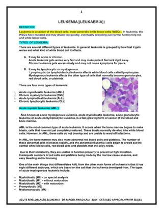
  • 1. 1 ACUTE MYELOBLASTIC LEUKEMIA DR MAGDI AWAD SASI 2014 DETAILED APPROACH WITH SLIDES LEUKEMIA((LEUKAEMIA)) DEFINITION: Leukemia is a cancer of the blood cells, most generally white blood cells (WBCs). In leukemia, the WBCs have mutated and may divide too quickly, eventually crowding out normal functioning red and white blood cells. TYPES OF LEUKEMIA: There are several different types of leukemia. In general, leukemia is grouped by how fast it gets worse and what kind of white blood cell it affects. A. It may be acute or chronic. Acute leukemia gets worse very fast and may make patient feel sick right away. Chronic leukemia gets worse slowly and may not cause symptoms for years. B. It may be lymphocytic or myelogenous. Lymphocytic (or lymphoblastic) leukemia affects white blood cells called lymphocytes. Myelogenous leukemia affects the other type of cells that normally become granulocytes, red blood cells, or platelets. There are four main types of leukemia:  Acute myeloblastic leukemia (AML)  Chronic myelocytic leukemia (CML)  Acute lymphoblasti leukemia (ALL)  Chronic lymphocytic leukemia (CLL) Acute myeloid leukemia( AML ): Also known as acute myelogenous leukemia, acute myeloblastic leukemia, acute granulocytic leukemia or acute nonlymphocytic leukemia, is a fast-growing form of cancer of the blood and bone marrow. AML is the most common type of acute leukemia. It occurs when the bone marrow begins to make blasts, cells that have not yet completely matured. These blasts normally develop into white blood cells. However, in AML, these cells do not develop and are unable to ward off infections. In AML, the bone marrow may also make abnormal red blood cells and platelets. The number of these abnormal cells increases rapidly, and the abnormal (leukemia) cells begin to crowd out the normal white blood cells, red blood cells and platelets that the body needs. Due to their immaturity, they are unable to function properly to prevent or fight infection. Inadequate numbers of red cells and platelets being made by the marrow cause anaemia, and easy bleeding and/or bruising One of the main things that differentiates AML from the other main forms of leukemia is that it has eight different subtypes, which are based on the cell that the leukemia developed from. The types of acute myelogenous leukemia include:  Myeloblastic (M0) - on special analysis  Myeloblastic (M1) - without maturation  Myeloblastic (M2) - with maturation  Promyeloctic (M3)  Myelomonocytic (M4)
  • 2. 2 ACUTE MYELOBLASTIC LEUKEMIA DR MAGDI AWAD SASI 2014 DETAILED APPROACH WITH SLIDES  Monocytic (M5)  Erythroleukemia (M6)  Megakaryocytic (M7) CHARACTERISTC FEATURES OF ACUTE LEUKEMIA: -The condition progresses rapidly and aggressively, requiring immediate treatment. -The site of the disease is bone marrow where all the cell lines may be depressed . -Bone marrow aspiration is mandatory for diagnosis (˃ 20% blasts ). -There is no enough time for the organs to be hugely enlarged (( may be palpable )). -The blood is circulating and malignant cells may invade the organs. -There is no surgical treatment for it. INCIDENCE: Acute leukaemia is an uncommon type of cancer. About 7,600 people are diagnosed with leukaemia each year in the UK. AML is an uncommon type of cancer. Around 2,600 people are diagnosed with the condition each year in the UK. AML can develop at any age, but it's more common in people over the age of 60. SYMPTOMS:  Many people often do not experience symptoms in the early stages of certain types of leukemia, or the symptoms develop slowly. Acute myeloid leukemia (AML) and acute lymphocytic leukemia (ALL) progress much faster and symptoms may worsen more quickly than with the chronic leukemias (CML and CLL).  Some leukemia symptoms, like night sweats, fever, fatigue and achiness, often resemble flu-like symptoms. If you have the flu, symptoms will likely subside as you get better. Make an appointment to see your doctor if the symptoms persist longer than expected.  Some general symptoms of leukemia include: Fever, chills Fatigue, weakness Loss of appetite, weight loss Night sweats Bone/joint pain Abdominal discomfort Headaches Shortness of breath Frequent infections Easy bruising or bleeding Petechiae (small red spots under the skin)
  • 3. 3 ACUTE MYELOBLASTIC LEUKEMIA DR MAGDI AWAD SASI 2014 DETAILED APPROACH WITH SLIDES AML symptoms: Bone marrow depression—anemia , bleeding , infection Bony pain with sternal tenderness DIC , gum hyperatrophy S&S: 1. ANEMIA : Pallor Easy fatigability Dyspnea on mild exertion Lassitude Syncopal attacks Headache Palpitation Anorexia Taste disturbance Tinnitus Dizziness Dimension of vision Insomnia Angina Parasthesia in fingers and toes 2. BLEEDING (( THROMBOCYTOPENIA)): Petechiae Bruisability Epistaxis Gingival bleeding (spongy bleeding) Melena Hematuria Purpura Spontaneous involving ---CNS , LUNG , GIT MUCOUS MEMBRANE Haemorrhage increased ˂ 20 x 10000/ml 3. INFECTION: Is inversely related to number of circulating neutrophils Major risk in patient with granulocyte ˂ 0.5 x 10000/ml Neutrophil may function abnormally and comprises host defenses. The leukemia and its treatment cause a breakdown of mucosal barrier & systemic infections usually develop from skin, throat, GIT.
  • 4. 4 ACUTE MYELOBLASTIC LEUKEMIA DR MAGDI AWAD SASI 2014 DETAILED APPROACH WITH SLIDES In leukocytosis, increase in blast cells of ˃ 100 x 10 000 /ml leads to leukostasis due to occlusion of the microcirculation by blast cells leading to hypoperfusion of lung and brain. Common sites of infections: Skin; Gingival; Ulcers in mouth and pharynx ;Perirectal tissues ;Lung;Urinary tract; Sore throat Septicemia usually occurs without an apparent cause. 4. HEPATOMEGALLY AND SPLENOMEGALLY: Rare in AML. The visceral involvement can produce symptoms of nausea , abdominal fullness and satiety. Lymphoadenopathy –AML 50% Practical point—extramedullary leukemia can precede detectable involvement of B.M. 5. EXTRAMEDULLARY TISSUES : Acute leukemia may infiltrate –skin, lung, eye, kidney ,nasopharynx CHLOROMAS----soft tissues masses of leukemic cells. 6. SYMPTOMS RELATED TO THE EXPANDING MALIGNANT CELL MASS: Bony pain and sternal tenderness This occur in half of patients Osteolytic lesions are rare Renal abnormalities due to : A. Leukemic infiltration B. Ureteral obstruction by urate nephropathy—enlarged LN, uric acid stone, infections ,haemorrhage. GIT----bleeding, distention ,satiety ,constipation((due to organomegally or leukemic infiltrate)) . 7. METABOLIC DISTURBANCE : Hypokalemia and hyponatremia due to renal involvement . LDH—lactic dehydrogenase may be increased. CLASSIFICATION OF ACUTE LEUKEMIA: Classification of acute leukemias used to rely entirely on morphology and histochemical changes, but now information regarding cytogenetic patterns and correlations with immunophenotype, oncogene expression, and gene mutation has changed the biology of this disease 1. FAB classification(French-American-British) :1970 A classification of acute leukemia produced by a three-nation joint collaboration. A schema that divides acute leukemias into lymphoid–ALL or myeloid–AML cell lines This is based on the study of microscopic features and cytochemistry of blast cells. Acute myelogenous leukemia is subdivided into eight types(M0-M7) based on the type of cell from which the leukemia developed and how mature the cells are.
  • 5. 5 ACUTE MYELOBLASTIC LEUKEMIA DR MAGDI AWAD SASI 2014 DETAILED APPROACH WITH SLIDES Acute myeloid leukemia (AML) Subtypes M0 through M5 all start in precursors of white blood cells. M6 AML starts in very early forms of red blood cells, while M7 AML starts in early forms of cells that make platelets. M1 Myeloblasts without maturation M2 Myeloblasts with maturation (best AML prognosis) M3 Hypergranular promyelocytic leukemia (faggot cells) M3V Variant, microgranular promyelocytic leukemia M4 Myelomonocytic leukocytes M5 Monocytic, subtype a. Poorly differentiated monocytic leukemia b. Well differentiated monocytic leukemia M6 Erythroleukemia/DiGuglielmo syndrome M7 Megakaryocytic leukemia Pleomorphic undifferentiated cells with cytoplasmic blebs; myelofibrosis or ↑ BM reticulin; positive for platelet peroxidase antifactor VIII 2. World Health Organization (WHO) classification of AML The FAB classification system is useful and is still commonly used to group AML into subtypes. But it doesn’t take into account many of the factors that are known to impact prognosis (outlook). In 2001, the World Health Organization (WHO) published a newer system that includes some of these factors to try to help better classify cases of AML based on a patient’s outlook. The WHO classification system divides AML into several broad groups: 1. AML with certain genetic abnormalities  AML with a translocation between chromosomes 8 and 21  AML with a translocation or inversion in chromosome 16  AML with changes in chromosome 11  APL (M3), which usually has translocation between chromosomes 15 and 17 2. AML with multilineage dysplasia (more than one abnormal myeloid cell type is involved) 3. AML related to previous chemotherapy or radiation 4. AML not otherwise specified (includes cases of AML that don’t fall into one of the above groups; similar to the FAB classification)  Undifferentiated AML (M0)  AML with minimal maturation (M1)  AML with maturation (M2)  Acute myelomonocytic leukemia (M4)  Acute monocytic leukemia (M5)  Acute erythroid leukemia (M6)
  • 6. 6 ACUTE MYELOBLASTIC LEUKEMIA DR MAGDI AWAD SASI 2014 DETAILED APPROACH WITH SLIDES  Acute megakaryoblastic leukemia (M7)  Acute basophilic leukemia  Acute panmyelosis with fibrosis  Myeloid sarcoma (also known as granulocytic sarcoma or chloroma) 5. Undifferentiated or biphenotypic acute leukemias (leukemias that have both lymphocytic and myeloid features). Sometimes called ALL with myeloid markers, AML with lymphoid markers, or mixed lineage leukemias. Morphological subtypes of AML: AML occurs more often in men and is usually diagnosed in individuals over 50, although it can occur at any age. M1  ((AML without maturation))  20% of AML  Myeloblast predominant  Distinct nucleoli  Few Auer rods , Azurophilic granules  ( > 90% type I and II blasts ) of marrow nonerythroid cells is present. Most of the blasts are agranular. Auer rods are infrequent.  Staining: Relatively few blasts (5-10%) are MPO (myeloperoxidase) positive. A minimum of 3% MPO positive blasts are required for diagnosis. NSE and PAS are generally negative.  Immunophenotype: Variably positive for CD13, CD14, CD11b, CD33, and HLA-DR.  Chromosome Abnormalities: t(9;22) Philadelphia chromosome, 8+, -5, and -7.
  • 7. 7 ACUTE MYELOBLASTIC LEUKEMIA DR MAGDI AWAD SASI 2014 DETAILED APPROACH WITH SLIDES M2 AML with maturation ((acute myeloblastic with maturation)) The most common (20-40%) type of AML Maturation: Type II blasts ((Myeloblast)) are common and Auer rods are frequent (promyelocytes- myelocytes). Positive to peroxidase sudan black Staining: The blasts are largely MPO peroxidase sudan black positive. NSE and PAS are generally negative.  Immunophenotype: Variable positivity for CD13, CD33, and HLA-DR, but are -ve for CD14 and CD11b.  Chromosome Abnormalities: t(8;21), 8+, -5, and -7.  Cell Morphology: In some cases the immature cells have abundant, frequently basophilic cytoplasm, with variable numbers of often indistinct, sometimes coalescent granules. If such immature cells are < 10% the diagnosis is M1, but if > 10% the diagnosis becomes M2. M3 o Acute Promyelocytic Leukemia (APL)  Maturation: the majority of promyelocytes are hypergranular. Auer rods are frequent and often multiple.  Staining: The cells are MPO and chloroacetate esterase (CAE) positive, but generally negative for NSE (NSE positive in 25%).  Immunophenotype: Positivity for CD13 and CD33, but are usually negative for HLA-DR.  Chromosome Abnormalities: The t(15;17) is unique to promyelocytic leukemia.  DIC is seen in M3 by thromboplastic material released by leukemic cells & exacerbated by chemotherapy.  Treatment Options: Why is it critical to distinguish M3 from other myeloid leukemias?  The t(15;17) splits the retinoic acid receptor (RAR) alpha-gene on 17. This is replaced by the myl gene from 15, producing a novel protein: myl/RAR-a. Myl/RAR-alpha may suppress the normal RAR- aalpha allele thus blocking expression of retinoic acid genes needed for myeloid differentiation.  Recognition of M3 leukemias is critical because M3s can be initially treated with retinoic acid rather than toxic chemotherapy.  Microgranular Variant:  In the microgranular variant, M3v, the leukemic cells have a monocytic appearance with clefted angel-wing nuclei and
  • 8. 8 ACUTE MYELOBLASTIC LEUKEMIA DR MAGDI AWAD SASI 2014 DETAILED APPROACH WITH SLIDES abundant cytoplasm having at best indistinct cytoplasmic granulation.  The cytochemical, immunophenotypic and chromosomal features are indentical to the classic M3.  Both forms of acute promyelocytic leukemia are associated with a high incidence of disseminated intravascular coagulation (DIC) and hemorrhage. M4  Acute Myelomonocytic Leukemia (AMML) – ((30%))  Maturation: Differentiation along both myeloid and monocytic lines. Monocytes and promonocytes represent > 20%, but < 80% of the marrow differential.  Staining: More than 20% of the blasts should be MPO + and more than 20% should be NSE + .  Diagnosis Aides: I. High serum lysozyme (3x normal) II. A peripheral monocytosis of > 5x10/L in an otherwise M2 marrow and increased lysozyme III. A peripheral monocytosis of >5 x10/L in M2 marrow and >20% NSE + marrow blasts.  Chromosome Abnormalities: t(4;11), t(9;11), 8+ and -7.  Variant: M4e variant in which eosinophils (> 5%) are increased in number and abnormal associated with abnormalities of chromosome 16.  Staining of Variant: CAE, usually negative in eosinophils, is frequently positive in the abnormal eosinophils of M4e. M5 o Acute Monocytic Leukemia (AMoL) – ((10%)) o Differentiatiation: Monocytic------Two subtypes: 1. M5a is the poorly differentiated form 2. M5b is the well-differentiated form o Chromosome Abnormalities: t(9;11), 8+, -5, and -7. Chromosome abnormalities of 11q are closely associated with M5a o Cell Morphology: Note the nuclear folds and the relatively large nucleoli typical of monoblasts at right. Monoblast Myeloblasts fewer granules more granules occasionally will see slight nuclear fold typically round nucleus few, large nucleoli more nucleoli
  • 9. 9 ACUTE MYELOBLASTIC LEUKEMIA DR MAGDI AWAD SASI 2014 DETAILED APPROACH WITH SLIDES M5a  This is the M5a or poorly differentiated form of acute monoblastic leukemia. >80% of cells are monoblasts.  Staining: The blasts in M5a are generally MPO negative, but strongly positive for the nonspecific esterase (NSE) and inhibited by Fluoride. M5b This is the M5b or differentiated form of acute monoblastic leukemia in which > 80% of the leukemic cells are monoblasts, promonocytes, and monocytes. Note the large nucleoli so typical of monoblasts Staining: The blasts of M5b usually contain some peroxidase activity and, of course, are positive for nonspecific esterase (NSE) and inhibited by Fluoride. M4 & M5 have extramedullary involvement of skin ,gingival and CNS. M6 o Acute Erythroblastic Leukemia  Maturation: Erythroleukemia is rare and difficult to diagnose. More than 50%* of the nucleated marrow cells are abnormal nucleated red blood cells.  Morphology: The leukemic red cells are frequently bizarre with extreme dysplastic features including: giant forms, multinucleation, cytoplasmic vacuolization, cytoplasmic buds, and megaloblastoid changes.  30% considered adequate by some  Staining: The blasts are MPO negative, but often positive for NSE. The malignant red cells are PAS positive, (forming PAS positive lakes or containing coarse chunks of PAS positive material).  Immunophenotype: Positive for glycophorin A.  Chromosome Abnormalities: 8+, -5, del(5q), and -7.  Differential Diagnosis:
  • 10. 10 ACUTE MYELOBLASTIC LEUKEMIA DR MAGDI AWAD SASI 2014 DETAILED APPROACH WITH SLIDES 1. Congenital dyserythropoietic anemia 2. Myelodysplastic syndrome 3. Sideroblastic anemia 4. Megaloblastic anemia. M7  Acute Megakaryocytic Leukemia (AMKL) – ((5%))  M7 blasts are often resemble lymphoblasts, although M7 leukemias may be accompanied by atypical megakaryocytes. The marrow is often fibrotic.  Morphology: M7 blasts may have granular cytoplasm and shed 'platelets'. M7 blasts often clump together Immunoperoxidase staining (brown) for Factor VIII related protein identifies the blasts as being of megakaryocyte lineage  Staining: M7 blasts are MPO negative and variably positive for PAS and NSE.  Diagnosis: Determination of an M7 subtype is dependent on immunologic evidence or electron microscopic ultracytochemical identification of platelet peroxidase (PPO).  Immunophenotypic studies of M7 are positive for glycoproteins GP Ib andGP IIb/IIIa.  Factor VIII related protein is usually found in the megakaryoblast cytoplasm.  Chromosome Abnormalities: t(1;22), have been associated with M7 in infants. Myeloid Leukemia with Minimal Differentiation - FAB M0:  M0 is a recently defined FAB nonlymphoid leukemia dependent on the immunologic identification of myeloid antigens. Morphologic and cytochemical studies show no distinguishing features.  Staining: M0 blasts are nondescript (no Auer rods) and are MPO, PAS and NSE negative (<3%).
  • 11. 11 ACUTE MYELOBLASTIC LEUKEMIA DR MAGDI AWAD SASI 2014 DETAILED APPROACH WITH SLIDES  Electron microscopic ultra cytochemical studies for platelet peroxidase are negative.  Immunophenotype: M0 blasts are negative for B and T lymphoid antigens, platelet glycoproteins GP Ib and GP IIb/IIIa, and erythroid glycophorin A. Myeloid antigens such as CD13, CD33 and CD11b are variably positive. CD34 and HLA-DR are generally positive.  Chromosome Abnormalities: No particular association. ADDITIONAL INFORMATION REGARDING LEUKEMIA: Eosinophilic leukemia is a rare variant of acute myeloid leukemia in which blasts and immature eosinophils proliferate. CNS involvement appears to be common. Should be distinguished from CML with large numbers of eosinophils. Basophilic leukemia is a rare subset of AML associated with t(6;9) and abnormal 12p. Ultrastructural studies of the immature basophilic granules may be necessary. Philadelphia chromosome positive cases may be related to the blast crisis of CML Blast crisis of chronic leukemia refers to a phase of a chronic leukemia resembling an acute leukemia ( in which blasts are >30%). In chronic myelogenous leukemia (CML), blast crisis signals the terminal phase of the disease for which there is no effective therapy. Secondary (therapy-induced) acute leukemia is nearly always myeloid and is usually preceeded by a myelodysplastic syndrome involving all three hematopoietic cell lines (panmyeloisis). Abnormalities of chromosomes 5, 7, and 11 are common in therapy-induced leukemias. This is most common after alkylating chemotherapy or radiation therapy, with an average latent period of 5 years (range1- 15 years). Hypocellular acute leukemia is a situation in which the marrow cellularity (< 30%) often resembles aplastic anemia, but with numerous (>30%) blasts in the interstitium of the marrow. About 5-10% of acute myeloid leukemias present in this form. Extramedullary myeloid cell tumor (EMCT) or granulocytic sarcoma or chloroma is an extramedullary tissue mass of blasts and immature myeloid cells. The surface of a freshly cut EMCT turns light green (hence the name chloroma) upon exposure to air as large amounts of peroxidase are oxidized. EMCT may represent the initial manifestation of acute leukemia or signal relapse. The blast crisis of chronic myeloid leukemia may present as EMCT. Masses of monoblasts as may be seen in M5 leukemias are also sometimes referred to as EMCT. The surface of this subcutaneous chloroma turned pale green after exposure to air. The patient had been in remission following Rx for AML. Acute biphenotypic leukemia – Cells contain both markers (lymphoid and myeloid) It is a leukemia of primitive pleuripotent stem cells. To differentiate ALL from AML ( M1 from L2 ): 1. Cytochemical stains 2. Analysis of myeloid and lymphoid antigen 3. Electron microscopy to detect promyelocytic granules Leukemia in BM can’t be detected when cell mass ˂1000/ml
  • 12. 12 ACUTE MYELOBLASTIC LEUKEMIA DR MAGDI AWAD SASI 2014 DETAILED APPROACH WITH SLIDES Clinical criteria of complete remission: 1. ˂5% blast cells in the BM with absence of leukemic cells in peripheral blood 2. The restoration of normal peripheral blood count 3. The absence of physical findings attributable to extramedullary involvement leukemia THE SUMMARY SHEET OF AML FAB: Morphological Features of AML TYPE(FAB) CHARACTERISTIC MORPHOLOGY Acute myelocytic leukemia (M1) Cells very undifferentiated, occasionally cytoplasmic granules, some promyelocytes seen. Acute myelocytic leukemia (M2) Granulated blasts predominate, small number of monocytoid cells may be present, differentiation beyond promyelocytic stage evident, +/- Aeur bodies Acute promyelocytic leukemia (M3) Typically, hypergranular promyelocytes predominate, cells have large basophilic and esoinophilic granules, +/- Auder bodies Acute myelomonocytic leukemia (M4) Monocytic and granulocytic precursors seen, serum lysozyme elevated, +/- Auer bodies Acute monoblastic leukemia (M5) Large monoblasts with abundant, agranular cytoplasm that may be vacuolated and basophilic Erythroleukemia (M6) Megaloblastoid red cell precursors predominate, myeloid blasts also seen, multinucleated red cell precursors common Megakaryocytic leukemia (M7) Variable morphology, megakaryocytic features may not be seen with light microscopy  Identification of AML subtypes is important because several new drugs have more activity against some varieties than against others.  Prognosis and some clinical features may differ considerably among the various AML subtypes. Histochemical Features of AML FAB HISTOCHEMISTRY M1 Occasional peroxidate+ granules, PAS- M2 Strongly peroxidase+, PAS- M3 Strongly peroxidase+, PAS- M4 Strongly peroxidase+, some cells may be PAS+ M5 Many be peroxidase+ and PAS+, nonspecific esterase stains are strongly + and inhibited by NAF M6 Red cell precursors are PAS+, ringed sideroblasts are seen with iron stains M7 Variable, platelet peroxidase can be demonstrated by electron microscopy  Distinction among the various subgroups of AML is now possible with greater precision because of new histochemical studies and immunologic techniques.  Immunophenotyping and terminal deoxytransferase (TDT) determination may help distinguish between AML and ALL, and between the various subtypes of AML.
  • 13. 13 ACUTE MYELOBLASTIC LEUKEMIA DR MAGDI AWAD SASI 2014 DETAILED APPROACH WITH SLIDES AML MARKERS  Morphologic ----------Auer bodies AND "malignant" primary granules  Cytoplasmic organelles o chloracetate esterase o Sudan B o myeloperoxidase  Cytogenetic o t(8;21) o t(15;17) o -5,-7  Immunologic/biochemical o lysozyme o cell surface markers QUESTIONS FOR TRAINING: 1. A 12-year-old girl is noted to be pale and lethragic by her parents. You note some petechiae on her legs. A chest roentgenogram is normal. There is no lymphadenopathy. A CBC revealed a WBC of 25.0 x10 /L with 15% blasts.The bone marrow aspirate differential count included 32% type I blasts; 13% type II blasts; 10% type III blasts; 10% promyelocytes; 25% myelocytes; 3% PMNs and 7% monocytes. Many of the blasts contained Auer rods. There were fewer than 50% erythroblasts. With the results of this information you make the following diagnosis: A. Acute myelocytic leukemia, M1 B. Acute myelocytic leukemia, M2 C. Acute promyelocytic leukemia, M3 D. Acute myelocytic leukemia, M4 E. Acute myelocytic leukemia, M5 ANSWER In this case >30% blasts and >10% cells with maturation ( promyelocytes - polymorphonuclear leukocytes) are seen. Type II blasts are common and Auer rods are frequently, all features of acute myelocytic leukemia with maturation - M2. Too much maturation (too many Type III blasts, promyelocytes and myelocytes) for an M1 Fewer than 20% were monocytic cells, thus not an M4 or M5. 2. A 25-year-old woman comes to you because of a severe headache and epistaxis. She is pale and has petechiae on her arms and chest. There is no lymphadenopathy or hepatosplenomegaly. A chest roentgenogram is normal. The WBC was 18.0 x10 /L with 10% blasts. On the bone marrow aspirate it was difficult to distinguish between Type II and III blasts and promyelocytes. There were an estimated15% type I blasts and 75% immature cells (type II and type III blasts and promyelocytes). The remainder were dysplastic myelocytes. There were fewer than 50% erythroblasts. Cytogenetic studies showed a t(15;17) chromosome abnormality. With the results of this information you make the following diagnosis: A. Acute myelocytic leukemia, M1 B. Acute myelocytic leukemia, M2 C. Acute promyelocytic leukemia, M3 D. Acute myelocytic leukemia, M4 E. Acute myelocytic leukemia, M5 ANSWER Because of the difficulty of distinguishing between Type II and III blasts and promyelocytes (75% type II and type III blasts and promyelocytes) the differential is between an FAB M2 and an FAB M3 myeloid leukemia. The key is the cytogenetic study showing a t(15;17) chromosome abnormality which is diagnositic of promyelocytic leukemia (FAB-M3).
  • 14. 14 ACUTE MYELOBLASTIC LEUKEMIA DR MAGDI AWAD SASI 2014 DETAILED APPROACH WITH SLIDES 3. A 45-year-old woman is referred to you from her dentist because of swollen and bleeding gums. Her conjunctiva were pale and she had been feeling exceptionally wornout. There is mild splenomegaly. A chest roentgenogram is normal. The WBC was 23.0 x10 /L with 44% blasts. The bone marrow aspirate showed > 55% large blasts with folded nuclei and large nucleoli. The remainder were promonocytes, monocytes and a few dysplastic myelocytes. There were fewer than 50% erythroblasts. Cytogenetic studies showed a t(9;11) chromosome abnormality. With the results of this information you make the following diagnosis: A. Acute myelocytic leukemia, M1 B. Acute myelocytic leukemia, M2 C. Acute promyelocytic leukemia, M3 D. Acute myelomonocytic leukemia, M4 E. Acute monocytic leukemia, M5 ANSWER The finding of > 55% large blasts with folded nuclei and large nucleoli accompanied by numerous promonocytes, monocytes and a few dysplastic myelocytes indicates a monoblastic leukemia. An M4 might also be considered as monocytes are part of the M4 milieu. Key is the cytogenetic study showing a t(9;11) chromosome abnormality which is closely associated with acute monocytic leukemia (FAB M5). 4. A 37-year-old man comes to you complaining of fatigue and increasing SOB, and now a fever (the fever is what got him to see you). He has had to stop jogging. He is pale and diaphoretic. There is no lymphadenopathy or hepatosplenomegaly. A chest roentgenogram is shows dense bilateral infiltrates c/w pneumonia. The WBC is 78.0 x10 /L with 20% blasts. On the bone marrow aspirate there were an estimated 25% type I blasts and 55% immature cells (type II and type III blasts and promyelocytes). The remainder were dysplastic myelocytes.Cytogenetic studies showed a t(8;21) chromosome abnormality. With the results of this information you make the following diagnosis: A. Acute myelocytic leukemia, M1 B. Acute myelocytic leukemia, M2 C. Acute promyelocytic leukemia, M3 D. Acute myelocytic leukemia, M4 E. Acute lymphoblastic leukemia ANSWER The difficulty of distinguishing among type II blasts, type III blasts and promyelocytes can make the morphologic distinction between an M2 and an M3 (and possibly an M4) leukemia difficult. The finding of a t(8;21) chromosome abnormality is of great help in classifying this leukemia, a t(8;21) abnormality is associated with acute myelocytic leukemia with differentiation. Other chromosome abnormalities commonly associated with M2 AMLs are 8+, -5, and -7. 5. A 32-year-old man comes to you because of a persistent cough x3 weeks and noted some shortness of breath. He is pale and has several ecchymoses and petechiae on his back and legs. He has several small axillary nodes and an ill-defined mediastinal mass. The WBC is 114.0 x10 /L with 90% blasts. The marrow aspirate showed 54% blasts; 8% promyelocytes; 15% myelocytes; 3% bands;12% segmented neutrophils; 5% eosinophils, and 3% basophils. The blasts are negative for myeloperoxidase; nonspecific esterase and PAS. Chromosome studies showed a t(10;14)(q24;q11) abnormality. Immunologic marker studies are pending. With the results of this information you make the following diagnosis: A. Acute myelocytic leukemia, M1 B. Acute myelocytic leukemia, M2
  • 15. 15 ACUTE MYELOBLASTIC LEUKEMIA DR MAGDI AWAD SASI 2014 DETAILED APPROACH WITH SLIDES C. Acute promyelocytic leukemia, M3 D. Acute myelocytic leukemia, M4 E. Acute lymphoblastic leukemia ANSWER A 32-year-old man comes to you because of a persistent cough x3 weeks and noted some shortness of breath. He is pale and has several ecchymoses and petechiae on his back and legs. The ill-defined mediastinal mass and the high peripheral blood blast count > 100 x10 /L is typical of acute lymphoblastic leukemia of T-cell type. The nonmyeloid nature of the blasts is indicated by the negative myeloperoxidase, nonspecific esterase and PAS. The chromosome studies showing a t(10;14)(q24;q11) abnormality is further evidence of the T-lymphoblastic nature of this lesion. AML ---- THE PRESENCE OF AUER RODS DIAGNOSTIC-----BY Romanorsky dye and cytochemical stains MORE COMMON IN ADULT POLYCLONAL SEVER BLEEDING MAY OCCUR Cytogenatic studies -----the most powerful prognostic factor Favorable in AML---t(8 , 21) , t(15 , 17) , inv 169 Favorable in ALL --- Hyperlipoid state Risk Factors for Acute Myeloid Leukemia If something increases your risk of getting a disease, it's called a risk factor. Risk factors don't tell the whole story. For example, you can have few risk factors and still get a disease or have several and not get it. Acute myeloid leukemia risk factors include: 1. Smoking 2. Exposure to certain chemicals such as benzene , certain cleaning products, detergents, and paint strippers. 3. Treatment with certain chemotherapy drugs used to treat other cancers, such as mechlorethamine, procarbazine, and chlorambucil -- especially when combined with radiation therapy 4. Exposure to high doses of radiation 5. Certain blood disorders, such as myeloproliferative disorders (for example, chronic myelogenous leukemia) 6. Certain congenital syndromes, such as Down syndrome 7. Being male 8. For most people, the cause of AML is unknown. There is not a way to prevent it, but you may reduce your risk by quitting smoking and avoiding exposure to chemicals. Diagnostically Important AML Markers  Morphologic o Auer bodies o "malignant" primary granules  Cytoplasmic organelles o chloracetate esterase o Sudan B
  • 16. 16 ACUTE MYELOBLASTIC LEUKEMIA DR MAGDI AWAD SASI 2014 DETAILED APPROACH WITH SLIDES o myeloperoxidase  Cytogenetic o t(8;21) o t(15;17) o -5,-7  Immunologic/biochemical o lysozyme o cell surface markers  A variety of morphologic, cytoplasmic, cytochemical, and biochemical features are associated with AML that can aid in diagnosis.  No available marker is either totally specific or sensitive enough to be used in all cases.  Cytogenetic analyses should be performed at diagnosis in all patients with AML. Approximately 65% of patients will have abnormal karyotypes.  Cytogenetic abnormalities may help determine prognosis Work-Up for Patient with AML:  Complete history: o family, work, and medical history ---radiation and chemical exposure history  Complete physical examination, with special attention to: o Temperature, Iymph node-bearing areas and splenic area, optic fundi and cranial nerves o Potential sites of infection:  Skin , including axillae  Oropharynx , including gingivae  Lungs  Perianal area o Peripheral blood studies:  Hematocrit , leukocyte count, platelet count  Leukocyte differential count o Bone marrow examination:  Perform biopsy to determine cellularity  Aspirate smears stained with Wright's, Sudan black, and esterase stains; periodic acid-Schiff reagent; iron stain; and immunofluorescent stain for terminal transferase  Aspirate for karyotyping and immunophenotyping  Blood chemistries and other studies:  Serum electrolytes, uric acid, blood urea nitrogen, and muramidase (lysozyme)  Coagulation profile, including fibrinogen level, prothrombin time, and partial thromboplastin time  Liver function analysis  Chest radiographs:  chest posteroanterior and lateral radiographic films  Transfusion work-up:  determine blood type and human leulocyte antigen type if patient has circulating lymphocytes  do same for family members who are willing to serve as platelet, granulocyte, or potential marrow donors Peripheral Blood Features of AML
  • 17. 17 ACUTE MYELOBLASTIC LEUKEMIA DR MAGDI AWAD SASI 2014 DETAILED APPROACH WITH SLIDES  The peripheral white blood cell count may be increased, decreased, or normal with approximately equal frequency.  Granulocytopenia is very common. Approximately 1/2 of all patients will have granulocyte counts < 1,000/uL.  Thrombocytopenia is frequently observed and platelet counts <20,000/uL are common.  The hematocrit is generally low but severe anemia is uncommon.  Circulating blast cells are absent from the peripheral blood in approximately 15% of AML patients initially, and in 1/2 of patients presenting with leukopenia Certain factors affect prognosis (chance of recovery) and treatment options: The prognosis (chance of recovery) and treatment options depend on:  The age of the patient.  The subtype of AML.  Whether the patient received chemotherapy in the past to treat a different cancer.  Whether there is a history of a blood disorder such as myelodysplastic syndrome.  Whether the cancer has spread to the central nervous system.  Whether the cancer has been treated before or recurred (come back). There is no standard staging system for adult AML The disease is described as untreated, in remission, or recurrent. A. Untreated adult AML In untreated adult AML, the disease is newly diagnosed. It has not been treated except to relieve signs and symptoms such as fever, bleeding, or pain, and the following are true:  The complete blood count is abnormal.  At least 20% of the cells in the bone marrow are blasts (leukemia cells).  There are signs or symptoms of leukemia. B. Adult AML in remission In adult AML in remission, the disease has been treated and the following are true:  The complete blood count is normal.  Less than 5% of the cells in the bone marrow are blasts (leukemia cells).  There are no signs or symptoms of leukemia in the brain and spinal cord or elsewhere in the body. C. Recurrent Adult AML Recurrent AML is cancer that has recurred (come back) after it has been treated. The AML may come back in the blood or bone marrow.
  • 18. 18 ACUTE MYELOBLASTIC LEUKEMIA DR MAGDI AWAD SASI 2014 DETAILED APPROACH WITH SLIDES Treatment for Acute Myeloid Leukemia This is a disease which need to be treated by haematologist in specialized center. If the patient had pancytopenia , supportive measures are used to correct them by packed red blood cells for anemia , granulocyte colony stimulating factor for leucopenia , and platelet concentrate for thrombocytopenia. The patient should be admitted to haematology department and quick screening for external orifices should be carried out to look for infections. Because AML is a group of related diseases, treatment depends upon each subtype as well as on other factors. It is important to start treatment as soon as possible after diagnosis. The 2 treatment phases of adult AML are:  Remission induction therapy: This is the first phase of treatment. The goal is to kill the leukemic cells in the blood and bone marrow. This puts the leukemia into remission.  Post-remission therapy: This is the second phase of treatment. It begins after the leukemia is in remission. The goal of post-remission therapy is to kill any remaining leukemia cells that may not be active but could begin to regrow and cause a relapse. This phase is also called remission continuation therapy. You may have more than one type of treatment, including: Chemotherapy Chemotherapy is a cancer treatment that uses drugs to stop the growth of cancer cells, either by killing the cells or by stopping them from dividing. When chemotherapy is taken by mouth or injected into a vein or muscle, the drugs enter the bloodstream and can reach cancer cells throughout the body (systemic chemotherapy). When chemotherapy is placed directly into the cerebrospinal fluid(intrathecal chemotherapy), an organ, or a body cavity such as the abdomen, the drugs mainly affect cancer cells in those areas (regional chemotherapy). Intrathecal chemotherapy may be used to treat adult AML that has spread, or may spread to the brain and spinal cord. Combination chemotherapy is treatment using more than one anticancer drug. The way the chemotherapy is given depends on thesubtype of the cancer being treated and whether it has spread to the brain and spinal cord. AML treatment consists of two parts: induction therapy and consolidation therapy, which are both done in the hospital. The goal of induction therapy is to achieve remission by: 1. Killing as many AML cells as possible 2. Returning blood counts to normal over time 3. Ridding the body of signs of disease for a long time Even if remission is achieved, you will likely still need more treatment. Consolidation therapy attacks leukemia cells that common blood or marrow tests can't find. This therapy may include chemotherapy, which may be different drugs than those used earlier, and possibly a bone marrow transplant. Age, health, and other factors clearly need to be taken into account when considering treatment options. Doctors are also trying to determine whether people with certain gene or chromosome changes are more likely to benefit from more intensive treatment. In younger patients, such as those under 60, induction often involves treatment with 2 chemo drugs, cytarabine (ara-C) and an anthracycline drug such as daunorubicin (daunomycin) or idarubicin. Sometimes a third drug, cladribine(Leustatin, 2-CdA), is given as well. The chemo is usually given in the hospital and lasts about a week. Patients with poor heart function can’t be treated with anthracyclines, and so may be treated with another chemo drug, such as fludarabine (Fludara) or topotecan. Induction destroys most of the normal bone marrow cells as well as the leukemia cells. Most patients develop dangerously low blood counts, and the patient may be very ill.
  • 19. 19 ACUTE MYELOBLASTIC LEUKEMIA DR MAGDI AWAD SASI 2014 DETAILED APPROACH WITH SLIDES Most patients need antibiotics and blood product transfusions. Drugs to raise white blood cell counts may also be used. Blood counts tend to stay down for weeks. Usually, the patient stays in the hospital during this time. About 1 or 2 weeks after chemo is done, the doctor will check a bone marrow biopsy. It should show few bone marrow cells (hypocellular bone marrow) and only a few blasts (less than 10%). Over the next few weeks, normal bone marrow cells will return and start making new blood cells. The doctor may check other bone marrow biopsies during that time. When the blood cell counts recover, the doctor will check cells in a bone marrow sample to see if the leukemia is in remission. If the biopsy done a week or 2 after the chemo treatment shows that there are still leukemia cells in the bone marrow, more chemo may be given. Sometimes a stem cell transplant is recommended at this point. If it isn’t clear looking at the first bone marrow whether the leukemia is still there, another bone marrow biopsy may be done again in about a week. Remission induction usually does not destroy all the leukemia cells, and a small number often persist. Without consolidation treatment, the leukemia is likely to return within several months. Chemotherapy, the use of anticancer drugs -- often two or three -- such as cytarabine, anthracycline drugs, 6- thioguanine, hydroxyurea, or prednisone. Consolidation (post-remission therapy):  Induction is considered successful if remission is achieved. Further treatment is then given to try to destroy any remaining leukemia cells and help prevent a relapse. This is called consolidation.  For younger patients, the main options for AML consolidation therapy are: I. Several cycles of high-dose cytarabine (ara-C) chemo (this is sometimes known as HiDAC) II. Allogeneic (donor) stem cell transplant III. Autologous stem cell transplant  Consolidation chemo differs from induction therapy in that usually only cytarabine is used. The drug is given at very high doses, typically over 5 days. This is repeated about every 4 weeks, usually for a total of 3 or 4 cycles.  Another approach after successful induction therapy is a stem cell transplant. Patients first receive very high doses of chemo to destroy all bone marrow cells. This is followed by either an allogeneic (from a donor) or autologous (patient’s own) stem cell transplant to restore blood cell production. Stem cell transplants have been found to reduce the risk of leukemia coming back more than standard chemo, but they are also more likely to have serious complications, including an increased risk of death from treatment.  Older patients or those in poor health may not be able to tolerate such intensive consolidation treatment. Often, giving them more intensive therapy raises the risk of serious side effects (including treatment-related death) without providing much more of a benefit. These patients may be treated with: 1. 1 or 2 cycles of higher dose cytarabine (usually not quite as high as in younger patients) 2. 1 or 2 cycles of standard dose cytarabine, possibly along with idarubicin or daunorubicin  Non-myeloablative stem cell transplant (mini-transplant)  It is not always clear which of the treatment options is best for consolidation. Each has pros and cons. Doctors look at several different factors when recommending what type of post-remission therapy a patient should receive.  These include: 1. How many courses (cycles) of chemo it took to bring about a remission? If it took more than one course, some doctors recommend that the patient receive a more intensive program, which might involve a stem cell transplant. 2. The availability of a brother, sister, or an unrelated donor who matches the patient’s tissue type? If a close enough tissue match is found, an allogeneic (donor) stem cell transplant may be an option, especially for younger patients. 3. The potential of collecting leukemia-free bone marrow cells from the patient? If lab tests show that a patient is in remission, collecting stem cells from the patient’s bone marrow or blood for an autologous stem cell transplant may be an option. Stem cells collected from the patient would be purged (treated in the lab to try to remove or kill any remaining leukemia cells) to lower the chances of relapse. 4. The presence of one or more adverse prognostic factors, such as certain gene or chromosome changes, a very high initial white blood cell count, AML that develops from a previous blood disorder or after treatment for an earlier cancer, or spread to the central nervous system?
  • 20. 20 ACUTE MYELOBLASTIC LEUKEMIA DR MAGDI AWAD SASI 2014 DETAILED APPROACH WITH SLIDES These factors might lead doctors to recommend more aggressive therapy, such as a stem cell transplant. On the other hand, for people with good prognostic factors, such as favorable gene or chromosome changes, many doctors might advise holding off on a stem cell transplant unless the disease recurs. 5. The age of the patient. Older patients may not be able to tolerate some of the severe side effects that can occur with high-dose chemo or stem cell transplants. 6. The patient’s wishes. There are many issues that revolve around quality of life that must be discussed. An important issue is the higher chance of early death from high-dose chemo or a stem cell transplant. This and other issues must be discussed between the patient and the doctor. Radiation therapy  Radiation therapy is a cancer treatment that uses high-energy x-rays or other types of radiation to kill cancer cells or keep them from growing. There are two types of radiation therapy. External radiationtherapy uses a machine outside the body to send radiation toward the cancer. Internal radiationtherapy uses a radioactive substance sealed in needles, seeds, wires, or catheters that are placed directly into or near the cancer. The way the radiation therapy is given depends on the type and stage of the cancer being treated.  Radiation therapy, the use of high-energy radiation to kill cancer cells. This is not used often for AML but may be used to treat leukemia in the brain, bone, or testicles; before a stem cell transplant; or, in rare cases, to shrink a tumor that's pressing on the windpipe.  A bone marrow transplant, which involves use of high doses ofchemotherapy and possibly radiation, followed by a transplant of bone-forming stem cells. Stem cells usually come from a donor. Or, less likely, they come from your own bone marrow or peripheral blood. If you cannot tolerate high doses of chemotherapy and radiation, lower doses may be used with a "mini-transplant." What are the survival rates for acute myeloid leukemia? The five-year survival rate overall is about 24%. Surviving at least five years indicates a good chance of long-term survival. Survival rates may be better now as this statistic reflects outcomes from 1999 to 2006. . Treating frail, older adults Treatment of AML in people under 60 is fairly standard. It involves cycles of intensive chemo (discussed above). Many patients older than 60 are healthy enough to be treated in the same way, although sometimes the chemo may be less intense. People who are much older or are in poor health may not be able to tolerate this intense treatment. In fact, intense chemo could actually shorten their lives. In some cases, doctors may recommend low-intensity chemo with a low dose of cytarabine given in cycles. In some cases, this may induce remission. In others, it may control the leukemia for a time. Treatment of these patients is often not divided into induction and consolidation, but may be given every so often as long as it seems helpful. Sometimes, these patients may be treated with drugs used to treat myelodysplastic syndrome, like 5-azacytidine(Vidaza) or decitabine (Dacogen). These drugs are not approved to treat AML, but still may be helpful. Some patients decide against chemo and other drugs and instead choose supportive care. This focuses on treating any symptoms or complications that arise and keeping the person as comfortable as possible.
  • 21. 21 ACUTE MYELOBLASTIC LEUKEMIA DR MAGDI AWAD SASI 2014 DETAILED APPROACH WITH SLIDES