More Related Content
Similar to Acs0108 Preparation Of The Operating Room
Similar to Acs0108 Preparation Of The Operating Room (20)
More from medbookonline (20)
Acs0108 Preparation Of The Operating Room
- 1. © 2008 BC Decker Inc ACS Surgery: Principles and Practice
1 BASIC SURGICAL AND PERIOPERATIVE CONSIDERATIONS 8 PREPARATION OF THE OPERATING
ROOM — 1
8 PREPARATION OF THE OPERATING
ROOM
T. Forcht Dagi, MD, MPH, FACS, FCCM, and William Schecter, MD, FACS, FCCM
This chapter concentrates on the general principles of operat- area of the cubital tunnel must be well padded to avoid injury
ing room (OR) design and operation. The OR is designed to the ulnar nerve. The surgeon generally stands on the
to permit sterile, safe, painless, and effective surgical inter- patient’s right and the assistant on the left. For pelvic proce-
vention to improve the lives of patients. Advances in science dures, the right-handed surgeon may elect to stand on the
and technology have significantly increased the complexity patient’s left. The scrub nurse stands next to the instrument
of the OR environment. With further advances, the OR will tray placed on a Mayo stand over the patient’s legs. The back
continue to evolve. table containing additional instruments is placed at the foot
The basic aspects of OR design have not changed since the of the bed (Figure 1A).
late 19th century, except for changes necessitated by the Two sets of ceiling lights are positioned over the field. In
introduction of complex surgical imaging and monitoring many cases, a head light worn by the surgeon provides addi-
equipment. Effective integration of this equipment into the tional focused illumination. For pelvic procedures, access to
confined OR space is essential. This chapter focuses on the perineum is either helpful or essential. The patient should
general principles of planning and operation rather than the be placed in the Lloyd-Davies position. The patient’s legs are
specific requirements of individual surgical specialties. placed in well-padded leg holders attached to the side of the
Efficient operation of the OR depends on many individu- OR table. The distal portion of the table is lowered to form
als, some seldom seen, whose skills are essential. They are a right angle with the proximal table. The patient is moved
rarely acknowledged and easily overlooked. Continuous, distally so that the sacrum is positioned adjacent to the table
respectful communication is essential for safe and successful brake. Care is taken to protect the airway, arms, and legs
outcomes. during repositioning. A foam pad is placed between the
sacrum and the OR table. The leg holders are positioned to
avoid abduction of the hips. Care is taken to ensure padding
General Principles of OR Design and Construction in the area of the peroneal nerves, feet, and calves. Peroneal
physical layout nerve palsy and compartment syndrome attributable to
prolonged pressure are risks. Morbid obesity is a relative
The basic physical design of the OR is determined first by contraindication to the Lloyd-Davies position because of the
requirements for patient positioning, illumination, anesthesia, risk of a traction injury to the sciatic nerve.
and instrumentation and second by considerations of storage,
patient and staff movement, and communication. Basic OR Thoracotomy
design has not changed substantially over the past century. The patient is placed in the full lateral position. A bean bag
More recently, however, advances in minimally invasive is a useful device to hold the patient in position. It holds its
surgery, intraoperative imaging, patient monitoring, surgical shape when a suction device is applied. An axillary roll, either
navigation, surgical robotics, and data connectivity have a rolled sheet or an IV bag, is placed to protect the brachial
stimulated surgeons and architects to rethink the layout of the plexus. Alternatively, a large pillow may be placed under the
OR. axilla and chest. Pillows are useful for supporting the legs.
The bottom leg should be slightly flexed and the top leg
Positioning the Patient extended. The position should be secured by the application
The anesthesia machine is positioned at the head of the of 2-inch tape to the area of the iliac crest. Care must be
table so that the anesthesiologist has an unimpeded view of taken to protect the skin from the tape to avoid blister
the machine, monitors, airway, and intravenous (IV) fluids. formation. The arms are extended in front of the patient.
The suction tubing and electrocautery cords are usually They may rest on an arm board protected by pillows or
brought off the field toward the feet. However, they may be sheets. Alternatively, the top arm may be supported by a
brought off the side or head of the patient in selected cases well-placed “airplane splint.” As in laparotomy, the surgeon
depending on equipment location and room orientation. and assistant stand on either side of the patient and the scrub
nurse stands toward the feet. Two lights are positioned over
Laparotomy the patient. The use of a head light by the operating surgeon
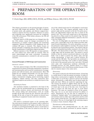
The patient is positioned supine on the operating table. is routine in many situations (Figure 1B).
The arms may be tucked in at the patient’s sides or placed on
well-padded arm boards at 90° angles to the table. Care must Thyroidectomy and Parathyroidectomy
be taken to avoid hyperextending the arms to avoid injury to Thyroidectomy and parathyroidectomy are the most
the brachial plexus. If the arms are tucked in at the side, the common neck procedures performed by general surgeons.
DOI 10.2310/7800.S01C08
10/08
- 2. © 2008 BC Decker Inc ACS Surgery: Principles and Practice
1 BASIC SURGICAL AND PERIOPERATIVE CONSIDERATIONS 8 PREPARATION OF THE OPERATING
ROOM — 2
a b
c
Figure 1 Patient positioning: (a) laparotomy,
(b) thoracotomy, and (c) thyroidectomy.
Preoperative evaluation should include assessment of cervical such as the Occupational Safety and Health Administration
spine mobility. The patient is positioned supine on the OR (OSHA) and the Nuclear Regulatory Commission.2–5
table. After endotracheal intubation, a sheet roll is placed Numerous articles and books discuss the various aspects
posterior to the scapula. The head is extended and placed on of OR design.6–10 The American Institute of Architects has
a foam “donut” pad, avoiding excess tension on the cervical published a comprehensive set of guidelines for health care
spine. Extension of the head should be avoided in the pres- facility design that includes a detailed discussion of OR
ence of limited cervical spine mobility. The head of the table design.11 Along with such guidelines and recommendations
is elevated to reduce venous pressure. Two ceiling lights are obtained from specialty surgical, anesthesiology, biomedical
positioned over the neck. Most surgeons use a head light to engineering and nursing associations, the design of new ORs
provide additional focused illumination. The scrub nurse must consider local needs and perspectives. The design of a
stands next to the Mayo stand holding the instrument tray new OR should be a collaborative effort reflecting the efforts
over the patient’s feet (Figure 1C). of clinical services, support services, and administration.
A similar position is employed for neck explorations. For
unilateral neck exploration, the patient‘s head is turned away design process and considerations
from the side of the planned incision. Designs must accommodate work flow and patient move-
ment. Important considerations include the mix of inpatient
design standards and outpatient surgery, the design of the hospital or institu-
Standards for new construction and major remodeling of tion (including proximity to clinical services such as radiology
ORs in the United States generally fall under the jurisdiction and pathology), the surgical specialties to be served, accessi-
of state and local agencies. These agencies, in turn, often bility to perioperative care units, accessibility of supplies,
incorporate guidelines published by the Department of Health and removal of waste materials. The need for intraoperative
and Human Services,1 as well as other groups and agencies, fluoroscopy and sectional imaging, shielding, ceiling-mounted
10/08
- 3. © 2008 BC Decker Inc ACS Surgery: Principles and Practice
1 BASIC SURGICAL AND PERIOPERATIVE CONSIDERATIONS 8 PREPARATION OF THE OPERATING
ROOM — 3
microscopes, and surgical robotics and similar advanced including nuclear, biological, chemical, and radiation
technologies will be influential. It is important to allow for incidents, are not addressed in this chapter.
flexibility and to anticipate the introduction of new
technologies over the life of the design. Time motion studies, physical requirements
simulations, and models may prove helpful and contribute to The fundamental model for OR design is a quadrangular
OR efficiency long term. Balancing flexibility and cost is a room with minimum dimensions of 20 × 20 ft or 7 × 7 m.
continuing challenge.12 More often, the dimensions are closer to 30 × 30 ft or
The geography and physical relationship of clean and less 10 × 10 m, so as to accommodate more specialized cardiac,
clean areas in the operating suite determine many other neurosurgical, minimally invasive, orthopedic, and multiteam
aspects of the OR suite. There are two designs in common procedures. Smaller rooms are generally adequate for minor
use. The first involves one or more clean hubs. The ORs are surgery and for procedures such as cystoscopy and eye
situated centrifugally in spokelike fashion. Clean and sterile surgery.
equipment and supplies that must be on hand for immediate Ceiling height should be at least 10 ft or 3.5 m to allow
use are stored in the hub. A peripheral corridor typically ceiling mounting of operating lights, microscopes, robots,
affords access to perioperative care units, instrument and navigational systems, and other equipment. An additional 1
anesthesia workrooms, clean storage, staff lounges, and other to 2 ft or 0.5 to 0.75 m of ceiling height may be advisable if
facilities. Entry and egress from the suite and access to the x-ray or other boom equipment is to be permanently mounted.
OR front desk and reception area also involve the peripheral In ORs designed to accept intraoperative sectional imaging
corridor. In the simplest embodiment, four ORs surround a devices (computed tomography [CT] and magnetic reso-
hub. The number can be greater or smaller. Controlled nance imaging [MRI]), the floors may be elevated to accom-
movement through the hub or hubs drives this design. modate cabling and wiring for power and data connections.
The second model uses corridors rather than hubs and When floors are elevated, the overall height of the OR has to
spokes. Supply rooms are typically situated between adjacent be increased.
rooms. A less common variation builds a separate supply Some institutions design or redesign the entire OR suite
with the potential to accommodate oversized equipment as
room next to each individual OR. Older ORs were designed
well as conduits and cables in both the ceiling and floor.
with one door only. Current design favors two, one connect-
Others prefer to customize individual rooms.
ing to the more sterile area and the other to the less sterile
OR and perioperative facilities may be specially constructed
area.
to withstand environmental hazards such as earthquakes,
The OR must balance the restrictions on access needed for
tornadoes, and floods or hardened to contend with hostile
safety and efficiency with freedom of movement for personnel
military or terrorist attack. Standards and specifications
and patients. This balance is equally important in emergency
for this type of construction fall outside the scope of this
situations and during complex and lengthy operations.
chapter.
Should ORs be dedicated to specific surgical specialties?
There are practical and logistical advantages to dedicated computerization, communication, and data
rooms, especially in cardiac surgery, neurosurgery, trauma, exchange: voice, video, and data
and ophthalmology. However, specialty-specific rooms can Computers, telephones, imaging, and other systems for
limit scheduling flexibility. Although it is hard to imagine data capture, analysis, and exchange should be integrated
justifying a room so narrowly designed that it absolutely into the OR design. The ubiquitous deployment of radiology
cannot be used for more than one specialty, dedicated rooms Picture Archiving and Communication Systems (PACS) and
might be emphasized in one institution and versatility in the potential of digital pathology to improve surgical pathol-
another. Each approach has its advocates. Design and ogy services mandate the installation of high-speed broad-
equipment should ultimately reflect expected and projected band connections. Two-way audio with teleconferencing
case mix. capabilities enables teleconsultation and teaching from and
Finally, the design of the OR suite must facilitate cleaning, in the OR.
disinfection and efficient turnover, and the installation and Effective communication systems capable of connecting
maintenance of installed equipment. the OR team, the OR front desk, and the rest of the hospital
storage must be accessible. Emergency communication channels
must be tested at regular intervals. Most importantly, systems
The OR design must provide adequate storage space for for communication, data exchange, and data storage retrieval
urgently required supplies. Storage in hallways and inside the must be available, preferably off-site. Off-site data storage
OR can create obstructions and hazards. backup is essential for hospitals likely to respond to mass
casualty events or subject to environmental hazards.
critical devices and emergency equipment
Critical devices, as well as emergency supplies and instru- assimilating new technologies
ments, must be prepared and positioned for immediate Advances in the medical device and information technol-
deployment. Accessories should be stored next to the ogy (IT) sectors stimulate a constant stream of innovation.
instruments that need them. Nevertheless, the undisciplined introduction of new technol-
Some hospitals have mass casualty response as part of their ogies can be distracting, contraproductive, and expensive.
mission. These institutions will have special requirements for ORs should be focused on measurable benefit and integration
versatility, equipment, storage, and supplies. The specific rather than novelty. The introduction of new technologies
design requirements for response to mass casualty events, should reduce complexity and tangibly increase therapeutic
10/08
- 4. © 2008 BC Decker Inc ACS Surgery: Principles and Practice
1 BASIC SURGICAL AND PERIOPERATIVE CONSIDERATIONS 8 PREPARATION OF THE OPERATING
ROOM — 4
benefits and options for patients in ways that have been The risks associated with malpositioning deserve further
recognized for more than a decade.13 comment. Any bony prominence is subject to injury from
The OR of the future will be characterized by increasing excessive pressure. The fragile skin of infants and older
dependence on technology. Although it is essential to track patients may be injured by dragging rather than lifting into
new technologies, they should be assessed and introduced position. The American Society of Anesthesiologists (ASA)
in a disciplined manner. Technology acquisition should be practice advisory on the prevention of perioperative periph-
strategic. A full discussion of technology assessment in eral neuropathies recommends that when practical, patients
surgery is outside the purview of this chapter, but the central should be placed in the intended position before induction of
principles are straightforward. First, the magnitude of unmet anesthesia to test for comfort.25 Uncomfortable positions
needs should be detailed and quantified. Second, the should be modified. This is especially important with older
cost-effectiveness of a new technology should be assessed. patients or patients with degenerative spine and joint disease
Next, competitive solutions, including substitutes for the or skeletal instability.
candidate technology acquisition, should be considered. Two major preventable consequences of malpositioning
Last, the marginal benefits and burdens of new technology are neuropathy or plexopathy and skin burns or ulceration.
introduction should be quantified. Approximately 80% of surgical procedures are performed
with patients in the supine position. Ulnar neuropathy and
brachial plexopathy are the two most common complications
Patient Safety, OR Efficiency, and Quality in this position and constitute 28% and 20% of claims,
Improvement respectively, in recent closed claims studies.26–31 Padding of
The OR is a high-stress environment with inherent risk in the precondylar groove of the humerus and tucking of the
arms or, at the least, restriction of abduction to less than 90°
which members of multiple disciplines temporarily congre-
help prevent this problem.25
gate to treat surgical patients. The potential for error and
The lithotomy position is used in approximately 9% of
inefficiency is great. Conversion of this group of individuals
cases and is the next most commonly used position.27,29
from disciplines with differing professional cultures and skill
Damage to the obturator, sciatic, lateral femoral cutaneous,
sets into a smoothly functioning surgical team is the key and peroneal nerves has been observed. These injuries
to increasing patient safety and efficiency.14 Surgical team account for 5% of claims for nerve damage in the Closed
training is an effective method to achieve a “team culture” Claims Data Base. They represent a small but not insignifi-
resulting in improved communication and decreased risk cant risk.26 Other patient positions have also been associated
of error.15 The use of perioperative care protocols reduces with malpositioning injuries.
failures of communication.16
The Joint Commission Universal Protocol became effective occupational injury to the health care team
on July 1, 2004. The key principles of the Universal Protocol Professionals engaged in perioperative and surgical care
are (1) the preoperative verification process, (2) marking the are exposed to biologic, ergonomic, chemical, physical, and
correct surgical site, (3) taking a “time-out” prior to surgery, psychosocial occupational risks.
and (4) adapting the requirements to non-OR settings. The
elements of the time-out concept are evolving and may Biologic Risks
include introduction of team members; patient identification; These risks include (1) parenteral and mucocutaneous
review of the patient’s history, risk factors, medications, and exposure to pathogens, (2) respiratory tract exposure to
allergies; availability of appropriate images and equipment; pathogens, (3) exposure to the biologic components of surgi-
goals of surgery; and potential intra- and postoperative cal smoke, and (4) exposure to allergens in latex gloves.32
problems. The role of a discussion of planned postoperative Strategies to reduce mucocutaneous exposure to pathogens
care at the completion of the surgical procedure prior to the include double-gloving, blunt suture needles, a neutral zone
emergence from anesthesia is not clear at the present time.17 for passage of sharps, and engineered sharps injury preven-
The use of a protocol during the hand-over of surgical tion devices.33 The use of N-95 respirator masks reduces the
patients to intensive care results in improved care.18 Extend- risk of occupational exposure to aerosolized Mycobacterium
tuberculosis and particulate matter greater than 1 micron in
ing the team concept to postanesthesia care has resulted in
size.34 Health care workers with allergies to latex should avoid
improved quality of care as measured by intubation times and
contact with latex gloves and tubing.
hospital stay following cardiac surgery.19 The use of specialty
teams during surgery to improve communication and effi- Ergonomic Risks
ciency is logical, but at present, there is a paucity of studies Health problems related to ergonomics, especially back
documenting its efficacy. Continuous monitoring is essential pain, are common. These problems are associated with
to maintain quality.20 awkward posture at the operating table, standing for long
Patient safety21 begins with protection of patients from periods of time, and back injury incurred by lifting patients.
misidentification, physical and chemical hazards, medication The OR table, monitors, imaging screens, and equipment
error, operative error, and improper positioning.22 The sur- should be positioned so that the OR team’s posture and posi-
geon is responsible for positioning the patient and preventing tion are as comfortable as possible within the limits of patient
positioning injury.23 The OR team is responsible for procur- safety. Lifting injury can be minimized by using proper patient
ing and maintaining the equipment for proper positioning transfer techniques and obtaining additional assistance when
and monitoring the patient intraoperatively. The entire team moving patients in the OR. Repetitive motion injury is a less
is responsible for preventing falls.24 common but not insignificant risk.32
10/08
- 5. © 2008 BC Decker Inc ACS Surgery: Principles and Practice
1 BASIC SURGICAL AND PERIOPERATIVE CONSIDERATIONS 8 PREPARATION OF THE OPERATING
ROOM — 5
Chemical Risks Another study considered risks to the fetus of the pregnant
Health care workers are exposed to numerous potentially surgeon. This risk is of particular importance early in gesta-
hazardous chemicals in the OR, including anesthetic gases, tion, before the pregnancy is recognized. In this study, the
disinfecting and sterilizing chemicals, specimen preservatives, calculated accumulative dose received intraoperatively
and chemotherapeutic drugs.32 Scavenger systems are exceeded the threshold dose for the induction of radiation
deployed broadly and have significantly reduced exposure to injury by at least two orders of magnitude. Approximately
anesthetic gases.35 Protective gowns and gloves prevent 2100 hours of fluoroscopy are required to reach this
exposure to other chemicals in the OR. threshold.
Childhood cancer is another documented risk of low-dose
Physical Risks fetal radiation exposure. The extent of this risk remains
Physical risks to patients and the OR team include controversial. Nevertheless, the ICRP has recommended a
fire, electrocution, radiation exposure, and laser energy maximum dose of 2 mSv and preferably less than 1 mSv
exposure.32 during the declared pregnancy. With the protection afforded
by a 0.5 mm lead-equivalent apron, however, the calculated
Fire The first principle of fire prevention is control of expected cumulative fetal dose is less than 0.37 mSv, a
the three elements of the fire triangle: oxygen, fuel, and an figure that extrapolates to a significantly lower dose, and
ignition source.36 Air/oxygen mixtures should be adjusted to consequently a lower risk, than that derived from natural
reduce the fraction of inspired oxygen (FiO2) to the lowest background radiation.
level consistent with patient safety and satisfactory hemoglo- Surgeons exposed to intraoperative ionizing radiation may
bin oxygen saturation.36 Light, heat, and electrical sources of avail themselves of several protective strategies. A 1 mm lead-
ignition should be carefully monitored and kept away from equivalent whole-body shield or apron provides 99% protec-
paper drapes. The use of alcohol-based skin preparation tion. A 0.5 mm lead-equivalent free-standing shield reduces
solutions should be minimized if possible.37 The use of the exposure by 90%. A neck (thyroid) shield with 0.25 mm lead
electrocautery or laser energy in the airway or in the unpre- equivalence reduces exposure by 60%. Radiation-protective
pared intestinal lumen in the presence of high concentrations gloves reduce dosage to the hand by up to 40%.37,41
of oxygen increases the risk of fire. Maintaining distance is also protective. Radiation attenu-
ates with the square of the distance. For any given distance
Radiation exposure Exposure to ionizing radiation is from the source of radiation, if the intensity of the radiation
inherently dangerous. For this reason, the extent of exposure
at a distance of d = 1, the intensity at any other distance is
must be minimized through the use of all protective means
equal to 1/d2. Intensity diminishes as a function of the square
available. Although the hazards of radiation were witnessed
of distance.
early in the 20th century, how exposure was to be calculated
The effects of distance are manifested quickly. Staff
and what acute and cumulative doses were dangerous could
exposure at 1 m distance from a C arm is 1/1000th that of
not be determined until later. In 1928, the International
the patient, even without shielding. In a model of femoral
Commission on Radiological Protection (ICRP) was formed
fracture, moving from the treated hip side of a patient to the
to study and set standards for radiation protection. The
contralateral side reduces exposure by a factor of 57 for
National Council on Radiation Protection (NCRP) was estab-
the surgeon, 13 for the nurse, and 21 for the anesthetist. If
lished in the United States to help set US policy on radiation
the surgeon remains ipsilateral to the operative site but moves
protection.37 Until recently, guidelines for radiation exposure
as little as 0.5 m away (cephalad) from the beam, the dose is
have been based on annual exposure limits (maximal permis-
sible yearly dose). The historical means of dosimetry under- reduced by a factor of 13, and at 1.5 m away, by a factor of
estimated exposure because of technical limitations and 26. At 2 m from the source of radiation, the scattered radia-
sampling error. For this reason, guidelines now focus on life- tion received by staff is very small.42 Nevertheless, because of
time accumulated exposure as a more useful surrogate for the the problem of accumulated lifetime exposure, no degree of
real risks in those with chronic or prolonged exposure. exposure is ever deemed truly innocuous.
Concessions to higher annual dose limits were made Image-guided surgery now plays a major role in many sur-
for workers allowed higher radiation exposure because of gical disciplines, including orthopedics, vascular surgery, and
occupational responsibilities. These were called “classified” urology. The cumulative radiation exposure associated with
workers, and they were subjected to strict monitoring and image guidance may be hazardous. The use of radionuclides
regular examination. Radiologists have been considered for identification of sentinel nodes and intraoperative brachy-
classified workers, but not surgeons. Classified workers were therapy for the treatment of certain malignancies also creates
permitted more than three times the annual dose than the potential for radiation exposure.43
nonclassified.38 Exposure to radiation comes from background radioactiv-
Empirical data suggest that the typical spinal surgeon per- ity and from radiation sources in the workplace. The ICRP
forming fluoroscopically guided procedures may receive as has established recommended yearly limits of radiation expo-
much as 1.3-fold the annual whole-body radiation dose allow- sure for these two categories [see Table 1].44 Recent studies of
able for radiologists, although the level of thyroid exposure is OR staff indicate that radiation exposure during surgical
less than 10% of the applicable limit specified by the NCRP.39 procedures employing radiation imaging techniques are well
A study of endovascular surgeons, in contrast, showed that within the ICRP limits.45–47
their radiation exposure fell well within these limits, as well All OR personnel should wear radiation safety badges to
as the limits established by the ICRP.40 allow monitoring of radiation exposure on a monthly basis.
10/08
- 6. © 2008 BC Decker Inc ACS Surgery: Principles and Practice
1 BASIC SURGICAL AND PERIOPERATIVE CONSIDERATIONS 8 PREPARATION OF THE OPERATING
ROOM — 6
Table 1 ICRP-Recommended Radiation Dose Limits44 OR Design
Advances in videoendoscopic surgery and endoluminal
Dose Limit
vascular surgery since 1990 have led to the introduction of
Occupational Public many complex electronic devices into an OR environment
Effective dose 20 mSv/yr averaged over 1 mSv/yr designed at the end of the 19th century. The promise of
defined periods of 5 yr widespread robotics use in the future may further complicate
Annual equivalent dose in equipment and use of space within the OR. At the same time,
Lens of the eye 150 mSv 15 mSv the importance of IT in both the performance of surgery
Skin 500 mSv 50 mSv and its documentation will only increase. These changes
Hands and feet 500 mSv —
will inevitably require design adaptations in the OR to
ICRP = International Commission on Radiological Protection; mSv = ensure adequate power and communication infrastructure
millisievert.
support.56
The ideal OR of the future will have videoendoscopic
monitors, light sources, and CO2 insufflators mounted on
OR personnel should wear protective lead aprons (0.25– booms that can be introduced or withdrawn from patient care
0.5 mm in thickness) with thyroid shields during procedures without moving heavy carts or reconnecting cables and power
in which fluoroscopy is used. Personnel frequently exposed to sources. ORs with ceiling- or floor-mounted fluoroscopy
radiation should wear lead-containing lenses for eye protec- permit efficient use of catheter-based endoluminal and intra-
tion. A mobile shield is useful for additional protection during vascular techniques during surgical care. Boom-mounted
cine runs. equipment can be easily moved, permitting more rapid room
OR personnel should stand as far away from the x-ray turnover between cases.
beam as possible to limit exposure. Even a distance of several Designing rooms with the flexibility for multidisciplinary
feet can significantly reduce exposure. Equipment emitting use may be more cost effective for many hospitals. In the long
ionizing radiation should be maintained and monitored to run, however, team training for subspecialty fields employing
ensure proper function.48 The use of pulsed fluoroscopy and complex imaging equipment and catheter-based therapy may
prove to be a more important factor for patient safety and
effective collimation of images will help minimize the dose
efficiency than any particular OR design.57 This is a key
of radiation. Excessive and unnecessary imaging should be
element in program development. Hospital administration
eliminated.
must also commit the capital necessary to complete such
a project before proceeding with program development.
Laser exposure Surgical lasers are class 4 lasers that
Balancing OR efficiency, new technology, and cost is a
have the potential to irreparably damage the eye and burn the continuous challenge.
skin. A class 4 laser beam is 1 billion times brighter than a
100 W light bulb.49 All OR personnel must wear protective
glasses with lenses designed specifically for the wavelength of Environmental Issues in the OR
the specific laser being used. Glasses should be inspected
temperature and humidity
prior to use to ensure that there are no scratches on the lens
that could render the wearer vulnerable to an eye injury. The In Europe and North America, the OR is typically main-
windows of the room should be shielded to protect individu- tained at 18° to 26°C (64.4° to 78.8°F). A higher tempera-
als passing by. The number of personnel in a laser therapy ture is necessary during operations on infants and burn
room should be kept to a minimum. patients to conserve body heat. Fully gowned surgeons prefer
the lower temperature of 18°C (64.4°F), but anesthesiologists
Noise and light exposure General environmental prefer 21.5°C (70.7°F).58 The optimum ambient temperature
in the OR reflects a compromise between the comfort of
hazards include high levels of noise and intense levels of
gowned and ungowned staff. The ubiquitous deployment of
light.50–52 These hazards are of equal significance to patients
patient warming devices has all but eliminated the problem
and to staff. One early study has indicated that elevated noise
of hypothermia in the adult during elective surgery.
levels during orthopedic procedures (above the recommended
Recent publications have indicated neurologic benefits
limits of 110 dBA) were associated with significant hearing from controlled hypothermia in the region of 34°C (93.2°F)
loss in orthopedic surgeons.51 Noise, excessive chatter, in certain cases of asystole, cardiopulmonary arrest, and
and even music can also pose a hazard to the patient by resuscitation.59–61 The equipment required for therapeutic
distracting the attention of OR personnel. hypothermia can be cumbersome.
Humidity in the OR is generally maintained between 50
Psychosocial risks Perhaps the most underappreciated and 60%. Humidity greater than 60% may cause condensa-
risks to the perioperative professional are psychosocial. Long tion on cool surfaces. Humidity less than 50% may not
hours and nighttime emergencies lead to fatigue. The high suppress static electricity. Static electricity can adversely
level of responsibility, the consequences of error, and the affect the function of modern computers and smart medical
level of oversight have led to high levels of stress and burn- devices.
out.53 Unfortunately, violence in the health care workplace,
both verbal and physical, is common.54 Stress reduction can lighting
be achieved by team training.15 Hospitals should have zero ORs are no longer provided with skylights and northern
tolerance for workplace violence.55 exposure to maximize the benefit of natural illumination.
10/08
- 7. © 2008 BC Decker Inc ACS Surgery: Principles and Practice
1 BASIC SURGICAL AND PERIOPERATIVE CONSIDERATIONS 8 PREPARATION OF THE OPERATING
ROOM — 7
Nevertheless, effective lighting remains an important factor in interface as a function of resistance to the flow of current.
reducing stress by providing clear views of the operative field, The vaporization of intracellular water content leads to disin-
the anesthesia and monitoring systems, and the nursing tegration of the cell. Heat leads to the coagulation of proteins
area. and hemostasis. Cellular debris is aerosolized. The smoke
The standards for OR illumination were outlined by Dr. that is generated is comparable to that accompanying laser
William Beck and the Illuminating Engineering Society.62,63 A use. The smoke and its particulate content may be carcino-
general illumination brightness of up to 200 footcandles (ft-c) genic, allergenic, inflammatory, and infectious. There is little
is desirable in new construction. The lighting sources should difference between laser plume and electrosurgical smoke.65
be free of glare and nonreflective.64 The electrosurgical instrument requires close supervision.66
The amount of light required during an operation varies It is very easy to become complacent in its use. The unit
with the surgeon and the operative site. Quantities of light generates an electrical arc. In the past, these have been asso-
can be measured as a function of the amount beamed on a ciated with explosions. This risk has been lessened because
subject (incident light) or the amount reflected (reflected explosive anesthetic agents are no longer used. Nevertheless,
light). In one study, general surgeons operating on the explosion of hydrogen and methane gases in the large bowel
common bile duct found 300 ft-c sufficient because the remains a threat, especially when surgery must be performed
reflectance of this tissue area is 15%. The corresponding inci- on an unprepared bowel.67 Because the unit and its arc
dent light level would be 2,000 ft-c.21 Surgeons performing generate a broad and unfiltered band of radiofrequencies,
coronary bypass operations require an incident level of electrosurgical units may interfere with monitoring devices,
3,500 ft-c.21 Whether changes in the color of light can improve most notably the electrocardiographic (ECG) monitor.
discrimination of different tissues is unknown. Interference with cardiac pacemaker activity may occur.68
The powerful lights installed for surgery generate heat Skin burns are the most frequently reported complication
through the projection of infrared energy. This is an in use. Rarely are they fatal, but they are painful and may
inefficient but inevitable byproduct of most forms of light require skin grafts, raising the possibility of litigation. The
appropriate for the OR. Much of the infrared energy can burn site can be at the dispersive electrode, ECG monitoring
be eliminated by filtration or by heat-diverting dichroic leads, esophageal or rectal temperature probes, or areas of
reflectors. body contact with grounded objects. The dispersive electrode
There are several types of overhead lighting designs. For should be firmly attached to a broad area of dry, hairless skin,
preferably over a large muscle mass.67,68
small ORs, a single large overhead light with one or two small
Alcohol-based hair care products should be avoided during
satellites may suffice. Larger ORs may benefit more from
head and neck surgery because the risk of combustion during
several large lights with broader orbits. The larger the OR,
the use of electrocautery.
the more important the installation of multiple light sources
Preparation of the OR should include verification of the
that can concentrate light in different areas of the operating
proper function and grounding of the unit and proper place-
field. This consideration is of particular importance where
ment of the dispersive electrode. Smoke evacuators are highly
large body areas are exposed or where multiple sites are
recommended. The National Institute for Occupational
accessed simultaneously.
Safety and Health (NIOSH) recommends a system that can
The illumination provided by overhead lighting can be
pull 50 cubic feet per minute with a capture velocity of 100
usefully amplified and improved with headlights. Headlights
to 150 feet per minute at the inlet nozzle and positioned
have substituted for mobile lights in many OR suites, but within 2 inches of the surgical site.69 Filters should be installed
both have their place. When headlights are used, spare bulbs to capture the contents of the smoke. Used filters are deemed
must be kept on hand and cabling examined for damage and biohazardous. Routine, unfiltered suction is not adequate for
properly stored after each use. this purpose.69
During microsurgical and laparoscopic procedures, the
surgical field is illuminated primarily by dint of the instru- lasers and laser safety
ments used for visualization. The importance of ambient Lasers generate focused energy. They have been implicated
lighting for the remainder of the OR must not be forgotten. in skin burns, retinal injuries, endotracheal tube fires,
All ORs should be equipped with battery-powered backup pneumothorax, and damage to viscera and the vasculature.70
illumination and flashlights for emergency use. Both patients and staff have suffered injuries.
or equipment The laser-safe OR requires specific modifications. The OR
should not have windows. Walls, ceilings, and equipment
The modern OR is replete with medical devices designed should be nonreflective. Equipment used in the operative
to facilitate the work of the surgical team and to support, field should be nonreflective and nonflammable. A sign
position, and protect the patient [see Table 2]. The equipment warning personnel that a laser is in use should be posted.
in the OR must be chosen and maintained with three Anesthetic technique should also take the use of lasers into
concerns in mind: patient safety, surgical efficiency, and consideration. When lasers are used in the vicinity of the face,
reduction of occupational hazards. airway, or chest cavity, the concentration of O2 and N2O in
the inhaled gases should be reduced to decrease the possibil-
electrosurgical devices
ity of fire. Personnel should wear appropriate eye protection.
The common electrosurgical device is a 500 W radiofre- A smoke evacuator will improve visualization, reduce objec-
quency generator used to cut and coagulate tissue. A current tionable odor, and decrease the potential for papillomavirus
passes from the cutting or coagulating surgical instrument infection or ingestion of toxic fumes from the laser smoke
to a dispersion electrode. Heat is generated at the tissue plume.65,71–74
10/08
- 8. © 2008 BC Decker Inc ACS Surgery: Principles and Practice
1 BASIC SURGICAL AND PERIOPERATIVE CONSIDERATIONS 8 PREPARATION OF THE OPERATING
ROOM — 8
Table 2 Devices Used in the Operating Room
Function Device
Support of patient Anesthesia delivery devices
Ventilator
Physiologic monitoring devices
Warming devices
IV fluid warmers and infusers
Support of surgeon Sources of mechanical, electrical, and internal power, including power tools and electrocautery, as well as
laser and ultrasound instruments
Mechanical retractors
Lights mounted in various locations
Suction devices and smoke evacuators
Electromechanical and computerized assistive devices, such as robotic assistants
Visualization equipment, including microscopes, endoscopic video cameras, and display devices such as
video monitors, projection equipment, and head-mounted displays
Data, sound, and video storage and transmission equipment
Diagnostic imaging devices (e.g., for fluoroscopy, ultrasonography, MRI, and CT)
Support of OR team Surgical instruments, usually packaged in case carts before each operation but occasionally stored in
nearby fixed or mobile modules
Tables for display of primary and secondary surgical instruments
Containers for disposal of single-use equipment, gowns, drapes, etc.
Workplace for charting and record keeping
Communication equipment
CT = computed tomography; IV = intravenous; MRI = magnetic resonance imaging; OR = operating room.
powered devices Improperly balanced instruments have been known to twist
The surgical table must be properly positioned and adjusted or fall, causing injury to patients and staff.
to ensure the safety of the patient and the efficient work of Many commercial microscope drapes are available. Where
the surgical team. Manually adjustable tables are simple, but the laser is mounted to the microscope, it is essential to
those with electrical controls are easier to manage. OR table confirm that the drape is out of the line of fire to avoid
attachments, such as the arm boards and leg stirrups used to contamination, melting, or kindling.
position patients, must be properly maintained and secured intraoperative imaging and navigation
to prevent injuries to patients or staff. During transfer to and
from the OR table, care must be taken to ensure that the The advent of less invasive surgery requires accurate intra-
patient is not injured and that life support, monitoring, and operative confirmation of surgical anatomy through the use
intravenous (IV) systems remain connected. Proper transfer of x-ray, CT, MRI, and ultrasonography. These modalities
technique, adequate staff, and the use of assistive devices are increasingly accompanied by and aligned with the use of
such as rollers will help prevent musculoskeletal injuries to surgical navigation systems. Intraoperative ultrasonography
the OR staff during this maneuver. Patients with fragile skin, requires a high-quality portable ultrasound unit and special-
including infants, small children, the ill, and the elderly, ized probes. Depending on the procedure and the training of
should be lifted and not dragged into position. the surgeon, the presence of a radiologist and an ultrasound
Other powered surgical instruments common in the OR technician may be requested.
include those used to obtain skin grafts, open the sternum The ultrasound unit should be positioned near the patient,
and the cranium, and perform orthopedic procedures. and the surgical team must be able to view the image
Powered saws and drills can aerosolize body fluids, thereby comfortably. In some cases, the image may be displayed on
creating a potential infection and contamination hazard for OR monitors by means of a video mixing device. Ultrasound
OR personnel.75 units may also be used to modify the reference image in real
time in surgical navigation systems.76
operating microscope Fluoroscopic and x-ray units may be portable or dedicated.
Microsurgery is a routine part of many surgical specialties, Open radiologic units are usually installed either in the OR
including neurosurgery, plastic surgery, hand surgery, gyne- proper or immediately adjacent to the OR to permit intraop-
cologic surgery, and ophthalmology. Microscopes may be erative imaging of the selected body area. Intraoperative angi-
moved into the OR on wheeled stands or mounted on the ography and MRI devices are typically installed in dedicated
ceiling, wall, or floor. Built-in microscopes are best employed ORs.77 Intraoperative CT for neurosurgery, for example, may
in dedicated ORs.46 All controls and displays must be involve either portable or fixed devices.78
properly positioned at or below the user’s line of sight to The use of sequential compression stockings has become
allow comfortable and unobstructed viewing. the standard of care for the prevention of venous thrombo-
Modern operating microscopes are servo controlled and embolism in the majority of operations for which direct access
counterbalanced. The sequence for properly balancing a to the lower extremities is not required [see 6:6 Venous
microscope should be carefully followed prior to every Thromboembolism].79 The air pump often must be placed near
procedure and each time accessories are added or removed. the patient on the floor or on a nearby cart. The pressure
10/08
- 9. © 2008 BC Decker Inc ACS Surgery: Principles and Practice
1 BASIC SURGICAL AND PERIOPERATIVE CONSIDERATIONS 8 PREPARATION OF THE OPERATING
ROOM — 9
tubing from the stockings to the pump must be carefully use a foot pedal to select various imaging and recording
routed so as not to interfere with surgical access. modes and to play back selected images and sequences.
Suction devices are ubiquitous in the OR. A typical suction Unlike fixed systems, which require patients to be moved
apparatus consists of a set of canisters on a wheeled base on a floating table to change the field of view, C-arm systems
receiving suction from a wall- or ceiling-mounted source. The require the image intensifier to be moved from station to
surgeon’s aspirating cannula is connected sterilely to these station over a fixed patient. Although more cumbersome than
canisters. Suction tubing is a common tripping hazard in the fixed systems, the newest C-arms have increased mobility
OR. In some procedures, the suction canisters may require and maneuverability. Patients must be placed on a special
repeated changing. nonmetallic carbon fiber table. To provide a sufficient field of
view and permit panning from head to toe, the tables must
equipment be supported at one end and completely clear beneath.
Although these tables do not flex, they are sufficient for most
Imaging Equipment
operations. Furthermore, they are mobile and may be replaced
Imaging equipment for endovascular surgery can be fixed with conventional operating tables when the endovascular
to the ceiling or portable. The fixed system is also employed suite is being used for standard open surgical procedures.
in catheterization laboratories and dedicated interventional
radiology suites. Portable systems use C-arms with dedicated Interventional Equipment
vascular software packages designed for optimal endovascular The performance of endovascular procedures in the OR
imaging. Each of these systems has advantages and disadvan- requires familiarity with a wide range of devices that may
tages. be unfamiliar to OR personnel. These include guidewires,
Fixed ceiling-mounted systems have higher power output sheaths, specialized catheters, angioplasty balloons, stents,
and smaller focal spot size and provide the highest-quality and stent grafts [see Table 3]. In a busy endovascular OR,
images. Larger image intensifiers (up to 16 in.) make possible much of this equipment must be stocked for everyday use,
larger visual fields for diagnostic arteriograms. Fewer runs are with the remainder ordered on a case-by-case basis. The
necessary, with the advantage of reducing the quantity of expense of establishing the necessary inventory of equipment
contrast injected and radiation exposure. can be substantial and can place a considerable burden on
Fixed systems are accompanied by floating angiography smaller hospitals that are already spending sizable amounts
tables, which allow the surgeon to move the patient easily on stocking similar devices for their catheterization laborato-
beneath the fixed image intensifier. The variable distance ries and interventional suites. Fortunately, many companies
between the x-ray tube and the image intensifier allows the are willing to supply equipment on a consignment and case
basis, allowing hospitals to pay for devices as they are used
intensifier to be placed close to the patient if desired, thereby
and reducing the volume of devices stocked.
improving image quality and decreasing radiation scatter.
It is generally accepted that such systems afford the surgeon ORs in Other Specialties
the most control and permit the most effective imaging of
Specialty equipment is generally available for permanent
patients. installation in ophthalmologic, orthopedic, trauma, neurosur-
Fixed ceiling-mounted systems are quite expensive (typi- gical, and urologic ORs. The observations offered regarding
cally $1 to $1.5 million), and major structural renovations are microscopes, imaging devices and lasers, storage, and physi-
often required for installation in a typical OR. These systems cal layout are broadly applicable. The cardiovascular OR and
are not particularly flexible. The floating angiography tables the neurosurgical OR often have complex needs that are more
and the immobility of the image intensifiers render the rooms difficult to fill simply by equipping a general-purpose OR.
unsuitable for most conventional open surgical procedures. Thus, a neurosurgical OR may need to be equipped for
As a result, fixed imaging systems are generally restricted to neuronavigation, laser and microsurgery, intraoperative angi-
high-volume centers where use rates justify the construction ography, neurointerventional surgery, and pediatric surgery.
of dedicated endovascular ORs. In addition, it may need shielding for neurophysiologic
The imaging capability and versatility of portable digital recordings necessary for cranial electrostimulation. The pres-
C-arms have increased dramatically. State-of-the-art portable ence of fixed or mounted instrumentation per se rarely poses
C-arms are considerably less expensive than fixed systems an insurmountable difficulty in adapting a general-purpose
($175,000 to $225,000) while retaining many of their advan- OR to specialty use, but the reverse is not the case.
tages. The variable image intensifier size (from 6 to 12 in.)
offers valuable flexibility, with excellent resolution at the Surgeon’s Control of Equipment: Touch Panels, Voice
smaller end and an adequate field of view at the larger end. Activation, and Robotics
With some portable C-arm systems, it is possible to vary the Inherent shortcomings in the design of OR equipment have
distance between the image intensifier and the x-ray tube, as been exacerbated by the spread of minimally invasive proce-
with a fixed system (see above). Pulsed fluoroscopy, image dures. Because most of the equipment needed for minimally
collimation, and filtration are standard features for improving invasive surgery resides outside the sterile field, the circulat-
imaging and decreasing radiation exposure. Sophisticated ing nurse becomes the point person for critical control. Often
software packages allow high-resolution digital subtraction the circulating nurse is out of the room at the precise moment
angiography, variable magnification, road mapping (i.e., the important equipment adjustments must be made.
superimposition of live fluoroscopy over a saved digital Surgeons grow frustrated at the inevitable delays that
arteriogram), image fusion, and a number of other useful follow, and nurses often weary of these additional and some-
features. Improvements in C-arm design allow the surgeon to times distracting responsibilities. As a result, there is great
10/08
- 10. © 2008 BC Decker Inc ACS Surgery: Principles and Practice
1 BASIC SURGICAL AND PERIOPERATIVE CONSIDERATIONS 8 PREPARATION OF THE OPERATING
ROOM — 10
Table 3 Standard Equipment for Endovascular Operating Rooms
Diagnostic arteriography Entry needle (16-gauge beveled)
Entry wire (J wire or floppy-tip wire)
Arterial sheath (5 Fr)
Catheters
Multipurpose nonselective (pigtail, tennis racquet, straight, etc.)
Selective (cobra head, shepherd’s crook, etc.)
Guidewires (floppy, steerable, angled, hydrophilic, etc.)
Contrast agent (nonionic preferred)
Power injector
Balloon stent angioplasty Sheaths (various lengths and diameters)
Guide catheters
Balloons (various lengths and diameters)
Stents
Balloon expandable (various lengths and diameters)
Self-expanding (various lengths and diameters)
Inflation device
Endovascular aneurysm repair Large-caliber sheaths (12–24 Fr)
Super-stiff guidewires
Endovascular stent grafts
Main body and contralateral iliac limb
Aortic and iliac extension grafts
Endovascular arterial coils
interest in improving the interface of surgical devices with the provides the OR team with critical information about the pro-
surgeon. Sterile touchscreens offered the first solution, but gress of the operation. To operate a device, the surgeon must
many two-handed surgical procedures still make it difficult take approximately 20 minutes to train the recognition system
for a surgeon to control ancillary equipment manually. Voice to his or her voice patterns and must wear an audio headset
activation technology offers a potential solution. to relay commands. The learning curve for voice control is
Development of voice activation began in the late 1960s. minimal (two or three cases, on average). Devices that can
The goal was a simple, safe, and universally acceptable voice now be controlled by voice activation software include
recognition system that flawlessly carried out the verbal cameras, light sources, digital image capture and documenta-
requests of the user. However, attempts to construct a system tion devices, printers, insufflators, OR ambient and surgical
capable of accurately recognizing a wide array of speech lighting systems, operating tables, and electrocauteries.
patterns faced formidable technological hurdles that are only
now beginning to be overcome. Infection Control in the OR
In 1998, the first Food and Drug Administration (FDA)- Infection control is a major concern in health care in
approved voice activation system, Hermes (Computer general, but it is a particularly important issue in the sterile
Motion, Santa Barbara, CA), was introduced into the OR. environment of the OR, where patients undergo surgical
Designed to provide surgeons with direct access and control procedures and are at significant risk for perioperative
of surgical devices, Hermes is operated via either a handheld nosocomial infection. Even the best OR design will not
pendant or voice commands from the surgeon. The chal- compensate for improper surgical technique or failure to pay
lenges of advanced laparoscopic surgery provide a fertile attention to infection prevention.
ground for demonstrating the benefits of voice activation [see Surgical site infection (SSI) is a major cause of patient
Table 4]. morbidity, mortality, and health care costs. In the United
Voice activation offers surgeons immediate access to States, according to the Centers for Disease Control and
and direct control of surgical devices. At the same time, it Prevention (CDC), approximately 2.9% of nearly 30 million
Table 4 Benefits of Voice Activation Technology in the Laparoscopic Operating Room
Benefits to surgical team Gives surgeons direct and immediate control of devices
Frees nursing staff from dull, repetitive tasks
Reduces miscommunication and frustration between surgeons and staff
Increases OR efficiency
Alerts staff when device is malfunctioning or setting off alarm
Benefits to hospital Saves money, allowing shorter, more efficient operations
Contributes to better OR use and, potentially, performance of more surgical procedures
Lays foundation for expanded use of voice activation in ORs
Allows seamless working environment
Benefit to patient Reduces operating time, which—coupled with improved optics, ergonomics, and
efficiency—leads to better surgical outcome
OR = operating room.
10/08
- 11. © 2008 BC Decker Inc ACS Surgery: Principles and Practice
1 BASIC SURGICAL AND PERIOPERATIVE CONSIDERATIONS 8 PREPARATION OF THE OPERATING
ROOM — 11
operations are complicated by SSIs each year. This percent- of surgical gloves rather than a single pair provides an addi-
age may, in fact, be an underestimate, given the known tional barrier and further reduces the risk of contamination.
inherent problems with surgeons’ voluntary self-reporting of A 2002 Cochrane Review concluded that wearing two pairs
infections occurring in the ambulatory surgical setting.80 Each of latex gloves significantly reduced the risk of hand exposure.
infection is estimated to increase total hospital stay by several The rate of single-glove perforations is approximately 15%.
days and add materially to costs and charges. The rate of inner glove perforation when two pairs of gloves
SSIs pose an increasingly difficult problem for institutions are worn is approximately 5%.86
with a large Medicare population. Recently, the Centers OSHA requires that personal protective equipment be
for Medicare and Medicaid Services (CMS) published the available in the health care setting, and these requirements
Inpatient Prospective Payment System (IPPS) FY 2009 final are specified in the OSHA standard on occupational exposure
rule expanding the list of hospital-acquired conditions (HACs) to bloodborne pathogens, which went into effect in 1992.
that will reduce Medicare payments. A number of SSIs figure Among the requirements is the implementation of the CDC’s
prominently on this list.81 State Medicaid directors have also universal precautions, designed to prevent transmission of
been authorized to deny payment for certain HACs. HIV, hepatitis B virus, and other bloodborne pathogens.87
The epidemiology and management of SSIs in older adults These precautions dictate the use of protective barriers (e.g.,
are receiving increasing attention.82 SSIs have been divided gloves, gowns, aprons, masks, and protective eyewear) to
by the CDC into three broad categories: superficial incisional reduce the risk that the health care worker’s skin or mucous
SSI, deep incisional SSI, and organ or space SSI [see Table 5 membranes will be exposed to potentially infectious
and 1:1 Prevention of Postoperative Infection].83 materials.
Factors that contribute to the development of SSI include Performance standards for protective barriers are the
responsibility of the FDA’s Center for Devices and
(1) the patient’s health status, (2) the physical environment
Radiological Health. FDA standards define the performance
where surgical care is provided, and (3) clinical interventions
properties that these products must exhibit, including mini-
that increase the patient’s inherent risk. Careful patient
mum strength, barrier protection, and fluid resistance. The
selection and preparation, including judicious use of antibi-
current CDC recommendation is to use surgical gowns
otic prophylaxis, can decrease the overall risk of infection,
and drapes that resist liquid penetration and remain effective
especially after clean-contaminated and contaminated
barriers when wet.
operations.
These standards are intuitively appealing, but few studies
hand hygiene address the comparative benefits of specific types of drapes.
A meta-analysis of over 5,000 surgical cases indicated that
Hand antisepsis plays a significant role in preventing noso- adhesive drapes increase the risk of SSI (relative risk = 1.23;
comial infections. When outbreaks of infection occur in the p = .03) and iodine-impregnated drapes had no effect.88
perioperative period, careful assessment of the adequacy of Compliance with universal precautions and barrier
hand hygiene among OR personnel is important. Agents protection is a challenge. Educational efforts aimed at OR
used for surgical hand scrubs should substantially reduce personnel improved compliance significantly, particularly
microorganisms on intact skin, contain a nonirritating antimi- with regard to the use of protective eyewear and double-
crobial preparation, possess broad-spectrum activity, and be gloving. These efforts were associated with a reduced
fast-acting and persistent.84 The CDC offers the following incidence of blood and body fluid exposure.89
guidelines85:
antimicrobial prophylaxis
• Surgical hand antisepsis using either an antimicrobial
SSIs are established several hours after contamination.90
soap or an alcohol-based hand rub with persistent activity
There is a well-recognized “golden period” during which
is recommended before donning sterile gloves when
prophylactic antibiotics can be effective. Administration of
performing surgical procedures (evidence level IB).
antibiotics before contamination reduces the risk of infection
• When performing surgical hand antisepsis using an antimi-
but is subsequently of little value.91 Selective use of short-
crobial soap, scrub hands and forearms for the length of
duration, narrow-spectrum antibiotic agents should be con-
time recommended by the manufacturer, usually 2 to 6 sidered for appropriate patients to cover the usual pathogens
minutes. Long scrub times (e.g., 10 minutes) are not isolated from SSIs [see Table 6].92
necessary (evidence level IB). Recommendations for antibiotic prophylaxis are addressed
• When using an alcohol-based surgical hand-scrub product in more detail elsewhere [see 1:1 Prevention of Postoperative
with persistent activity, follow the manufacturer’s instruc- Infection]. In brief, the principles of optimal surgical
tions. Before applying the alcohol solution, prewash hands antimicrobial prophylaxis include (1) appropriate choice of
and forearms with a nonantimicrobial soap and dry hands an antimicrobial agent, (2) proper timing of antibiotic admin-
and forearms completely. After application of the alcohol- istration before incision, and (3) limited duration of antibiotic
based product, allow hands and forearms to dry thoroughly administration after operation.
before donning sterile gloves. When a preoperative antibiotic is indicated, a single dose
of therapeutic strength, administered shortly before incision,
gloves and protective barriers
usually suffices.93 The dose may have to be increased if the
There is a high risk of pathogen transfer during surgery. patient is morbidly obese.94 A second dose is indicated if the
This is a risk from which both the patient and the surgical procedure is longer than two half-lives of the drug or if exten-
team must be protected. The risk can be reduced by using sive blood loss occurs. Continuation of prophylaxis beyond
protective barriers, such as surgical gloves. Wearing two pairs 24 hours is not recommended.
10/08
- 12. © 2008 BC Decker Inc ACS Surgery: Principles and Practice
1 BASIC SURGICAL AND PERIOPERATIVE CONSIDERATIONS 8 PREPARATION OF THE OPERATING
ROOM — 12
Table 5 Criteria for Defining a Surgical Site Infection83
Superficial incisional SSI
Infection occurs within 30 days after the operation, and infection involves only skin or subcutaneous tissue of the incisions, and at least
one of the following:
1. Purulent drainage, with or without laboratory confirmation, from the superficial incision
2. Organisms isolated from an aseptically obtained culture of fluid or tissue from the superficial incision
3. At least one of the following signs or symptoms of infection: pain or tenderness, localized swelling, redness, or heat; and superficial
incision is deliberately opened by surgeon, unless incision is culture negative
4. Diagnosis of superficial incisional SSI by the surgeon or attending physician
Do not report the following conditions as SSI:
1. Stitch abscess (minimal inflammation and discharge confined to the points of suture penetration)
2. Infection of an episiotomy or newborn circumcision site
3. Infected burn wound
4. Incisional SSI that extends into the fascial and muscle layers (see deep incisional SSI)
Note: Specific criteria are used for identifying infected episiotomy and circumcision sites and burn wounds.
Deep incisional SSI
Infection occurs within 30 days after the operation if no implant* is left in place or within 1 yr if an implant is in place and the infection
appears to be related to the operation, and infection involves deep soft tissues (e.g., fascial and muscle layers) on the incision, and at
least one of the following:
1. Purulent drainage from the deep incision but not from the organ/space component of the surgical site
2. A deep incision spontaneously dehisces or is deliberately opened by a surgeon when the patient has at least one of the following
signs or symptoms: fever (> 38°C [100.4°F]), localized pain, or tenderness, unless the site is culture negative
3. An abscess or other evidence of infection involving the deep incision is found on direct examination, during reoperation, or by
histopathologic or radiologic examination
4. Diagnosis of a deep incisional SSI by a surgeon or attending physician
Notes
1. Report infection that involves both superficial and deep incision sites as deep incisional SSI
2. Report an organ/space SSI that drains through the incision as a deep incisional SSI
Organ/space SSI
Infection occurs within 30 days after the operation if no implant* is left in place or within 1 yr if an implant is in place and the infection
appears to be related to the operation, and infection involves any part of the anatomy (e.g., organs or spaces), other than the incision,
which was opened or manipulated during an operation, and at least one of the following:
1. Purulent drainage from a drain that is placed through a stab wound† into the organ/space
2. Organisms isolated from an aseptically obtained culture of fluid or tissue in the organ/space
3. An abscess or other evidence of infection involving the organ/space that is found on direct examination, during reoperation, or by
histopathologic or radiologic examination
4. Diagnosis of an organ/space SSI by a surgeon or attending physician
SSI = surgical site infection.
*National Nosocomial Infection Surveillance definition; a non–human-derived implantable foreign body (e.g., prosthetic heart valve, nonhuman vascular graft,
mechanical heart, or hip prosthesis) that is permanantly placed in a patient during surgery.
†
If the area around a stab wound becomes infected, it is not an SSI. It is considered a skin or soft tissue infection, depending on its depth.
The provision of prophylactic antibiotics, where indicated, published in 2001, determined that warming patients before
has been accepted as a measure of surgical quality aimed at short-duration clean procedures (breast, varicose vein, or
a reduction in SSIs.95,96 A strong economic case has been hernia) reduced infection (p = .001) and reduced wound
made for infection control, including prophylactic antibiotics, scores (p = .007) in this setting as well.104
benefiting the hospital as well as the patient.97 The perio- The safest and most effective way of protecting patients
perative nursing team has a central role, together with the from hypothermia is to use forced-air warmers with special-
anesthesiologist, in monitoring the administration of ized blankets placed over the upper or lower body. Alterna-
preoperative prophylactic antibiotics.98–100 tives include placing a warming water mattress under the
patient and draping the patient with an aluminized blanket.
nonpharmacologic preventive measures Second-line therapy involves warming all IV fluids. Any
Several studies have confirmed that certain nonpharmaco- irrigation fluids used in a surgical procedure should be at or
logic measures, including maintenance of perioperative slightly above body temperature before use. Radiant heating
normothermia and provision of supplemental perioperative devices placed above the operative field may be especially
oxygen, are efficacious in preventing SSIs.101 useful during operations on infants. Use of a warmer on
the inhalation side of the anesthetic gas circuit can also
Perioperative Normothermia help maintain the patient’s body temperature during an
A 1996 study showed that warming patients during colorec- operation.
tal surgery reduced infection rates.102 This finding was
confirmed in a subsequent observational cohort study that Supplemental Perioperative Oxygen
reported a significantly increased incidence of SSI with Destruction by oxidation, or oxidative killing, is the body’s
hypothermia.103 A randomized, controlled trial of 421 patients, most important defense against surgical pathogens. This
10/08
- 13. © 2008 BC Decker Inc ACS Surgery: Principles and Practice
1 BASIC SURGICAL AND PERIOPERATIVE CONSIDERATIONS 8 PREPARATION OF THE OPERATING
ROOM — 13
Table 6 Distribution of Pathogens Isolated from Surgical Site Infections: National Nosocomial Infections Surveillance
System, 1986–199683
Pathogen Percentage of Isolates*
1986–1989 (N = 16,727) 1990–1996 (N = 17,671)
Staphylococcus aureus 17 20
Coagulase-negative staphylococci 12 14
Enterococcus species 13 12
Escherichia coli 10 8
Pseudomonas aeruginosa 8 8
Enterobacter species 8 7
Proteus mirabilis 4 3
Klebsiella pneumoniae 3 3
Other Streptococcus species 3 3
Candida albicans 2 3
Group D streptococci (nonenterococci) — 2
Other gram-positive aerobes — 2
Bacteroides fragilis — 2
*Pathogens representing less than 2% of isolates are excluded.
defensive response depends on oxygen tension in contami- decrease in mortality rate generally in critically ill patients.111,112
nated tissue. An easy method of improving oxygen tension in The effects are not restricted to diabetics. In fact, there is
adequately perfused tissue is to increase the FiO2. Supple- evidence to conclude that nondiabetics enjoy more protection
mental perioperative oxygen (i.e., an FiO2 of 80% instead of than diabetics.113
30%) significantly overcomes the decrease in phagocytosis Some questions remain about just how tight control must
and bacterial killing usually associated with anesthesia and be to have an effect. There is a small but measurable risk
surgery (and significantly reduces postoperative nausea and of hypoglycemia with intensive IV insulin therapy, but this
vomiting). Oxygen tension in wound tissue has been found to risk can be mitigated with continuous or near-continuous
be a good predictor of SSI risk.105 glucometry or dynamic scale nomograms.114
Avoidance of Blood Transfusion
Housekeeping Procedures
The association between blood transfusion and increased
perioperative infection rates is well documented. In a 1997 floors and walls
study, geriatric hip fracture patients undergoing surgical
Despite detailed recommendations for cleaning the OR [see
repair who received blood transfusion had significantly higher
Table 7], the procedures that are optimal to provide a clean
rates of perioperative infection than those who did not (27%
environment while still being cost-effective have not been
versus 15%), and this effect was confirmed on multivariate
critically analyzed.115
analysis.106 Another study yielded similar findings in colorec-
Only a few studies have attempted to correlate surface
tal cancer: the relative risk of infection was 1.6 for transfusion contamination of the OR with SSI risk. In one randomized,
of one to three units of blood and 3.6 for transfusion of more
controlled study, for example, the control rooms were cleaned
than three units.107
with a germicidal agent and wet-vacuumed before the first
A large prospective cohort study published in 2003 evalu-
case of the day and between cases, whereas in the experimen-
ated the association between anemia, blood transfusion, and
tal rooms, cleaning consisted only of wiping up grossly visible
perioperative infection.108 Regression analysis confirmed that
contamination after clean and clean-contaminated cases.116
intraoperative transfusion of packed red blood cells was an
Both rooms had complete floor cleanup after contaminated
independent risk factor for perioperative infection (odds ratio
1.06; confidence interval 1.01 to 1.11; p > .0001). Further- or dirty and infected cases. Bacterial colony counts obtained
more, transfusion of more than four units of packed red blood directly from the floors were lower in the control rooms, but
cells was associated with a ninefold increased risk of infection counts obtained from other horizontal surfaces did not differ
(confidence interval 5.74 to 15.00; p < .0001). between the two OR groups. Wound infection rates were the
same in the control rooms and the experimental rooms and
Tight Glycemic Control were comparable with rates reported in other series.
Hyperglycemia is common under conditions of physiologic Another study found that floor disinfectants decreased
stress, in the intensive care unit (ICU) and in the OR. Tight bacterial concentration on the floor for 2 hours only. Colony
glycemic control using intensive insulin therapy and continu- counts return to pretreatment levels as personnel walked
ous glucose monitoring has been shown to reduce infection about.117
in the ICU and is emerging as a standard in surgical critical Even when an OR floor is contaminated, the rate of redis-
care and intraoperatively.109,110 It has been associated with a persal of bacteria into the air is low, and the clearance rate is
10/08