SlideShare una empresa de Scribd logo
1 de 13
© 2006 WebMD, Inc. All rights reserved.                                                                   ACS Surgery: Principles and Practice
5 GASTROINTESTINAL TRACT AND ABDOMEN                                                                                          2 Abdominal Mass — 1


2          ABDOMINAL MASS
Wilbur B. Bowne, M.D., and Michael E. Zenilman, M.D., F.A.C.S.


Evaluation of an Abdominal Mass
                                                                                       Clinical Evaluation
Abdominal masses are commonly addressed by surgeons, as well
as by members of many clinical subspecialties. In terms of clinical                       In general, the term abdominal mass
importance, abdominal masses cover a broad spectrum: some                              refers to a palpable mass that lies anterior
have few or no apparent consequences, others significantly impair                       to the paraspinous muscles in a region bor-
quality of life, and still others represent severe conditions that are                 dered by the costal margins, the iliac crests,
associated with poor outcomes and high mortalities. For each                           and the pubic symphysis. One method of
patient, therefore, it is essential to formulate a management                          description divides the abdomen into nine
approach that is tailored to the particular clinical situation.                        areas: epigastric, umbilical, suprapubic,
Effective decision-making in this regard involves establishing the                     right hypochondriac, left hypochondriac,
correct diagnosis, introducing an effective treatment plan, elimi-                     right lumbar, left lumbar, right inguinal,
nating risks and complicating factors, initiating preventive mea-                      and left inguinal.3 Our preferred method
sures, and determining the prognosis.                                                  divides the abdominal cavity into four quadrants—right upper,
   The history of the abdominal mass in the medical literature is                      right lower, left upper, and left lower—and makes specific refer-
ancient, dating back to the Egyptians.The varied differential diag-                    ence to the epigastrium and the hypogastrium as necessary. This
nosis of such masses was discussed in the Papyrus Ebers (ca. 1500                      method of description also includes masses discovered within the
B.C.).1 Egyptian medical scholars kept detailed notes chronicling                      retroperitoneum and the abdominal wall. For practical purposes,
conditions encountered and describing methods of abdominal                             the abdominal wall begins from the diaphragm superiorly and
examination that were based on studies of basic anatomy and                            continues inferiorly to the pelvic cavity through the pelvic inlet.
embalming practices. Centuries later, in his Book of Prognostics, the                  The anterior, posterior, and lateral boundaries of the abdominal
Greek physician Hippocrates (ca. 400 B.C.) discussed the prog-                         wall should be familiar to surgeons. Further anatomic detail is
nostic significance of various types of abdominal masses:                               available in other sources.4,5
                                                                                          A sound understanding of the normal anatomy in each
  The state of the hypochondrium is best when it is free from pain, soft, and of       abdominal quadrant is essential for the evaluation of the abdom-
  equal size on the right side and the left. But if inflamed, or painful, or distend-   inal mass. Particular abnormalities tend to be associated with
  ed; or when the right and left sides are of disproportionate sizes; all of these
                                                                                       particular regions or quadrants of the abdomen, and these asso-
  appearances are to be dreaded. A swelling in the hypochondrium, that is hard
                                                                                       ciations should be considered first in the differential diagnosis.
  and painful, is very bad…. Such swellings at the commencement of disease
  prognosticate speedy death. Such swellings as are soft, free from pain, and
                                                                                       Commonly, an abnormal enlargement or mass in the abdomen
  yield to the finger, occasion more protracted crises, and are less dangerous          comes to the clinician’s attention in one of three ways: it is
  than others.2                                                                        detected and reported by the patient, it is discovered by the clin-
                                                                                       ician on physical examination, or it is noticed as an unrelated
Along with the basic methods of clinical evaluation known since                        incidental finding on a radiographic study. Subsequent clinical
antiquity, the modern surgeon has an armamentarium of sophis-                          decision making is then influenced by whether the lesion is intra-
ticated diagnostic studies that aid in the detection, diagnosis, and                   abdominal, pelvic, retroperitoneal, or situated within the abdom-
appropriate treatment of abdominal masses.                                             inal wall. In certain cases, a prompt diagnosis can be made after
   In this chapter, we begin with essential definitions and anatom-                     the physical examination, with no further investigation required;
ic considerations and then outline our fundamental approach to                         obesity, ascites, pregnancy, hernias, infection or abscess, cysts,
evaluating patients with an abdominal mass, which integrates the                       and lipomas are examples of conditions that can generally be
clinical history, the physical examination, and various investigative                  diagnosed at this point.
studies. In particular, we address current developments in inves-                         Of the various factors that go into making the diagnosis and
tigative techniques, including radiographic and molecular imaging                      implementing therapy, clinical experience is undoubtedly para-
studies that facilitate anatomic evaluation, diagnosis, and determi-                   mount. Nevertheless, even the most experienced physicians are
nation of the biologic significance of the abdominal mass; we also                      subject to some degree of clinical inaccuracy. A randomized study
address minimally invasive diagnostic interventions. Throughout,                       from 1981 found that even when experienced clinicians were cer-
we emphasize an algorithmic, evidence-based approach to detec-                         tain about the presence of a mass, there was still an appreciable
tion and evaluation of abdominal masses. Specific perioperative                         (22%) chance that further investigation would not reveal any
and operative strategies for addressing particular diagnoses are                       abnormality.6 The evaluation of abdominal masses continues to
outlined in other chapters.                                                            pose many clinical challenges for the surgeon.There is no magical
© 2006 WebMD, Inc. All rights reserved.                                                       ACS Surgery: Principles and Practice
5 GASTROINTESTINAL TRACT AND ABDOMEN                                                                                    2 Abdominal Mass — 2


          Evaluation of an Abdominal Mass


                                         Patient presents with abdominal mass

                                         Obtain complete history.
                                         Generate differential diagnosis.
                                         Perform thorough physical examination.


              Patient has generalized abdominal                                            Patient has discrete mass
              swelling

             Consider common causes of generalized
             abdominal enlargement or swelling:
             Fat, F luid, F latus, F etus, Feces, and
             F atal growths.


                         Working or presumed diagnosis                  Diagnosis is unknown
                         is generated
                                                                        Initiate investigative studies:
                         Once diagnosis is established,                 • Laboratory tests (e.g., chemistry
                         manage as appropriate.                           profile, CBC with differential, urinalysis,
                                                                          occult blood, tumor markers, LDH).
                                                                        • Imaging (e.g., plain film, US, CT, MRI,
                                                                          PET, PET/CT).


                                                     Diagnosis remains unknown               Diagnosis is established

                                                     Perform image-guided                    Manage as appropriate.
                                                     percutaneous biopsy (US,
                                                     EUS, CT, MRI).


                                                  Diagnosis remains unknown                    Diagnosis is established

                                                  Perform diagnostic laparoscopy               Manage as appropriate.
                                                  and biopsy, supplemented by
                                                  laparoscopic US or peritoneal
                                                  cytology if indicated. If diagnosis is
                                                  still unclear, consider exploratory
                                                  laparotomy.
                                                  Once diagnosis is established,
                                                  manage as appropriate.
© 2006 WebMD, Inc. All rights reserved.                                                       ACS Surgery: Principles and Practice
5 GASTROINTESTINAL TRACT AND ABDOMEN                                                                              2 Abdominal Mass — 3


                                                                          DIFFERENTIAL DIAGNOSIS
formula for mastering the necessary diagnostic skills; the closest
thing to such a formula is an approach that combines knowledge                For practical purposes, the differential diagnosis for an abdom-
and application of fundamental anatomic principles with continu-          inal mass is divided into categories corresponding to the anatom-
ous development and appropriate utilization of new diagnostic             ic divisions of the abdomen (i.e., the four quadrants, the epigastri-
modalities. For accurate assessment of the origin and character of        um, and the hypogastrium) [see Figure 1]. The challenge for the
the abdominal mass, it is essential to possess a thorough under-          modern surgeon is how to narrow down the diagnostic possibili-
standing of the normal anatomy, the anatomic variations that may          ties while avoiding needlessly extensive and expensive evaluations.
be observed, and the distortions that may be caused by the vari-          To accomplish this goal with efficiency, the surgeon must draw
ous potential disease processes. As has been said of many profes-         both on his or her own reservoir of fundamental knowledge and
sions besides surgery, “You must know the territory.” Ultimately,         on the available patient data (e.g., age, gender, associated symp-
whether a correct diagnosis calls for further intervention or for         toms, and comorbidities).
referral to colleagues with complementary technical expertise                 After obtaining a thorough clinical history, the surgeon should
depends on the experience of the practitioner.                            be able to generate a differential diagnosis.The physical examina-
   Fundamental to the successful diagnosis of any abdominal mass          tion may then help confirm or rule out diagnostic possibilities. For
are a detailed medical and surgical history and a meticulous phys-        example, the presence or absence of pain or tenderness may dis-
ical examination (see below).                                             tinguish an inflammatory or nonneoplastic process from a neo-
                                                                          plastic one (e.g., cholecystitis from Courvoisier gallbladder or, per-
HISTORY
                                                                          haps, diverticulitis from carcinoma of the colon). Likewise, the
   Establishing a solid surgeon-patient relationship is vital for         acuteness of the condition may help eliminate diagnostic possibil-
building patient trust and confidence, particularly during a period        ities, as when an incarcerated abdominal wall hernia is distin-
of great uncertainty and vulnerability in the patient’s life.             guished from a lipomatous mass. So too may the nature of the
Accordingly, our philosophy in dealing with an abdominal mass is          process, as when a pulsatile mass such as an aneurysm is distin-
to evaluate the patient first and then consider radiographic and           guished from a nonpulsatile one such as a hematoma or a cyst.
laboratory studies if the initial assessment does not yield a diagno-         Masses of the abdominal wall commonly are subcutaneous
sis. A careful and methodical clinical history should be taken that       lipomas, and care should be taken to differentiate them from neo-
includes all factors pertaining to the lesion. Information about the      plastic lesions such as desmoid tumors,13 dermatofibrosarcoma
lesion’s mode of onset, duration, character, chronology, and loca-        protuberans (DFSP),14 and other related15 or nonrelated
tion should be obtained, as well as confirmation of the presence or        tumors.16,17 When an abdominal mass is associated with uncom-
absence of associated symptoms.                                           mon or unexpected findings, the surgeon must be alert to the pos-
   Interviewing strategies for collecting clinical data may vary from     sibility of an uncommon or unexpected disease process.18,19 It
surgeon to surgeon.7 For example, some prefer to conduct a clin-          remains true, however, that knowledge of the most common dis-
ical history while sitting rather than standing because this posture      ease processes associated with region-specific abdominal masses,
tends to suggest the absence of undue haste and the presence of           combined with familiarity with the characteristic signs and symp-
appropriate concern and empathy. A focused, comprehensive                 toms, is the foundation of the clinical assessment of such masses.
interview usually provides all the information necessary for mak-
                                                                          PHYSICAL EXAMINATION
ing the correct diagnosis. Our practice is to start by asking nondi-
rective questions—for example, “When did you first notice the                  The physical examination plays an essential role in the evalua-
mass on your left side?” or “How long did you experience this pain        tion and workup of an abdominal mass. Current investigative
in your abdomen?” It is important to allow patients to describe the       studies are also important in this setting, but all too often, clini-
history in their own words. It is also important to avoid questions       cians become overly reliant on various imaging modalities, some-
with a built-in degree of bias—for example, “Didn’t you know the          times overlooking the importance of a careful and thorough exam-
mass was on your left side?” or “The pain must have been there            ination. Such overreliance can increase the chances of missing
for some time?” Such questions can lead to biased answers that            subtle physical findings—such as an enlarged lymph node, subcu-
may misrepresent the chronology or the true natural history of the        taneous irregularity, or referred pain—that could have a significant
disease. In most cases, we then proceed to ask questions designed         effect on the management of the abdominal mass. Our practice in
to elicit more specific information (e.g., previous operations, pre-       examining patients with an abdominal mass is to follow an orga-
vious medical conditions or therapies, family medical history, or         nized, systematic approach consisting of inspection, auscultation,
recent travel). It is sometimes necessary to fill in the details by ask-   percussion, and palpation, in that order. More detailed discussions
ing direct questions about particular points not already mentioned        of these specific maneuvers are available elsewhere.20
by the patient. For example, an inquiry regarding gastrointestinal            The physical examination has three main objectives. First, the
symptoms associated with the abdominal mass may be either non-            examiner must evaluate the patient’s condition as it directly or
specific (e.g., concerned with nausea, vomiting, diarrhea, or con-         indirectly relates to the mass (e.g., by noting associated systemic
stipation) or specific (e.g., concerned with jaundice, melena,             illness, pain, malaise, or cachexia). Second, the examiner must
hematochezia, hematemesis, hematuria, or changes in stool cal-            assess the acuteness of the patient’s condition (e.g., by determin-
iber). Non-GI symptoms (including urologic, gynecologic or                ing whether a left upper quadrant mass is likely to be a ruptured
obstetric, vascular, and endocrinologic symptoms) should not be           spleen or simply a long-standing mass in the abdominal wall),
overlooked. A history of surgery, trauma, or neoadjuvant or adju-         which will dictate whether the next step is immediate treatment or
vant cancer therapy may be diagnostically important.8 For                 further evaluation. Third, the examiner must carefully examine
instance, the presence of an abdominal mass representing recur-           each abdominal quadrant, assessing both normal and abnormal
rent cancer raises important clinical questions concerning the            anatomic relations as possible sources of the presumed mass.
advisability of additional therapy or palliative measures, which              How to distinguish a normal abdominal mass or swelling from
may carry significant morbidity and mortality.9-12                         an abnormal one remains a common challenge for the surgeon.
© 2006 WebMD, Inc. All rights reserved.                                                   ACS Surgery: Principles and Practice
5 GASTROINTESTINAL TRACT AND ABDOMEN                                                                          2 Abdominal Mass — 4


                                                                                                     EPIGASTRIUM
                                                                                                     Omental Hernia
                                                                                                     Pancreatic Tumor
    RIGHT UPPER QUADRANT
                                                                                                     Pancreatic Cyst
    Tender                                                                                           Gastric Carcinoma
      Liver in Hepatitis                                                                             Gastrointestinal Stromal Tumor (GIST)
      Congestive Heart Failure                                                                       Pyloric Stenosis
      Gallbladder in Cholecystitis                                                                   Aortic Aneurysm
      Subphrenic Abscess                                                                             Retroperitoneal Sarcoma
      Perinephric Abscess                                                                            Hepatomegaly
      Colonic Tumor
      Abdominal Wall Hematoma                                                                        LEFT UPPER QUADRANT
    Nontender                                                                                        Splenomegaly
      Hepatomegaly                                                                                   Abdominal Wall Hematoma
      Renal Tumor                                                                                    Pancreatic Tumor
      Adrenal Tumor                                                                                  Pancreatic Cyst
      Courvoisier’s Gallbladder                                                                      Gastric Tumor
      Hydrops of Gallbladder                                                                         Colonic Tumor
      Fecal Impaction                                                                                Renal Tumor or Enlargement
                                                                                                     Fecal Impaction
    RIGHT LOWER QUADRANT
    Tender                                                                                           LEFT LOWER QUADRANT
      Appendiceal Abscess                                                                            Sigmoid Diverticulitis
      Psoas Abscess                                                                                  Carcinoma of Colon
      Pyosalpinx                                                                                     Ovarian Tumor
      Regional Ileitis                                                                               Pyosalpinx
      Intussusception
    Nontender                                                                                        HYPOGASTRIUM
      Carcinoma of Colon                                                                             Bladder
      Ovarian Tumor                                                                                  Gravid Uterus
                                                                                                     Uterine Fibroids
                                                                                                     Regional Ileitis
                                                                                                     Urachal Cyst

           Figure 1 Schema represents differential diagnosis of an abdominal mass by quadrant or region. Fundamental
           knowledge of normal anatomy and clinical presentations is the basis for distinguishing the various disease
           processes. Abdominal wall hernia is considered a possibility in every region or quadrant.


Physical findings on examination are sometimes variable and can            Palpable or discrete masses should always be localized with
be affected by factors such as obesity, body habitus, associated       respect to the previously described landmarks (see above), and
medical conditions, and the patient’s ability to cooperate. For        they should, if possible, be described in terms of size, shape, con-
example, the normal aorta is often palpable within the epigastrium     sistency, contour, presence or absence of tenderness, pulsatility,
and may be slightly tender; in elderly, asthenic patients, the nor-    and fixation. Knowledge of the location of the mass in the
mal aorta may be mistaken for an aneurysm. Likewise, the cecum         abdomen shortens the list of structures or organs to be considered
and the descending colon, both of which are usually palpable in        and may give insight into the nature and extent of the pathologic
thin patients (especially when they contain feces), sometimes mas-     process. Frequently, however, the mass’s location can only be
querade as a cancerous mass; subsequent disimpaction causes            vaguely outlined, particularly when fluid is present, when the
such “masses” to resolve. Obesity may preclude evaluation of a         abdomen is tender or tense, or when the patient is obese. Gastric
potential abdominal mass: it can be difficult to identify discrete      neoplasms, pancreatic neoplasms, colonic neoplasms, sarcomas,
palpable masses amid the often remarkable adiposity present with-      pancreatic cysts, and distended gallbladders may be palpable, typ-
in the abdominal wall and the surrounding structures. Ascites may      ically at advanced stages of disease. Recognition of such masses
also obscure abdominal masses, making examination more prob-           can be facilitated by repeating the abdominal examination after
lematic. Transient gaseous distention or intestinal bloating occa-     analgesics have been administered or after the patient has been
sionally presents a similar problem, but it usually resolves sponta-   anesthetized in preparation for a procedure.
neously, except in cases of intestinal obstruction. Either gastric
dilatation or intestinal obstruction may lead to abdominal disten-
tion that is severe enough to necessitate nasogastric decompres-       Working or Presumed Diagnosis
sion. Not uncommonly, in women of childbearing age, a lower               Once a thorough clinical history has
abdominal mass may represent a gravid uterus. In such cases, a         been obtained and a careful physical exam-
gynecologic examination must be conducted and a pregnancy test         ination conducted, it is usually possible to
performed before further studies are ordered. The multiplicity of      generate a working diagnosis. Once the
potential benign causes notwithstanding, the possibility of a neo-     working diagnosis has been established,
plasm (single or multiple) clearly remains a matter of considerable    subsequent management is considered in
concern in the evaluation of any patient with abdominal disten-        light of its appropriateness for the presumed
tion. A convenient method of recalling the main causes of gener-       condition. Sometimes, however, the diag-
alized enlargement or distention of the abdomen is to use the so-      nosis remains unknown even after a com-
called “six Fs” mnemonic device: Fat, Fluid, Flatus, Fetus, Feces,     prehensive clinical history and physical examination; in such cases,
and Fatal growths.21-23                                                further studies are required. A wide range of laboratory and imag-
© 2006 WebMD, Inc. All rights reserved.                                                      ACS Surgery: Principles and Practice
5 GASTROINTESTINAL TRACT AND ABDOMEN                                                                             2 Abdominal Mass — 5


ing studies are now available for establishing the diagnosis. If these   improved and refined for use in evaluating abdominal masses.
studies do not resolve the diagnostic uncertainty, additional pro-       In particular, advances in cross-sectional imaging techniques,
cedures, including image-guided percutaneous biopsy, diagnostic          such as ultrasonography (US), computed tomography, magnet-
laparoscopy, and exploratory laparotomy, may be employed as              ic resonance imaging, and positron emission tomography
necessary.                                                               (PET), have made it possible to assess these lesions more pre-
                                                                         cisely. Consequently, whenever the surgeon is confronted with
                                                                         the scenario of a clinically suspected or palpable abdominal
Investigative Studies                                                    mass, accurate diagnostic imaging is of paramount importance.
   Surgeons are in a unique position to                                  The appropriate use of different imaging modalities in the eval-
care for patients presenting with an                                     uation of the palpable abdominal mass is well described by the
abdominal mass and should guide the                                      American College of Radiology guidelines,25,26 which are up-
collaborative management effort and the                                  dated every 6 years.
choice of appropriate investigative stud-                                   The use of noninvasive US and CT as first-line procedures for
ies. It is therefore essential that surgeons                             the evaluation of palpable masses has received considerable clini-
be familiar with every available method                                  cal attention.6,27-30 Investigators have found both US and CT to
for efficient and cost-effective diagnosis of                             be excellent for affirming or excluding a clinically suspected
an abdominal mass. For any given situa-                                  abdominal mass, with sensitivity and specificity values exceeding
tion, the selection of investigative studies should be based on          95%.This finding is particularly noteworthy because in only 16%
the preferences of the patient, the knowledge and judgment of            to 38% of patients referred for a suspected abdominal mass will
the surgeon, and the capabilities of the institution. In this way,       the diagnosis be corroborated by an imaging study.31 Both US and
surgeons who practice outside large, specialized referral cen-           CT are also capable of visualizing the organ from which the mass
ters will still be able to provide integral leadership for most dis-     arises: US successfully determines the organ of origin approxi-
ease management efforts arising from the diagnosis of an                 mately 88% to 91% of the time, and CT does so approximately
abdominal mass.                                                          93% of the time. Prediction of the pathologic diagnosis of an
                                                                         abdominal mass, however, remains a challenge for both modali-
LABORATORY STUDIES
                                                                         ties. US correctly predicts the pathologic diagnosis in 77% to 81%
    The diagnostic workup of an abdominal mass usually includes          of cases, whereas CT suggests the diagnosis in 88% of cases.
laboratory evaluation. If the cause of the mass remains unknown,         Further advancements in cross-sectional imaging (e.g., multide-
preliminary laboratory analysis should include a chemistry profile        tector CT [MDCT] with three-dimensional reconstruction and
(electrolyte, blood urea nitrogen [BUN], and creatinine concen-          magnetic resonance angiography [MRA]) and the addition of
trations, as well as liver function tests), a complete blood count       molecular and functional imaging modalities (e.g., PET) will
(CBC) with differential, and urinalysis. An abnormal laboratory          undoubtedly improve the predictive abilities of CT and US. At
value sometimes plays an important role in establishing the iden-        any rate, the current state of imaging technology affords clinicians
tity or pathogenesis of an abdominal mass. For example, an ele-          the ability to distinguish benign from malignant processes, to
vated alkaline phosphatase or liver transaminase level may suggest       assess tumor biology, and to detect lesions that impose a minimal
metastasis to the liver. Likewise, an elevated serum amylase con-        disease burden. As a consequence, clinicians are more likely to
centration may be suggestive of a pancreatic pseudocyst rather           detect clinically occult disease or discover it incidentally.
than a cystic neoplasm or an adenocarcinoma; however, an elevat-            Employing an integrative assessment approach (which includes
ed total serum bilirubin level (i.e., > 10 mg/dl) may be more sug-       clinical history, physical examination, and investigative studies)
gestive of a malignant process secondary to adenocarcinoma of            should lead to more targeted, efficient, and cost-effective strategies
the pancreatic head or cholangiocarcinoma. Routine testing for           for evaluating abdominal masses. For example, the surgeon can
occult blood in the stool should not be overlooked. Tumor mark-          correlate the clinical location of the abdominal mass with perti-
ers (e.g., carcinoembryonic antigen [CEA], the cancer antigens           nent findings from the history and laboratory studies to determine
CA 19-9 and CA 125, and α-fetoprotein [AFP]) may also help               which imaging modality is the most expeditious and cost-effective
differentiate between benign disease processes and malignant             for a given circumstance. Each imaging modality has unique
ones, distinguish high-level disease from low-level disease, and, in     strengths and weaknesses.
some cases, establish a disease diagnosis (e.g., elevated AFP levels
in patients with hepatocellular carcinoma). Similarly, an elevated         Plain Abdominal Radiographs
serum lactate dehydrogenase (LDH) level may prove invaluable in             By definition, a plain film is a radiograph made without the use
the staging and prognosis of certain diseases (e.g., melanoma)           of an artificially introduced contrast substance.32 Commonly
connected with an abdominal mass.24 Furthermore, the ability to          employed for initial surveillance of the abdomen, the plain film
distinguish between functional abdominal masses and nonfunc-             still has an important place within the investigative armamentari-
tional ones (e.g., adrenal tumors) also has important implications       um. Otherwise known as a KUB (kidney-ureter-bladder) study,
for evaluation and management.                                           this low-cost technique may reveal nonspecific or indirect evi-
    In some cases, when the type of mass remains unknown, need-          dence of an abdominal mass, such as variations in the size and
less and expensive laboratory analysis can and should be avoided         density of an organ or displacement of normal structures or fat
if it appears that other studies may prove more beneficial.               planes. Furthermore, the radiolucency of air within the bowel may
                                                                         also prove helpful for recognizing worrisome displacement of vis-
IMAGING
                                                                         cera as a result of a large abdominal mass. Occasionally, a simple
  Diagnostic radiology is a dynamic specialty that has under-            plain radiograph can assist the surgeon in making a specific diag-
gone rapid change in conjunction with the ongoing evolution of           nosis, such as calcified aortic aneurysm, acute gastric distention,
imaging technology. Not only has the number of imaging                   fecal impaction, porcelain gallbladder, and certain malignancies
modalities increased, but each modality continues to be                  [see Figure 2].
© 2006 WebMD, Inc. All rights reserved.                                                    ACS Surgery: Principles and Practice
5 GASTROINTESTINAL TRACT AND ABDOMEN                                                                            2 Abdominal Mass — 6


                                                                        Novel approaches (e.g., CT virtual colonoscopy), in conjunction
                                                                        with advances in cross-sectional imaging, may eventually render
                                                                        conventional GI imaging unnecessary.
                                                                          Ultrasonography
                                                                           Compared with other modalities, US has several advantages in
                                                                        the evaluation of suspected abdominal masses, including wide-
                                                                        spread availability, speed of use, the absence of ionizing radiation,
                                                                        low cost, and the ability to document the size, consistency (solid
                                                                        or cystic), and origin of a mass with real-time images.27,33 When
                                                                        directed at solving a specific clinical problem, US generally pro-
                                                                        vides more diagnostic information. Moreover, the necessary
                                                                        equipment can easily be transported to the patient’s bedside or
                                                                        another clinical setting; thus, no patient preparation is required,
                                                                        and only minimal patient cooperation is needed.
                                                                           We consider US indispensable in the assessment of abdominal
                                                                        masses. At the same time, we acknowledge that one disadvantage
                                                                        of US is the extent to which the quality of the results depends on
                                                                        the technical proficiency and diligence of the operator or techni-
                                                                        cian (though this disadvantage can actually become an advantage
                                                                        when personnel are well trained and experienced). In the hands of
                                                                        an inexperienced operator, US may yield inconclusive or untrust-
                                                                        worthy results that contribute to delayed diagnosis or even misdi-
                                                                        agnosis. In an effort to help minimize this problem, we encourage
                                                                        the surgeons at our institution (who are trained in US) to perform
                                                                        their own studies in the clinic and the operating room. This
                                                                        approach further expedites recognition of disease [see Figure 3],
                                                                        positively influences management, and facilitates operative deci-
                                                                        sion making regarding abdominal masses [see Figure 4].
                                                                           Another disadvantage of US is its inability to visualize the entire
                                                                        abdominal cavity as a consequence of the acoustic barriers present-
                                                                        ed by gas-containing structures (e.g., the bowel) and the absorptive
                                                                        interfaces (acoustic shadowing) provided by soft tissue and bone.
Figure 2 Plain abdominal radiograph shows a 10 cm functional            For optimal visualization of abdominal masses, US should be per-
left adrenocortical carcinoma. Calcifications creating a rim
                                                                        formed through “acoustic windows” that allow adequate transmis-
enhancement are easily identified. The diagnosis was confirmed
                                                                        sion of sound. Accordingly, US is most effective as a tool for evalu-
by means of laboratory analysis and abdominal CT.
                                                                        ating masses in those regions of the abdomen where an acoustic
                                                                        window exists (e.g., the right and left upper quadrants and the
  Conventional Gastrointestinal Imaging
   As a consequence of the technical advances in cross-sectional
imaging and endoscopy, conventional GI contrast studies are now
largely relegated to more adjunctive roles in the evaluation of
abdominal masses. In the upper and middle portions of the
abdomen, we occasionally use upper GI studies, small bowel fol-
low-through (SBFT), or enteroclysis to evaluate inflammatory
masses (e.g., lesions arising from Crohn disease), masses that are
inaccessible to endoscopy, or unusual masses with uncertain diag-
noses. For such lesions, we employ single- or double-contrast bar-
ium protocols to ensure that significant pathology is not missed;
however, these studies are notoriously insensitive and do not pro-
vide an opportunity for tissue diagnosis. In the lower portion of the
abdomen, barium studies still play a significant role in the evalua-
tion of masses whose history includes GI symptoms (e.g., anemia
and weight loss) suggestive of a colonic neoplasm, as well as for
evaluating inflammatory masses arising from diverticular disease.
In certain cases, we employ a single-contrast barium enema for
masses that are causing near-complete obstruction; this study is
also helpful for assessing the remaining large bowel for synchro-
nous disease. For small lesions (masses < 1 cm), we typically favor     Figure 3 Sagittal ultrasonogram of the pancreas demonstrates a
a double-contrast barium enema.                                         large mass in the pancreatic head of a 71-year-old patient
   Currently, in the evaluation of an abdominal mass, barium            referred for “gallstones” after experiencing a 10 lb weight loss.
studies are used mainly to complement colonoscopy and CT.               The mass lies anterior to the inferior vena cava.
© 2006 WebMD, Inc. All rights reserved.                                                      ACS Surgery: Principles and Practice
5 GASTROINTESTINAL TRACT AND ABDOMEN                                                                             2 Abdominal Mass — 7


a                                                                        b


              Figure 4 (a) Transverse ultrasonogram of the liver of a 72-year-old cirrhotic patient with a hepatitis C
              infection shows a 4.0 × 3.5 cm hepatoma, nestled between the right and middle hepatic veins and closely
              apposed to the inferior vena cava. (b) Color Doppler ultrasonogram from the same patient displays blood
              flow in the middle hepatic vein and the surrounding liver parenchyma. Blood flow toward the transducer is
              usually displayed in shades of red, whereas blood flow away from the transducer is displayed in shades of
              blue. Color Doppler ultrasonography allows evaluation of the patency and flow characteristics of the hepatic
              circulation as it relates to the mass.

pelvis). Fortunately, the shortcomings of US can be compensated          and delineating the relations between the abdominal mass and
for by employing other cross-sectional imaging modalities.               adjacent structures [see Figure 5]. Such data are essential for guid-
                                                                         ing diagnostic procedures, determining whether operative manage-
    Computed Tomography                                                  ment is indicated, and selecting the optimal operative approach.
   At present, helical (spiral) CT is the most efficient and cost-        Although modalities such as MRI, PET, and endoscopic ultra-
effective imaging modality for the evaluation of abdominal mass-         sonography (EUS) have advantages over CT in one area or anoth-
es.6,27,34,35 Unlike US, CT provides cross-sectional images with         er, CT continues to be superior overall for assessing abdominal mass-
excellent spatial resolution and exquisite density discrimination that   es and remains our preferred imaging method for this purpose.
are unaffected by bowel gas, bone, or excessive abdominal fat. CT           The use of contrast during the acquisition of CT scans is vital.
routinely visualizes the abdominal wall, the viscera, the mesentery,     Opacification of the bowel enables the examiner to distinguish the
and the retroperitoneum, clearly defining important tissue planes         abdominal mass from surrounding viscera or other adjacent struc-
                                                                         tures. Contrast-enhanced scans also allow delineation of the rele-
                                                                         vant vascular anatomy; in fact, CT angiography has now relegat-
                                                                         ed conventional angiography to a minimal role in the evaluation
                                                                         of certain abdominal masses.36,37 Triple-phase or multiphase scan-
                                                                         ning that includes noncontrast images is now recommended.
                                                                         Such scans achieve optimal definition and characterization of liver
                                                                         and pancreatic masses. This achievement is of significant clinical
                                                                         value: state-of-the-art CT imaging of malignant pancreatic mass-
                                                                         es, as well as of other malignancies, has the potential to improve
                                                                         outcome not only by correctly detecting the mass but also by
                                                                         accurately assessing the extent of disease, thereby helping deter-
                                                                         mine which patients may benefit from surgical management or
                                                                         neoadjuvant therapy [see Figure 6].
                                                                            The advent of MDCT technology offers the possibility of even
                                                                         better imaging of abdominal masses than standard contrast CT
                                                                         provides. MDCT scanners can image specific organs or masses
                                                                         with 1 mm slices in less than 20 seconds, and the resultant data can
                                                                         be displayed not only as an axial image but also in a three-dimen-
                                                                         sional representation that includes detailed vascular mapping.35
                                                                         Studies suggest that MDCT may be the most useful modality for
                                                                         preoperative assessment of the resectability of pancreatic and other
Figure 5 CT scan of a 65-year-old man with a large retroperi-            abdominal masses.36 MDCT has a sensitivity of 90% and a speci-
toneal leiyomyosarcoma clearly demonstrates close association of         ficity of 99%, respectively, and it is not observer dependent.
this mass with the right hemiliver, as well as displacement of the          Currently, although MRI (see below) offers unique tissue con-
inferior vena cava.                                                      trast and inherent multiplanar capabilities for imaging abdominal
© 2006 WebMD, Inc. All rights reserved.                                                       ACS Surgery: Principles and Practice
5 GASTROINTESTINAL TRACT AND ABDOMEN                                                                              2 Abdominal Mass — 8


                                                                           is a better choice than CT for evaluating an abdominal mass. An
                                                                           example is a case in which the use of iodinated contrast material
                                                                           is contraindicated. The extracellular gadolinium chelates used in
                                                                           MRI are very safe and can be given to patients with mild to mod-
                                                                           erate azotemia without causing renal impairment. MRI has
                                                                           unique characteristics that can be effectively employed to distin-
                                                                           guish normal from pathologic tissue in a patient with an abdomi-
                                                                           nal mass.38
                                                                              Detailed information about the principles and practices of
                                                                           abdominal MRI is beyond the scope of this chapter and is read-
                                                                           ily available elsewhere.39 A brief technical summary may, how-
                                                                           ever, be worthwhile. The abdomen and its contents are subject-
                                                                           ed to a momentary radiofrequency pulse, then allowed to return
                                                                           to a state of equilibrium. During the return to equilibrium, the
                                                                           nuclei within each specific tissue will emit specific radiofrequen-
                                                                           cy signals. The strength and type of the emitted signal deter-
                                                                           mine the image intensity. The way in which the different tissues
                                                                           are visually rendered depends on (1) the longitudinal relaxation
                                                                           time (T1) and the transverse relaxation time (T2) of the nuclei
                                                                           in the tissues and (2) the method of image weighting employed.
Figure 6 CT angiography performed to evaluate vascular inva-               By convention, tissues with short T1 values (such as solid struc-
sion in a 58-year-old patient with a pancreatic mass demon-
                                                                           tures) appear bright on T1-weighted images, whereas structures
strates nearly complete encasement of the superior mesenteric
vein. The superior mesenteric artery is not involved with the
                                                                           with long T2 values (e.g., fluid-containing tissues) appear bright
mass.                                                                      on T2-weighted images. The tissue contrast and multiplanar
                                                                           capabilities of MRI allow surgeons and radiologists to distin-
                                                                           guish not only obvious but also subtle differences between
masses, CT has several advantages—high resolution, short scan              abdominal masses and normal anatomy. For example, T1-
times, and fast patient throughput—that make it a more widely              weighted images may be valuable for detecting abdominal mass-
preferred imaging modality for this purpose.                               es that contain fluid (e.g., cystic masses or masses containing
                                                                           necrotic tissue), whereas T2-weighted images may be useful for
    Magnetic Resonance Imaging                                             characterizing these masses as either benign or malignant [see
   Since its introduction in the mid-1980s, MRI has become one             Figure 7]. Similarly, magnetic resonance cholangiopancreatog-
of radiology’s great success stories (though, because it still is not as   raphy (MRCP) uses T2-weighted images to distinguish masses
widely available as US or CT, its cost-effectiveness has yet to be         with different signal intensities in the pancreas, the liver, and the
determined). Few would dispute the enormous impact MRI has                 biliary tract.40
had on our ability to diagnose pathologic conditions of the brain,
                                                                               Positron Emission Tomography
the spine, and the musculoskeletal system.Whereas MRI has clear
advantages over CT in these areas of the body, this is not the case           In 1930, Warburg reported that cancer cells show higher rates
in the abdomen. Nevertheless, there are situations in which MRI            of glycolysis than normal cells do.41 This discovery has stood the

a                                                                          b


                 Figure 7 (a) Gadolinium-enhanced, T1-weighted MRI shows a large mass that appears dark and well-cir-
                 cumscribed in comparison with the normal-appearing enhanced liver and spleen. This abnormal mass
                 clearly contains some fluid. The fluid-filled stomach also appears dark. (b) T2-weighted MRI of the same
                 patient details subtle inhomogeneities characteristic of a malignant mass (less organized appearance with
                 an enhanced necrotic component). Subsequent biopsy showed this mass to be a poorly differentiated adeno-
                 carcinoma from recurrent colon cancer.
© 2006 WebMD, Inc. All rights reserved.                                                     ACS Surgery: Principles and Practice
5 GASTROINTESTINAL TRACT AND ABDOMEN                                                                           2 Abdominal Mass — 9


                                                                         BIOPSY

                                                                            In many cases, the pathologist is the sur-
                                                                         geon’s greatest teacher. Despite the sur-
                                                                         geon’s most strenuous efforts, the biology
                                                                         of the disease or lesion will inevitably dic-
                                                                         tate the outcome. Nowhere is this state-
                                                                         ment more true than in the evaluation of
                                                                         the abdominal mass, and its truth becomes
                                                                         increasingly evident as ongoing refine-
                                                                         ments in molecular diagnosis permit ever
                                                                         more sophisticated discrimination among different tumor types
                                                                         and their respective behaviors.44 Aside from the treatment of
                                                                         lymphoma, in which the surgeon is frequently called on to pro-
                                                                         vide technical assistance in obtaining tissue for diagnosis, the
                                                                         decision whether to perform a biopsy (as well as when and how
                                                                         to do so) rests on the surgeon’s understanding of the probable
                                                                         disease. For example, surgeons who treat pancreatic cancer usu-
                                                                         ally proceed to surgery without biopsy if the evidence for malig-
                                                                         nancy is strong. In other cases, biopsy is performed to confirm
                                                                         what is already suspected on the basis of clinical and radiograph-
                                                                         ic findings. Moreover, establishing the type of tumor or mass
                                                                         present has important implications for the use of neoadjuvant or
                                                                         adjuvant therapy, as well as for the planning of the surgical
                                                                         approach. We view the biopsy of an abdominal mass as the first
                                                                         stage of surgery. This procedure, though seemingly innocuous,
                                                                         has the potential to contaminate tissue planes and must there-
                                                                         fore be performed carefully. Accordingly, in order to make the
                                                                         appropriate choice when confronted with an abdominal mass,
                                                                         the surgeon must possess a thorough understanding of the vari-
                                                                         ous methods of obtaining an accurate and safe biopsy. Factors
    Figure 8 18FDG PET scan demonstrates a large metaboli-
                                                                         related to the size and location of the abdominal mass, as well as
    cally active non-Hodgkin lymphoma giving rise to an
    abdominal mass.
                                                                         factors related to institutional preference and experience, may
                                                                         influence the choice of biopsy technique.
                                                                           Image-Guided Percutaneous Biopsy
test of time and now serves as the theoretical rationale for the use        The value of image-guided percutaneous biopsy in the evalu-
of 18F-fluorodeoxyglucose (18FDG) PET imaging to assess                   ation of the abdominal mass is well established.45,46 In practice,
abdominal masses caused by cancer. Briefly, 18FDG is a glucose            the procedure begins with identification of the mass by means of
analogue that crosses the cell membrane by sharing the glucose           a cross-sectional imaging modality such as US, CT, or MRI.
transporter molecules used by glucose. Like glucose, it undergoes        Often, three-dimensional imaging reconstructions are generated
phosphorylation by the enzyme hexokinase. The resulting mole-            to detail the relations of the abdominal mass to the surrounding
cule, 18FDG-6-phosphate, is polar and is unable to cross cell            anatomy. Once the mass is identified, decisions are made regard-
membranes or serve as a substrate for metabolism. The net effect         ing the safest approach and the most appropriate technique.The
is that 18FDG both accumulates in and is retained by cancer cells.       biopsy needle is then inserted percutaneously under the guid-
    The molecular information obtained from PET, as measured by          ance of US, CT, or MRI.The choice among the different modal-
standard uptake values (SUV), allows identification of hypermeta-         ities depends on several factors, including the size and location
bolic (18FDG-avid) abdominal masses (typically arising from lym-         of the mass, the surgeon’s judgment regarding which method is
phomas, melanomas, or certain GI malignancies [see Figure 8]).42         best in the circumstances, and the availability of the various
PET may also prove to be an important surrogate modality for dis-        modalities at a particular institution. The most important con-
tinguishing malignant abdominal masses from benign ones.43               sideration, however, is the personal preference and experience of
When PET is used alone, it has the disadvantage of being unable          the radiologist performing the biopsy.We favor either US or CT,
to provide sufficient anatomic information to guide biopsy or fur-        both of which yield good results.
ther therapy.When PET is used with CT in PET/CT fusion imag-                In general, we prefer US-guided biopsy for large, superficial,
ing, however, the functional advantages of PET and the structural        and cystic masses. This technique is also appropriate for lesions
advantages of CT combine to enhance the detection rate for               lying at moderate depths in thin to average-size persons. In some
abdominal masses.42 If a mass is anatomically evident but metabol-       cases, US can be employed to guide biopsy of small, deep, and
ically inactive, it will be detected by CT. If it shows increased gly-   solid abdominal masses; however, US-guided biopsy of these
colysis but few or no CT abnormalities, it will be detected by PET.      deep-seated masses (as well as of masses in obese patients) often
The apparent advantages of PET/CT notwithstanding, prospec-              proves difficult because of inadequate visualization resulting from
tive, randomized validation is necessary before the widespread           sound attenuation in the soft tissues. Similarly, lesions located
application of this approach to the evaluation of abdominal mass-        within or behind bone or gas-filled bowel cannot be easily visual-
es can be justified. At present, the use of PET/CT is mostly restrict-    ized (a consequence of the nearly complete reflection of sound
ed to large tertiary referral centers.                                   from bone or air interfaces).
© 2006 WebMD, Inc. All rights reserved.                                                       ACS Surgery: Principles and Practice
5 GASTROINTESTINAL TRACT AND ABDOMEN                                                                             2 Abdominal Mass — 10


                                                                           laceration or of damage from tearing. Such needles are often used
                                                                           to confirm tumor recurrence or metastasis in patients with a
                                                                           pathologically confirmed primary malignancy. Large-caliber nee-
                                                                           dles are typically used to obtain greater amounts of material for
                                                                           histologic or cytologic analysis.49 In practice, the choice of a biop-
                                                                           sy needle is often influenced by whether the suspected pathology
                                                                           is benign or malignant. For example, large-caliber needles may be
                                                                           necessary to obtain a sufficiently large histologic specimen when
                                                                           certain types of malignancies (e.g., lymphoma) are suspected.
                                                                           When an inflammatory mass is suspected and material is needed
                                                                           for culture, however, a small-caliber needle may be preferred.
                                                                              Additional considerations for image-guided biopsy include the
                                                                           accuracy, safety, and potential complications of the proposed tech-
                                                                           nique. These considerations are essential for an evidence-based
                                                                           approach to diagnosis of an abdominal mass.
                                                                              The reported accuracy of US-guided biopsy ranges from 66%
                                                                           to 97%. The location, size, and histologic origin of the abdominal
                                                                           mass appear to influence the diagnostic accuracy of the proce-
                                                                           dure.47 In a series that included 126 consecutive small (< 3 cm)
                                                                           solid masses distributed among various anatomic locations and
                                                                           histologic types, US-guided biopsies showed an overall accuracy of
Figure 9 In a percutaneous biopsy of a large abdominal mass,
CT guidance is a reliable means of determining the direction and           91%.47 Biopsy results improved as the size of the mass increased:
depth of the needle.                                                       accuracy rose from 79% in masses 1 cm or less in diameter to 98%
                                                                           in masses 2 to 3 cm in diameter.The accuracy of US-guided biop-
                                                                           sy in the liver, where most of the biopsies were performed, exceed-
   US possesses several strengths as a guidance modality for percu-        ed 96%. Another study found US-guided biopsy to be 91% accu-
taneous biopsy. It is readily available, inexpensive, and portable, and    rate for abdominal masses less than 2.5 cm in diameter.50 Two
it provides guidance in multiple transverse, longitudinal, or oblique      organ-specific reviews concluded that US-guided biopsy of hepat-
planes. Moreover, it offers real-time visualization of the needle tip as   ic masses had an accuracy of 94%51 and that US-guided biopsy of
it passes through tissue planes into the target area,47 thereby allow-     pancreatic masses had an accuracy of 95%.52
ing the surgeon to place the needle precisely and to avoid important          The reported accuracy of CT-guided biopsy ranges from 80%
intervening structures. In addition, color flow Doppler imaging can         to 100%. As with US-guided biopsy, the size, location, and histo-
help prevent complications of needle placement by identifying the          logic origin of the mass influence the results.53-55 In a study of 200
blood vessels involved with the mass, as well as any vessels lying         consecutive CT-guided needle biopsies, the overall accuracy for all
within the needle path. Because of its real-time capabilities, US          sites biopsied was 95%.The reported organ-specific accuracy was
guidance has the potential to allow quicker, more accurate, and less       as follows: kidneys, 100%; liver, 99%; retroperitoneum, 87.5%;
expensive biopsies than CT guidance does.48 In theory, any mass            and pancreas, 82%.56 In a prospective study of 1,000 consecutive
that is well visualized with US should be amenable to US-guided            CT-guided biopsies, the reported sensitivity was 91.8% and the
biopsy. In practice, however, this modality remains best suited for        specificity 98.9%.55 At our institution, as well as others, CT-guid-
superficial to moderately deep abdominal masses and for patients            ed biopsy is now considered a reliable tool for the diagnosis and
with a thin to average body habitus.                                       classification of malignant abdominal lymphomas.57
   The utility of US notwithstanding, CT remains indispensable at             The safety of image-guided percutaneous biopsy is well docu-
our institution as a guidance method for percutaneous biopsy of            mented. Several large multi-institutional reviews reported major
most regions in the body. It is particularly useful when an abdom-         complication rates ranging from 0.05% to 0.18% and mortalities
inal mass is in a location that is inaccessible to US as a result of       ranging from 0.008% to 0.031%.58-60 A large prospective study of
bowel gas or body habitus. In the abdomen, CT provides excellent           3,393 biopsies (1,825 US-guided; 1,568 CT-guided) documented
spatial resolution of all structures between the skin and the mass,        an overall mortality of 0.06%, a major complication rate of 0.34%
regardless of body habitus or lesion depth, and it provides an accu-       (0.3% with US; 0.5% with CT), and a minor complication rate of
rate image of the needle tip.We favor CT guidance for abdominal            2.9% (2.4% with US; 3.3% with CT).47 Procedure-related mor-
masses that are located deep in the abdomen or in the retroperi-           bidity and mortality appear to be largely unaffected by whether a
toneum.The only limitation of CT in this setting is that it does not       small-caliber or a large-caliber biopsy needle is used. A review of
offer continuous visualization of the needle during insertion and          11,700 patients who underwent percutaneous abdominal biopsy
biopsy. In most cases, however, CT guidance can reliably establish         with 20- to 23-gauge needles found an overall complication rate of
the direction and depth of the needle [see Figure 9].                      only 0.05% and an overall mortality of only 0.008%.58 A single-
   Numerous different needles, covering a broad spectrum of cal-           institution review of 8,000 US-guided needle biopsies performed
ibers, lengths, and tip designs, are commercially available for use        with both small- and large-caliber needles reported equivalent
in percutaneous image-guided fine-needle aspiration (FNA) biop-             results: a major complication rate of 0.187% and a mortality of
sy. For convenience, these needles can be grouped into two main            0.038%.61 Of the rare major complications that occur, hemorrhage
size categories: small caliber (20 to 25 gauge) and large caliber (14      is the most frequently reported; pneumothorax, pancreatitis, bile
to 19 gauge). Small-caliber needles are used primarily for cytolog-        leakage, peritonitis, and needle track seeding may also develop.
ic analysis but may also be employed to obtain small pieces of tis-           Needle-track seeding remains an important theoretical consid-
sue for histologic analysis. The flexible shaft of small-caliber nee-       eration when an abdominal mass appears likely to be malignant.
dles allows them to be passed with minimal risk of tissue or organ         According to some investigators, percutaneous needle biopsy has
© 2006 WebMD, Inc. All rights reserved.                                                       ACS Surgery: Principles and Practice
5 GASTROINTESTINAL TRACT AND ABDOMEN                                                                             2 Abdominal Mass — 11


the potential to seed between 103 to 104 tumor cells into the nee-       ease,75 and the number of samples obtained. In one series that
dle track.62,63 Nevertheless, tumor dissemination after percuta-         included more than 200 patients with esophageal or gastric mass-
neous biopsy remains exceedingly rare: with fewer than 100 cases         es, a diagnosis was made in 70% of patients after the first biopsy,
reported in the world literature, it has an estimated frequency of       95% of patients after the fourth biopsy, and 98.9% of patients after
0.005%,64-66 mostly occurring after biopsy of pancreatic, hepatic,       the seventh biopsy.76 Several other studies have confirmed the high
or retroperitoneal masses. Poorly planned biopsies of malignant          sensitivity and specificity of EUS-guided biopsy (especially for the
abdominal masses have the potential to exert adverse effects on          diagnosis of extraluminal abdominal masses) and verified the safe-
subsequent surgery and to compromise local tumor control; for-           ty of the procedure (reported complication rates range from 0.3%
tunately, such negative consequences remain rare.                        to 2%).68-71,77 It is worth noting that in the resection of a potential-
                                                                         ly curable abdominal mass, concern about needle-track contami-
  EUS-Guided Imaging and Biopsy                                          nation is obviated when the path of the needle is removed as part
   EUS provides unique imaging information because it involves           of the surgical specimen (as in pancreaticoduodenectomy for a
the close apposition of a high-frequency ultrasound transducer,          pancreatic head mass or gastrectomy for a stomach mass).
called an echoendoscope (whereby image resolution is directly               We consider EUS-guided biopsy for the diagnosis of masses
related to frequency), to the structures being studied. As a result,     that are not readily accessible to percutaneous biopsy, on the
it can delineate abdominal masses and associated structures with         grounds that it can obviate more invasive procedures (e.g.,
greater anatomic detail than standard transcutaneous ultrasonog-         laparoscopy and laparotomy). In a 10-year study of the impact of
raphy can. In general, EUS-guided biopsy is well suited for              EUS on patient management, 86% of patients required no further
abdominal masses that are too small for visualization by means of        imaging, and 25% were able to avoid unnecessary laparotomy.77
other cross-sectional imaging modalities or that are inaccessible to     Overall, EUS changed clinical management significantly in as
percutaneous biopsy.67 The most frequently used EUS device is            many as one third of the 537 patients studied.77 Nevertheless,
the radial echoendoscope, which creates a 360º tomographic               despite the high diagnostic yield achieved with EUS-guided biop-
image perpendicular to the scope. The circumferential view ob-           sy, results that are negative for tumor should not always be inter-
tained with this instrument facilitates orientation and therefore is     preted as proving that no tumor is present; laparoscopic or open
more efficient for diagnostic imaging. Alternatively, the linear-         biopsy may still be indicated.
array echoendoscope, which generates an image parallel to the
                                                                         DIAGNOSTIC LAPAROSCOPY
shaft of the scope, may be used. This instrument produces high-
quality gray-scale images, as well as color and duplex images.               The available evidence now clearly sup-
EUS-guided biopsy with a linear scanning system offers clear and         ports the role of laparoscopy in the diagno-
consistent visualization of the biopsy needle along its entire path      sis and management of abdominal masses.
in real time, with excellent delineation of intervening tissues and      We and others advocate the liberal use of
without any interference from intestinal gas.                            laparoscopy as a primary staging tool for
   EUS has proved to be superior to other cross-sectional imaging        upper and lower GI malignancies, believing
modalities for detection and staging of pancreatic, gastric, and         it to be a safe, cost-effective tool that offers a
esophageal masses.68-71 For instance, in a patient with a pancreat-      clear benefit in more than 20% of patients
ic mass, EUS not only identifies the size of the mass and the peri-       with these diseases.78,79 Preventing unneces-
pancreatic lymph nodes but also delineates the relations of these        sary laparotomy in selected patients by performing diagnostic
structures to major blood vessels. EUS has also proved to be help-       laparoscopy is associated with shorter hospital stays and earlier ini-
ful in selecting patients for various neoadjuvant protocols.             tiation of locoregional or systemic therapy. Moreover, laparoscopic
Furthermore, the availability of high-frequency catheter-based           ultrasonography80 and peritoneal cytology81 are known to provide
intraductal ultrasonography (IDUS) now enables surgeons to               added value in the staging of disease. Furthermore, diagnostic
visualize masses within the biliary tree and obtain biopsy speci-        laparoscopy can safely provide tissue samples from suspected lym-
mens from them.72                                                        phomatous masses for full diagnostic analysis.82 With the growth of
   Advantages notwithstanding, EUS technology has several                dedicated minimally invasive fellowships and the improved quality
important limitations. As with all forms of ultrasonography, a sub-      and availability of laparoscopic training for general surgery resi-
stantial period is required before the operator achieves proficien-       dents and related subspecialties, the skill sets required for diagnos-
cy. EUS is highly operator dependent; when it is done by an inex-        tic laparoscopy are coming to be more widely mastered, and the
perienced operator, the potential exists for serious misinterpreta-      concerns once commonly expressed regarding intra-abdominal
tions. For example, if an operator obtains only one view of a mass       adhesions and effective biopsy techniques for abdominal masses
in the head of the pancreas, the mass may appear to be invading          now appear to be less problematic.
vascular structures when it is not actually doing so. In the evalua-
tion of pancreatic masses around vessels, the operator should
always obtain multiple views. It cannot be overemphasized that           Indications for Exploratory Laparotomy
EUS and EUS-guided biopsy require personnel with sufficient                  Advances in diagnostic imaging, endoscopy, and minimally inva-
experience and skill in both ultrasonography and endoscopy.              sive surgery have nearly eliminated the need for open exploration
   EUS is frequently employed for diagnosis and staging of upper         for the sole purpose of establishing a diagnosis in patients with an
GI malignancies. In a large single-institution study of 267 pancre-      abdominal mass. In selected cases, however, exploratory laparoto-
atic masses that were sampled by means of EUS-guided biopsy              my may still help in the assessment of abdominal masses that were
and subsequently resected, the overall diagnostic accuracy was           initially misinterpreted on preoperative evaluation. In general,
95.6%, the sensitivity was 94.6%, and the specificity was 100%.73         exploratory laparotomy should be reserved for those rare instances
In studies of gastric and esophageal masses, diagnostic accuracy         in which other modalities have failed to yield crucial information
was related to the location of the biopsy,74 the histology of the dis-   needed for evaluation and diagnosis of an abdominal mass.
© 2006 WebMD, Inc. All rights reserved.                                                                               ACS Surgery: Principles and Practice
5 GASTROINTESTINAL TRACT AND ABDOMEN                                                                                                       2 Abdominal Mass — 12


References

 1. Ancient Egyptian Medicine–Smith Papyrus–Ebers            23. DeGowin EL, DeGowin RL: Bedside diagnostic               45. Gazelle GS, Haaga JR: Guided percutaneous biop-
    Papyrus                                                      examination. Macmillan, New York, 1976, p 471                sy of intraabdominal lesions. AJR Am J Radiol 153:
    http://crystalinks.com/egyptmedicine.html                24. Balch CM, Soong SJ, Atkins MB, et al: An evi-                929, 1989
 2. Hippocrates: The Book of Prognostics. Francis                dence-based staging system for cutaneous                 46. Welch TJ, Reading CC: Imaging-guided biopsy.
    Adams, Transl.                                               melanoma. CA Cancer J Clin 54:131, 2004                      Mayo Clin Proc 64:1295, 1989
    http://etext.library.adelaide.edu.au/h/hippocrates/      25. DiSantis DJ, Ralls PW, Balfe DM, et al: Imaging          47. Caspers JM, Reading CC, McGahan JP, et al:
    h7w/prognost.html                                            evaluation of the palpable abdominal mass.                   Ultrasound-guided biopsy and drainage of the
 3. Swartz MH: Textbook of Physical Diagnosis:                   American College of Radiology. ACR Appropriate-              abdomen and pelvis. Diagnostic Ultrasound, 2nd
    History and Examination, 5th ed. Saunders                    ness Criteria. Radiology 215(suppl):201, 2000                ed. Rumack CM,Wilson SR, Charboneau JW, Eds.
    Elsevier, Philadelphia, 2006, p 479                      26. Grollman J, Bettman MA, Boxt LM, et al: Pulsatile            Mosby, St Louis, 1998, p 600
 4. Wood WC, Skandalakis JE: Anatomic Basis of Tumor             abdominal mass. American College of Radiology.           48. Sheafor DH, Paulson EK, Simmons CM, et al:
    Surgery. Quality Medical Publishing, St. Louis,              ACR Appropriateness Criteria. Radiology 215(suppl):          Abdominal percutaneous interventional proce-
    1999, p 307                                                  55, 2000                                                     dures: comparison of CT and US guidance.
 5. Hart FD: French’s Index of Differential Diagnosis,       27. Williams MP, Scott IHK, Dixon AK: Computed                   Radiology 207:705, 1998
    11th ed.Year Book Medical, Chicago, 1979, p 9                tomography in 101 patients with a palpable abdom-        49. Silverman JF, Geisinger KR: Interventional radiolo-
 6. Dixon AK, Kingham JGC, Fry IK, et al: Computed               inal mass. Clin Radiol 35:293, 1984                          gy of deep organs. Fine Needle Aspiration Cytology
    tomography in patients with an abdominal mass:           28. Holm HH, Gammelgaard J, Jensen F, et al:                     of the Thorax and Abdomen. Churchhill
    effective and efficient? A controlled trial. Lancet           Ultrasound in the diagnosis of a palpable abdomi-            Livingstone, New York, 1996, p 263
    1:1199, 1981                                                 nal mass: a prospective study of 107 patients.           50. Downey DB, Wilson SR: Ultrasonographically
 7. Walker HK, Hall WD, Hurst JW: Clinical Methods:              Gastrointest Radiol 7:149, 1982                              guided biopsy of small intra-abdominal masses.
    The History, Physical, and Laboratory Examina-           29. Barker CS, Lindsell DR: Ultrasound of the palpable           Can Assoc Radiol J 44:350, 1993
    tions, 3rd ed. Butterworth, Stoneham, Massachusetts,         abdominal mass. Clin Radiol 41:98, 1990                  51. Buscarini L, Fornari F, Bolondi L, et al:
    1990, p 415                                              30. Aspelin P, Hildell J, Karlsson S, et al: Ultrasonic          Ultrasound-guided fine-needle biopsy of focal liver
 8. Brady MS, Gaynor JJ, Brennan MF: Radiation-                  evaluation of palpable abdominal masses. Acta Chir           lesions: technique, diagnostic accuracy and compli-
    associated sarcoma of bone and soft tissue. Arch             Scand 156:501, 1980                                          cations: a retrospective study on 2091 biopsies. J
    Surg 127:1379, 1992                                                                                                       Hepatology 11:344, 1990
                                                             31. Colquhoun IR, Saywell WR, Dewbury KC: An
 9. Bowne WB, Lee B, Wong WD, et al: Operative sal-              analysis of referrals for primary diagnostic abdomi-     52. Brandt KR, Charboneau JW, Stephens DH, et al:
    vage for locoregional recurrent colon cancer: an             nal ultrasound to a general X-ray department. Br J           CT- and US-guided biopsy of the pancreas.
    analysis of 100 cases. Dis Colon Rectum 5:897,               Radiol 61:297                                                Radiology 187:99, 1993
    2003                                                     32. Squire LF, Novelline RA: Fundamentals of                 53. Sundaram M, Wolverson MK, Heiberg E, et al:
10. Miner TJ, Jaques DP, Karpeh MS, et al: Defining               Radiology, 4th ed. Harvard University Press, 1988,           Utility of CT-guided abdominal aspiration proce-
    palliative surgery in patients receiving non-curative        p 156                                                        dures. AJR Am J Radiol 139:1111, 1982
    resections for gastric cancer. J Am Coll Surg            33. Barker CS, Lindsell DRM: Ultrasound of the pal-          54. Smith C, Butler JA: Efficacy of directed percuta-
    198:1013, 2004                                               pable abdominal mass. Clin Radiol 41:98, 1990                neous fine-needle aspiration cytology in the diagno-
11. Miner TJ, Jaques DP, Shriver C: A prospective eval-                                                                       sis of intra-abdominal masses. Arch Surg 123:820,
                                                             34. Gore RM: Palpable abdominal masses. Diagnostic               1988
    uation of patients undergoing surgery for palliation         Imaging: An Algorithmic Approach. Eisenberg RL,
    of an advanced malignancy. Ann Surg Oncol 9:696,             Ed. JB Lippincott, Philadelphia, 1988, p 214             55. Welch TJ, Sheedy PF, Johnson CD, et al: CT-guid-
    2002                                                                                                                      ed biopsy: prospective analysis of 1,000 procedures.
                                                             35. Lawler LP, Fishman EK: Three-dimensional CT                  Radiology 171:493, 1989
12. Miner TJ, Brennan MF, Jaques DP: A prospective,              angiography with multidetector CT data: study
    symptom related outcomes analysis of 1022 pallia-            optimization, protocol design, and clinical applica-     56. Staab EV, Jaques PF, Partain CL: Percutaneous
    tive procedures for advanced cancer. Ann Surg                tions in the abdomen. Crit Rev Comput Tomogr                 biopsy in the management of solid intra-abdominal
    240:719, 2004                                                43:77, 2002                                                  masses of unknown etiology. Radiol Clin North Am
13. Stojadinovic A, Hoos A, Karpoff HM, et al: Soft tis-                                                                      17:435, 1979
                                                             36. Fishman EK, Horton KM: Imaging pancreatic can-
    sue tumors of the abdominal wall: analysis of dis-           cer: the role of multidetector CT with three-dimen-      57. Balestreri L, Morassut S, Bernardi D, et al: Efficacy
    ease patterns and treatment. Arch Surg 136:70,               sional CT angiography. Pancreatology 1:610, 2001             of CT-guided percutaneous needle biopsy in the
    2001                                                                                                                      diagnosis of malignant lymphoma at first presenta-
                                                             37. Murugiah M, Windsor JA, Redhead DN, et al: The               tion. Clin Imaging 29:123, 2005
14. Bowne WB, Antonescu CR, Leung DH, et al:                     role of selective visceral angiography in the manage-
    Dermatofibrosarcoma protuberans: a clinicopatho-              ment of pancreatic and periampullary cancer.World        58. Livraghi T, Damascelli B, Lombardi C, et al: Risk in
    logic analysis of patients treated and followed at a         J Surg 17:796, 1993                                          fine-needle abdominal biopsy. J Clin Ultrasound
    single institution. Cancer 88:2711, 2000                                                                                  11:77, 1983
                                                             38. Brown JJ: Body MR: no longer optional. European
15. Lewis JJ, Brennan MF: Soft tissue sarcomas. Curr             Society of Gastrointestinal and Abdominal                59. Fornari F, Civardi G, Cavanna L, et al: Compli-
    Probl Surg 33:817, 1996                                      Radiology 2006                                               cations of ultrasonically guided fine-needle abdom-
16. Reeves WM, Coit DG: Melanoma: a multidiscipli-               http://www.diagnosticimaging.com/bodymri/                    inal biopsy: results of a multi-centre Italian study
    nary approach for the general surgeon. Surg Clin             body.jhtml                                                   and a review of the literature (The Cooperative
    North Am 80:581, 2000                                                                                                     Italian Study Group). Scand J Gastroenterol 24:
                                                             39. Weisskoff RM, Edelman RR: Basic principles of                949, 1989
17. Allen PJ, Bowne WB, Jaques DP: Merkel cell carci-            MRI. Clinical Magnetic Resonance Imaging, 2nd
    noma: prognosis and treatment of patients from a             ed. Edelman RR, Hesselink JR, Zlatkin MB, Eds.           60. Smith EH: Complications of percutaneous abdom-
    single institution. J Clin Oncol 23:2300, 2005               WB Saunders, Philadelphia, 1990, p 3                         inal fine needle biopsy. Radiology 178:253, 1991

18. Brooks AD, Bowne WB, Delgado R, et al: Soft tis-         40. Schwartz LH, DeCorato DR: Magnetic resonance             61. Nolsoe C, Nielsen L, Torp-Pedersen S, et al: Major
    sue sarcomas of the groin: diagnosis, management,            imaging of the liver and biliary tract. Surgery of the       complications and deaths due to interventional
    and prognosis. 193:130, 2001                                 Liver and Biliary Tract, 3rd ed. Blumgart LH, Fong           ultrasonography: a review of 8000 cases. J Clin
                                                                 Y, Eds. WB Saunders, Edinburgh, 2003, p 341                  Ultrasound 18:179, 1990
19. Bowne WB, Lewis JJ, Filippa DA, et al: The man-
    agement of unicentric and multicentric Castleman’s       41. Warburg O: The metabolism of tumors. Richard R           62. Ryd W, Hagmar B, Eriksson O: Local tumor cell
    disease: a report of 16 cases and a review of the lit-       Smith, New York, 1931, p 129                                 seeding by fine-needle aspiration biopsy: a semi-
    erature. Cancer 85:706, 1999                                                                                              quantitative study. Acta Pathol Microbiol Immunol
                                                             42. Schröder H, Larson SM,Yeung HWD: PET/CT in                   Scand [A] 91:17, 1983
20. Judge RD, Zuidema GD, Fitzgerald FT: Clinical                oncology: integration into clinical management of
    Diagnosis, 5th ed. Little, Brown and Co, Boston              lymphoma, melanoma, and gastrointestinal malig-          63. Eriksson O, Hagmar B, Ryd W: Effects of fine-nee-
    and Toronto, 1989, p 339                                     nancies. J Nucl Med 45(suppl):1, 2004                        dle aspiration and other biopsy procedures on
                                                                                                                              tumor dissemination in mice. Cancer 54:73, 1984
21. Schaffner F: Abdominal enlargement and masses.           43. Sperti C, Pasquali C, Chierichetti F, et al: Value of
    Gastroenterology. Haubrich WS, Schaffner F, Berk             18-fluorodeoxyglucose positron emission tomogra-          64. Smith EH: The hazards of fine-needle aspiration
    JE, Eds. WB Saunders, Philadelphia, 1998, p 138              phy in the management of patients with cystic                biopsy. Ultrasound Med Biol 10:629, 1984

22. Morales TG, Fennerty MB: Abdominal distention.               tumors of the pancreas. Ann Surg 234:675, 2001           65. Engzell U, Esposti PL, Rubio C, et al: Investigation
    Clinical Medicine, 2nd Ed. Greene HL, Fincher            44. Brennan MF: Pre-emptive surgery and increasing               on tumor spread in connection with aspiration
    RME, Johnson WP, et al, Eds. Mosby, St Louis,                demands for technical perfection. Br J Surg 90:3,            biopsy. Acta Radiol Ther Phys Biol 10:385, 1971
    1996, p 290                                                  2002                                                     66. Smith FP, Macdonald JS, Schein PS, et al: Cu-
© 2006 WebMD, Inc. All rights reserved.                                                                                  ACS Surgery: Principles and Practice
5 GASTROINTESTINAL TRACT AND ABDOMEN                                                                                                         2 Abdominal Mass — 13


     taneous seeding of pancreatic cancer by skinny-nee-    73. Mitsuhashi T, Ghafari S, Chang CY, et al: Endos-                nostic laparoscopy prior to planned hepatic resec-
     dle aspiration biopsy. Arch Intern Med 140:855,            copic ultrasound-guided fine-needle aspiration of                tion for colorectal metastases. Arch Surg 139:1326,
     1980                                                       the pancreas: cytomorphological evaluation with                 2004
                                                                emphasis on adequacy assessment, diagnostic crite-
67. Ingram M, Arregui ME: Endoscopic ultrasonogra-                                                                          80. Minnard EA, Conlon KC, Hoos A, et al: Lapar-
                                                                ria and contamination from the gastrointestinal tract.
    phy. Surg Clin North Am 84:1035, 2004                       Cytopathology 17:34, 2006                                       oscopic ultrasound enhances standard laparoscopy
68. Pfau PR, Chak A: Endoscopic ultrasonography.                                                                                in the staging of pancreatic cancer. Ann Surg
                                                            74. Hatfield AR, Slavin G, Segal AW, et al: Importance
    Endoscopy 34:21, 2002                                       of the site of endoscopic gastric biopsy in ulcerating          228:182, 1998
69. Harewood GC, Wiersema MJ: Endosonography-                   lesions of the stomach. Gut 16:884, 1975                    81. Bentrem D, Wilton A, Mazumdar M, et al: The
    guided fine needle aspiration biopsy in the evalua-      75. Winawer SJ, Posner G, Lightdale CJ, et al: Endo-                value of peritoneal cytology as a preoperative pre-
    tion of pancreatic masses. Am J Gastroenterol               scopic diagnosis of advanced gastric cancer: factors            dictor in patients with gastric carcinoma undergo-
    97:1386, 2002                                               influencing yield. Gastroenterology 69:1183, 1975                ing a curative resection. Ann Surg Oncol 12:347,
70. Catalano MF, Sial S, Chak A, et al: EUS-guided          76. Graham DY, Schwartz JT, Cain GD, et al: Pros-                   2005
    fine needle aspiration of idiopathic abdominal mass-         pective evaluation of biopsy number in the diagno-          82. Mann GB, Conlon KC, LaQuaglia M, et al:
    es. Gastrointest Endosc 55:854, 2002                        sis of esophageal and gastric carcinoma. Gastroen-
                                                                                                                                Emerging role of laparoscopy in the diagnosis of
71. Williams DB, Sahai AV, Aabakken L, et al:                   terology 82:228, 1982
                                                                                                                                lymphoma. J Clin Oncol 16:1909, 1998
    Endoscopic ultrasound guided fine needle aspira-         77. Kaffes AJ, Mishra A, Simpson SB, et al: Upper gas-
    tion biopsy: a large single centre experience. Gut          trointestinal endoscopic ultrasound and impact on
    44:720, 1999                                                patient management: 1990–2000. Intern Med J
72. Tamada K, Ido K, Ueno N, et al: Preoperative stag-
                                                                32:372, 2002                                                               Acknowledgments
    ing of extrahepatic bile duct cancer with intraductal   78. Conlon KC, Brennan MF: Laparoscopy for staging
    ultrasonography (IDUS). Am J Gastroenterol                  abdominal malignancies. Adv Surg 34:331, 2000               Figure 1 Tom Moore.
    89:239, 1994                                            79. Grobmyer SR, Fong Y, D’Angelica M, et al: Diag-             Figure 2 Courtesy of Bimal C. Ghosh, M.D., F.A.C.S.

Más contenido relacionado

La actualidad más candente

La actualidad más candente (20)

Obstructive jaundice management
Obstructive jaundice managementObstructive jaundice management
Obstructive jaundice management
 
Acute and chronic mesenteric ischaemia(1)
Acute and chronic mesenteric ischaemia(1)Acute and chronic mesenteric ischaemia(1)
Acute and chronic mesenteric ischaemia(1)
 
bariatric surgery
bariatric surgerybariatric surgery
bariatric surgery
 
Diverticulitis
DiverticulitisDiverticulitis
Diverticulitis
 
PERI-AMPULLARY CARCINOMA
PERI-AMPULLARY CARCINOMAPERI-AMPULLARY CARCINOMA
PERI-AMPULLARY CARCINOMA
 
Enterocutaneous fistulas
Enterocutaneous fistulasEnterocutaneous fistulas
Enterocutaneous fistulas
 
Surgery for gastric ca
Surgery for gastric caSurgery for gastric ca
Surgery for gastric ca
 
Acute abdomen
Acute abdomenAcute abdomen
Acute abdomen
 
Mesenteric Ischemia
Mesenteric Ischemia Mesenteric Ischemia
Mesenteric Ischemia
 
pH monitoring of the esophagus
pH monitoring of the esophaguspH monitoring of the esophagus
pH monitoring of the esophagus
 
Approach to Management of Upper Gastrointestinal (GI) Bleeding
Approach to Management of Upper Gastrointestinal (GI) BleedingApproach to Management of Upper Gastrointestinal (GI) Bleeding
Approach to Management of Upper Gastrointestinal (GI) Bleeding
 
Phyllodes tumor
Phyllodes tumorPhyllodes tumor
Phyllodes tumor
 
Acute abdomen
Acute abdomenAcute abdomen
Acute abdomen
 
Acute Calculous Cholecystitis
Acute Calculous CholecystitisAcute Calculous Cholecystitis
Acute Calculous Cholecystitis
 
Acute pancreatitis atlanta classification & management
Acute pancreatitis   atlanta classification & managementAcute pancreatitis   atlanta classification & management
Acute pancreatitis atlanta classification & management
 
Types of intestinal stomas and management
Types of intestinal stomas and management Types of intestinal stomas and management
Types of intestinal stomas and management
 
Lower gi bleeding
Lower gi bleeding Lower gi bleeding
Lower gi bleeding
 
Length of roux
Length of rouxLength of roux
Length of roux
 
Diverticular disease of the colon
Diverticular disease of the colonDiverticular disease of the colon
Diverticular disease of the colon
 
ACUTE MESENTERIC ISCHAEMIA
ACUTE MESENTERIC ISCHAEMIAACUTE MESENTERIC ISCHAEMIA
ACUTE MESENTERIC ISCHAEMIA
 

Similar a Acs0502 Abdominal Mass 2006

Acs0501 Acute Abdominal Pain 2006
Acs0501 Acute Abdominal Pain 2006Acs0501 Acute Abdominal Pain 2006
Acs0501 Acute Abdominal Pain 2006medbookonline
 
Acs0818 Intra Abdominal Infection
Acs0818 Intra Abdominal InfectionAcs0818 Intra Abdominal Infection
Acs0818 Intra Abdominal Infectionmedbookonline
 
Acs0309 Benign Breast Disease
Acs0309 Benign Breast DiseaseAcs0309 Benign Breast Disease
Acs0309 Benign Breast Diseasemedbookonline
 
Patient Gastrointestinal.docx
Patient Gastrointestinal.docxPatient Gastrointestinal.docx
Patient Gastrointestinal.docx4934bk
 
Practice management guidelines for selective nonoperative manegement of penet...
Practice management guidelines for selective nonoperative manegement of penet...Practice management guidelines for selective nonoperative manegement of penet...
Practice management guidelines for selective nonoperative manegement of penet...precirujanos
 
Drs. Penzler, Ricker, and Ahmad’s CMC Abdominal Imaging Mastery Project: Dece...
Drs. Penzler, Ricker, and Ahmad’s CMC Abdominal Imaging Mastery Project: Dece...Drs. Penzler, Ricker, and Ahmad’s CMC Abdominal Imaging Mastery Project: Dece...
Drs. Penzler, Ricker, and Ahmad’s CMC Abdominal Imaging Mastery Project: Dece...Sean M. Fox
 
Imaging of Acute Abdomen
Imaging of Acute AbdomenImaging of Acute Abdomen
Imaging of Acute AbdomenAbhineet Dey
 
Drs. Lena, Avery, and Davis’s CMC Abdominal Imaging Mastery Project: November...
Drs. Lena, Avery, and Davis’s CMC Abdominal Imaging Mastery Project: November...Drs. Lena, Avery, and Davis’s CMC Abdominal Imaging Mastery Project: November...
Drs. Lena, Avery, and Davis’s CMC Abdominal Imaging Mastery Project: November...Sean M. Fox
 
Bohomolets 4th year Surgery DDx of Appendicitis
Bohomolets 4th year Surgery DDx of AppendicitisBohomolets 4th year Surgery DDx of Appendicitis
Bohomolets 4th year Surgery DDx of AppendicitisDr. Rubz
 
update of IBD 2016 by Mohammed Hussien Ahmed
 update of IBD 2016 by Mohammed Hussien Ahmed  update of IBD 2016 by Mohammed Hussien Ahmed
update of IBD 2016 by Mohammed Hussien Ahmed Kafrelsheiekh University
 
ROJoson PEP Talk: Abdominal Mass - Management - Fundamentals and Generalities
ROJoson PEP Talk: Abdominal Mass - Management - Fundamentals and Generalities ROJoson PEP Talk: Abdominal Mass - Management - Fundamentals and Generalities
ROJoson PEP Talk: Abdominal Mass - Management - Fundamentals and Generalities Reynaldo Joson
 
Acs0516 Motility Disorders 2005
Acs0516 Motility Disorders 2005Acs0516 Motility Disorders 2005
Acs0516 Motility Disorders 2005medbookonline
 
Medical Management of Chronic Pelvic Pain: The Evidence.
Medical Management of Chronic Pelvic Pain: The Evidence.Medical Management of Chronic Pelvic Pain: The Evidence.
Medical Management of Chronic Pelvic Pain: The Evidence.Alex Swanton
 
Consensus Guidelines For The Management Of Chronic Pelvic Pain
Consensus Guidelines For The Management Of Chronic Pelvic PainConsensus Guidelines For The Management Of Chronic Pelvic Pain
Consensus Guidelines For The Management Of Chronic Pelvic PainEliana Cordero
 
Pharmacologic therapies.docx
Pharmacologic therapies.docxPharmacologic therapies.docx
Pharmacologic therapies.docxwrite5
 
Tumor Board Presentation- Appendiceal Mucinous Neoplasm .pdf
Tumor Board Presentation- Appendiceal Mucinous Neoplasm .pdfTumor Board Presentation- Appendiceal Mucinous Neoplasm .pdf
Tumor Board Presentation- Appendiceal Mucinous Neoplasm .pdfElvinLouieLisondra
 

Similar a Acs0502 Abdominal Mass 2006 (20)

Acs0501 Acute Abdominal Pain 2006
Acs0501 Acute Abdominal Pain 2006Acs0501 Acute Abdominal Pain 2006
Acs0501 Acute Abdominal Pain 2006
 
Acs0818 Intra Abdominal Infection
Acs0818 Intra Abdominal InfectionAcs0818 Intra Abdominal Infection
Acs0818 Intra Abdominal Infection
 
Acs0309 Benign Breast Disease
Acs0309 Benign Breast DiseaseAcs0309 Benign Breast Disease
Acs0309 Benign Breast Disease
 
Gastroduodenal tumors
Gastroduodenal tumorsGastroduodenal tumors
Gastroduodenal tumors
 
Patient Gastrointestinal.docx
Patient Gastrointestinal.docxPatient Gastrointestinal.docx
Patient Gastrointestinal.docx
 
Practice management guidelines for selective nonoperative manegement of penet...
Practice management guidelines for selective nonoperative manegement of penet...Practice management guidelines for selective nonoperative manegement of penet...
Practice management guidelines for selective nonoperative manegement of penet...
 
Drs. Penzler, Ricker, and Ahmad’s CMC Abdominal Imaging Mastery Project: Dece...
Drs. Penzler, Ricker, and Ahmad’s CMC Abdominal Imaging Mastery Project: Dece...Drs. Penzler, Ricker, and Ahmad’s CMC Abdominal Imaging Mastery Project: Dece...
Drs. Penzler, Ricker, and Ahmad’s CMC Abdominal Imaging Mastery Project: Dece...
 
Imaging of Acute Abdomen
Imaging of Acute AbdomenImaging of Acute Abdomen
Imaging of Acute Abdomen
 
Abdominal mass .pptx
Abdominal mass .pptxAbdominal mass .pptx
Abdominal mass .pptx
 
Drs. Lena, Avery, and Davis’s CMC Abdominal Imaging Mastery Project: November...
Drs. Lena, Avery, and Davis’s CMC Abdominal Imaging Mastery Project: November...Drs. Lena, Avery, and Davis’s CMC Abdominal Imaging Mastery Project: November...
Drs. Lena, Avery, and Davis’s CMC Abdominal Imaging Mastery Project: November...
 
Bohomolets 4th year Surgery DDx of Appendicitis
Bohomolets 4th year Surgery DDx of AppendicitisBohomolets 4th year Surgery DDx of Appendicitis
Bohomolets 4th year Surgery DDx of Appendicitis
 
Acute appendicitis
Acute appendicitisAcute appendicitis
Acute appendicitis
 
update of IBD 2016 by Mohammed Hussien Ahmed
 update of IBD 2016 by Mohammed Hussien Ahmed  update of IBD 2016 by Mohammed Hussien Ahmed
update of IBD 2016 by Mohammed Hussien Ahmed
 
ROJoson PEP Talk: Abdominal Mass - Management - Fundamentals and Generalities
ROJoson PEP Talk: Abdominal Mass - Management - Fundamentals and Generalities ROJoson PEP Talk: Abdominal Mass - Management - Fundamentals and Generalities
ROJoson PEP Talk: Abdominal Mass - Management - Fundamentals and Generalities
 
Acs0516 Motility Disorders 2005
Acs0516 Motility Disorders 2005Acs0516 Motility Disorders 2005
Acs0516 Motility Disorders 2005
 
Medical Management of Chronic Pelvic Pain: The Evidence.
Medical Management of Chronic Pelvic Pain: The Evidence.Medical Management of Chronic Pelvic Pain: The Evidence.
Medical Management of Chronic Pelvic Pain: The Evidence.
 
Consensus Guidelines For The Management Of Chronic Pelvic Pain
Consensus Guidelines For The Management Of Chronic Pelvic PainConsensus Guidelines For The Management Of Chronic Pelvic Pain
Consensus Guidelines For The Management Of Chronic Pelvic Pain
 
Pharmacologic therapies.docx
Pharmacologic therapies.docxPharmacologic therapies.docx
Pharmacologic therapies.docx
 
Acute abdomen new
Acute abdomen newAcute abdomen new
Acute abdomen new
 
Tumor Board Presentation- Appendiceal Mucinous Neoplasm .pdf
Tumor Board Presentation- Appendiceal Mucinous Neoplasm .pdfTumor Board Presentation- Appendiceal Mucinous Neoplasm .pdf
Tumor Board Presentation- Appendiceal Mucinous Neoplasm .pdf
 

Más de medbookonline

Acs0522 procedures for benign and malignant biliary tract disease-2005
Acs0522 procedures for benign and malignant biliary tract disease-2005Acs0522 procedures for benign and malignant biliary tract disease-2005
Acs0522 procedures for benign and malignant biliary tract disease-2005medbookonline
 
Acs0525 splenectomy-2005
Acs0525 splenectomy-2005Acs0525 splenectomy-2005
Acs0525 splenectomy-2005medbookonline
 
Hemigastrectomy, billroth I stapled
Hemigastrectomy, billroth I stapledHemigastrectomy, billroth I stapled
Hemigastrectomy, billroth I stapledmedbookonline
 
Hemigastrectomy, billroth I method
Hemigastrectomy, billroth I methodHemigastrectomy, billroth I method
Hemigastrectomy, billroth I methodmedbookonline
 
Closure of perforation
Closure of perforationClosure of perforation
Closure of perforationmedbookonline
 
A C S0103 Perioperative Considerations For Anesthesia
A C S0103  Perioperative  Considerations For  AnesthesiaA C S0103  Perioperative  Considerations For  Anesthesia
A C S0103 Perioperative Considerations For Anesthesiamedbookonline
 
A C S0105 Postoperative Management Of The Hospitalized Patient
A C S0105  Postoperative  Management Of The  Hospitalized  PatientA C S0105  Postoperative  Management Of The  Hospitalized  Patient
A C S0105 Postoperative Management Of The Hospitalized Patientmedbookonline
 
A C S0106 Postoperative Pain
A C S0106  Postoperative  PainA C S0106  Postoperative  Pain
A C S0106 Postoperative Painmedbookonline
 
A C S0104 Bleeding And Transfusion
A C S0104  Bleeding And  TransfusionA C S0104  Bleeding And  Transfusion
A C S0104 Bleeding And Transfusionmedbookonline
 
A C S0812 Brain Failure And Brain Death
A C S0812  Brain  Failure And  Brain  DeathA C S0812  Brain  Failure And  Brain  Death
A C S0812 Brain Failure And Brain Deathmedbookonline
 
Acs0906 Organ Procurement
Acs0906 Organ ProcurementAcs0906 Organ Procurement
Acs0906 Organ Procurementmedbookonline
 

Más de medbookonline (20)

Acs0522 procedures for benign and malignant biliary tract disease-2005
Acs0522 procedures for benign and malignant biliary tract disease-2005Acs0522 procedures for benign and malignant biliary tract disease-2005
Acs0522 procedures for benign and malignant biliary tract disease-2005
 
Acs0525 splenectomy-2005
Acs0525 splenectomy-2005Acs0525 splenectomy-2005
Acs0525 splenectomy-2005
 
Gastrostomy
GastrostomyGastrostomy
Gastrostomy
 
Hemigastrectomy, billroth I stapled
Hemigastrectomy, billroth I stapledHemigastrectomy, billroth I stapled
Hemigastrectomy, billroth I stapled
 
Hemigastrectomy, billroth I method
Hemigastrectomy, billroth I methodHemigastrectomy, billroth I method
Hemigastrectomy, billroth I method
 
Gastrostomy
GastrostomyGastrostomy
Gastrostomy
 
Gastrojejunostomy
GastrojejunostomyGastrojejunostomy
Gastrojejunostomy
 
Closure of perforation
Closure of perforationClosure of perforation
Closure of perforation
 
A C S0103 Perioperative Considerations For Anesthesia
A C S0103  Perioperative  Considerations For  AnesthesiaA C S0103  Perioperative  Considerations For  Anesthesia
A C S0103 Perioperative Considerations For Anesthesia
 
A C S0105 Postoperative Management Of The Hospitalized Patient
A C S0105  Postoperative  Management Of The  Hospitalized  PatientA C S0105  Postoperative  Management Of The  Hospitalized  Patient
A C S0105 Postoperative Management Of The Hospitalized Patient
 
A C S0106 Postoperative Pain
A C S0106  Postoperative  PainA C S0106  Postoperative  Pain
A C S0106 Postoperative Pain
 
A C S0104 Bleeding And Transfusion
A C S0104  Bleeding And  TransfusionA C S0104  Bleeding And  Transfusion
A C S0104 Bleeding And Transfusion
 
A C S0812 Brain Failure And Brain Death
A C S0812  Brain  Failure And  Brain  DeathA C S0812  Brain  Failure And  Brain  Death
A C S0812 Brain Failure And Brain Death
 
A C S9906
A C S9906A C S9906
A C S9906
 
Acs9903
Acs9903Acs9903
Acs9903
 
Acs9905
Acs9905Acs9905
Acs9905
 
Acs9904
Acs9904Acs9904
Acs9904
 
Acs0906 Organ Procurement
Acs0906 Organ ProcurementAcs0906 Organ Procurement
Acs0906 Organ Procurement
 
Acs9902
Acs9902Acs9902
Acs9902
 
Acs9901
Acs9901Acs9901
Acs9901
 

Acs0502 Abdominal Mass 2006

  • 1. © 2006 WebMD, Inc. All rights reserved. ACS Surgery: Principles and Practice 5 GASTROINTESTINAL TRACT AND ABDOMEN 2 Abdominal Mass — 1 2 ABDOMINAL MASS Wilbur B. Bowne, M.D., and Michael E. Zenilman, M.D., F.A.C.S. Evaluation of an Abdominal Mass Clinical Evaluation Abdominal masses are commonly addressed by surgeons, as well as by members of many clinical subspecialties. In terms of clinical In general, the term abdominal mass importance, abdominal masses cover a broad spectrum: some refers to a palpable mass that lies anterior have few or no apparent consequences, others significantly impair to the paraspinous muscles in a region bor- quality of life, and still others represent severe conditions that are dered by the costal margins, the iliac crests, associated with poor outcomes and high mortalities. For each and the pubic symphysis. One method of patient, therefore, it is essential to formulate a management description divides the abdomen into nine approach that is tailored to the particular clinical situation. areas: epigastric, umbilical, suprapubic, Effective decision-making in this regard involves establishing the right hypochondriac, left hypochondriac, correct diagnosis, introducing an effective treatment plan, elimi- right lumbar, left lumbar, right inguinal, nating risks and complicating factors, initiating preventive mea- and left inguinal.3 Our preferred method sures, and determining the prognosis. divides the abdominal cavity into four quadrants—right upper, The history of the abdominal mass in the medical literature is right lower, left upper, and left lower—and makes specific refer- ancient, dating back to the Egyptians.The varied differential diag- ence to the epigastrium and the hypogastrium as necessary. This nosis of such masses was discussed in the Papyrus Ebers (ca. 1500 method of description also includes masses discovered within the B.C.).1 Egyptian medical scholars kept detailed notes chronicling retroperitoneum and the abdominal wall. For practical purposes, conditions encountered and describing methods of abdominal the abdominal wall begins from the diaphragm superiorly and examination that were based on studies of basic anatomy and continues inferiorly to the pelvic cavity through the pelvic inlet. embalming practices. Centuries later, in his Book of Prognostics, the The anterior, posterior, and lateral boundaries of the abdominal Greek physician Hippocrates (ca. 400 B.C.) discussed the prog- wall should be familiar to surgeons. Further anatomic detail is nostic significance of various types of abdominal masses: available in other sources.4,5 A sound understanding of the normal anatomy in each The state of the hypochondrium is best when it is free from pain, soft, and of abdominal quadrant is essential for the evaluation of the abdom- equal size on the right side and the left. But if inflamed, or painful, or distend- inal mass. Particular abnormalities tend to be associated with ed; or when the right and left sides are of disproportionate sizes; all of these particular regions or quadrants of the abdomen, and these asso- appearances are to be dreaded. A swelling in the hypochondrium, that is hard ciations should be considered first in the differential diagnosis. and painful, is very bad…. Such swellings at the commencement of disease prognosticate speedy death. Such swellings as are soft, free from pain, and Commonly, an abnormal enlargement or mass in the abdomen yield to the finger, occasion more protracted crises, and are less dangerous comes to the clinician’s attention in one of three ways: it is than others.2 detected and reported by the patient, it is discovered by the clin- ician on physical examination, or it is noticed as an unrelated Along with the basic methods of clinical evaluation known since incidental finding on a radiographic study. Subsequent clinical antiquity, the modern surgeon has an armamentarium of sophis- decision making is then influenced by whether the lesion is intra- ticated diagnostic studies that aid in the detection, diagnosis, and abdominal, pelvic, retroperitoneal, or situated within the abdom- appropriate treatment of abdominal masses. inal wall. In certain cases, a prompt diagnosis can be made after In this chapter, we begin with essential definitions and anatom- the physical examination, with no further investigation required; ic considerations and then outline our fundamental approach to obesity, ascites, pregnancy, hernias, infection or abscess, cysts, evaluating patients with an abdominal mass, which integrates the and lipomas are examples of conditions that can generally be clinical history, the physical examination, and various investigative diagnosed at this point. studies. In particular, we address current developments in inves- Of the various factors that go into making the diagnosis and tigative techniques, including radiographic and molecular imaging implementing therapy, clinical experience is undoubtedly para- studies that facilitate anatomic evaluation, diagnosis, and determi- mount. Nevertheless, even the most experienced physicians are nation of the biologic significance of the abdominal mass; we also subject to some degree of clinical inaccuracy. A randomized study address minimally invasive diagnostic interventions. Throughout, from 1981 found that even when experienced clinicians were cer- we emphasize an algorithmic, evidence-based approach to detec- tain about the presence of a mass, there was still an appreciable tion and evaluation of abdominal masses. Specific perioperative (22%) chance that further investigation would not reveal any and operative strategies for addressing particular diagnoses are abnormality.6 The evaluation of abdominal masses continues to outlined in other chapters. pose many clinical challenges for the surgeon.There is no magical
  • 2. © 2006 WebMD, Inc. All rights reserved. ACS Surgery: Principles and Practice 5 GASTROINTESTINAL TRACT AND ABDOMEN 2 Abdominal Mass — 2 Evaluation of an Abdominal Mass Patient presents with abdominal mass Obtain complete history. Generate differential diagnosis. Perform thorough physical examination. Patient has generalized abdominal Patient has discrete mass swelling Consider common causes of generalized abdominal enlargement or swelling: Fat, F luid, F latus, F etus, Feces, and F atal growths. Working or presumed diagnosis Diagnosis is unknown is generated Initiate investigative studies: Once diagnosis is established, • Laboratory tests (e.g., chemistry manage as appropriate. profile, CBC with differential, urinalysis, occult blood, tumor markers, LDH). • Imaging (e.g., plain film, US, CT, MRI, PET, PET/CT). Diagnosis remains unknown Diagnosis is established Perform image-guided Manage as appropriate. percutaneous biopsy (US, EUS, CT, MRI). Diagnosis remains unknown Diagnosis is established Perform diagnostic laparoscopy Manage as appropriate. and biopsy, supplemented by laparoscopic US or peritoneal cytology if indicated. If diagnosis is still unclear, consider exploratory laparotomy. Once diagnosis is established, manage as appropriate.
  • 3. © 2006 WebMD, Inc. All rights reserved. ACS Surgery: Principles and Practice 5 GASTROINTESTINAL TRACT AND ABDOMEN 2 Abdominal Mass — 3 DIFFERENTIAL DIAGNOSIS formula for mastering the necessary diagnostic skills; the closest thing to such a formula is an approach that combines knowledge For practical purposes, the differential diagnosis for an abdom- and application of fundamental anatomic principles with continu- inal mass is divided into categories corresponding to the anatom- ous development and appropriate utilization of new diagnostic ic divisions of the abdomen (i.e., the four quadrants, the epigastri- modalities. For accurate assessment of the origin and character of um, and the hypogastrium) [see Figure 1]. The challenge for the the abdominal mass, it is essential to possess a thorough under- modern surgeon is how to narrow down the diagnostic possibili- standing of the normal anatomy, the anatomic variations that may ties while avoiding needlessly extensive and expensive evaluations. be observed, and the distortions that may be caused by the vari- To accomplish this goal with efficiency, the surgeon must draw ous potential disease processes. As has been said of many profes- both on his or her own reservoir of fundamental knowledge and sions besides surgery, “You must know the territory.” Ultimately, on the available patient data (e.g., age, gender, associated symp- whether a correct diagnosis calls for further intervention or for toms, and comorbidities). referral to colleagues with complementary technical expertise After obtaining a thorough clinical history, the surgeon should depends on the experience of the practitioner. be able to generate a differential diagnosis.The physical examina- Fundamental to the successful diagnosis of any abdominal mass tion may then help confirm or rule out diagnostic possibilities. For are a detailed medical and surgical history and a meticulous phys- example, the presence or absence of pain or tenderness may dis- ical examination (see below). tinguish an inflammatory or nonneoplastic process from a neo- plastic one (e.g., cholecystitis from Courvoisier gallbladder or, per- HISTORY haps, diverticulitis from carcinoma of the colon). Likewise, the Establishing a solid surgeon-patient relationship is vital for acuteness of the condition may help eliminate diagnostic possibil- building patient trust and confidence, particularly during a period ities, as when an incarcerated abdominal wall hernia is distin- of great uncertainty and vulnerability in the patient’s life. guished from a lipomatous mass. So too may the nature of the Accordingly, our philosophy in dealing with an abdominal mass is process, as when a pulsatile mass such as an aneurysm is distin- to evaluate the patient first and then consider radiographic and guished from a nonpulsatile one such as a hematoma or a cyst. laboratory studies if the initial assessment does not yield a diagno- Masses of the abdominal wall commonly are subcutaneous sis. A careful and methodical clinical history should be taken that lipomas, and care should be taken to differentiate them from neo- includes all factors pertaining to the lesion. Information about the plastic lesions such as desmoid tumors,13 dermatofibrosarcoma lesion’s mode of onset, duration, character, chronology, and loca- protuberans (DFSP),14 and other related15 or nonrelated tion should be obtained, as well as confirmation of the presence or tumors.16,17 When an abdominal mass is associated with uncom- absence of associated symptoms. mon or unexpected findings, the surgeon must be alert to the pos- Interviewing strategies for collecting clinical data may vary from sibility of an uncommon or unexpected disease process.18,19 It surgeon to surgeon.7 For example, some prefer to conduct a clin- remains true, however, that knowledge of the most common dis- ical history while sitting rather than standing because this posture ease processes associated with region-specific abdominal masses, tends to suggest the absence of undue haste and the presence of combined with familiarity with the characteristic signs and symp- appropriate concern and empathy. A focused, comprehensive toms, is the foundation of the clinical assessment of such masses. interview usually provides all the information necessary for mak- PHYSICAL EXAMINATION ing the correct diagnosis. Our practice is to start by asking nondi- rective questions—for example, “When did you first notice the The physical examination plays an essential role in the evalua- mass on your left side?” or “How long did you experience this pain tion and workup of an abdominal mass. Current investigative in your abdomen?” It is important to allow patients to describe the studies are also important in this setting, but all too often, clini- history in their own words. It is also important to avoid questions cians become overly reliant on various imaging modalities, some- with a built-in degree of bias—for example, “Didn’t you know the times overlooking the importance of a careful and thorough exam- mass was on your left side?” or “The pain must have been there ination. Such overreliance can increase the chances of missing for some time?” Such questions can lead to biased answers that subtle physical findings—such as an enlarged lymph node, subcu- may misrepresent the chronology or the true natural history of the taneous irregularity, or referred pain—that could have a significant disease. In most cases, we then proceed to ask questions designed effect on the management of the abdominal mass. Our practice in to elicit more specific information (e.g., previous operations, pre- examining patients with an abdominal mass is to follow an orga- vious medical conditions or therapies, family medical history, or nized, systematic approach consisting of inspection, auscultation, recent travel). It is sometimes necessary to fill in the details by ask- percussion, and palpation, in that order. More detailed discussions ing direct questions about particular points not already mentioned of these specific maneuvers are available elsewhere.20 by the patient. For example, an inquiry regarding gastrointestinal The physical examination has three main objectives. First, the symptoms associated with the abdominal mass may be either non- examiner must evaluate the patient’s condition as it directly or specific (e.g., concerned with nausea, vomiting, diarrhea, or con- indirectly relates to the mass (e.g., by noting associated systemic stipation) or specific (e.g., concerned with jaundice, melena, illness, pain, malaise, or cachexia). Second, the examiner must hematochezia, hematemesis, hematuria, or changes in stool cal- assess the acuteness of the patient’s condition (e.g., by determin- iber). Non-GI symptoms (including urologic, gynecologic or ing whether a left upper quadrant mass is likely to be a ruptured obstetric, vascular, and endocrinologic symptoms) should not be spleen or simply a long-standing mass in the abdominal wall), overlooked. A history of surgery, trauma, or neoadjuvant or adju- which will dictate whether the next step is immediate treatment or vant cancer therapy may be diagnostically important.8 For further evaluation. Third, the examiner must carefully examine instance, the presence of an abdominal mass representing recur- each abdominal quadrant, assessing both normal and abnormal rent cancer raises important clinical questions concerning the anatomic relations as possible sources of the presumed mass. advisability of additional therapy or palliative measures, which How to distinguish a normal abdominal mass or swelling from may carry significant morbidity and mortality.9-12 an abnormal one remains a common challenge for the surgeon.
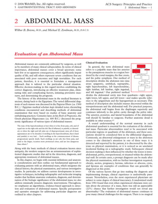
  • 4. © 2006 WebMD, Inc. All rights reserved. ACS Surgery: Principles and Practice 5 GASTROINTESTINAL TRACT AND ABDOMEN 2 Abdominal Mass — 4 EPIGASTRIUM Omental Hernia Pancreatic Tumor RIGHT UPPER QUADRANT Pancreatic Cyst Tender Gastric Carcinoma Liver in Hepatitis Gastrointestinal Stromal Tumor (GIST) Congestive Heart Failure Pyloric Stenosis Gallbladder in Cholecystitis Aortic Aneurysm Subphrenic Abscess Retroperitoneal Sarcoma Perinephric Abscess Hepatomegaly Colonic Tumor Abdominal Wall Hematoma LEFT UPPER QUADRANT Nontender Splenomegaly Hepatomegaly Abdominal Wall Hematoma Renal Tumor Pancreatic Tumor Adrenal Tumor Pancreatic Cyst Courvoisier’s Gallbladder Gastric Tumor Hydrops of Gallbladder Colonic Tumor Fecal Impaction Renal Tumor or Enlargement Fecal Impaction RIGHT LOWER QUADRANT Tender LEFT LOWER QUADRANT Appendiceal Abscess Sigmoid Diverticulitis Psoas Abscess Carcinoma of Colon Pyosalpinx Ovarian Tumor Regional Ileitis Pyosalpinx Intussusception Nontender HYPOGASTRIUM Carcinoma of Colon Bladder Ovarian Tumor Gravid Uterus Uterine Fibroids Regional Ileitis Urachal Cyst Figure 1 Schema represents differential diagnosis of an abdominal mass by quadrant or region. Fundamental knowledge of normal anatomy and clinical presentations is the basis for distinguishing the various disease processes. Abdominal wall hernia is considered a possibility in every region or quadrant. Physical findings on examination are sometimes variable and can Palpable or discrete masses should always be localized with be affected by factors such as obesity, body habitus, associated respect to the previously described landmarks (see above), and medical conditions, and the patient’s ability to cooperate. For they should, if possible, be described in terms of size, shape, con- example, the normal aorta is often palpable within the epigastrium sistency, contour, presence or absence of tenderness, pulsatility, and may be slightly tender; in elderly, asthenic patients, the nor- and fixation. Knowledge of the location of the mass in the mal aorta may be mistaken for an aneurysm. Likewise, the cecum abdomen shortens the list of structures or organs to be considered and the descending colon, both of which are usually palpable in and may give insight into the nature and extent of the pathologic thin patients (especially when they contain feces), sometimes mas- process. Frequently, however, the mass’s location can only be querade as a cancerous mass; subsequent disimpaction causes vaguely outlined, particularly when fluid is present, when the such “masses” to resolve. Obesity may preclude evaluation of a abdomen is tender or tense, or when the patient is obese. Gastric potential abdominal mass: it can be difficult to identify discrete neoplasms, pancreatic neoplasms, colonic neoplasms, sarcomas, palpable masses amid the often remarkable adiposity present with- pancreatic cysts, and distended gallbladders may be palpable, typ- in the abdominal wall and the surrounding structures. Ascites may ically at advanced stages of disease. Recognition of such masses also obscure abdominal masses, making examination more prob- can be facilitated by repeating the abdominal examination after lematic. Transient gaseous distention or intestinal bloating occa- analgesics have been administered or after the patient has been sionally presents a similar problem, but it usually resolves sponta- anesthetized in preparation for a procedure. neously, except in cases of intestinal obstruction. Either gastric dilatation or intestinal obstruction may lead to abdominal disten- tion that is severe enough to necessitate nasogastric decompres- Working or Presumed Diagnosis sion. Not uncommonly, in women of childbearing age, a lower Once a thorough clinical history has abdominal mass may represent a gravid uterus. In such cases, a been obtained and a careful physical exam- gynecologic examination must be conducted and a pregnancy test ination conducted, it is usually possible to performed before further studies are ordered. The multiplicity of generate a working diagnosis. Once the potential benign causes notwithstanding, the possibility of a neo- working diagnosis has been established, plasm (single or multiple) clearly remains a matter of considerable subsequent management is considered in concern in the evaluation of any patient with abdominal disten- light of its appropriateness for the presumed tion. A convenient method of recalling the main causes of gener- condition. Sometimes, however, the diag- alized enlargement or distention of the abdomen is to use the so- nosis remains unknown even after a com- called “six Fs” mnemonic device: Fat, Fluid, Flatus, Fetus, Feces, prehensive clinical history and physical examination; in such cases, and Fatal growths.21-23 further studies are required. A wide range of laboratory and imag-
  • 5. © 2006 WebMD, Inc. All rights reserved. ACS Surgery: Principles and Practice 5 GASTROINTESTINAL TRACT AND ABDOMEN 2 Abdominal Mass — 5 ing studies are now available for establishing the diagnosis. If these improved and refined for use in evaluating abdominal masses. studies do not resolve the diagnostic uncertainty, additional pro- In particular, advances in cross-sectional imaging techniques, cedures, including image-guided percutaneous biopsy, diagnostic such as ultrasonography (US), computed tomography, magnet- laparoscopy, and exploratory laparotomy, may be employed as ic resonance imaging, and positron emission tomography necessary. (PET), have made it possible to assess these lesions more pre- cisely. Consequently, whenever the surgeon is confronted with the scenario of a clinically suspected or palpable abdominal Investigative Studies mass, accurate diagnostic imaging is of paramount importance. Surgeons are in a unique position to The appropriate use of different imaging modalities in the eval- care for patients presenting with an uation of the palpable abdominal mass is well described by the abdominal mass and should guide the American College of Radiology guidelines,25,26 which are up- collaborative management effort and the dated every 6 years. choice of appropriate investigative stud- The use of noninvasive US and CT as first-line procedures for ies. It is therefore essential that surgeons the evaluation of palpable masses has received considerable clini- be familiar with every available method cal attention.6,27-30 Investigators have found both US and CT to for efficient and cost-effective diagnosis of be excellent for affirming or excluding a clinically suspected an abdominal mass. For any given situa- abdominal mass, with sensitivity and specificity values exceeding tion, the selection of investigative studies should be based on 95%.This finding is particularly noteworthy because in only 16% the preferences of the patient, the knowledge and judgment of to 38% of patients referred for a suspected abdominal mass will the surgeon, and the capabilities of the institution. In this way, the diagnosis be corroborated by an imaging study.31 Both US and surgeons who practice outside large, specialized referral cen- CT are also capable of visualizing the organ from which the mass ters will still be able to provide integral leadership for most dis- arises: US successfully determines the organ of origin approxi- ease management efforts arising from the diagnosis of an mately 88% to 91% of the time, and CT does so approximately abdominal mass. 93% of the time. Prediction of the pathologic diagnosis of an abdominal mass, however, remains a challenge for both modali- LABORATORY STUDIES ties. US correctly predicts the pathologic diagnosis in 77% to 81% The diagnostic workup of an abdominal mass usually includes of cases, whereas CT suggests the diagnosis in 88% of cases. laboratory evaluation. If the cause of the mass remains unknown, Further advancements in cross-sectional imaging (e.g., multide- preliminary laboratory analysis should include a chemistry profile tector CT [MDCT] with three-dimensional reconstruction and (electrolyte, blood urea nitrogen [BUN], and creatinine concen- magnetic resonance angiography [MRA]) and the addition of trations, as well as liver function tests), a complete blood count molecular and functional imaging modalities (e.g., PET) will (CBC) with differential, and urinalysis. An abnormal laboratory undoubtedly improve the predictive abilities of CT and US. At value sometimes plays an important role in establishing the iden- any rate, the current state of imaging technology affords clinicians tity or pathogenesis of an abdominal mass. For example, an ele- the ability to distinguish benign from malignant processes, to vated alkaline phosphatase or liver transaminase level may suggest assess tumor biology, and to detect lesions that impose a minimal metastasis to the liver. Likewise, an elevated serum amylase con- disease burden. As a consequence, clinicians are more likely to centration may be suggestive of a pancreatic pseudocyst rather detect clinically occult disease or discover it incidentally. than a cystic neoplasm or an adenocarcinoma; however, an elevat- Employing an integrative assessment approach (which includes ed total serum bilirubin level (i.e., > 10 mg/dl) may be more sug- clinical history, physical examination, and investigative studies) gestive of a malignant process secondary to adenocarcinoma of should lead to more targeted, efficient, and cost-effective strategies the pancreatic head or cholangiocarcinoma. Routine testing for for evaluating abdominal masses. For example, the surgeon can occult blood in the stool should not be overlooked. Tumor mark- correlate the clinical location of the abdominal mass with perti- ers (e.g., carcinoembryonic antigen [CEA], the cancer antigens nent findings from the history and laboratory studies to determine CA 19-9 and CA 125, and α-fetoprotein [AFP]) may also help which imaging modality is the most expeditious and cost-effective differentiate between benign disease processes and malignant for a given circumstance. Each imaging modality has unique ones, distinguish high-level disease from low-level disease, and, in strengths and weaknesses. some cases, establish a disease diagnosis (e.g., elevated AFP levels in patients with hepatocellular carcinoma). Similarly, an elevated Plain Abdominal Radiographs serum lactate dehydrogenase (LDH) level may prove invaluable in By definition, a plain film is a radiograph made without the use the staging and prognosis of certain diseases (e.g., melanoma) of an artificially introduced contrast substance.32 Commonly connected with an abdominal mass.24 Furthermore, the ability to employed for initial surveillance of the abdomen, the plain film distinguish between functional abdominal masses and nonfunc- still has an important place within the investigative armamentari- tional ones (e.g., adrenal tumors) also has important implications um. Otherwise known as a KUB (kidney-ureter-bladder) study, for evaluation and management. this low-cost technique may reveal nonspecific or indirect evi- In some cases, when the type of mass remains unknown, need- dence of an abdominal mass, such as variations in the size and less and expensive laboratory analysis can and should be avoided density of an organ or displacement of normal structures or fat if it appears that other studies may prove more beneficial. planes. Furthermore, the radiolucency of air within the bowel may also prove helpful for recognizing worrisome displacement of vis- IMAGING cera as a result of a large abdominal mass. Occasionally, a simple Diagnostic radiology is a dynamic specialty that has under- plain radiograph can assist the surgeon in making a specific diag- gone rapid change in conjunction with the ongoing evolution of nosis, such as calcified aortic aneurysm, acute gastric distention, imaging technology. Not only has the number of imaging fecal impaction, porcelain gallbladder, and certain malignancies modalities increased, but each modality continues to be [see Figure 2].
  • 6. © 2006 WebMD, Inc. All rights reserved. ACS Surgery: Principles and Practice 5 GASTROINTESTINAL TRACT AND ABDOMEN 2 Abdominal Mass — 6 Novel approaches (e.g., CT virtual colonoscopy), in conjunction with advances in cross-sectional imaging, may eventually render conventional GI imaging unnecessary. Ultrasonography Compared with other modalities, US has several advantages in the evaluation of suspected abdominal masses, including wide- spread availability, speed of use, the absence of ionizing radiation, low cost, and the ability to document the size, consistency (solid or cystic), and origin of a mass with real-time images.27,33 When directed at solving a specific clinical problem, US generally pro- vides more diagnostic information. Moreover, the necessary equipment can easily be transported to the patient’s bedside or another clinical setting; thus, no patient preparation is required, and only minimal patient cooperation is needed. We consider US indispensable in the assessment of abdominal masses. At the same time, we acknowledge that one disadvantage of US is the extent to which the quality of the results depends on the technical proficiency and diligence of the operator or techni- cian (though this disadvantage can actually become an advantage when personnel are well trained and experienced). In the hands of an inexperienced operator, US may yield inconclusive or untrust- worthy results that contribute to delayed diagnosis or even misdi- agnosis. In an effort to help minimize this problem, we encourage the surgeons at our institution (who are trained in US) to perform their own studies in the clinic and the operating room. This approach further expedites recognition of disease [see Figure 3], positively influences management, and facilitates operative deci- sion making regarding abdominal masses [see Figure 4]. Another disadvantage of US is its inability to visualize the entire abdominal cavity as a consequence of the acoustic barriers present- ed by gas-containing structures (e.g., the bowel) and the absorptive interfaces (acoustic shadowing) provided by soft tissue and bone. Figure 2 Plain abdominal radiograph shows a 10 cm functional For optimal visualization of abdominal masses, US should be per- left adrenocortical carcinoma. Calcifications creating a rim formed through “acoustic windows” that allow adequate transmis- enhancement are easily identified. The diagnosis was confirmed sion of sound. Accordingly, US is most effective as a tool for evalu- by means of laboratory analysis and abdominal CT. ating masses in those regions of the abdomen where an acoustic window exists (e.g., the right and left upper quadrants and the Conventional Gastrointestinal Imaging As a consequence of the technical advances in cross-sectional imaging and endoscopy, conventional GI contrast studies are now largely relegated to more adjunctive roles in the evaluation of abdominal masses. In the upper and middle portions of the abdomen, we occasionally use upper GI studies, small bowel fol- low-through (SBFT), or enteroclysis to evaluate inflammatory masses (e.g., lesions arising from Crohn disease), masses that are inaccessible to endoscopy, or unusual masses with uncertain diag- noses. For such lesions, we employ single- or double-contrast bar- ium protocols to ensure that significant pathology is not missed; however, these studies are notoriously insensitive and do not pro- vide an opportunity for tissue diagnosis. In the lower portion of the abdomen, barium studies still play a significant role in the evalua- tion of masses whose history includes GI symptoms (e.g., anemia and weight loss) suggestive of a colonic neoplasm, as well as for evaluating inflammatory masses arising from diverticular disease. In certain cases, we employ a single-contrast barium enema for masses that are causing near-complete obstruction; this study is also helpful for assessing the remaining large bowel for synchro- nous disease. For small lesions (masses < 1 cm), we typically favor Figure 3 Sagittal ultrasonogram of the pancreas demonstrates a a double-contrast barium enema. large mass in the pancreatic head of a 71-year-old patient Currently, in the evaluation of an abdominal mass, barium referred for “gallstones” after experiencing a 10 lb weight loss. studies are used mainly to complement colonoscopy and CT. The mass lies anterior to the inferior vena cava.
  • 7. © 2006 WebMD, Inc. All rights reserved. ACS Surgery: Principles and Practice 5 GASTROINTESTINAL TRACT AND ABDOMEN 2 Abdominal Mass — 7 a b Figure 4 (a) Transverse ultrasonogram of the liver of a 72-year-old cirrhotic patient with a hepatitis C infection shows a 4.0 × 3.5 cm hepatoma, nestled between the right and middle hepatic veins and closely apposed to the inferior vena cava. (b) Color Doppler ultrasonogram from the same patient displays blood flow in the middle hepatic vein and the surrounding liver parenchyma. Blood flow toward the transducer is usually displayed in shades of red, whereas blood flow away from the transducer is displayed in shades of blue. Color Doppler ultrasonography allows evaluation of the patency and flow characteristics of the hepatic circulation as it relates to the mass. pelvis). Fortunately, the shortcomings of US can be compensated and delineating the relations between the abdominal mass and for by employing other cross-sectional imaging modalities. adjacent structures [see Figure 5]. Such data are essential for guid- ing diagnostic procedures, determining whether operative manage- Computed Tomography ment is indicated, and selecting the optimal operative approach. At present, helical (spiral) CT is the most efficient and cost- Although modalities such as MRI, PET, and endoscopic ultra- effective imaging modality for the evaluation of abdominal mass- sonography (EUS) have advantages over CT in one area or anoth- es.6,27,34,35 Unlike US, CT provides cross-sectional images with er, CT continues to be superior overall for assessing abdominal mass- excellent spatial resolution and exquisite density discrimination that es and remains our preferred imaging method for this purpose. are unaffected by bowel gas, bone, or excessive abdominal fat. CT The use of contrast during the acquisition of CT scans is vital. routinely visualizes the abdominal wall, the viscera, the mesentery, Opacification of the bowel enables the examiner to distinguish the and the retroperitoneum, clearly defining important tissue planes abdominal mass from surrounding viscera or other adjacent struc- tures. Contrast-enhanced scans also allow delineation of the rele- vant vascular anatomy; in fact, CT angiography has now relegat- ed conventional angiography to a minimal role in the evaluation of certain abdominal masses.36,37 Triple-phase or multiphase scan- ning that includes noncontrast images is now recommended. Such scans achieve optimal definition and characterization of liver and pancreatic masses. This achievement is of significant clinical value: state-of-the-art CT imaging of malignant pancreatic mass- es, as well as of other malignancies, has the potential to improve outcome not only by correctly detecting the mass but also by accurately assessing the extent of disease, thereby helping deter- mine which patients may benefit from surgical management or neoadjuvant therapy [see Figure 6]. The advent of MDCT technology offers the possibility of even better imaging of abdominal masses than standard contrast CT provides. MDCT scanners can image specific organs or masses with 1 mm slices in less than 20 seconds, and the resultant data can be displayed not only as an axial image but also in a three-dimen- sional representation that includes detailed vascular mapping.35 Studies suggest that MDCT may be the most useful modality for preoperative assessment of the resectability of pancreatic and other Figure 5 CT scan of a 65-year-old man with a large retroperi- abdominal masses.36 MDCT has a sensitivity of 90% and a speci- toneal leiyomyosarcoma clearly demonstrates close association of ficity of 99%, respectively, and it is not observer dependent. this mass with the right hemiliver, as well as displacement of the Currently, although MRI (see below) offers unique tissue con- inferior vena cava. trast and inherent multiplanar capabilities for imaging abdominal
  • 8. © 2006 WebMD, Inc. All rights reserved. ACS Surgery: Principles and Practice 5 GASTROINTESTINAL TRACT AND ABDOMEN 2 Abdominal Mass — 8 is a better choice than CT for evaluating an abdominal mass. An example is a case in which the use of iodinated contrast material is contraindicated. The extracellular gadolinium chelates used in MRI are very safe and can be given to patients with mild to mod- erate azotemia without causing renal impairment. MRI has unique characteristics that can be effectively employed to distin- guish normal from pathologic tissue in a patient with an abdomi- nal mass.38 Detailed information about the principles and practices of abdominal MRI is beyond the scope of this chapter and is read- ily available elsewhere.39 A brief technical summary may, how- ever, be worthwhile. The abdomen and its contents are subject- ed to a momentary radiofrequency pulse, then allowed to return to a state of equilibrium. During the return to equilibrium, the nuclei within each specific tissue will emit specific radiofrequen- cy signals. The strength and type of the emitted signal deter- mine the image intensity. The way in which the different tissues are visually rendered depends on (1) the longitudinal relaxation time (T1) and the transverse relaxation time (T2) of the nuclei in the tissues and (2) the method of image weighting employed. Figure 6 CT angiography performed to evaluate vascular inva- By convention, tissues with short T1 values (such as solid struc- sion in a 58-year-old patient with a pancreatic mass demon- tures) appear bright on T1-weighted images, whereas structures strates nearly complete encasement of the superior mesenteric vein. The superior mesenteric artery is not involved with the with long T2 values (e.g., fluid-containing tissues) appear bright mass. on T2-weighted images. The tissue contrast and multiplanar capabilities of MRI allow surgeons and radiologists to distin- guish not only obvious but also subtle differences between masses, CT has several advantages—high resolution, short scan abdominal masses and normal anatomy. For example, T1- times, and fast patient throughput—that make it a more widely weighted images may be valuable for detecting abdominal mass- preferred imaging modality for this purpose. es that contain fluid (e.g., cystic masses or masses containing necrotic tissue), whereas T2-weighted images may be useful for Magnetic Resonance Imaging characterizing these masses as either benign or malignant [see Since its introduction in the mid-1980s, MRI has become one Figure 7]. Similarly, magnetic resonance cholangiopancreatog- of radiology’s great success stories (though, because it still is not as raphy (MRCP) uses T2-weighted images to distinguish masses widely available as US or CT, its cost-effectiveness has yet to be with different signal intensities in the pancreas, the liver, and the determined). Few would dispute the enormous impact MRI has biliary tract.40 had on our ability to diagnose pathologic conditions of the brain, Positron Emission Tomography the spine, and the musculoskeletal system.Whereas MRI has clear advantages over CT in these areas of the body, this is not the case In 1930, Warburg reported that cancer cells show higher rates in the abdomen. Nevertheless, there are situations in which MRI of glycolysis than normal cells do.41 This discovery has stood the a b Figure 7 (a) Gadolinium-enhanced, T1-weighted MRI shows a large mass that appears dark and well-cir- cumscribed in comparison with the normal-appearing enhanced liver and spleen. This abnormal mass clearly contains some fluid. The fluid-filled stomach also appears dark. (b) T2-weighted MRI of the same patient details subtle inhomogeneities characteristic of a malignant mass (less organized appearance with an enhanced necrotic component). Subsequent biopsy showed this mass to be a poorly differentiated adeno- carcinoma from recurrent colon cancer.
  • 9. © 2006 WebMD, Inc. All rights reserved. ACS Surgery: Principles and Practice 5 GASTROINTESTINAL TRACT AND ABDOMEN 2 Abdominal Mass — 9 BIOPSY In many cases, the pathologist is the sur- geon’s greatest teacher. Despite the sur- geon’s most strenuous efforts, the biology of the disease or lesion will inevitably dic- tate the outcome. Nowhere is this state- ment more true than in the evaluation of the abdominal mass, and its truth becomes increasingly evident as ongoing refine- ments in molecular diagnosis permit ever more sophisticated discrimination among different tumor types and their respective behaviors.44 Aside from the treatment of lymphoma, in which the surgeon is frequently called on to pro- vide technical assistance in obtaining tissue for diagnosis, the decision whether to perform a biopsy (as well as when and how to do so) rests on the surgeon’s understanding of the probable disease. For example, surgeons who treat pancreatic cancer usu- ally proceed to surgery without biopsy if the evidence for malig- nancy is strong. In other cases, biopsy is performed to confirm what is already suspected on the basis of clinical and radiograph- ic findings. Moreover, establishing the type of tumor or mass present has important implications for the use of neoadjuvant or adjuvant therapy, as well as for the planning of the surgical approach. We view the biopsy of an abdominal mass as the first stage of surgery. This procedure, though seemingly innocuous, has the potential to contaminate tissue planes and must there- fore be performed carefully. Accordingly, in order to make the appropriate choice when confronted with an abdominal mass, the surgeon must possess a thorough understanding of the vari- ous methods of obtaining an accurate and safe biopsy. Factors Figure 8 18FDG PET scan demonstrates a large metaboli- related to the size and location of the abdominal mass, as well as cally active non-Hodgkin lymphoma giving rise to an abdominal mass. factors related to institutional preference and experience, may influence the choice of biopsy technique. Image-Guided Percutaneous Biopsy test of time and now serves as the theoretical rationale for the use The value of image-guided percutaneous biopsy in the evalu- of 18F-fluorodeoxyglucose (18FDG) PET imaging to assess ation of the abdominal mass is well established.45,46 In practice, abdominal masses caused by cancer. Briefly, 18FDG is a glucose the procedure begins with identification of the mass by means of analogue that crosses the cell membrane by sharing the glucose a cross-sectional imaging modality such as US, CT, or MRI. transporter molecules used by glucose. Like glucose, it undergoes Often, three-dimensional imaging reconstructions are generated phosphorylation by the enzyme hexokinase. The resulting mole- to detail the relations of the abdominal mass to the surrounding cule, 18FDG-6-phosphate, is polar and is unable to cross cell anatomy. Once the mass is identified, decisions are made regard- membranes or serve as a substrate for metabolism. The net effect ing the safest approach and the most appropriate technique.The is that 18FDG both accumulates in and is retained by cancer cells. biopsy needle is then inserted percutaneously under the guid- The molecular information obtained from PET, as measured by ance of US, CT, or MRI.The choice among the different modal- standard uptake values (SUV), allows identification of hypermeta- ities depends on several factors, including the size and location bolic (18FDG-avid) abdominal masses (typically arising from lym- of the mass, the surgeon’s judgment regarding which method is phomas, melanomas, or certain GI malignancies [see Figure 8]).42 best in the circumstances, and the availability of the various PET may also prove to be an important surrogate modality for dis- modalities at a particular institution. The most important con- tinguishing malignant abdominal masses from benign ones.43 sideration, however, is the personal preference and experience of When PET is used alone, it has the disadvantage of being unable the radiologist performing the biopsy.We favor either US or CT, to provide sufficient anatomic information to guide biopsy or fur- both of which yield good results. ther therapy.When PET is used with CT in PET/CT fusion imag- In general, we prefer US-guided biopsy for large, superficial, ing, however, the functional advantages of PET and the structural and cystic masses. This technique is also appropriate for lesions advantages of CT combine to enhance the detection rate for lying at moderate depths in thin to average-size persons. In some abdominal masses.42 If a mass is anatomically evident but metabol- cases, US can be employed to guide biopsy of small, deep, and ically inactive, it will be detected by CT. If it shows increased gly- solid abdominal masses; however, US-guided biopsy of these colysis but few or no CT abnormalities, it will be detected by PET. deep-seated masses (as well as of masses in obese patients) often The apparent advantages of PET/CT notwithstanding, prospec- proves difficult because of inadequate visualization resulting from tive, randomized validation is necessary before the widespread sound attenuation in the soft tissues. Similarly, lesions located application of this approach to the evaluation of abdominal mass- within or behind bone or gas-filled bowel cannot be easily visual- es can be justified. At present, the use of PET/CT is mostly restrict- ized (a consequence of the nearly complete reflection of sound ed to large tertiary referral centers. from bone or air interfaces).
  • 10. © 2006 WebMD, Inc. All rights reserved. ACS Surgery: Principles and Practice 5 GASTROINTESTINAL TRACT AND ABDOMEN 2 Abdominal Mass — 10 laceration or of damage from tearing. Such needles are often used to confirm tumor recurrence or metastasis in patients with a pathologically confirmed primary malignancy. Large-caliber nee- dles are typically used to obtain greater amounts of material for histologic or cytologic analysis.49 In practice, the choice of a biop- sy needle is often influenced by whether the suspected pathology is benign or malignant. For example, large-caliber needles may be necessary to obtain a sufficiently large histologic specimen when certain types of malignancies (e.g., lymphoma) are suspected. When an inflammatory mass is suspected and material is needed for culture, however, a small-caliber needle may be preferred. Additional considerations for image-guided biopsy include the accuracy, safety, and potential complications of the proposed tech- nique. These considerations are essential for an evidence-based approach to diagnosis of an abdominal mass. The reported accuracy of US-guided biopsy ranges from 66% to 97%. The location, size, and histologic origin of the abdominal mass appear to influence the diagnostic accuracy of the proce- dure.47 In a series that included 126 consecutive small (< 3 cm) solid masses distributed among various anatomic locations and histologic types, US-guided biopsies showed an overall accuracy of Figure 9 In a percutaneous biopsy of a large abdominal mass, CT guidance is a reliable means of determining the direction and 91%.47 Biopsy results improved as the size of the mass increased: depth of the needle. accuracy rose from 79% in masses 1 cm or less in diameter to 98% in masses 2 to 3 cm in diameter.The accuracy of US-guided biop- sy in the liver, where most of the biopsies were performed, exceed- US possesses several strengths as a guidance modality for percu- ed 96%. Another study found US-guided biopsy to be 91% accu- taneous biopsy. It is readily available, inexpensive, and portable, and rate for abdominal masses less than 2.5 cm in diameter.50 Two it provides guidance in multiple transverse, longitudinal, or oblique organ-specific reviews concluded that US-guided biopsy of hepat- planes. Moreover, it offers real-time visualization of the needle tip as ic masses had an accuracy of 94%51 and that US-guided biopsy of it passes through tissue planes into the target area,47 thereby allow- pancreatic masses had an accuracy of 95%.52 ing the surgeon to place the needle precisely and to avoid important The reported accuracy of CT-guided biopsy ranges from 80% intervening structures. In addition, color flow Doppler imaging can to 100%. As with US-guided biopsy, the size, location, and histo- help prevent complications of needle placement by identifying the logic origin of the mass influence the results.53-55 In a study of 200 blood vessels involved with the mass, as well as any vessels lying consecutive CT-guided needle biopsies, the overall accuracy for all within the needle path. Because of its real-time capabilities, US sites biopsied was 95%.The reported organ-specific accuracy was guidance has the potential to allow quicker, more accurate, and less as follows: kidneys, 100%; liver, 99%; retroperitoneum, 87.5%; expensive biopsies than CT guidance does.48 In theory, any mass and pancreas, 82%.56 In a prospective study of 1,000 consecutive that is well visualized with US should be amenable to US-guided CT-guided biopsies, the reported sensitivity was 91.8% and the biopsy. In practice, however, this modality remains best suited for specificity 98.9%.55 At our institution, as well as others, CT-guid- superficial to moderately deep abdominal masses and for patients ed biopsy is now considered a reliable tool for the diagnosis and with a thin to average body habitus. classification of malignant abdominal lymphomas.57 The utility of US notwithstanding, CT remains indispensable at The safety of image-guided percutaneous biopsy is well docu- our institution as a guidance method for percutaneous biopsy of mented. Several large multi-institutional reviews reported major most regions in the body. It is particularly useful when an abdom- complication rates ranging from 0.05% to 0.18% and mortalities inal mass is in a location that is inaccessible to US as a result of ranging from 0.008% to 0.031%.58-60 A large prospective study of bowel gas or body habitus. In the abdomen, CT provides excellent 3,393 biopsies (1,825 US-guided; 1,568 CT-guided) documented spatial resolution of all structures between the skin and the mass, an overall mortality of 0.06%, a major complication rate of 0.34% regardless of body habitus or lesion depth, and it provides an accu- (0.3% with US; 0.5% with CT), and a minor complication rate of rate image of the needle tip.We favor CT guidance for abdominal 2.9% (2.4% with US; 3.3% with CT).47 Procedure-related mor- masses that are located deep in the abdomen or in the retroperi- bidity and mortality appear to be largely unaffected by whether a toneum.The only limitation of CT in this setting is that it does not small-caliber or a large-caliber biopsy needle is used. A review of offer continuous visualization of the needle during insertion and 11,700 patients who underwent percutaneous abdominal biopsy biopsy. In most cases, however, CT guidance can reliably establish with 20- to 23-gauge needles found an overall complication rate of the direction and depth of the needle [see Figure 9]. only 0.05% and an overall mortality of only 0.008%.58 A single- Numerous different needles, covering a broad spectrum of cal- institution review of 8,000 US-guided needle biopsies performed ibers, lengths, and tip designs, are commercially available for use with both small- and large-caliber needles reported equivalent in percutaneous image-guided fine-needle aspiration (FNA) biop- results: a major complication rate of 0.187% and a mortality of sy. For convenience, these needles can be grouped into two main 0.038%.61 Of the rare major complications that occur, hemorrhage size categories: small caliber (20 to 25 gauge) and large caliber (14 is the most frequently reported; pneumothorax, pancreatitis, bile to 19 gauge). Small-caliber needles are used primarily for cytolog- leakage, peritonitis, and needle track seeding may also develop. ic analysis but may also be employed to obtain small pieces of tis- Needle-track seeding remains an important theoretical consid- sue for histologic analysis. The flexible shaft of small-caliber nee- eration when an abdominal mass appears likely to be malignant. dles allows them to be passed with minimal risk of tissue or organ According to some investigators, percutaneous needle biopsy has
  • 11. © 2006 WebMD, Inc. All rights reserved. ACS Surgery: Principles and Practice 5 GASTROINTESTINAL TRACT AND ABDOMEN 2 Abdominal Mass — 11 the potential to seed between 103 to 104 tumor cells into the nee- ease,75 and the number of samples obtained. In one series that dle track.62,63 Nevertheless, tumor dissemination after percuta- included more than 200 patients with esophageal or gastric mass- neous biopsy remains exceedingly rare: with fewer than 100 cases es, a diagnosis was made in 70% of patients after the first biopsy, reported in the world literature, it has an estimated frequency of 95% of patients after the fourth biopsy, and 98.9% of patients after 0.005%,64-66 mostly occurring after biopsy of pancreatic, hepatic, the seventh biopsy.76 Several other studies have confirmed the high or retroperitoneal masses. Poorly planned biopsies of malignant sensitivity and specificity of EUS-guided biopsy (especially for the abdominal masses have the potential to exert adverse effects on diagnosis of extraluminal abdominal masses) and verified the safe- subsequent surgery and to compromise local tumor control; for- ty of the procedure (reported complication rates range from 0.3% tunately, such negative consequences remain rare. to 2%).68-71,77 It is worth noting that in the resection of a potential- ly curable abdominal mass, concern about needle-track contami- EUS-Guided Imaging and Biopsy nation is obviated when the path of the needle is removed as part EUS provides unique imaging information because it involves of the surgical specimen (as in pancreaticoduodenectomy for a the close apposition of a high-frequency ultrasound transducer, pancreatic head mass or gastrectomy for a stomach mass). called an echoendoscope (whereby image resolution is directly We consider EUS-guided biopsy for the diagnosis of masses related to frequency), to the structures being studied. As a result, that are not readily accessible to percutaneous biopsy, on the it can delineate abdominal masses and associated structures with grounds that it can obviate more invasive procedures (e.g., greater anatomic detail than standard transcutaneous ultrasonog- laparoscopy and laparotomy). In a 10-year study of the impact of raphy can. In general, EUS-guided biopsy is well suited for EUS on patient management, 86% of patients required no further abdominal masses that are too small for visualization by means of imaging, and 25% were able to avoid unnecessary laparotomy.77 other cross-sectional imaging modalities or that are inaccessible to Overall, EUS changed clinical management significantly in as percutaneous biopsy.67 The most frequently used EUS device is many as one third of the 537 patients studied.77 Nevertheless, the radial echoendoscope, which creates a 360º tomographic despite the high diagnostic yield achieved with EUS-guided biop- image perpendicular to the scope. The circumferential view ob- sy, results that are negative for tumor should not always be inter- tained with this instrument facilitates orientation and therefore is preted as proving that no tumor is present; laparoscopic or open more efficient for diagnostic imaging. Alternatively, the linear- biopsy may still be indicated. array echoendoscope, which generates an image parallel to the DIAGNOSTIC LAPAROSCOPY shaft of the scope, may be used. This instrument produces high- quality gray-scale images, as well as color and duplex images. The available evidence now clearly sup- EUS-guided biopsy with a linear scanning system offers clear and ports the role of laparoscopy in the diagno- consistent visualization of the biopsy needle along its entire path sis and management of abdominal masses. in real time, with excellent delineation of intervening tissues and We and others advocate the liberal use of without any interference from intestinal gas. laparoscopy as a primary staging tool for EUS has proved to be superior to other cross-sectional imaging upper and lower GI malignancies, believing modalities for detection and staging of pancreatic, gastric, and it to be a safe, cost-effective tool that offers a esophageal masses.68-71 For instance, in a patient with a pancreat- clear benefit in more than 20% of patients ic mass, EUS not only identifies the size of the mass and the peri- with these diseases.78,79 Preventing unneces- pancreatic lymph nodes but also delineates the relations of these sary laparotomy in selected patients by performing diagnostic structures to major blood vessels. EUS has also proved to be help- laparoscopy is associated with shorter hospital stays and earlier ini- ful in selecting patients for various neoadjuvant protocols. tiation of locoregional or systemic therapy. Moreover, laparoscopic Furthermore, the availability of high-frequency catheter-based ultrasonography80 and peritoneal cytology81 are known to provide intraductal ultrasonography (IDUS) now enables surgeons to added value in the staging of disease. Furthermore, diagnostic visualize masses within the biliary tree and obtain biopsy speci- laparoscopy can safely provide tissue samples from suspected lym- mens from them.72 phomatous masses for full diagnostic analysis.82 With the growth of Advantages notwithstanding, EUS technology has several dedicated minimally invasive fellowships and the improved quality important limitations. As with all forms of ultrasonography, a sub- and availability of laparoscopic training for general surgery resi- stantial period is required before the operator achieves proficien- dents and related subspecialties, the skill sets required for diagnos- cy. EUS is highly operator dependent; when it is done by an inex- tic laparoscopy are coming to be more widely mastered, and the perienced operator, the potential exists for serious misinterpreta- concerns once commonly expressed regarding intra-abdominal tions. For example, if an operator obtains only one view of a mass adhesions and effective biopsy techniques for abdominal masses in the head of the pancreas, the mass may appear to be invading now appear to be less problematic. vascular structures when it is not actually doing so. In the evalua- tion of pancreatic masses around vessels, the operator should always obtain multiple views. It cannot be overemphasized that Indications for Exploratory Laparotomy EUS and EUS-guided biopsy require personnel with sufficient Advances in diagnostic imaging, endoscopy, and minimally inva- experience and skill in both ultrasonography and endoscopy. sive surgery have nearly eliminated the need for open exploration EUS is frequently employed for diagnosis and staging of upper for the sole purpose of establishing a diagnosis in patients with an GI malignancies. In a large single-institution study of 267 pancre- abdominal mass. In selected cases, however, exploratory laparoto- atic masses that were sampled by means of EUS-guided biopsy my may still help in the assessment of abdominal masses that were and subsequently resected, the overall diagnostic accuracy was initially misinterpreted on preoperative evaluation. In general, 95.6%, the sensitivity was 94.6%, and the specificity was 100%.73 exploratory laparotomy should be reserved for those rare instances In studies of gastric and esophageal masses, diagnostic accuracy in which other modalities have failed to yield crucial information was related to the location of the biopsy,74 the histology of the dis- needed for evaluation and diagnosis of an abdominal mass.
  • 12. © 2006 WebMD, Inc. All rights reserved. ACS Surgery: Principles and Practice 5 GASTROINTESTINAL TRACT AND ABDOMEN 2 Abdominal Mass — 12 References 1. Ancient Egyptian Medicine–Smith Papyrus–Ebers 23. DeGowin EL, DeGowin RL: Bedside diagnostic 45. Gazelle GS, Haaga JR: Guided percutaneous biop- Papyrus examination. Macmillan, New York, 1976, p 471 sy of intraabdominal lesions. AJR Am J Radiol 153: http://crystalinks.com/egyptmedicine.html 24. Balch CM, Soong SJ, Atkins MB, et al: An evi- 929, 1989 2. Hippocrates: The Book of Prognostics. Francis dence-based staging system for cutaneous 46. Welch TJ, Reading CC: Imaging-guided biopsy. Adams, Transl. melanoma. CA Cancer J Clin 54:131, 2004 Mayo Clin Proc 64:1295, 1989 http://etext.library.adelaide.edu.au/h/hippocrates/ 25. DiSantis DJ, Ralls PW, Balfe DM, et al: Imaging 47. Caspers JM, Reading CC, McGahan JP, et al: h7w/prognost.html evaluation of the palpable abdominal mass. Ultrasound-guided biopsy and drainage of the 3. Swartz MH: Textbook of Physical Diagnosis: American College of Radiology. ACR Appropriate- abdomen and pelvis. Diagnostic Ultrasound, 2nd History and Examination, 5th ed. Saunders ness Criteria. Radiology 215(suppl):201, 2000 ed. Rumack CM,Wilson SR, Charboneau JW, Eds. Elsevier, Philadelphia, 2006, p 479 26. Grollman J, Bettman MA, Boxt LM, et al: Pulsatile Mosby, St Louis, 1998, p 600 4. Wood WC, Skandalakis JE: Anatomic Basis of Tumor abdominal mass. American College of Radiology. 48. Sheafor DH, Paulson EK, Simmons CM, et al: Surgery. Quality Medical Publishing, St. Louis, ACR Appropriateness Criteria. Radiology 215(suppl): Abdominal percutaneous interventional proce- 1999, p 307 55, 2000 dures: comparison of CT and US guidance. 5. Hart FD: French’s Index of Differential Diagnosis, 27. Williams MP, Scott IHK, Dixon AK: Computed Radiology 207:705, 1998 11th ed.Year Book Medical, Chicago, 1979, p 9 tomography in 101 patients with a palpable abdom- 49. Silverman JF, Geisinger KR: Interventional radiolo- 6. Dixon AK, Kingham JGC, Fry IK, et al: Computed inal mass. Clin Radiol 35:293, 1984 gy of deep organs. Fine Needle Aspiration Cytology tomography in patients with an abdominal mass: 28. Holm HH, Gammelgaard J, Jensen F, et al: of the Thorax and Abdomen. Churchhill effective and efficient? A controlled trial. Lancet Ultrasound in the diagnosis of a palpable abdomi- Livingstone, New York, 1996, p 263 1:1199, 1981 nal mass: a prospective study of 107 patients. 50. Downey DB, Wilson SR: Ultrasonographically 7. Walker HK, Hall WD, Hurst JW: Clinical Methods: Gastrointest Radiol 7:149, 1982 guided biopsy of small intra-abdominal masses. The History, Physical, and Laboratory Examina- 29. Barker CS, Lindsell DR: Ultrasound of the palpable Can Assoc Radiol J 44:350, 1993 tions, 3rd ed. Butterworth, Stoneham, Massachusetts, abdominal mass. Clin Radiol 41:98, 1990 51. Buscarini L, Fornari F, Bolondi L, et al: 1990, p 415 30. Aspelin P, Hildell J, Karlsson S, et al: Ultrasonic Ultrasound-guided fine-needle biopsy of focal liver 8. Brady MS, Gaynor JJ, Brennan MF: Radiation- evaluation of palpable abdominal masses. Acta Chir lesions: technique, diagnostic accuracy and compli- associated sarcoma of bone and soft tissue. Arch Scand 156:501, 1980 cations: a retrospective study on 2091 biopsies. J Surg 127:1379, 1992 Hepatology 11:344, 1990 31. Colquhoun IR, Saywell WR, Dewbury KC: An 9. Bowne WB, Lee B, Wong WD, et al: Operative sal- analysis of referrals for primary diagnostic abdomi- 52. Brandt KR, Charboneau JW, Stephens DH, et al: vage for locoregional recurrent colon cancer: an nal ultrasound to a general X-ray department. Br J CT- and US-guided biopsy of the pancreas. analysis of 100 cases. Dis Colon Rectum 5:897, Radiol 61:297 Radiology 187:99, 1993 2003 32. Squire LF, Novelline RA: Fundamentals of 53. Sundaram M, Wolverson MK, Heiberg E, et al: 10. Miner TJ, Jaques DP, Karpeh MS, et al: Defining Radiology, 4th ed. Harvard University Press, 1988, Utility of CT-guided abdominal aspiration proce- palliative surgery in patients receiving non-curative p 156 dures. AJR Am J Radiol 139:1111, 1982 resections for gastric cancer. J Am Coll Surg 33. Barker CS, Lindsell DRM: Ultrasound of the pal- 54. Smith C, Butler JA: Efficacy of directed percuta- 198:1013, 2004 pable abdominal mass. Clin Radiol 41:98, 1990 neous fine-needle aspiration cytology in the diagno- 11. Miner TJ, Jaques DP, Shriver C: A prospective eval- sis of intra-abdominal masses. Arch Surg 123:820, 34. Gore RM: Palpable abdominal masses. Diagnostic 1988 uation of patients undergoing surgery for palliation Imaging: An Algorithmic Approach. Eisenberg RL, of an advanced malignancy. Ann Surg Oncol 9:696, Ed. JB Lippincott, Philadelphia, 1988, p 214 55. Welch TJ, Sheedy PF, Johnson CD, et al: CT-guid- 2002 ed biopsy: prospective analysis of 1,000 procedures. 35. Lawler LP, Fishman EK: Three-dimensional CT Radiology 171:493, 1989 12. Miner TJ, Brennan MF, Jaques DP: A prospective, angiography with multidetector CT data: study symptom related outcomes analysis of 1022 pallia- optimization, protocol design, and clinical applica- 56. Staab EV, Jaques PF, Partain CL: Percutaneous tive procedures for advanced cancer. Ann Surg tions in the abdomen. Crit Rev Comput Tomogr biopsy in the management of solid intra-abdominal 240:719, 2004 43:77, 2002 masses of unknown etiology. Radiol Clin North Am 13. Stojadinovic A, Hoos A, Karpoff HM, et al: Soft tis- 17:435, 1979 36. Fishman EK, Horton KM: Imaging pancreatic can- sue tumors of the abdominal wall: analysis of dis- cer: the role of multidetector CT with three-dimen- 57. Balestreri L, Morassut S, Bernardi D, et al: Efficacy ease patterns and treatment. Arch Surg 136:70, sional CT angiography. Pancreatology 1:610, 2001 of CT-guided percutaneous needle biopsy in the 2001 diagnosis of malignant lymphoma at first presenta- 37. Murugiah M, Windsor JA, Redhead DN, et al: The tion. Clin Imaging 29:123, 2005 14. Bowne WB, Antonescu CR, Leung DH, et al: role of selective visceral angiography in the manage- Dermatofibrosarcoma protuberans: a clinicopatho- ment of pancreatic and periampullary cancer.World 58. Livraghi T, Damascelli B, Lombardi C, et al: Risk in logic analysis of patients treated and followed at a J Surg 17:796, 1993 fine-needle abdominal biopsy. J Clin Ultrasound single institution. Cancer 88:2711, 2000 11:77, 1983 38. Brown JJ: Body MR: no longer optional. European 15. Lewis JJ, Brennan MF: Soft tissue sarcomas. Curr Society of Gastrointestinal and Abdominal 59. Fornari F, Civardi G, Cavanna L, et al: Compli- Probl Surg 33:817, 1996 Radiology 2006 cations of ultrasonically guided fine-needle abdom- 16. Reeves WM, Coit DG: Melanoma: a multidiscipli- http://www.diagnosticimaging.com/bodymri/ inal biopsy: results of a multi-centre Italian study nary approach for the general surgeon. Surg Clin body.jhtml and a review of the literature (The Cooperative North Am 80:581, 2000 Italian Study Group). Scand J Gastroenterol 24: 39. Weisskoff RM, Edelman RR: Basic principles of 949, 1989 17. Allen PJ, Bowne WB, Jaques DP: Merkel cell carci- MRI. Clinical Magnetic Resonance Imaging, 2nd noma: prognosis and treatment of patients from a ed. Edelman RR, Hesselink JR, Zlatkin MB, Eds. 60. Smith EH: Complications of percutaneous abdom- single institution. J Clin Oncol 23:2300, 2005 WB Saunders, Philadelphia, 1990, p 3 inal fine needle biopsy. Radiology 178:253, 1991 18. Brooks AD, Bowne WB, Delgado R, et al: Soft tis- 40. Schwartz LH, DeCorato DR: Magnetic resonance 61. Nolsoe C, Nielsen L, Torp-Pedersen S, et al: Major sue sarcomas of the groin: diagnosis, management, imaging of the liver and biliary tract. Surgery of the complications and deaths due to interventional and prognosis. 193:130, 2001 Liver and Biliary Tract, 3rd ed. Blumgart LH, Fong ultrasonography: a review of 8000 cases. J Clin Y, Eds. WB Saunders, Edinburgh, 2003, p 341 Ultrasound 18:179, 1990 19. Bowne WB, Lewis JJ, Filippa DA, et al: The man- agement of unicentric and multicentric Castleman’s 41. Warburg O: The metabolism of tumors. Richard R 62. Ryd W, Hagmar B, Eriksson O: Local tumor cell disease: a report of 16 cases and a review of the lit- Smith, New York, 1931, p 129 seeding by fine-needle aspiration biopsy: a semi- erature. Cancer 85:706, 1999 quantitative study. Acta Pathol Microbiol Immunol 42. Schröder H, Larson SM,Yeung HWD: PET/CT in Scand [A] 91:17, 1983 20. Judge RD, Zuidema GD, Fitzgerald FT: Clinical oncology: integration into clinical management of Diagnosis, 5th ed. Little, Brown and Co, Boston lymphoma, melanoma, and gastrointestinal malig- 63. Eriksson O, Hagmar B, Ryd W: Effects of fine-nee- and Toronto, 1989, p 339 nancies. J Nucl Med 45(suppl):1, 2004 dle aspiration and other biopsy procedures on tumor dissemination in mice. Cancer 54:73, 1984 21. Schaffner F: Abdominal enlargement and masses. 43. Sperti C, Pasquali C, Chierichetti F, et al: Value of Gastroenterology. Haubrich WS, Schaffner F, Berk 18-fluorodeoxyglucose positron emission tomogra- 64. Smith EH: The hazards of fine-needle aspiration JE, Eds. WB Saunders, Philadelphia, 1998, p 138 phy in the management of patients with cystic biopsy. Ultrasound Med Biol 10:629, 1984 22. Morales TG, Fennerty MB: Abdominal distention. tumors of the pancreas. Ann Surg 234:675, 2001 65. Engzell U, Esposti PL, Rubio C, et al: Investigation Clinical Medicine, 2nd Ed. Greene HL, Fincher 44. Brennan MF: Pre-emptive surgery and increasing on tumor spread in connection with aspiration RME, Johnson WP, et al, Eds. Mosby, St Louis, demands for technical perfection. Br J Surg 90:3, biopsy. Acta Radiol Ther Phys Biol 10:385, 1971 1996, p 290 2002 66. Smith FP, Macdonald JS, Schein PS, et al: Cu-
  • 13. © 2006 WebMD, Inc. All rights reserved. ACS Surgery: Principles and Practice 5 GASTROINTESTINAL TRACT AND ABDOMEN 2 Abdominal Mass — 13 taneous seeding of pancreatic cancer by skinny-nee- 73. Mitsuhashi T, Ghafari S, Chang CY, et al: Endos- nostic laparoscopy prior to planned hepatic resec- dle aspiration biopsy. Arch Intern Med 140:855, copic ultrasound-guided fine-needle aspiration of tion for colorectal metastases. Arch Surg 139:1326, 1980 the pancreas: cytomorphological evaluation with 2004 emphasis on adequacy assessment, diagnostic crite- 67. Ingram M, Arregui ME: Endoscopic ultrasonogra- 80. Minnard EA, Conlon KC, Hoos A, et al: Lapar- ria and contamination from the gastrointestinal tract. phy. Surg Clin North Am 84:1035, 2004 Cytopathology 17:34, 2006 oscopic ultrasound enhances standard laparoscopy 68. Pfau PR, Chak A: Endoscopic ultrasonography. in the staging of pancreatic cancer. Ann Surg 74. Hatfield AR, Slavin G, Segal AW, et al: Importance Endoscopy 34:21, 2002 of the site of endoscopic gastric biopsy in ulcerating 228:182, 1998 69. Harewood GC, Wiersema MJ: Endosonography- lesions of the stomach. Gut 16:884, 1975 81. Bentrem D, Wilton A, Mazumdar M, et al: The guided fine needle aspiration biopsy in the evalua- 75. Winawer SJ, Posner G, Lightdale CJ, et al: Endo- value of peritoneal cytology as a preoperative pre- tion of pancreatic masses. Am J Gastroenterol scopic diagnosis of advanced gastric cancer: factors dictor in patients with gastric carcinoma undergo- 97:1386, 2002 influencing yield. Gastroenterology 69:1183, 1975 ing a curative resection. Ann Surg Oncol 12:347, 70. Catalano MF, Sial S, Chak A, et al: EUS-guided 76. Graham DY, Schwartz JT, Cain GD, et al: Pros- 2005 fine needle aspiration of idiopathic abdominal mass- pective evaluation of biopsy number in the diagno- 82. Mann GB, Conlon KC, LaQuaglia M, et al: es. Gastrointest Endosc 55:854, 2002 sis of esophageal and gastric carcinoma. Gastroen- Emerging role of laparoscopy in the diagnosis of 71. Williams DB, Sahai AV, Aabakken L, et al: terology 82:228, 1982 lymphoma. J Clin Oncol 16:1909, 1998 Endoscopic ultrasound guided fine needle aspira- 77. Kaffes AJ, Mishra A, Simpson SB, et al: Upper gas- tion biopsy: a large single centre experience. Gut trointestinal endoscopic ultrasound and impact on 44:720, 1999 patient management: 1990–2000. Intern Med J 72. Tamada K, Ido K, Ueno N, et al: Preoperative stag- 32:372, 2002 Acknowledgments ing of extrahepatic bile duct cancer with intraductal 78. Conlon KC, Brennan MF: Laparoscopy for staging ultrasonography (IDUS). Am J Gastroenterol abdominal malignancies. Adv Surg 34:331, 2000 Figure 1 Tom Moore. 89:239, 1994 79. Grobmyer SR, Fong Y, D’Angelica M, et al: Diag- Figure 2 Courtesy of Bimal C. Ghosh, M.D., F.A.C.S.