
Recomendados
Más contenido relacionado
La actualidad más candente
La actualidad más candente (20)
Destacado
Similar a Development Of Vertebral Column Group 11
Similar a Development Of Vertebral Column Group 11 (20)
Más de Anan
Más de Anan (20)
Último
Último (20)
Development Of Vertebral Column Group 11
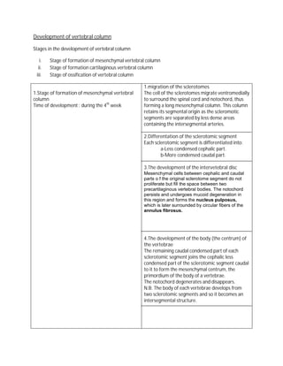
- 1. Development of vertebral column Stages in the development of vertebral column i. Stage of formation of mesenchymal vertebral column ii. Stage of formation cartilaginous vertebral column iii. Stage of ossification of vertebral column 1.migration of the sclerotomes 1.Stage of formation of mesenchymal vertebral The cell of the sclerotomes migrate ventromedially column to surround the spinal cord and notochord, thus Time of development : during the 4th week forming a long mesenchymal column. This column retains its segmental origin as the scleromotic segments are separated by less dense areas containing the intersegmental arteries. 2.Differentation of the sclerotomic segment Each sclerotomic segment is differentiated into: a-Less condensed cephalic part. b-More condensed caudal part. 3.The development of the intervetebral disc Mesenchymal cells between cephalic and caudal parts o f the original sclerotome segment do not proliferate but fill the space between two precartilaginous vertebral bodies. The notochord persists and undergoes mucoid degeneration in this region and forms the nucleus pulposus, which is later surrounded by circular fibers of the annulus fibrosus. 4.The development of the body (the centrum) of the vertebrae The remaining caudal condensed part of each sclerotomic segment joins the cephalic less condensed part of the sclerotomic segment caudal to it to form the mesenchymal centrum, the primordium of the body of a vertebrae. The notochord degenerates and disappears. N.B: The body of each vertebrae develops from two sclerotomic segments and so it becomes an intersegmental structure.
- 2. 5.The development of the neural arch Sclerotomic tissue migrates backwards from both sides of the centrum of the vertebrae to surround the neural tube (two pedicles and two lamina). The neural spine forms at the point of meeting of the neural arch, posteriorly. Sclerotomic tissue ,also, extends laterally from both sides of the centrum to form two processes: a-Costal process ventrally b-Transverse process dorsally 2.Stage of formation of cartilaginous vertebral Process of chondrofication: column -Two centers of chodrofication appear in the Time of appearance of the chondrofication centrum of the vertebrae. They fused together at centers: during the 6th week. the end of the embryonic period (8th week) -Centers of chondrofication appear in the neural arches. They fuse with each other and with the centrum. -The spinous and transverse processes develop from extensions of chondrofication centers in the neural arch. 3.Stage of ossification of vertebral column i. The primary ossification centers: Time of development: At the end of the 8th week. Number: -Three primary ossification centers are present by the end of the embryonic period -One in the centrum -One in each half of the neural arch. Process of Ossification: -At birth each vertebrae consists of three bony parts connected by cartilages. -The bony halves of the vertebral arch fuse together during the first 3 to 5 years. -The arches articulate with the centrum at cartilaginous neurocentral joints. These joints disappear when the vertebral arch fuses with the centrum during the third to sixth year. ii.The secondary ossification centers: Time of development: after puberty Number : Five secondary ossification centers appear: 1.One for the tip of the spinous process. 2.One for the tip of each tranverse process. 3.One for the superior rim of the vertebral body. 4.One for the inferior rim of the vertebral body.
- 3. Fate of notochord: 1.The most cranial part 3.The part of the of the notochord is notochord in-between incorporated in the 2.The parts of the the bodies of the basilar part of occipital notochord in the bodies vertebrae undergo bone and the posterior of the vertebrae undergo mucoid degeneration to part of body of sphenoid degeneration and form the nucleus bone. disappear. pulposus. 1.In the cervical Fate of costal processes: 4.In the upper sacral region: They form region: They unite to the anterior and form the anterior lateral boundary of 2.In the 3.In the portion of the ala of the foramen thoracic lumbar region: sacrum. transversum region: They fuse with They form the the ribs. transverses processes. Anomalies of the vertebral column 1)The spina bifida Cause : non-fusion of the embryonic halves of the vertebral arches. 2)Hemivertebrae Cause: failure of one of the chondrofication centers to appear and subsequent failure of half of the vertebrae to form. Feature: These defective vertebrae produce scoliosis (lateral curvature) 3)Sacralization of the fifth lumbar vertebrae Cause: The 5th lumbar vertebrae is fused with the sacrum. Feature: Number of lumbar vertebrae is 4 and the sacrum is formed of 6 vertebrae. 4)Lumbrization of the first piece of the sacrum to Cause: Separation of the first piece of the sacrum form a separate vertebrae to form a separate vertebrae. Feature: Number of lumbar vertebrae is 6 and the sacrum is only formed of 4 sacral vertebrae.
- 4. 5)Congenital kyphosis Feature: abnormality increased convexity in curvature of the thoracic spine as viewed from side 6)Congenital scoliosis Feature: Lateral curvature of the vertebral column