Ground-Glass Opacities: Patterns and Causes
•Download as PPTX, PDF•
36 likes•7,867 views

This document discusses ground-glass opacities seen on CT scans. It defines ground-glass opacities as a hazy increase in lung opacity while preserving bronchial and vascular markings. Various pathologies can cause ground-glass opacities by partially filling the airspaces. The document then describes different patterns of ground-glass opacities including diffuse, patchy, focal, halo, and peripheral distributions. For each pattern, common diseases that may present with that appearance are listed and briefly characterized.

Recommended
More Related Content
What's hot
What's hot (20)
Viewers also liked
Viewers also liked (20)
Similar to Ground-Glass Opacities: Patterns and Causes
Similar to Ground-Glass Opacities: Patterns and Causes (20)
Recently uploaded
Recently uploaded (20)
Ground-Glass Opacities: Patterns and Causes
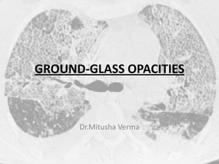
- 2. Definition… • Non-specific increased opacity / hazziness of the lung parenchyma due to change in relative propotions of air and alveolar walls with preservation of bronchial and vascular markings.
- 3. Pathologic basis • Partial filling of air spaces with- fluid, macrophages,neutrophils, amorphus materials. • Interstial thickening. • Partial collapse of alveoli. • Normal expiration. • Increased capillary blood volume.
- 4. False Positve / Pitfalls • Artificial Blooming- Narrow window width. • Volume averaging- Thicker collimation. • Expiratory phase. • Cardiac and Respiratory motion. • Microatelectasis- In gravity dependent positions.
- 6. DIFFUSE Acute lung transplant rejection. ARDS Edema Extrinsic allergic alveolitis Hemorrhage Infectious pneumonia.
- 7. Acute rejection of lung transplant • HRCT 65% sensitive & 85% specific •GGO Mild rejection –Patchy & localised Severe rejection –Widespread DDs- Reperfusion edema Infections- CMV
- 8. Acute Respiratory Distress Syndrome •Non Hydrosatatic pulmonary edema •Leaky capillary membranes •Etiology- Aspiration,contusion,smoke, sepsis. •CT –Bilateral gravity dependentclung opacities.
- 9. Pulmonary Edema Venous / Lymphatic ostruction Increased capillary permeability Hypoproteinemia CT- interlobular septal thickening increased vascular calibre peribronchovascular interstitial thickening, pleural effusion, thickening of fissures.
- 10. Extrinsic allergic alveolitis Also known as Hypersensitive Pneumonitis. Complex immunologic reaction Of lung to inhaled organic Antigens. Acute, Sub acute ,Chronic. CT- GGO(82%) , Small Nodules, Reticular pattern, Air trapping.
- 11. Diffuse Alveolar Haemorrhage May be Diffuse , patchy or focal Acute phase- GGO / Consolidation Sub acute- uniformly distributed 1-3mm nodules with GGO & interstial septal thickening.
- 12. Infectious Pneumonia Bacterial, Viral, mycobacterial, Fungal, Parasitic. A diffuse pattern – CMV & PCP CMV with HIV -dense consolidation, Bronchiectasis,interstitial reticulations. CMV post transplant - small nodules, Irregular lines.
- 13. Infectious Pneumonia Presence of isolated GGO without additional findings in patient with AIDS highly suggstive of Pneumocystis carinii.
- 14. Patchy Bronchiolitis obliterans organising pneumonia. Bronchio-alveolar cell carcinoma. Pulmonary alveolar proteinosis. Acute lung transplant rejection. ARDS Extrinsic allergic alveolitis Hemorrhage Infectious pneumonia.
- 15. Pulmonary alveolar proteinosis Filling of alveoli with PAS positive Proteinacious material. CT – Crazy paving DDs- lipoid pneumonia, ARDS, PCP.
- 16. FOCAL Bronchoalveolar Lavage Bronchiolitis obliterans organising pneumonia Bronchio-alveolar carcinoma Hemorrhage Pulmonary infection.
- 17. HALO Pattern Invasive pulmonary aspergillosis Neoplasm,haemorrhagic Post-Biopsy pseudonodule.
- 18. Invasive Aspergillosis. Peripheral ring of haemorrhage or haemorrhagic infarction surrounding target lesion of Aspergillosis.
- 19. Peripheral Pattern. Collagen vascular disease Contusions Desquamative interstitial pneumonitis Drug toxicity Eosinophilic pneumonia Fibrosis Sarcoidosis BOOP.
- 20. Bronchiolitis obliterans organising pneumonia. Histologically- granulation tissue plugs within respiratory bronchioles and alveolar ducts with Organising pneumonia extending into the surrounding alveoli. CT –pachy GGO,nodules, consolidtion in peripheral distribution Bilateral, non-segmental.
- 21. Pulmonary contusions Bleeding into lung interstitium and air spaces. CT- ill defined areas of GGO, Peripheral, non-anatomic distribution.
- 22. Desquamative interstitial pneumonitis Alveoli filling with macrophages. CT –lower lung zones peripheral UIP –similar with more honeycombing & traction bronchiectasis.
- 23. Collagen vascular disease Multisystem disorders characterized By vascular changes, fibrosis, Inflammation of connective tissue. SLE, RA , Polymyositis, Sjogren’s. CT- GGO (63-100%) Is a sign of ACTIVE inflammation In absence of significant Honeycombing, bronchiectasis,fibrosis. Site of Biopsy Treatment Planning. Response to Treatment.
- 24. Centrilobular / Bronchovascular • Eosinophilic pneumonia • Sarcoidosis • Extrinsic allergic alveolitis • Respiratory Bronchiolitis.
- 25. To Conclude…
- 26. • Thank you…