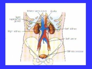
11. Excretion urography (I.V.U.).
The IVU consists of a series of films taken
after the administration of intravenous
injection of CM. The choice of whether to
use an ionic or nonionic contrast medium
depends on patient risk and economics.
It demonstrate both the function and
structure of the urinary system.
12. The main indications for the
IVU are:
•
•
•
•
Haematuria.
Ureteric calculi.
Ureteric fistula and stricture.
Urinary tract infection (UTI).
13. • Before the examination is started, the
procedure is explained to the pt to be more
cooperated and the patient history and
blood chemistry level should be checked.
(BUN= 8-25 mg/100 ml – creatinine = 0.61.5 mg/100ml).
14. Patient preparation:
•
Bowel is purged with strong laxative
and gas-absorbent tabs.
• Patient should take nothing by mouth
after midnight before the day of
examination.
16. • They should be well hydrated (they are at
increased risk for CM induced renal
failure if they are Dehydrated).
17. Technique:
• KUB film is done to check:
- Exposure factors.
- Patient preparation.
- Site of kidneys.
- Centering.
obvious pathology (UT calcification).
18. • The CM is injected through vein.
• Adult dose = 50 mm and pediatric
dose = 1 mm per kg
• Most reaction to contrast media
within the first 5 minutes after
administration. (Should not be left
alone).
19. Films:
1.
Immediate film (nephrogram). AP of
the renal areas (14-15 S = arm-to-kidney
time). It aims to show the renal
parenchyma opicified by C.M. in the
renal tubules.
2. 5 minutes film. AP of the renal areas.
This film is taken to determine if
excretion is symmetrical.
20. 1.
A compression band is now applied
around the patient’s abdomen at the level
of ASIS. Its aim is to inhibit ureteric
drainage and promote distension of the
pelvicalyceal systems (optimizing their
visualization).