SlideShare una empresa de Scribd logo
1 de 28
Descargar para leer sin conexión
-1 -

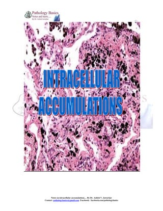
Notes on intracellular accumulations… By Dr. Ashish V. Jawarkar
Contact: pathologybasics@gmail.com Facebook: facebook.com/pathologybasics
-2 -

OVERVIEW CHART
INTRACELLULAR ACCUMULATIONS

METABOLIC PRODUCTS

PIGMENTS

1. Lipid metabolism disorder products
a. Fatty change
b. Cholesterol metabolism
- Atherosclerosis
- Xanthomas
- Cholesterosis
- Neimann-Pick disease type C

exogenous
endogenous
1. Coal
1.lipofuscin
a. CWP
2.Hemosiderin
b. anthracosis
3.Melanin
4.Homogent
2. Tattooing
isic acid
2. Protein metabolism disorder products
5. Bilirubin
a. Resorption droplets in proximal renal tubules
b. Russel Bodies
c. Alpha 1 antitrypsin deficiency
d. Cytoskeletal proteins
e. Amyloidosis
3. Carbohydrate metabolism disorder products
a. Glycogen storage disorders/Lysosomal storage
disorders
b. Diabetes Mellitus

Notes on intracellular accumulations… By Dr. Ashish V. Jawarkar
Contact: pathologybasics@gmail.com Facebook: facebook.com/pathologybasics
-3 -

* Lipid Metabolism Disorder Products:

1. Fatty change (steatosis)
Normal mechanism of fat metabolism:

Causes of lipid accumulation in liver
1. Excessive entry of free fatty acids in liver
1. starvation
2. Inhibition of catabolism in liver
1. alcohol
2. hypoxia
3. Decreased synthesis of apoproteins
CCl4
1.
2. Protein malnutrition

Notes on intracellular accumulations… By Dr. Ashish V. Jawarkar
Contact: pathologybasics@gmail.com Facebook: facebook.com/pathologybasics
-4 -

Morphology (Liver,Heart)
LIVER
Gross: 1. Enlarged (2-4 times)
2. Cut surface – bright yellow, soft, greasy

Diffuse hepatic steatosis results in pale, yellow parenchyma (as seen here) with greasy cut surfaces due
to lipid accumulation.

Microscopy:
In parenchymal cells
Early – small vacuoles in cytoplasm around the nucleus

Circled area shows early fatty change

Notes on intracellular accumulations… By Dr. Ashish V. Jawarkar
Contact: pathologybasics@gmail.com Facebook: facebook.com/pathologybasics
-5 -

Late – Vacuoles coalesce and create clear spaces that displace nucleus to periphery of cell
Contiguous cells coalesce and produce fatty cysts

Circled area shows late fatty change – fat displacing nucleus to periphery
Special stains for fat demonstration
1. Sudan IV

Notes on intracellular accumulations… By Dr. Ashish V. Jawarkar
Contact: pathologybasics@gmail.com Facebook: facebook.com/pathologybasics
-6 -

2. oil red O

3. PAS stain with diastase – cleared

glycogen

Not cleared with diastase

fat

Notes on intracellular accumulations… By Dr. Ashish V. Jawarkar
Contact: pathologybasics@gmail.com Facebook: facebook.com/pathologybasics
-7 -

Frozen section

Ultrastructure
Early – minute membrane bound inclusions (liposomes) closely applied to ER

EM photos of a Kupffer cell loaded with fat

Notes on intracellular accumulations… By Dr. Ashish V. Jawarkar
Contact: pathologybasics@gmail.com Facebook: facebook.com/pathologybasics
-8 -

HEART
Gross:
Bands of yellow myocardium alternating with unaffected myocardium – tigered
effect

Microscopy

Notes on intracellular accumulations… By Dr. Ashish V. Jawarkar
Contact: pathologybasics@gmail.com Facebook: facebook.com/pathologybasics
-9 -

2. Cholesterol metabolism
A. Atherosclerosis

Plaques
Cholesterol accumulates in intimal layers
of aorta and large arteries
Accumulation is in smooth muscle cells
and macrophages

Cholesterol clefts
Cholesterol esters accumulate in form of
needle shaped crystals

Mechanism

Notes on intracellular accumulations… By Dr. Ashish V. Jawarkar
Contact: pathologybasics@gmail.com Facebook: facebook.com/pathologybasics
- 10 -

Atherosclerotic plaque

Atherosclerotic plaque

Notes on intracellular accumulations… By Dr. Ashish V. Jawarkar
Contact: pathologybasics@gmail.com Facebook: facebook.com/pathologybasics
- 11 -

Cholesterol clefts
B. Xanthomas
Clusters of foamy cells in subepithelial connective tissue and tendons producing
tumorous masses

Xanthomas

Notes on intracellular accumulations… By Dr. Ashish V. Jawarkar
Contact: pathologybasics@gmail.com Facebook: facebook.com/pathologybasics
- 12 -

Achilles tendon xanthoma

Notes on intracellular accumulations… By Dr. Ashish V. Jawarkar
Contact: pathologybasics@gmail.com Facebook: facebook.com/pathologybasics
- 13 -

C. Cholesterosis
Focal accumulation of cholesterol laden macrophages in lamina propria of gall bladder

Gall bladder cholesterosis
D. Neimann Pick disease Type C
Lysosomal storage disorder characterized by cholesterol accumulation in multiple organs
Niemann Pick Disease type C (NPC) is an autosomal recessive lipidosis that is
characterized by lysosomal storage of cholesterol and glycosphingolipids. In normal
mammalian cells, low-density lipoprotein (LDL) is bound and internalized by cell surface
receptors and is hydrolyzed in the endocytic compartment. The cholesterol that is
released is transported to the cell surface and endoplasmic reticulum.

Notes on intracellular accumulations… By Dr. Ashish V. Jawarkar
Contact: pathologybasics@gmail.com Facebook: facebook.com/pathologybasics
- 14 -

Depiction of the cholesterol transport pathways in normal and NPC cells.
In NPC cells, LDL-derived cholesterol accumulates in lysosomes and endosomes, LDLcholesterol transport from endocytic compartments to other cellular compartments is
delayed

Notes on intracellular accumulations… By Dr. Ashish V. Jawarkar
Contact: pathologybasics@gmail.com Facebook: facebook.com/pathologybasics
- 15 -

*Protein metabolism disorder products
A. Resorption droplets in proximal renal tubules
Proteins filtered through glomerulus are reabsorbed by pinocytosis through proximal
tubules
Seen as pink hyaline droplets within cytoplasm of tubular cells in proteinuric states

In proteinuric states, the proximal tubular epithelial cells reabsorbs proteins that has
leaked through the glomerular filter

Notes on intracellular accumulations… By Dr. Ashish V. Jawarkar
Contact: pathologybasics@gmail.com Facebook: facebook.com/pathologybasics
- 16 -

B. Russel bodies
In plasma cell myelomas, ER becomes hugely distended with homogenous pink (on HE)
eosinophilic inclusions – Russel bodies
These inclusions basically consist of immunoglobulins

Russel bodies on HE

Russel bodies on MGG

Notes on intracellular accumulations… By Dr. Ashish V. Jawarkar
Contact: pathologybasics@gmail.com Facebook: facebook.com/pathologybasics
- 17 -

3. α1 antitrypsin deficiency
Protein folding defect

Build up of partially folded proteins

Aggregate in ER of liver / also results in emphysema

the synthesized protein lacks the ability to migrate from endoplasmic reticulum (ER) to
Golgi zone and thus accumulates inside ER as hyaline globules

Notes on intracellular accumulations… By Dr. Ashish V. Jawarkar
Contact: pathologybasics@gmail.com Facebook: facebook.com/pathologybasics
- 18 -

4. Accumulation of cytoskeletal proteins
ALCOHOLIC HYALINE (MALLORY HYALINE)
Eosinophilic globules seen in liver cells, consists predominantly of keratin intermediate
filaments
Seen to be accumulated in alcoholic liver disease

At high magnification can be seen globular red hyaline material within hepatocytes.
NEUROFIBRILLARY TANGLE
Seen in Alzheimers disease

Neurofibrillary Tangles (NFTs) are aggregates of hyperphosphorylated tau protein that
are most commonly known as a primary marker of Alzheimer's Disease.

Notes on intracellular accumulations… By Dr. Ashish V. Jawarkar
Contact: pathologybasics@gmail.com Facebook: facebook.com/pathologybasics
- 19 -

5. Amyloidosis
See notes on amyloidosis

Notes on intracellular accumulations… By Dr. Ashish V. Jawarkar
Contact: pathologybasics@gmail.com Facebook: facebook.com/pathologybasics
- 20 -

* Carbohydrate metabolism disorder products
Glycogen
Glycgoen accumulates in renal tubular epithelial cells, liver cells and islets of langerhans,
heart cells in diabetes mellitus and glycogen storage disorders.
It is cleared by diastase unlike fat.
PAS gives it a rose violet color.

Glycogen storage disease (GSD, also glycogenosis and dextrinosis) is the result of
defects in the processing of glycogen synthesis or breakdown within muscles, liver, and
other cell types

Notes on intracellular accumulations… By Dr. Ashish V. Jawarkar
Contact: pathologybasics@gmail.com Facebook: facebook.com/pathologybasics
- 21 -

* PIGMENTS
EXOGENOUS PIGMENTS
1. Carbon
Gets deposited in Coal worker’s pneumoconiosis and anthracosis.
Coal workers' pneumoconiosis (CWP), colloquially referred to as black lung disease, is
caused by long exposure to coal dust. It is a common affliction of coal miners and others
who work with coal, similar to both silicosis from inhaling silica dust, and to the longterm effects of tobacco smoking[citation needed]. Inhaled coal dust progressively builds up in
the lungs and is unable to be removed by the body; that leads to inflammation, fibrosis,
and in worse cases, necrosis.
Coal workers' pneumoconiosis, severe state, develops after the initial, milder form of the
disease known as anthracosis (anthrac — coal, carbon). This is often asymptomatic and
is found to at least some extent in all urban dwellers[1] due to air pollution.

Coal workers pneumoconiosis

Notes on intracellular accumulations… By Dr. Ashish V. Jawarkar
Contact: pathologybasics@gmail.com Facebook: facebook.com/pathologybasics
- 22 -

2. Tattooing
Pigments injected reside in dermal macrophages
No inflammatory response

Pigment in dermal macrophages

Notes on intracellular accumulations… By Dr. Ashish V. Jawarkar
Contact: pathologybasics@gmail.com Facebook: facebook.com/pathologybasics
- 23 -

ENDOGENOUS PIGMENTS
1. Lipofuscin
Lipofuscin is the name given to finely granular yellow-brown pigment granules
composed of lipid-containing residues of lysosomal digestion. It is considered to be one
of the aging or "wear-and-tear" pigments, found in the liver, kidney, heart muscle, retina,
adrenals, nerve cells, and ganglion cells. It is specifically arranged around the nucleus,
and is a type of lipochrome.

Neuronal lipofuscin

Notes on intracellular accumulations… By Dr. Ashish V. Jawarkar
Contact: pathologybasics@gmail.com Facebook: facebook.com/pathologybasics
- 24 -

2. Hemosiderin
Normally hemosiderin is seen in mononuclear phagocytes of bone marrow, spleen and
liver engaged in red cell break down.
Local deposition of hemosiderin can be seen in bruises
Systemic excess of iron as in increased dietry iron, hemolytic anemias and blood
transfusion leads to deposition in 1. Initially in phagocytes of spleen, liver, bone marrow and lymphnodes
(hemosiderosis)
2. Then in parenchymal cells of liver, pancreas (bronze diabetes), heart and
endocrine organs
It is a golden brown pigment on HE
Prussian blue stain is used to stain hemosiderin
Hemosiderosis
Due to iron excess due to various reasons
Deposition in phagocytes

Hemochromatosis
Due to inherited defect in iron transporter
Deposition in parenchymal cells

The hepatocytes and Kupffer cells here are full of granular brown deposits of
hemosiderin from accumulation of excess iron in the liver.

Notes on intracellular accumulations… By Dr. Ashish V. Jawarkar
Contact: pathologybasics@gmail.com Facebook: facebook.com/pathologybasics
- 25 -

A Prussian blue iron stain demonstrates the blue granules of hemosiderin in hepatocytes
and Kupffer cells.

Notes on intracellular accumulations… By Dr. Ashish V. Jawarkar
Contact: pathologybasics@gmail.com Facebook: facebook.com/pathologybasics
- 26 -

3. Melanin
Melanin is a derivative of the amino acid tyrosine, however it is not itself made of amino
acids and is not a protein. The pigment is produced in a specialized group of cells known
as melanocytes.
In humans, melanin is the primary determinant of skin color. It is also found in hair, the
pigmented tissue underlying the iris of the eye, and the stria vascularis of the inner ear. In
the brain, tissues with melanin include the medulla and pigment-bearing neurons within
areas of the brainstem, such as the locus coeruleus and the substantia nigra. It also occurs
in the zona reticularis of the adrenal gland.
Melanin is brown, non-refractile, and finely granular with individual granules having a
diameter of less than 800 nanometers. This differentiates melanin from common blood
breakdown pigments, which are larger, chunky, and refractile, and range in color from
green to yellow or red-brown. In heavily pigmented lesions, dense aggregates of melanin
can obscure histologic detail. A dilute solution of potassium permanganate is an effective
melanin bleach.

Pigmented melanoma

Notes on intracellular accumulations… By Dr. Ashish V. Jawarkar
Contact: pathologybasics@gmail.com Facebook: facebook.com/pathologybasics
- 27 -

4. Homogentisic acid
Alkaptonuria (black urine disease or alcaptonuria) is a rare inherited genetic disorder of
phenylalanine and tyrosine metabolism.
This is an autosomal recessive condition that is due to a defect in the enzyme
homogentisate 1,2-dioxygenase which participates in the degradation of tyrosine.
As a result, homogentisic acid and its oxide, called alkapton, accumulate in the blood and
are excreted in urine in large amounts (hence -uria).
Excessive homogentisic acid causes damage to cartilage (ochronosis, leading to
osteoarthritis) and heart valves as well as precipitating as kidney stones

Notes on intracellular accumulations… By Dr. Ashish V. Jawarkar
Contact: pathologybasics@gmail.com Facebook: facebook.com/pathologybasics
- 28 -

6. Bilirubin
Accumulates in jaundice

Notes on intracellular accumulations… By Dr. Ashish V. Jawarkar
Contact: pathologybasics@gmail.com Facebook: facebook.com/pathologybasics

Más contenido relacionado

La actualidad más candente

La actualidad más candente (20)

2 hyperemia-congestion
2 hyperemia-congestion2 hyperemia-congestion
2 hyperemia-congestion
 
4
44
4
 
Hyperemia and congestion edema
Hyperemia and congestion edema Hyperemia and congestion edema
Hyperemia and congestion edema
 
Infarction
InfarctionInfarction
Infarction
 
Hemodynamics congestion & hyperemia
Hemodynamics  congestion & hyperemiaHemodynamics  congestion & hyperemia
Hemodynamics congestion & hyperemia
 
INTRACELLULAR ACCUMULATIONS A SHORT REVIEW
INTRACELLULAR ACCUMULATIONS A SHORT REVIEWINTRACELLULAR ACCUMULATIONS A SHORT REVIEW
INTRACELLULAR ACCUMULATIONS A SHORT REVIEW
 
INFARCTION
INFARCTION INFARCTION
INFARCTION
 
Pathologic Calcification
Pathologic CalcificationPathologic Calcification
Pathologic Calcification
 
INTRACELLULAR ACCUMULATIONS
INTRACELLULAR ACCUMULATIONSINTRACELLULAR ACCUMULATIONS
INTRACELLULAR ACCUMULATIONS
 
Amyloidosis ppt
Amyloidosis pptAmyloidosis ppt
Amyloidosis ppt
 
Intracellular accumulations
Intracellular accumulations Intracellular accumulations
Intracellular accumulations
 
Pathology of Acute Inflammation
Pathology of Acute InflammationPathology of Acute Inflammation
Pathology of Acute Inflammation
 
Cell injury 2
Cell injury 2Cell injury 2
Cell injury 2
 
Cell injury: causes, pathogenesis, Morphology of reversible cell injury
Cell injury: causes, pathogenesis, Morphology of reversible cell injuryCell injury: causes, pathogenesis, Morphology of reversible cell injury
Cell injury: causes, pathogenesis, Morphology of reversible cell injury
 
Apoptosis
ApoptosisApoptosis
Apoptosis
 
Neoplasia
NeoplasiaNeoplasia
Neoplasia
 
Inflammation 6
Inflammation 6Inflammation 6
Inflammation 6
 
Cvc lung, liver and spleen
Cvc lung, liver and spleenCvc lung, liver and spleen
Cvc lung, liver and spleen
 
Pigments
PigmentsPigments
Pigments
 
Pathology neoplasm
Pathology  neoplasmPathology  neoplasm
Pathology neoplasm
 

Destacado

Destacado (6)

Cell injury: CELL ADAPTATIONS
Cell injury: CELL ADAPTATIONSCell injury: CELL ADAPTATIONS
Cell injury: CELL ADAPTATIONS
 
cell injury
cell injurycell injury
cell injury
 
Cell injury
Cell injuryCell injury
Cell injury
 
Pathology cell injury i
Pathology   cell injury iPathology   cell injury i
Pathology cell injury i
 
General pathology lecture 1 introduction & cell injury
General pathology lecture 1 introduction & cell injuryGeneral pathology lecture 1 introduction & cell injury
General pathology lecture 1 introduction & cell injury
 
Cell Injury Patho
Cell Injury PathoCell Injury Patho
Cell Injury Patho
 

Similar a Intracellular Accumulation Guide

Intracellular accumulations 1
Intracellular accumulations 1Intracellular accumulations 1
Intracellular accumulations 1EshaMehmood2
 
My lecture Accu-Calci-Gangrene.pptx
My lecture Accu-Calci-Gangrene.pptxMy lecture Accu-Calci-Gangrene.pptx
My lecture Accu-Calci-Gangrene.pptxchiragkumarmenapara
 
Cellular Reactions to Injury.pptx
Cellular  Reactions  to Injury.pptxCellular  Reactions  to Injury.pptx
Cellular Reactions to Injury.pptxMohammedAbdela7
 
Intracellular accumulations
Intracellular accumulations Intracellular accumulations
Intracellular accumulations abdul aziz
 
Lecture 2 Degenerative changes and cellular injury
Lecture 2 Degenerative changes and cellular injuryLecture 2 Degenerative changes and cellular injury
Lecture 2 Degenerative changes and cellular injuryGreen-book
 
Degeneration, necrosis, and pathological pigmentation
Degeneration, necrosis, and pathological pigmentationDegeneration, necrosis, and pathological pigmentation
Degeneration, necrosis, and pathological pigmentationBruno Mmassy
 
LESSON 2-ACCUMULATION OF THE OF ENDOGENOUS AND EXOGENOUS PRODUCT_09091039.ppt
LESSON 2-ACCUMULATION OF THE OF ENDOGENOUS AND EXOGENOUS PRODUCT_09091039.pptLESSON 2-ACCUMULATION OF THE OF ENDOGENOUS AND EXOGENOUS PRODUCT_09091039.ppt
LESSON 2-ACCUMULATION OF THE OF ENDOGENOUS AND EXOGENOUS PRODUCT_09091039.pptmdaquib14
 
Basic Principles of Cell Injury and Adaptation.pptx
Basic Principles of Cell Injury and Adaptation.pptxBasic Principles of Cell Injury and Adaptation.pptx
Basic Principles of Cell Injury and Adaptation.pptxsanjayudps2016
 
Cell accumulations foundation block pathology
Cell accumulations foundation block pathologyCell accumulations foundation block pathology
Cell accumulations foundation block pathologyMulazim Bukhari
 
Cell adaptation and injury
Cell adaptation and injuryCell adaptation and injury
Cell adaptation and injuryAmi Febriza
 
ANTIOXIDANTS FOR HEPATIC CIRRHOSIS.pptx
ANTIOXIDANTS FOR HEPATIC CIRRHOSIS.pptxANTIOXIDANTS FOR HEPATIC CIRRHOSIS.pptx
ANTIOXIDANTS FOR HEPATIC CIRRHOSIS.pptxPriyansha Singh
 
cell injury1.pptx
cell injury1.pptxcell injury1.pptx
cell injury1.pptxSirnaEmana1
 
Cell injury cell swelling necrosis fatty change
Cell injury cell swelling necrosis fatty changeCell injury cell swelling necrosis fatty change
Cell injury cell swelling necrosis fatty changeKhalid Yousuf
 
Cellular adaptations & accumulations
Cellular adaptations & accumulationsCellular adaptations & accumulations
Cellular adaptations & accumulationsHabibah Chaudhary
 
Morphology of cell injury and Intracellular accumulationspptx
Morphology of cell injury and Intracellular accumulationspptxMorphology of cell injury and Intracellular accumulationspptx
Morphology of cell injury and Intracellular accumulationspptxvaishaliwasnik
 

Similar a Intracellular Accumulation Guide (20)

Intracellular accumulations 1
Intracellular accumulations 1Intracellular accumulations 1
Intracellular accumulations 1
 
My lecture Accu-Calci-Gangrene.pptx
My lecture Accu-Calci-Gangrene.pptxMy lecture Accu-Calci-Gangrene.pptx
My lecture Accu-Calci-Gangrene.pptx
 
Cellular Reactions to Injury.pptx
Cellular  Reactions  to Injury.pptxCellular  Reactions  to Injury.pptx
Cellular Reactions to Injury.pptx
 
Intracellular accumulations
Intracellular accumulations Intracellular accumulations
Intracellular accumulations
 
Apoptosis made easy
Apoptosis made easyApoptosis made easy
Apoptosis made easy
 
Lecture 2 Degenerative changes and cellular injury
Lecture 2 Degenerative changes and cellular injuryLecture 2 Degenerative changes and cellular injury
Lecture 2 Degenerative changes and cellular injury
 
Degeneration, necrosis, and pathological pigmentation
Degeneration, necrosis, and pathological pigmentationDegeneration, necrosis, and pathological pigmentation
Degeneration, necrosis, and pathological pigmentation
 
NECROSIS by Shabistan.pptx
NECROSIS by Shabistan.pptxNECROSIS by Shabistan.pptx
NECROSIS by Shabistan.pptx
 
LESSON 2-ACCUMULATION OF THE OF ENDOGENOUS AND EXOGENOUS PRODUCT_09091039.ppt
LESSON 2-ACCUMULATION OF THE OF ENDOGENOUS AND EXOGENOUS PRODUCT_09091039.pptLESSON 2-ACCUMULATION OF THE OF ENDOGENOUS AND EXOGENOUS PRODUCT_09091039.ppt
LESSON 2-ACCUMULATION OF THE OF ENDOGENOUS AND EXOGENOUS PRODUCT_09091039.ppt
 
Basic Principles of Cell Injury and Adaptation.pptx
Basic Principles of Cell Injury and Adaptation.pptxBasic Principles of Cell Injury and Adaptation.pptx
Basic Principles of Cell Injury and Adaptation.pptx
 
Cell accumulations foundation block pathology
Cell accumulations foundation block pathologyCell accumulations foundation block pathology
Cell accumulations foundation block pathology
 
Cell adaptation and injury
Cell adaptation and injuryCell adaptation and injury
Cell adaptation and injury
 
ANTIOXIDANTS FOR HEPATIC CIRRHOSIS.pptx
ANTIOXIDANTS FOR HEPATIC CIRRHOSIS.pptxANTIOXIDANTS FOR HEPATIC CIRRHOSIS.pptx
ANTIOXIDANTS FOR HEPATIC CIRRHOSIS.pptx
 
cell injury1.pptx
cell injury1.pptxcell injury1.pptx
cell injury1.pptx
 
Cell injury cell swelling necrosis fatty change
Cell injury cell swelling necrosis fatty changeCell injury cell swelling necrosis fatty change
Cell injury cell swelling necrosis fatty change
 
Cellular adaptations & accumulations
Cellular adaptations & accumulationsCellular adaptations & accumulations
Cellular adaptations & accumulations
 
Amyloidosis
AmyloidosisAmyloidosis
Amyloidosis
 
Mod+11 2017
Mod+11 2017Mod+11 2017
Mod+11 2017
 
MID2163 TOPIC 2
MID2163 TOPIC 2MID2163 TOPIC 2
MID2163 TOPIC 2
 
Morphology of cell injury and Intracellular accumulationspptx
Morphology of cell injury and Intracellular accumulationspptxMorphology of cell injury and Intracellular accumulationspptx
Morphology of cell injury and Intracellular accumulationspptx
 

Más de Ashish Jawarkar

Ch 6 diseases of the immune system part 1
Ch 6 diseases of the immune system part 1Ch 6 diseases of the immune system part 1
Ch 6 diseases of the immune system part 1Ashish Jawarkar
 
MALE AND FEMALE GENITAL TRACT
MALE AND FEMALE GENITAL TRACTMALE AND FEMALE GENITAL TRACT
MALE AND FEMALE GENITAL TRACTAshish Jawarkar
 
Ch 2 adaptations, cell injury, cell death
Ch 2 adaptations, cell injury, cell deathCh 2 adaptations, cell injury, cell death
Ch 2 adaptations, cell injury, cell deathAshish Jawarkar
 
Ch 4 hemorragic disorders,thromboembolic diseases, shock
Ch 4 hemorragic disorders,thromboembolic diseases, shockCh 4 hemorragic disorders,thromboembolic diseases, shock
Ch 4 hemorragic disorders,thromboembolic diseases, shockAshish Jawarkar
 
Ch 3 inflammation and repair
Ch 3 inflammation and repairCh 3 inflammation and repair
Ch 3 inflammation and repairAshish Jawarkar
 
CYTOLOGY OF BREAST LESIONS??!
CYTOLOGY OF BREAST LESIONS??! CYTOLOGY OF BREAST LESIONS??!
CYTOLOGY OF BREAST LESIONS??! Ashish Jawarkar
 
Immunohistochemistry in breast lesions
Immunohistochemistry in breast lesionsImmunohistochemistry in breast lesions
Immunohistochemistry in breast lesionsAshish Jawarkar
 
Immunohistochemistry of Prostatic lesions
Immunohistochemistry of Prostatic lesionsImmunohistochemistry of Prostatic lesions
Immunohistochemistry of Prostatic lesionsAshish Jawarkar
 
Robbins Chapter 1.. Cell as a unit of health and disease
Robbins Chapter 1.. Cell as a unit of health and diseaseRobbins Chapter 1.. Cell as a unit of health and disease
Robbins Chapter 1.. Cell as a unit of health and diseaseAshish Jawarkar
 
Pathology of the digestive system
Pathology of the digestive systemPathology of the digestive system
Pathology of the digestive systemAshish Jawarkar
 
Cellular adaptations, injury and death.. Lecture 1
Cellular adaptations, injury and death.. Lecture 1Cellular adaptations, injury and death.. Lecture 1
Cellular adaptations, injury and death.. Lecture 1Ashish Jawarkar
 
Quality control in clinical laboratories
Quality control in clinical laboratoriesQuality control in clinical laboratories
Quality control in clinical laboratoriesAshish Jawarkar
 
CSF - Cerebrospinal fluid examination - from tapping to pathological diagnosis
CSF - Cerebrospinal fluid examination - from tapping to pathological diagnosisCSF - Cerebrospinal fluid examination - from tapping to pathological diagnosis
CSF - Cerebrospinal fluid examination - from tapping to pathological diagnosisAshish Jawarkar
 
LABORATORY DIAGNOSIS OF HIV - AIDS
LABORATORY DIAGNOSIS OF HIV - AIDSLABORATORY DIAGNOSIS OF HIV - AIDS
LABORATORY DIAGNOSIS OF HIV - AIDSAshish Jawarkar
 
Cancer and its types - an introduction to cancer
Cancer and its types - an introduction to cancerCancer and its types - an introduction to cancer
Cancer and its types - an introduction to cancerAshish Jawarkar
 

Más de Ashish Jawarkar (20)

Ch 6 diseases of the immune system part 1
Ch 6 diseases of the immune system part 1Ch 6 diseases of the immune system part 1
Ch 6 diseases of the immune system part 1
 
MALE AND FEMALE GENITAL TRACT
MALE AND FEMALE GENITAL TRACTMALE AND FEMALE GENITAL TRACT
MALE AND FEMALE GENITAL TRACT
 
Ch 2 adaptations, cell injury, cell death
Ch 2 adaptations, cell injury, cell deathCh 2 adaptations, cell injury, cell death
Ch 2 adaptations, cell injury, cell death
 
Ch 4 hemorragic disorders,thromboembolic diseases, shock
Ch 4 hemorragic disorders,thromboembolic diseases, shockCh 4 hemorragic disorders,thromboembolic diseases, shock
Ch 4 hemorragic disorders,thromboembolic diseases, shock
 
Ch 3 inflammation and repair
Ch 3 inflammation and repairCh 3 inflammation and repair
Ch 3 inflammation and repair
 
CYTOLOGY OF BREAST LESIONS??!
CYTOLOGY OF BREAST LESIONS??! CYTOLOGY OF BREAST LESIONS??!
CYTOLOGY OF BREAST LESIONS??!
 
Histotechniques
HistotechniquesHistotechniques
Histotechniques
 
Immunohistochemistry in breast lesions
Immunohistochemistry in breast lesionsImmunohistochemistry in breast lesions
Immunohistochemistry in breast lesions
 
Immunohistochemistry of Prostatic lesions
Immunohistochemistry of Prostatic lesionsImmunohistochemistry of Prostatic lesions
Immunohistochemistry of Prostatic lesions
 
Cardiovascular system
Cardiovascular systemCardiovascular system
Cardiovascular system
 
Ch7 Neoplasia
Ch7 NeoplasiaCh7 Neoplasia
Ch7 Neoplasia
 
Amyloidosis
AmyloidosisAmyloidosis
Amyloidosis
 
Ch12 Heart Part 1
Ch12 Heart Part 1Ch12 Heart Part 1
Ch12 Heart Part 1
 
Robbins Chapter 1.. Cell as a unit of health and disease
Robbins Chapter 1.. Cell as a unit of health and diseaseRobbins Chapter 1.. Cell as a unit of health and disease
Robbins Chapter 1.. Cell as a unit of health and disease
 
Pathology of the digestive system
Pathology of the digestive systemPathology of the digestive system
Pathology of the digestive system
 
Cellular adaptations, injury and death.. Lecture 1
Cellular adaptations, injury and death.. Lecture 1Cellular adaptations, injury and death.. Lecture 1
Cellular adaptations, injury and death.. Lecture 1
 
Quality control in clinical laboratories
Quality control in clinical laboratoriesQuality control in clinical laboratories
Quality control in clinical laboratories
 
CSF - Cerebrospinal fluid examination - from tapping to pathological diagnosis
CSF - Cerebrospinal fluid examination - from tapping to pathological diagnosisCSF - Cerebrospinal fluid examination - from tapping to pathological diagnosis
CSF - Cerebrospinal fluid examination - from tapping to pathological diagnosis
 
LABORATORY DIAGNOSIS OF HIV - AIDS
LABORATORY DIAGNOSIS OF HIV - AIDSLABORATORY DIAGNOSIS OF HIV - AIDS
LABORATORY DIAGNOSIS OF HIV - AIDS
 
Cancer and its types - an introduction to cancer
Cancer and its types - an introduction to cancerCancer and its types - an introduction to cancer
Cancer and its types - an introduction to cancer
 

Último

Statistical modeling in pharmaceutical research and development.
Statistical modeling in pharmaceutical research and development.Statistical modeling in pharmaceutical research and development.
Statistical modeling in pharmaceutical research and development.ANJALI
 
Nutrition of OCD for my Nutritional Neuroscience Class
Nutrition of OCD for my Nutritional Neuroscience ClassNutrition of OCD for my Nutritional Neuroscience Class
Nutrition of OCD for my Nutritional Neuroscience Classmanuelazg2001
 
Presentation for Bella Mahl 2024-03-28-24-MW-Overview-Bella.pptx
Presentation for Bella Mahl 2024-03-28-24-MW-Overview-Bella.pptxPresentation for Bella Mahl 2024-03-28-24-MW-Overview-Bella.pptx
Presentation for Bella Mahl 2024-03-28-24-MW-Overview-Bella.pptxpdamico1
 
SWD (Short wave diathermy)- Physiotherapy.ppt
SWD (Short wave diathermy)- Physiotherapy.pptSWD (Short wave diathermy)- Physiotherapy.ppt
SWD (Short wave diathermy)- Physiotherapy.pptMumux Mirani
 
Informed Consent Empowering Healthcare Decision-Making.pptx
Informed Consent Empowering Healthcare Decision-Making.pptxInformed Consent Empowering Healthcare Decision-Making.pptx
Informed Consent Empowering Healthcare Decision-Making.pptxSasikiranMarri
 
Study on the Impact of FOCUS-PDCA Management Model on the Disinfection Qualit...
Study on the Impact of FOCUS-PDCA Management Model on the Disinfection Qualit...Study on the Impact of FOCUS-PDCA Management Model on the Disinfection Qualit...
Study on the Impact of FOCUS-PDCA Management Model on the Disinfection Qualit...MehranMouzam
 
Biomechanics- Shoulder Joint!!!!!!!!!!!!
Biomechanics- Shoulder Joint!!!!!!!!!!!!Biomechanics- Shoulder Joint!!!!!!!!!!!!
Biomechanics- Shoulder Joint!!!!!!!!!!!!ibtesaam huma
 
Presentation on Parasympathetic Nervous System
Presentation on Parasympathetic Nervous SystemPresentation on Parasympathetic Nervous System
Presentation on Parasympathetic Nervous SystemPrerana Jadhav
 
CEHPALOSPORINS.pptx By Harshvardhan Dev Bhoomi Uttarakhand University
CEHPALOSPORINS.pptx By Harshvardhan Dev Bhoomi Uttarakhand UniversityCEHPALOSPORINS.pptx By Harshvardhan Dev Bhoomi Uttarakhand University
CEHPALOSPORINS.pptx By Harshvardhan Dev Bhoomi Uttarakhand UniversityHarshChauhan475104
 
Big Data Analysis Suggests COVID Vaccination Increases Excess Mortality Of ...
Big Data Analysis Suggests COVID  Vaccination Increases Excess Mortality Of  ...Big Data Analysis Suggests COVID  Vaccination Increases Excess Mortality Of  ...
Big Data Analysis Suggests COVID Vaccination Increases Excess Mortality Of ...sdateam0
 
world health day presentation ppt download
world health day presentation ppt downloadworld health day presentation ppt download
world health day presentation ppt downloadAnkitKumar311566
 
Measurement of Radiation and Dosimetric Procedure.pptx
Measurement of Radiation and Dosimetric Procedure.pptxMeasurement of Radiation and Dosimetric Procedure.pptx
Measurement of Radiation and Dosimetric Procedure.pptxDr. Dheeraj Kumar
 
PULMONARY EDEMA AND ITS MANAGEMENT.pdf
PULMONARY EDEMA AND  ITS  MANAGEMENT.pdfPULMONARY EDEMA AND  ITS  MANAGEMENT.pdf
PULMONARY EDEMA AND ITS MANAGEMENT.pdfDolisha Warbi
 
VarSeq 2.6.0: Advancing Pharmacogenomics and Genomic Analysis
VarSeq 2.6.0: Advancing Pharmacogenomics and Genomic AnalysisVarSeq 2.6.0: Advancing Pharmacogenomics and Genomic Analysis
VarSeq 2.6.0: Advancing Pharmacogenomics and Genomic AnalysisGolden Helix
 
METHODS OF ACQUIRING KNOWLEDGE IN NURSING.pptx by navdeep kaur
METHODS OF ACQUIRING KNOWLEDGE IN NURSING.pptx by navdeep kaurMETHODS OF ACQUIRING KNOWLEDGE IN NURSING.pptx by navdeep kaur
METHODS OF ACQUIRING KNOWLEDGE IN NURSING.pptx by navdeep kaurNavdeep Kaur
 
Apiculture Chapter 1. Introduction 2.ppt
Apiculture Chapter 1. Introduction 2.pptApiculture Chapter 1. Introduction 2.ppt
Apiculture Chapter 1. Introduction 2.pptkedirjemalharun
 
SYNDESMOTIC INJURY- ANATOMICAL REPAIR.pptx
SYNDESMOTIC INJURY- ANATOMICAL REPAIR.pptxSYNDESMOTIC INJURY- ANATOMICAL REPAIR.pptx
SYNDESMOTIC INJURY- ANATOMICAL REPAIR.pptxdrashraf369
 
97111 47426 Call Girls In Delhi MUNIRKAA
97111 47426 Call Girls In Delhi MUNIRKAA97111 47426 Call Girls In Delhi MUNIRKAA
97111 47426 Call Girls In Delhi MUNIRKAAjennyeacort
 
epilepsy and status epilepticus for undergraduate.pptx
epilepsy and status epilepticus  for undergraduate.pptxepilepsy and status epilepticus  for undergraduate.pptx
epilepsy and status epilepticus for undergraduate.pptxMohamed Rizk Khodair
 

Último (20)

Statistical modeling in pharmaceutical research and development.
Statistical modeling in pharmaceutical research and development.Statistical modeling in pharmaceutical research and development.
Statistical modeling in pharmaceutical research and development.
 
Nutrition of OCD for my Nutritional Neuroscience Class
Nutrition of OCD for my Nutritional Neuroscience ClassNutrition of OCD for my Nutritional Neuroscience Class
Nutrition of OCD for my Nutritional Neuroscience Class
 
Presentation for Bella Mahl 2024-03-28-24-MW-Overview-Bella.pptx
Presentation for Bella Mahl 2024-03-28-24-MW-Overview-Bella.pptxPresentation for Bella Mahl 2024-03-28-24-MW-Overview-Bella.pptx
Presentation for Bella Mahl 2024-03-28-24-MW-Overview-Bella.pptx
 
SWD (Short wave diathermy)- Physiotherapy.ppt
SWD (Short wave diathermy)- Physiotherapy.pptSWD (Short wave diathermy)- Physiotherapy.ppt
SWD (Short wave diathermy)- Physiotherapy.ppt
 
Informed Consent Empowering Healthcare Decision-Making.pptx
Informed Consent Empowering Healthcare Decision-Making.pptxInformed Consent Empowering Healthcare Decision-Making.pptx
Informed Consent Empowering Healthcare Decision-Making.pptx
 
Study on the Impact of FOCUS-PDCA Management Model on the Disinfection Qualit...
Study on the Impact of FOCUS-PDCA Management Model on the Disinfection Qualit...Study on the Impact of FOCUS-PDCA Management Model on the Disinfection Qualit...
Study on the Impact of FOCUS-PDCA Management Model on the Disinfection Qualit...
 
Biomechanics- Shoulder Joint!!!!!!!!!!!!
Biomechanics- Shoulder Joint!!!!!!!!!!!!Biomechanics- Shoulder Joint!!!!!!!!!!!!
Biomechanics- Shoulder Joint!!!!!!!!!!!!
 
Presentation on Parasympathetic Nervous System
Presentation on Parasympathetic Nervous SystemPresentation on Parasympathetic Nervous System
Presentation on Parasympathetic Nervous System
 
CEHPALOSPORINS.pptx By Harshvardhan Dev Bhoomi Uttarakhand University
CEHPALOSPORINS.pptx By Harshvardhan Dev Bhoomi Uttarakhand UniversityCEHPALOSPORINS.pptx By Harshvardhan Dev Bhoomi Uttarakhand University
CEHPALOSPORINS.pptx By Harshvardhan Dev Bhoomi Uttarakhand University
 
Big Data Analysis Suggests COVID Vaccination Increases Excess Mortality Of ...
Big Data Analysis Suggests COVID  Vaccination Increases Excess Mortality Of  ...Big Data Analysis Suggests COVID  Vaccination Increases Excess Mortality Of  ...
Big Data Analysis Suggests COVID Vaccination Increases Excess Mortality Of ...
 
world health day presentation ppt download
world health day presentation ppt downloadworld health day presentation ppt download
world health day presentation ppt download
 
Measurement of Radiation and Dosimetric Procedure.pptx
Measurement of Radiation and Dosimetric Procedure.pptxMeasurement of Radiation and Dosimetric Procedure.pptx
Measurement of Radiation and Dosimetric Procedure.pptx
 
PULMONARY EDEMA AND ITS MANAGEMENT.pdf
PULMONARY EDEMA AND  ITS  MANAGEMENT.pdfPULMONARY EDEMA AND  ITS  MANAGEMENT.pdf
PULMONARY EDEMA AND ITS MANAGEMENT.pdf
 
VarSeq 2.6.0: Advancing Pharmacogenomics and Genomic Analysis
VarSeq 2.6.0: Advancing Pharmacogenomics and Genomic AnalysisVarSeq 2.6.0: Advancing Pharmacogenomics and Genomic Analysis
VarSeq 2.6.0: Advancing Pharmacogenomics and Genomic Analysis
 
METHODS OF ACQUIRING KNOWLEDGE IN NURSING.pptx by navdeep kaur
METHODS OF ACQUIRING KNOWLEDGE IN NURSING.pptx by navdeep kaurMETHODS OF ACQUIRING KNOWLEDGE IN NURSING.pptx by navdeep kaur
METHODS OF ACQUIRING KNOWLEDGE IN NURSING.pptx by navdeep kaur
 
Apiculture Chapter 1. Introduction 2.ppt
Apiculture Chapter 1. Introduction 2.pptApiculture Chapter 1. Introduction 2.ppt
Apiculture Chapter 1. Introduction 2.ppt
 
SYNDESMOTIC INJURY- ANATOMICAL REPAIR.pptx
SYNDESMOTIC INJURY- ANATOMICAL REPAIR.pptxSYNDESMOTIC INJURY- ANATOMICAL REPAIR.pptx
SYNDESMOTIC INJURY- ANATOMICAL REPAIR.pptx
 
97111 47426 Call Girls In Delhi MUNIRKAA
97111 47426 Call Girls In Delhi MUNIRKAA97111 47426 Call Girls In Delhi MUNIRKAA
97111 47426 Call Girls In Delhi MUNIRKAA
 
epilepsy and status epilepticus for undergraduate.pptx
epilepsy and status epilepticus  for undergraduate.pptxepilepsy and status epilepticus  for undergraduate.pptx
epilepsy and status epilepticus for undergraduate.pptx
 
Epilepsy
EpilepsyEpilepsy
Epilepsy
 

Intracellular Accumulation Guide

  • 1. -1 - Notes on intracellular accumulations… By Dr. Ashish V. Jawarkar Contact: pathologybasics@gmail.com Facebook: facebook.com/pathologybasics
  • 2. -2 - OVERVIEW CHART INTRACELLULAR ACCUMULATIONS METABOLIC PRODUCTS PIGMENTS 1. Lipid metabolism disorder products a. Fatty change b. Cholesterol metabolism - Atherosclerosis - Xanthomas - Cholesterosis - Neimann-Pick disease type C exogenous endogenous 1. Coal 1.lipofuscin a. CWP 2.Hemosiderin b. anthracosis 3.Melanin 4.Homogent 2. Tattooing isic acid 2. Protein metabolism disorder products 5. Bilirubin a. Resorption droplets in proximal renal tubules b. Russel Bodies c. Alpha 1 antitrypsin deficiency d. Cytoskeletal proteins e. Amyloidosis 3. Carbohydrate metabolism disorder products a. Glycogen storage disorders/Lysosomal storage disorders b. Diabetes Mellitus Notes on intracellular accumulations… By Dr. Ashish V. Jawarkar Contact: pathologybasics@gmail.com Facebook: facebook.com/pathologybasics
  • 3. -3 - * Lipid Metabolism Disorder Products: 1. Fatty change (steatosis) Normal mechanism of fat metabolism: Causes of lipid accumulation in liver 1. Excessive entry of free fatty acids in liver 1. starvation 2. Inhibition of catabolism in liver 1. alcohol 2. hypoxia 3. Decreased synthesis of apoproteins CCl4 1. 2. Protein malnutrition Notes on intracellular accumulations… By Dr. Ashish V. Jawarkar Contact: pathologybasics@gmail.com Facebook: facebook.com/pathologybasics
  • 4. -4 - Morphology (Liver,Heart) LIVER Gross: 1. Enlarged (2-4 times) 2. Cut surface – bright yellow, soft, greasy Diffuse hepatic steatosis results in pale, yellow parenchyma (as seen here) with greasy cut surfaces due to lipid accumulation. Microscopy: In parenchymal cells Early – small vacuoles in cytoplasm around the nucleus Circled area shows early fatty change Notes on intracellular accumulations… By Dr. Ashish V. Jawarkar Contact: pathologybasics@gmail.com Facebook: facebook.com/pathologybasics
  • 5. -5 - Late – Vacuoles coalesce and create clear spaces that displace nucleus to periphery of cell Contiguous cells coalesce and produce fatty cysts Circled area shows late fatty change – fat displacing nucleus to periphery Special stains for fat demonstration 1. Sudan IV Notes on intracellular accumulations… By Dr. Ashish V. Jawarkar Contact: pathologybasics@gmail.com Facebook: facebook.com/pathologybasics
  • 6. -6 - 2. oil red O 3. PAS stain with diastase – cleared glycogen Not cleared with diastase fat Notes on intracellular accumulations… By Dr. Ashish V. Jawarkar Contact: pathologybasics@gmail.com Facebook: facebook.com/pathologybasics
  • 7. -7 - Frozen section Ultrastructure Early – minute membrane bound inclusions (liposomes) closely applied to ER EM photos of a Kupffer cell loaded with fat Notes on intracellular accumulations… By Dr. Ashish V. Jawarkar Contact: pathologybasics@gmail.com Facebook: facebook.com/pathologybasics
  • 8. -8 - HEART Gross: Bands of yellow myocardium alternating with unaffected myocardium – tigered effect Microscopy Notes on intracellular accumulations… By Dr. Ashish V. Jawarkar Contact: pathologybasics@gmail.com Facebook: facebook.com/pathologybasics
  • 9. -9 - 2. Cholesterol metabolism A. Atherosclerosis Plaques Cholesterol accumulates in intimal layers of aorta and large arteries Accumulation is in smooth muscle cells and macrophages Cholesterol clefts Cholesterol esters accumulate in form of needle shaped crystals Mechanism Notes on intracellular accumulations… By Dr. Ashish V. Jawarkar Contact: pathologybasics@gmail.com Facebook: facebook.com/pathologybasics
  • 10. - 10 - Atherosclerotic plaque Atherosclerotic plaque Notes on intracellular accumulations… By Dr. Ashish V. Jawarkar Contact: pathologybasics@gmail.com Facebook: facebook.com/pathologybasics
  • 11. - 11 - Cholesterol clefts B. Xanthomas Clusters of foamy cells in subepithelial connective tissue and tendons producing tumorous masses Xanthomas Notes on intracellular accumulations… By Dr. Ashish V. Jawarkar Contact: pathologybasics@gmail.com Facebook: facebook.com/pathologybasics
  • 12. - 12 - Achilles tendon xanthoma Notes on intracellular accumulations… By Dr. Ashish V. Jawarkar Contact: pathologybasics@gmail.com Facebook: facebook.com/pathologybasics
  • 13. - 13 - C. Cholesterosis Focal accumulation of cholesterol laden macrophages in lamina propria of gall bladder Gall bladder cholesterosis D. Neimann Pick disease Type C Lysosomal storage disorder characterized by cholesterol accumulation in multiple organs Niemann Pick Disease type C (NPC) is an autosomal recessive lipidosis that is characterized by lysosomal storage of cholesterol and glycosphingolipids. In normal mammalian cells, low-density lipoprotein (LDL) is bound and internalized by cell surface receptors and is hydrolyzed in the endocytic compartment. The cholesterol that is released is transported to the cell surface and endoplasmic reticulum. Notes on intracellular accumulations… By Dr. Ashish V. Jawarkar Contact: pathologybasics@gmail.com Facebook: facebook.com/pathologybasics
  • 14. - 14 - Depiction of the cholesterol transport pathways in normal and NPC cells. In NPC cells, LDL-derived cholesterol accumulates in lysosomes and endosomes, LDLcholesterol transport from endocytic compartments to other cellular compartments is delayed Notes on intracellular accumulations… By Dr. Ashish V. Jawarkar Contact: pathologybasics@gmail.com Facebook: facebook.com/pathologybasics
  • 15. - 15 - *Protein metabolism disorder products A. Resorption droplets in proximal renal tubules Proteins filtered through glomerulus are reabsorbed by pinocytosis through proximal tubules Seen as pink hyaline droplets within cytoplasm of tubular cells in proteinuric states In proteinuric states, the proximal tubular epithelial cells reabsorbs proteins that has leaked through the glomerular filter Notes on intracellular accumulations… By Dr. Ashish V. Jawarkar Contact: pathologybasics@gmail.com Facebook: facebook.com/pathologybasics
  • 16. - 16 - B. Russel bodies In plasma cell myelomas, ER becomes hugely distended with homogenous pink (on HE) eosinophilic inclusions – Russel bodies These inclusions basically consist of immunoglobulins Russel bodies on HE Russel bodies on MGG Notes on intracellular accumulations… By Dr. Ashish V. Jawarkar Contact: pathologybasics@gmail.com Facebook: facebook.com/pathologybasics
  • 17. - 17 - 3. α1 antitrypsin deficiency Protein folding defect Build up of partially folded proteins Aggregate in ER of liver / also results in emphysema the synthesized protein lacks the ability to migrate from endoplasmic reticulum (ER) to Golgi zone and thus accumulates inside ER as hyaline globules Notes on intracellular accumulations… By Dr. Ashish V. Jawarkar Contact: pathologybasics@gmail.com Facebook: facebook.com/pathologybasics
  • 18. - 18 - 4. Accumulation of cytoskeletal proteins ALCOHOLIC HYALINE (MALLORY HYALINE) Eosinophilic globules seen in liver cells, consists predominantly of keratin intermediate filaments Seen to be accumulated in alcoholic liver disease At high magnification can be seen globular red hyaline material within hepatocytes. NEUROFIBRILLARY TANGLE Seen in Alzheimers disease Neurofibrillary Tangles (NFTs) are aggregates of hyperphosphorylated tau protein that are most commonly known as a primary marker of Alzheimer's Disease. Notes on intracellular accumulations… By Dr. Ashish V. Jawarkar Contact: pathologybasics@gmail.com Facebook: facebook.com/pathologybasics
  • 19. - 19 - 5. Amyloidosis See notes on amyloidosis Notes on intracellular accumulations… By Dr. Ashish V. Jawarkar Contact: pathologybasics@gmail.com Facebook: facebook.com/pathologybasics
  • 20. - 20 - * Carbohydrate metabolism disorder products Glycogen Glycgoen accumulates in renal tubular epithelial cells, liver cells and islets of langerhans, heart cells in diabetes mellitus and glycogen storage disorders. It is cleared by diastase unlike fat. PAS gives it a rose violet color. Glycogen storage disease (GSD, also glycogenosis and dextrinosis) is the result of defects in the processing of glycogen synthesis or breakdown within muscles, liver, and other cell types Notes on intracellular accumulations… By Dr. Ashish V. Jawarkar Contact: pathologybasics@gmail.com Facebook: facebook.com/pathologybasics
  • 21. - 21 - * PIGMENTS EXOGENOUS PIGMENTS 1. Carbon Gets deposited in Coal worker’s pneumoconiosis and anthracosis. Coal workers' pneumoconiosis (CWP), colloquially referred to as black lung disease, is caused by long exposure to coal dust. It is a common affliction of coal miners and others who work with coal, similar to both silicosis from inhaling silica dust, and to the longterm effects of tobacco smoking[citation needed]. Inhaled coal dust progressively builds up in the lungs and is unable to be removed by the body; that leads to inflammation, fibrosis, and in worse cases, necrosis. Coal workers' pneumoconiosis, severe state, develops after the initial, milder form of the disease known as anthracosis (anthrac — coal, carbon). This is often asymptomatic and is found to at least some extent in all urban dwellers[1] due to air pollution. Coal workers pneumoconiosis Notes on intracellular accumulations… By Dr. Ashish V. Jawarkar Contact: pathologybasics@gmail.com Facebook: facebook.com/pathologybasics
  • 22. - 22 - 2. Tattooing Pigments injected reside in dermal macrophages No inflammatory response Pigment in dermal macrophages Notes on intracellular accumulations… By Dr. Ashish V. Jawarkar Contact: pathologybasics@gmail.com Facebook: facebook.com/pathologybasics
  • 23. - 23 - ENDOGENOUS PIGMENTS 1. Lipofuscin Lipofuscin is the name given to finely granular yellow-brown pigment granules composed of lipid-containing residues of lysosomal digestion. It is considered to be one of the aging or "wear-and-tear" pigments, found in the liver, kidney, heart muscle, retina, adrenals, nerve cells, and ganglion cells. It is specifically arranged around the nucleus, and is a type of lipochrome. Neuronal lipofuscin Notes on intracellular accumulations… By Dr. Ashish V. Jawarkar Contact: pathologybasics@gmail.com Facebook: facebook.com/pathologybasics
  • 24. - 24 - 2. Hemosiderin Normally hemosiderin is seen in mononuclear phagocytes of bone marrow, spleen and liver engaged in red cell break down. Local deposition of hemosiderin can be seen in bruises Systemic excess of iron as in increased dietry iron, hemolytic anemias and blood transfusion leads to deposition in 1. Initially in phagocytes of spleen, liver, bone marrow and lymphnodes (hemosiderosis) 2. Then in parenchymal cells of liver, pancreas (bronze diabetes), heart and endocrine organs It is a golden brown pigment on HE Prussian blue stain is used to stain hemosiderin Hemosiderosis Due to iron excess due to various reasons Deposition in phagocytes Hemochromatosis Due to inherited defect in iron transporter Deposition in parenchymal cells The hepatocytes and Kupffer cells here are full of granular brown deposits of hemosiderin from accumulation of excess iron in the liver. Notes on intracellular accumulations… By Dr. Ashish V. Jawarkar Contact: pathologybasics@gmail.com Facebook: facebook.com/pathologybasics
  • 25. - 25 - A Prussian blue iron stain demonstrates the blue granules of hemosiderin in hepatocytes and Kupffer cells. Notes on intracellular accumulations… By Dr. Ashish V. Jawarkar Contact: pathologybasics@gmail.com Facebook: facebook.com/pathologybasics
  • 26. - 26 - 3. Melanin Melanin is a derivative of the amino acid tyrosine, however it is not itself made of amino acids and is not a protein. The pigment is produced in a specialized group of cells known as melanocytes. In humans, melanin is the primary determinant of skin color. It is also found in hair, the pigmented tissue underlying the iris of the eye, and the stria vascularis of the inner ear. In the brain, tissues with melanin include the medulla and pigment-bearing neurons within areas of the brainstem, such as the locus coeruleus and the substantia nigra. It also occurs in the zona reticularis of the adrenal gland. Melanin is brown, non-refractile, and finely granular with individual granules having a diameter of less than 800 nanometers. This differentiates melanin from common blood breakdown pigments, which are larger, chunky, and refractile, and range in color from green to yellow or red-brown. In heavily pigmented lesions, dense aggregates of melanin can obscure histologic detail. A dilute solution of potassium permanganate is an effective melanin bleach. Pigmented melanoma Notes on intracellular accumulations… By Dr. Ashish V. Jawarkar Contact: pathologybasics@gmail.com Facebook: facebook.com/pathologybasics
  • 27. - 27 - 4. Homogentisic acid Alkaptonuria (black urine disease or alcaptonuria) is a rare inherited genetic disorder of phenylalanine and tyrosine metabolism. This is an autosomal recessive condition that is due to a defect in the enzyme homogentisate 1,2-dioxygenase which participates in the degradation of tyrosine. As a result, homogentisic acid and its oxide, called alkapton, accumulate in the blood and are excreted in urine in large amounts (hence -uria). Excessive homogentisic acid causes damage to cartilage (ochronosis, leading to osteoarthritis) and heart valves as well as precipitating as kidney stones Notes on intracellular accumulations… By Dr. Ashish V. Jawarkar Contact: pathologybasics@gmail.com Facebook: facebook.com/pathologybasics
  • 28. - 28 - 6. Bilirubin Accumulates in jaundice Notes on intracellular accumulations… By Dr. Ashish V. Jawarkar Contact: pathologybasics@gmail.com Facebook: facebook.com/pathologybasics