SlideShare una empresa de Scribd logo
1 de 10
A & P Terminology
Bones and joints
Synovial – Freely moveable joints
Appendicular skeleton – bones of the limbs / Axial Skeleton – bones of the central core.
Hyaline Cartilage – covers the ends of bones to reduce friction and absorb shock
Joint capsule – tough fibrous covering of the joint that strengthens and secretes fluid
Ligament – strong bands connecting bone to bone
Bursae – Sac containing fluid that prevent friction where ligaments, bones and muscles rub
Meniscus – wedge of cartilage that improves the fit between bone ends
Pad of Fat – provides cushioning between bone and muscle
Types of joints- B&S, Hinge, Pivot (neck), Condyloid (wrist), Gliding (spine), Saddle (thumb)
Medial – towards the middle / Lateral –towards the outside
Flexion-reducing joint angle/ Extension-increasing joint angle
Horizontal flexion & extension – movement pointing at the horizon
Abduction / adduction – movement away from and towards the midline of body
Pronation / supination – palms down / bowl of soup
Dorsiflexion / plantarflexion – toes to shin / pointing toes

Muscles
Skeletal muscle –attaches to and moves the skeleton
Origin / insertion – muscle attachment to bones that stay still / move
Agonist / Antagonist – muscle that is responsible for movement / opposite muscle
Fixator muscle – stabilises the origin so movement occurs freely
Know major muscles working across joints and strengthening exercises
Isometric contraction – muscle exerts a force but there is no movement
Isotonic contraction - a muscle exerts a force but changes length by shortening (concentric –
usually against gravity) or lengthening (eccentric usually with gravity)
Slow oxidative muscles – red, small, lots of capillaries, slower and weaker in contraction, low
fatigue resistance eg. gastrocnemius of marathon runner.
Fast glycolytic muscles – white, big, few capillaries, fatser and stronger contractions, fatigue
quickly eg. Gastrocnemius of 100m sprinter
Fast oxidative glycolytic muscles – in the middle eg. gastrocnemius of1500m
Effects of a warm-up on muscle – Greater strength and speed of contraction due to improved
elasticity, nerve transmission, temperature, enzyme activity. Reduce injury.

 Motion
Linear motion –movement in a straight or curved line at the same distance, direction and speed
Eg. Toboggan or shot put in flight
Angular motion – when a body or part moves in a circle around an axis. Eg.Gymast on high bar
General motion – combination of above. Eg. Most sport (javelin thrower)
Force – push or pull that alters the state of motion by either causing a body at rest to move
(penalty kick), a moving body to change direction(snooker ball), accelerate (tennis serve) or
decelerate (Stopping on skis) and/or change it’s shape (squash ball)
Newtons 3 laws – Inertia (staying at rest or in motion), Acceleration (proportional force to
change in movement) and equal and opposite reactions (pushing off the ground to sidestep)
Centre of Mass – point at which the body is balanced in all directions. May be inside or outside
the body.
Stability –hoe difficult it is to disturb a body. Depends upon position of C of Mass, line of
gravity, base of support and mass of the athlete
Direct force – acts through the C of M creating linear motion (no spin)
Eccentric force – passes outside the C of M creating angular motion (topspin in tennis)
Cardiovascular system
Aerobic / anaerobic – exercise that takes place with or without oxygen
Aerobic exercise involves the interaction of respiratory, heart and vascular systems
Structures in the heart – atria, ventricles, valves, septum, veins and arteries entering/leaving
Pulmonary – linked to the lungs
Conduction system – myogenic electrical impulse that stimulated the heart to contract. SA
node (R atrium)-delay-AV node, bundle of His, left and right branches, Purkinje fibres
(ventricles)
Cardiac cycle – corresponding actions of diastole (relaxation) and systole (contraction) that
make up one heart beat. Happens because of the conduction system.
Bradycardia – a resting HR below 60.
Hypertrophy – increase in size of muscle ( can be heart or skeletal)
Cardiac output – the volume of blood ejected from the ventricles in one minute
Stroke volume – blood ejected from the ventricles in one beat
Max Heart rate = 220 –age, Q=SV x HR
Know resting and maximal values for SV, HR, Q
Max SV is limited by HR.
SV is limited by venous return (blood flow back to the heart) Starling’s Law. This may be
effected by exercise position.
HR graphs for maximal and submaximal exercise
Oxygen debt – additional oxygen required during recovery above that usually required at rest
CCC – medula oblongata, regulates HR by monitoring the receptors (sense organs that monitor
the body) and increase SA node firing (sympathetic nerve) or decrease firing (para
sympathetic)
Neural control – proprioceptors (movement), chemoreceptors ( CO2, O2, pH in blood),
Baroreceptors (stretch in blood vessel walls)
Hormonal control – adrenalin monitoring
Intrinsic control – temperature and venous return monitoring

Blood supply
Pulmonary and systemic circulation – blood flow to and from the heart, lungs and body
Arteries / Arterioles – O2 rich blood away from heart, large muscle wall (more pressure),
Vasodilate and vasoconstrict, Pre-capillary sphincters control blood flow into capillaries
Veins / venuoles – de-oxygenated blood back to heart, large lumen and thin walls, pocket
valves direct blood flow
Venous return – is the transport of blood through the veins back to the heart, Starling’s law
states that VR effects SV and hence Q so 5 mechanisms work to maintain venous return:
Pocket valves – one-way valves prevent back flow, muscle pump – skeletal muscle contracts
near veins helping to squeeze blood, respiratory pump- large veins in the abdomen being
squeezed by deeper/faster breathing, smooth muscle – muscles in vein walls contract,
Gravity – blood from upper body is pushed by gravity. Link with active cool-down.
Blood pooling – heavy legs caused by blood sitting in veins after exercise with no cool-down.
Q at rest vs exercise- rest 15% muscles, 85% organs, exercise 85% muscles, 15% organs.
Redistributing Q = vascular shunt. Brain gets the same amount, skin gets more in heavier
work (heat loss).
VCC - chemoreceptors ( CO2, O2, pH in blood), Baroreceptors (stretch in blood vessel walls) tell
VCC to open arterial and venous walls and pre-capillary sphincters to open during exercise.
O2 carried in blood by haemoglobin (97%) as oxyhaemoglobin, and blood plasma (3%).
CO2 carried in blood by RBC as Carbonic acid (70%) (acid in blood?) , haemoglobin as
carbaminohaemoglobin (23%) and plasma (7%).
Warm-up benefits on vascular system- vascular shunt (vaso constriction/dilation in muscles
and organs), Increase temp increases O2 dissociation (enzymes), decreases blood viscosity
and decrease OBLA (onset of blood lactic acid)
Cooldown benefits on VS – maintains respiratory/muscle pumps, blood pressure and removes
lactic acid.
Respiratory system
  1. Pulmonary ventilation- getting air in/out of lungs
  Lung structure- lobes, bronchii, bronchioles, alveoli (sacs). Alveoli gas exchange by large
  surface area, thin cell layer, lots of blood capillaries and moist lining.
  Pleura attach lungs to ribs allowing for movement.
  Mechanics – muscles (diaphragm, intercostals) create movement (ribs and sternum)
  changing thoracic cavity changes (bigger IN, smaller OUT) changing lung air pressure (low
  IN, high OUT) and movement of air. More muscles and force cause greater changes during
  exercise. Know table Page 95 and values P96
  VE (minute ventilation) = TV (tidal volume) x f (frequency). This and ventilation response to
  exercise (p 97) is very similar to the heart.
  Inspiratory and expiratory reserves – maximum capacities after a normal breath
  Know the graph of a respiratory trace

   2. External respiration – exchange of O2 and CO2 in lungs and blood
   Partial pressure – pressure of a gas within a mixture of gases PPO2, PPCO2
   Gases always move from areas of high to low pressure so O2 is drawn out of the air in CO2
   forced back into it as we breathe.
   PPO2 – atmosphere 159, alveoli 100, arteries 100, muscles (rest) 40, muscles (exercise) <5.
   PPCO2 – atmosphere 0.3, alveoli 40, veins (rest) 46, muscles (rest) 46, muscles (ex) 80?
   Diffusion gradients – (difference between high and low pressure) become greater during
   exercise – hence more gas is exchanged

   3. Internal respiration – exchange of O2 and CO2 in blood and muscle tissue.
    Haemoglobin transfer O2 to myoglobin for use in mitochondria as energy production
   Oxygen-haemoglobin dissociation curve – more gas is exchanged during exercise (the
   graph or S curve moves to the right) because of an increase in Temp, increase in acidity
   (pH Bohr effect), decrease in PPO2, increase in PPCO2.
   RCC - proprioceptors (movement), chemoreceptors ( CO2, O2, pH in blood), Baro (lung
   stretch) thermoreceptors monitor changes and change inspiration / expiration accordingly.
   Hering-breuer reflex – safety mechanism to stop over inflation of lungs
   Altitude effects respiration by lower PPO2 in air effecting diffusion gradients and causing
   fatigue and hyperventilation, colder air increases water loss – dehydration.
   Long term training increases Haemoglobin levels and RBC production increasing Ext. Resp.
   and O2 transport
PEShare.co.uk Shared Resource

Más contenido relacionado

La actualidad más candente

Exercise physiology 7
Exercise physiology 7 Exercise physiology 7
Exercise physiology 7 Sheldon Nelson
 
Circulation and exercise
Circulation and exerciseCirculation and exercise
Circulation and exerciseGabriel Zaharia
 
Cardiovascular responses to exercise
Cardiovascular responses to exerciseCardiovascular responses to exercise
Cardiovascular responses to exerciseSaeed Gholami
 
Cardio-Pulmonary Changes during Exercise
Cardio-Pulmonary Changes during ExerciseCardio-Pulmonary Changes during Exercise
Cardio-Pulmonary Changes during ExerciseShazeena Qaiser
 
Circulatory Adaptations to Exercise
Circulatory Adaptations to ExerciseCirculatory Adaptations to Exercise
Circulatory Adaptations to ExerciseFritzie Talidano
 
Lec 6b resp Physiology of Exercise
Lec 6b resp  Physiology of ExerciseLec 6b resp  Physiology of Exercise
Lec 6b resp Physiology of Exerciseangelickhan2
 
Cardiovascular System6
Cardiovascular System6Cardiovascular System6
Cardiovascular System6msu
 
Cardiac cycle and its abnormality
Cardiac cycle and its abnormalityCardiac cycle and its abnormality
Cardiac cycle and its abnormalitypankaj kushwaha
 
Respiratory adaptation to exercise
Respiratory adaptation to exerciseRespiratory adaptation to exercise
Respiratory adaptation to exerciseKhaled Alkhodary
 
Lec 5a circulati on exercise physiology
Lec 5a circulati on exercise physiologyLec 5a circulati on exercise physiology
Lec 5a circulati on exercise physiologyangelickhan2
 
Circulatory and respiration
Circulatory and respirationCirculatory and respiration
Circulatory and respirationNurNabilahShuib
 
PEShare.co.uk Shared Resource
PEShare.co.uk Shared ResourcePEShare.co.uk Shared Resource
PEShare.co.uk Shared Resourcepeshare.co.uk
 
Acute effect of steady level exerise on vital parameters
Acute effect of steady level exerise on vital parametersAcute effect of steady level exerise on vital parameters
Acute effect of steady level exerise on vital parametersnikita manjanwal
 

La actualidad más candente (19)

Exercise physiology 7
Exercise physiology 7 Exercise physiology 7
Exercise physiology 7
 
Circulation and exercise
Circulation and exerciseCirculation and exercise
Circulation and exercise
 
Cardiovascular responses to exercise
Cardiovascular responses to exerciseCardiovascular responses to exercise
Cardiovascular responses to exercise
 
Cardio-Pulmonary Changes during Exercise
Cardio-Pulmonary Changes during ExerciseCardio-Pulmonary Changes during Exercise
Cardio-Pulmonary Changes during Exercise
 
Circulatory Adaptations to Exercise
Circulatory Adaptations to ExerciseCirculatory Adaptations to Exercise
Circulatory Adaptations to Exercise
 
Cv System1
Cv System1Cv System1
Cv System1
 
Lec 6b resp Physiology of Exercise
Lec 6b resp  Physiology of ExerciseLec 6b resp  Physiology of Exercise
Lec 6b resp Physiology of Exercise
 
Cardiovascular System6
Cardiovascular System6Cardiovascular System6
Cardiovascular System6
 
Cardiac cycle and its abnormality
Cardiac cycle and its abnormalityCardiac cycle and its abnormality
Cardiac cycle and its abnormality
 
Respiratory adaptation to exercise
Respiratory adaptation to exerciseRespiratory adaptation to exercise
Respiratory adaptation to exercise
 
Effect of exercise on cardiovascular system
Effect of exercise on cardiovascular systemEffect of exercise on cardiovascular system
Effect of exercise on cardiovascular system
 
Heart
HeartHeart
Heart
 
Lec 5a circulati on exercise physiology
Lec 5a circulati on exercise physiologyLec 5a circulati on exercise physiology
Lec 5a circulati on exercise physiology
 
CVS in exercise ( sports physiology )
CVS in exercise ( sports physiology )CVS in exercise ( sports physiology )
CVS in exercise ( sports physiology )
 
Cardiac cycle by dr ambareesha
Cardiac cycle by dr ambareeshaCardiac cycle by dr ambareesha
Cardiac cycle by dr ambareesha
 
Circulatory and respiration
Circulatory and respirationCirculatory and respiration
Circulatory and respiration
 
Assignment 4&5
Assignment 4&5Assignment 4&5
Assignment 4&5
 
PEShare.co.uk Shared Resource
PEShare.co.uk Shared ResourcePEShare.co.uk Shared Resource
PEShare.co.uk Shared Resource
 
Acute effect of steady level exerise on vital parameters
Acute effect of steady level exerise on vital parametersAcute effect of steady level exerise on vital parameters
Acute effect of steady level exerise on vital parameters
 

Similar a PEShare.co.uk Shared Resource

PEShare.co.uk Shared Resource
PEShare.co.uk Shared ResourcePEShare.co.uk Shared Resource
PEShare.co.uk Shared Resourcepeshare.co.uk
 
PEShare.co.uk Shared Resource
PEShare.co.uk Shared ResourcePEShare.co.uk Shared Resource
PEShare.co.uk Shared Resourcepeshare.co.uk
 
Detailed powerpoint all topics part 2 excellent
Detailed powerpoint all topics part 2 excellentDetailed powerpoint all topics part 2 excellent
Detailed powerpoint all topics part 2 excellentmrnbewley
 
PEShare.co.uk Shared Resource
PEShare.co.uk Shared ResourcePEShare.co.uk Shared Resource
PEShare.co.uk Shared Resourcepeshare.co.uk
 
GCSE PE - Effects of exercise
GCSE PE - Effects of exerciseGCSE PE - Effects of exercise
GCSE PE - Effects of exerciseHaleema Begum
 
EXERCISE PERFORMANCE.pptx, Lung function
EXERCISE PERFORMANCE.pptx, Lung functionEXERCISE PERFORMANCE.pptx, Lung function
EXERCISE PERFORMANCE.pptx, Lung functionkrishnareddy157915
 
Cardiovascular system
Cardiovascular systemCardiovascular system
Cardiovascular systemrameshdaiya
 
Hft - Cardiovascular System
Hft  - Cardiovascular SystemHft  - Cardiovascular System
Hft - Cardiovascular Systemmrlchapman
 
Hepc01 cv system 2012
Hepc01 cv system 2012Hepc01 cv system 2012
Hepc01 cv system 2012BenJane
 
Physiological changes During Aerobic Exercise
Physiological changes During Aerobic ExercisePhysiological changes During Aerobic Exercise
Physiological changes During Aerobic ExerciseAnand Vaghasiya
 
Cardiovascular System
Cardiovascular SystemCardiovascular System
Cardiovascular Systempauline91
 
Transport
TransportTransport
Transportmiaceh
 
Anatomy 2012
Anatomy 2012Anatomy 2012
Anatomy 2012ravostulp
 
Presentation on physiology
Presentation on physiologyPresentation on physiology
Presentation on physiologyMUHAMMAD TARIQ
 
Cardiovascular system
Cardiovascular systemCardiovascular system
Cardiovascular systemManoj Kumar
 
Cardiovascular system ppt
Cardiovascular system pptCardiovascular system ppt
Cardiovascular system pptHari Sankar
 
Health Cardiorespiratory sx 2.1.ppt
Health Cardiorespiratory sx 2.1.pptHealth Cardiorespiratory sx 2.1.ppt
Health Cardiorespiratory sx 2.1.pptTamaraIngram4
 

Similar a PEShare.co.uk Shared Resource (20)

PEShare.co.uk Shared Resource
PEShare.co.uk Shared ResourcePEShare.co.uk Shared Resource
PEShare.co.uk Shared Resource
 
PEShare.co.uk Shared Resource
PEShare.co.uk Shared ResourcePEShare.co.uk Shared Resource
PEShare.co.uk Shared Resource
 
Detailed powerpoint all topics part 2 excellent
Detailed powerpoint all topics part 2 excellentDetailed powerpoint all topics part 2 excellent
Detailed powerpoint all topics part 2 excellent
 
PEShare.co.uk Shared Resource
PEShare.co.uk Shared ResourcePEShare.co.uk Shared Resource
PEShare.co.uk Shared Resource
 
Physiology of exercise 2009
Physiology of exercise 2009Physiology of exercise 2009
Physiology of exercise 2009
 
GCSE PE - Effects of exercise
GCSE PE - Effects of exerciseGCSE PE - Effects of exercise
GCSE PE - Effects of exercise
 
Aehlert emt 2e_ppt_ch06
Aehlert emt 2e_ppt_ch06Aehlert emt 2e_ppt_ch06
Aehlert emt 2e_ppt_ch06
 
EXERCISE PERFORMANCE.pptx, Lung function
EXERCISE PERFORMANCE.pptx, Lung functionEXERCISE PERFORMANCE.pptx, Lung function
EXERCISE PERFORMANCE.pptx, Lung function
 
Cardiovascular system
Cardiovascular systemCardiovascular system
Cardiovascular system
 
Hft - Cardiovascular System
Hft  - Cardiovascular SystemHft  - Cardiovascular System
Hft - Cardiovascular System
 
Hepc01 cv system 2012
Hepc01 cv system 2012Hepc01 cv system 2012
Hepc01 cv system 2012
 
ch 3&4.ppt
ch 3&4.pptch 3&4.ppt
ch 3&4.ppt
 
Physiological changes During Aerobic Exercise
Physiological changes During Aerobic ExercisePhysiological changes During Aerobic Exercise
Physiological changes During Aerobic Exercise
 
Cardiovascular System
Cardiovascular SystemCardiovascular System
Cardiovascular System
 
Transport
TransportTransport
Transport
 
Anatomy 2012
Anatomy 2012Anatomy 2012
Anatomy 2012
 
Presentation on physiology
Presentation on physiologyPresentation on physiology
Presentation on physiology
 
Cardiovascular system
Cardiovascular systemCardiovascular system
Cardiovascular system
 
Cardiovascular system ppt
Cardiovascular system pptCardiovascular system ppt
Cardiovascular system ppt
 
Health Cardiorespiratory sx 2.1.ppt
Health Cardiorespiratory sx 2.1.pptHealth Cardiorespiratory sx 2.1.ppt
Health Cardiorespiratory sx 2.1.ppt
 

Más de peshare.co.uk

PEShare.co.uk Shared Resource
PEShare.co.uk Shared ResourcePEShare.co.uk Shared Resource
PEShare.co.uk Shared Resourcepeshare.co.uk
 
PEShare.co.uk Shared Resource
PEShare.co.uk Shared ResourcePEShare.co.uk Shared Resource
PEShare.co.uk Shared Resourcepeshare.co.uk
 
PEShare.co.uk Shared Resource
PEShare.co.uk Shared ResourcePEShare.co.uk Shared Resource
PEShare.co.uk Shared Resourcepeshare.co.uk
 
PEShare.co.uk Shared Resource
PEShare.co.uk Shared ResourcePEShare.co.uk Shared Resource
PEShare.co.uk Shared Resourcepeshare.co.uk
 
PEShare.co.uk Shared Resource
PEShare.co.uk Shared ResourcePEShare.co.uk Shared Resource
PEShare.co.uk Shared Resourcepeshare.co.uk
 
PEShare.co.uk Shared Resource
PEShare.co.uk Shared ResourcePEShare.co.uk Shared Resource
PEShare.co.uk Shared Resourcepeshare.co.uk
 
PEShare.co.uk Shared Resource
PEShare.co.uk Shared ResourcePEShare.co.uk Shared Resource
PEShare.co.uk Shared Resourcepeshare.co.uk
 
PEShare.co.uk Shared Resource
PEShare.co.uk Shared ResourcePEShare.co.uk Shared Resource
PEShare.co.uk Shared Resourcepeshare.co.uk
 
PEShare.co.uk Shared Resource
PEShare.co.uk Shared ResourcePEShare.co.uk Shared Resource
PEShare.co.uk Shared Resourcepeshare.co.uk
 
PEShare.co.uk Shared Resource
PEShare.co.uk Shared ResourcePEShare.co.uk Shared Resource
PEShare.co.uk Shared Resourcepeshare.co.uk
 
PEShare.co.uk Shared Resource
PEShare.co.uk Shared ResourcePEShare.co.uk Shared Resource
PEShare.co.uk Shared Resourcepeshare.co.uk
 
PEShare.co.uk Shared Resource
PEShare.co.uk Shared ResourcePEShare.co.uk Shared Resource
PEShare.co.uk Shared Resourcepeshare.co.uk
 
PEShare.co.uk Shared Resource
PEShare.co.uk Shared ResourcePEShare.co.uk Shared Resource
PEShare.co.uk Shared Resourcepeshare.co.uk
 
PEShare.co.uk Shared Resource
PEShare.co.uk Shared ResourcePEShare.co.uk Shared Resource
PEShare.co.uk Shared Resourcepeshare.co.uk
 
PEShare.co.uk Shared Resource
PEShare.co.uk Shared ResourcePEShare.co.uk Shared Resource
PEShare.co.uk Shared Resourcepeshare.co.uk
 
PEShare.co.uk Shared Resource
PEShare.co.uk Shared ResourcePEShare.co.uk Shared Resource
PEShare.co.uk Shared Resourcepeshare.co.uk
 
PEShare.co.uk Shared Resource
PEShare.co.uk Shared ResourcePEShare.co.uk Shared Resource
PEShare.co.uk Shared Resourcepeshare.co.uk
 
PEShare.co.uk Shared Resource
PEShare.co.uk Shared ResourcePEShare.co.uk Shared Resource
PEShare.co.uk Shared Resourcepeshare.co.uk
 
PEShare.co.uk Shared Resource
PEShare.co.uk Shared ResourcePEShare.co.uk Shared Resource
PEShare.co.uk Shared Resourcepeshare.co.uk
 
PEShare.co.uk Shared Resource
PEShare.co.uk Shared ResourcePEShare.co.uk Shared Resource
PEShare.co.uk Shared Resourcepeshare.co.uk
 

Más de peshare.co.uk (20)

PEShare.co.uk Shared Resource
PEShare.co.uk Shared ResourcePEShare.co.uk Shared Resource
PEShare.co.uk Shared Resource
 
PEShare.co.uk Shared Resource
PEShare.co.uk Shared ResourcePEShare.co.uk Shared Resource
PEShare.co.uk Shared Resource
 
PEShare.co.uk Shared Resource
PEShare.co.uk Shared ResourcePEShare.co.uk Shared Resource
PEShare.co.uk Shared Resource
 
PEShare.co.uk Shared Resource
PEShare.co.uk Shared ResourcePEShare.co.uk Shared Resource
PEShare.co.uk Shared Resource
 
PEShare.co.uk Shared Resource
PEShare.co.uk Shared ResourcePEShare.co.uk Shared Resource
PEShare.co.uk Shared Resource
 
PEShare.co.uk Shared Resource
PEShare.co.uk Shared ResourcePEShare.co.uk Shared Resource
PEShare.co.uk Shared Resource
 
PEShare.co.uk Shared Resource
PEShare.co.uk Shared ResourcePEShare.co.uk Shared Resource
PEShare.co.uk Shared Resource
 
PEShare.co.uk Shared Resource
PEShare.co.uk Shared ResourcePEShare.co.uk Shared Resource
PEShare.co.uk Shared Resource
 
PEShare.co.uk Shared Resource
PEShare.co.uk Shared ResourcePEShare.co.uk Shared Resource
PEShare.co.uk Shared Resource
 
PEShare.co.uk Shared Resource
PEShare.co.uk Shared ResourcePEShare.co.uk Shared Resource
PEShare.co.uk Shared Resource
 
PEShare.co.uk Shared Resource
PEShare.co.uk Shared ResourcePEShare.co.uk Shared Resource
PEShare.co.uk Shared Resource
 
PEShare.co.uk Shared Resource
PEShare.co.uk Shared ResourcePEShare.co.uk Shared Resource
PEShare.co.uk Shared Resource
 
PEShare.co.uk Shared Resource
PEShare.co.uk Shared ResourcePEShare.co.uk Shared Resource
PEShare.co.uk Shared Resource
 
PEShare.co.uk Shared Resource
PEShare.co.uk Shared ResourcePEShare.co.uk Shared Resource
PEShare.co.uk Shared Resource
 
PEShare.co.uk Shared Resource
PEShare.co.uk Shared ResourcePEShare.co.uk Shared Resource
PEShare.co.uk Shared Resource
 
PEShare.co.uk Shared Resource
PEShare.co.uk Shared ResourcePEShare.co.uk Shared Resource
PEShare.co.uk Shared Resource
 
PEShare.co.uk Shared Resource
PEShare.co.uk Shared ResourcePEShare.co.uk Shared Resource
PEShare.co.uk Shared Resource
 
PEShare.co.uk Shared Resource
PEShare.co.uk Shared ResourcePEShare.co.uk Shared Resource
PEShare.co.uk Shared Resource
 
PEShare.co.uk Shared Resource
PEShare.co.uk Shared ResourcePEShare.co.uk Shared Resource
PEShare.co.uk Shared Resource
 
PEShare.co.uk Shared Resource
PEShare.co.uk Shared ResourcePEShare.co.uk Shared Resource
PEShare.co.uk Shared Resource
 

PEShare.co.uk Shared Resource

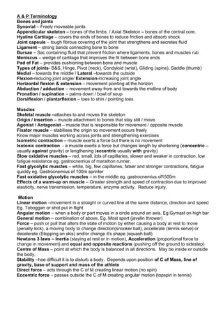
  • 1. A & P Terminology Bones and joints Synovial – Freely moveable joints Appendicular skeleton – bones of the limbs / Axial Skeleton – bones of the central core. Hyaline Cartilage – covers the ends of bones to reduce friction and absorb shock Joint capsule – tough fibrous covering of the joint that strengthens and secretes fluid Ligament – strong bands connecting bone to bone Bursae – Sac containing fluid that prevent friction where ligaments, bones and muscles rub Meniscus – wedge of cartilage that improves the fit between bone ends Pad of Fat – provides cushioning between bone and muscle Types of joints- B&S, Hinge, Pivot (neck), Condyloid (wrist), Gliding (spine), Saddle (thumb) Medial – towards the middle / Lateral –towards the outside Flexion-reducing joint angle/ Extension-increasing joint angle Horizontal flexion & extension – movement pointing at the horizon Abduction / adduction – movement away from and towards the midline of body Pronation / supination – palms down / bowl of soup Dorsiflexion / plantarflexion – toes to shin / pointing toes Muscles Skeletal muscle –attaches to and moves the skeleton Origin / insertion – muscle attachment to bones that stay still / move Agonist / Antagonist – muscle that is responsible for movement / opposite muscle Fixator muscle – stabilises the origin so movement occurs freely Know major muscles working across joints and strengthening exercises Isometric contraction – muscle exerts a force but there is no movement Isotonic contraction - a muscle exerts a force but changes length by shortening (concentric – usually against gravity) or lengthening (eccentric usually with gravity) Slow oxidative muscles – red, small, lots of capillaries, slower and weaker in contraction, low fatigue resistance eg. gastrocnemius of marathon runner. Fast glycolytic muscles – white, big, few capillaries, fatser and stronger contractions, fatigue quickly eg. Gastrocnemius of 100m sprinter Fast oxidative glycolytic muscles – in the middle eg. gastrocnemius of1500m Effects of a warm-up on muscle – Greater strength and speed of contraction due to improved elasticity, nerve transmission, temperature, enzyme activity. Reduce injury. Motion Linear motion –movement in a straight or curved line at the same distance, direction and speed Eg. Toboggan or shot put in flight Angular motion – when a body or part moves in a circle around an axis. Eg.Gymast on high bar General motion – combination of above. Eg. Most sport (javelin thrower) Force – push or pull that alters the state of motion by either causing a body at rest to move (penalty kick), a moving body to change direction(snooker ball), accelerate (tennis serve) or decelerate (Stopping on skis) and/or change it’s shape (squash ball) Newtons 3 laws – Inertia (staying at rest or in motion), Acceleration (proportional force to change in movement) and equal and opposite reactions (pushing off the ground to sidestep) Centre of Mass – point at which the body is balanced in all directions. May be inside or outside the body. Stability –hoe difficult it is to disturb a body. Depends upon position of C of Mass, line of gravity, base of support and mass of the athlete Direct force – acts through the C of M creating linear motion (no spin) Eccentric force – passes outside the C of M creating angular motion (topspin in tennis)
  • 2. Cardiovascular system Aerobic / anaerobic – exercise that takes place with or without oxygen Aerobic exercise involves the interaction of respiratory, heart and vascular systems Structures in the heart – atria, ventricles, valves, septum, veins and arteries entering/leaving Pulmonary – linked to the lungs Conduction system – myogenic electrical impulse that stimulated the heart to contract. SA node (R atrium)-delay-AV node, bundle of His, left and right branches, Purkinje fibres (ventricles) Cardiac cycle – corresponding actions of diastole (relaxation) and systole (contraction) that make up one heart beat. Happens because of the conduction system. Bradycardia – a resting HR below 60. Hypertrophy – increase in size of muscle ( can be heart or skeletal) Cardiac output – the volume of blood ejected from the ventricles in one minute Stroke volume – blood ejected from the ventricles in one beat Max Heart rate = 220 –age, Q=SV x HR Know resting and maximal values for SV, HR, Q Max SV is limited by HR. SV is limited by venous return (blood flow back to the heart) Starling’s Law. This may be effected by exercise position. HR graphs for maximal and submaximal exercise Oxygen debt – additional oxygen required during recovery above that usually required at rest CCC – medula oblongata, regulates HR by monitoring the receptors (sense organs that monitor the body) and increase SA node firing (sympathetic nerve) or decrease firing (para sympathetic) Neural control – proprioceptors (movement), chemoreceptors ( CO2, O2, pH in blood), Baroreceptors (stretch in blood vessel walls) Hormonal control – adrenalin monitoring Intrinsic control – temperature and venous return monitoring Blood supply Pulmonary and systemic circulation – blood flow to and from the heart, lungs and body Arteries / Arterioles – O2 rich blood away from heart, large muscle wall (more pressure), Vasodilate and vasoconstrict, Pre-capillary sphincters control blood flow into capillaries Veins / venuoles – de-oxygenated blood back to heart, large lumen and thin walls, pocket valves direct blood flow Venous return – is the transport of blood through the veins back to the heart, Starling’s law states that VR effects SV and hence Q so 5 mechanisms work to maintain venous return: Pocket valves – one-way valves prevent back flow, muscle pump – skeletal muscle contracts near veins helping to squeeze blood, respiratory pump- large veins in the abdomen being squeezed by deeper/faster breathing, smooth muscle – muscles in vein walls contract, Gravity – blood from upper body is pushed by gravity. Link with active cool-down. Blood pooling – heavy legs caused by blood sitting in veins after exercise with no cool-down. Q at rest vs exercise- rest 15% muscles, 85% organs, exercise 85% muscles, 15% organs. Redistributing Q = vascular shunt. Brain gets the same amount, skin gets more in heavier work (heat loss). VCC - chemoreceptors ( CO2, O2, pH in blood), Baroreceptors (stretch in blood vessel walls) tell VCC to open arterial and venous walls and pre-capillary sphincters to open during exercise. O2 carried in blood by haemoglobin (97%) as oxyhaemoglobin, and blood plasma (3%). CO2 carried in blood by RBC as Carbonic acid (70%) (acid in blood?) , haemoglobin as carbaminohaemoglobin (23%) and plasma (7%). Warm-up benefits on vascular system- vascular shunt (vaso constriction/dilation in muscles and organs), Increase temp increases O2 dissociation (enzymes), decreases blood viscosity and decrease OBLA (onset of blood lactic acid) Cooldown benefits on VS – maintains respiratory/muscle pumps, blood pressure and removes lactic acid.
  • 3. Respiratory system 1. Pulmonary ventilation- getting air in/out of lungs Lung structure- lobes, bronchii, bronchioles, alveoli (sacs). Alveoli gas exchange by large surface area, thin cell layer, lots of blood capillaries and moist lining. Pleura attach lungs to ribs allowing for movement. Mechanics – muscles (diaphragm, intercostals) create movement (ribs and sternum) changing thoracic cavity changes (bigger IN, smaller OUT) changing lung air pressure (low IN, high OUT) and movement of air. More muscles and force cause greater changes during exercise. Know table Page 95 and values P96 VE (minute ventilation) = TV (tidal volume) x f (frequency). This and ventilation response to exercise (p 97) is very similar to the heart. Inspiratory and expiratory reserves – maximum capacities after a normal breath Know the graph of a respiratory trace 2. External respiration – exchange of O2 and CO2 in lungs and blood Partial pressure – pressure of a gas within a mixture of gases PPO2, PPCO2 Gases always move from areas of high to low pressure so O2 is drawn out of the air in CO2 forced back into it as we breathe. PPO2 – atmosphere 159, alveoli 100, arteries 100, muscles (rest) 40, muscles (exercise) <5. PPCO2 – atmosphere 0.3, alveoli 40, veins (rest) 46, muscles (rest) 46, muscles (ex) 80? Diffusion gradients – (difference between high and low pressure) become greater during exercise – hence more gas is exchanged 3. Internal respiration – exchange of O2 and CO2 in blood and muscle tissue. Haemoglobin transfer O2 to myoglobin for use in mitochondria as energy production Oxygen-haemoglobin dissociation curve – more gas is exchanged during exercise (the graph or S curve moves to the right) because of an increase in Temp, increase in acidity (pH Bohr effect), decrease in PPO2, increase in PPCO2. RCC - proprioceptors (movement), chemoreceptors ( CO2, O2, pH in blood), Baro (lung stretch) thermoreceptors monitor changes and change inspiration / expiration accordingly. Hering-breuer reflex – safety mechanism to stop over inflation of lungs Altitude effects respiration by lower PPO2 in air effecting diffusion gradients and causing fatigue and hyperventilation, colder air increases water loss – dehydration. Long term training increases Haemoglobin levels and RBC production increasing Ext. Resp. and O2 transport