

Recomendados
Recomendados
Más contenido relacionado
La actualidad más candente
La actualidad más candente (20)
Destacado
Destacado (20)
Similar a 23204952
Similar a 23204952 (20)
Más de radgirl
Último
Último (20)
23204952
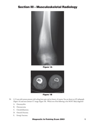
- 1. Section III – Musculoskeletal Radiology Figure 1A Figure 1B 90. A 15-year-old woman presents with aching knee pain and no history of trauma. You are shown an AP radiograph (Figure 1A) and non-contrast CT image (Figure 1B). Which one of the following is the MOST likely diagnosis? A. Osteomyelitis B. Osteosarcoma C. Chondroblastoma D. Osteoid Osteoma E. Ewing’s Sarcoma Diagnostic In-Training Exam 2003 1
- 2. Section III – Musculoskeletal Radiology Question #90 Findings: The standing AP radiograph of the femur shows a very dense but smooth and benign appearing periosteal reaction at the medial aspect of the distal femoral Meta diaphysis. No destructive or aggressive features are present on the image. The single axial, non-contrast CT image shows the dense reactive bone involving the medial and posterior cortex as well as a small lucent nidus with central calcification within the cortex itself. There is no soft tissue mass or any disruption of soft tissue planes on the CT image. Rationales: A) Incorrect. The pattern of osteomyelitis depends on the age of the patient and the mode of infection. In infants, perforating vessels cross the open growth plate and hematogenously spread infection can extend to the epiphysis. In childhood and early adolescence, those perforating vessels regress and there is not communication across the open growth plate and infection more commonly involves the metaphysis or Meta diaphysis. As the growth plate closes, there are again patent vessels, which allow communication between the metaphysis and epiphysis, but it is far more common for adults to acquire osteomyelitis from a source of infection in the contiguous soft tissues rather than from a hematogenous source. Radiographically, osteomyelitis usually presents as lucency in the bone. Depending on the infectious agent and the chronicity of infection, there may be more or less reactive bone near the lesion. With subacute or chronic infection, well-defined intraosseous abscesses with a sclerotic margin may develop (Brodie’s abscess) and sinus tracts may be seen. Bony sequestra, which are intracortical pieces of necrotic bone surrounded by granulation tissue, may also be seen. In the case of bone infection from an adjacent soft tissue source, the key diagnostic feature distinguishing osteomyelitis from reactive change to cellulitis is cortical destruction. In the early phases of infection, subtle changes in the bone may be imperceptible radiographically. MRI is both more sensitive and more specific for the bone changes in addition to identifying the accompanying soft tissue abnormalities. The specificity of MRI may be improved by the addition of nuclear medicine studies using labeled white cells to localize infection. In our case, the calcified nidus might possibly be taken for a small sequestrum and osteomyelitis is a differential diagnostic consideration here. However, the growth plate in this adolescent girl is closed, making the metaphysis an unlikely location for infection in this patient and the dense, benign reactive bone would be atypical for even the most chronic infection. B) Incorrect. Osteosarcoma is second only to myeloma in frequency as a primary malignancy of bone. Commonly presenting in the second and third decades, approximately 50-75% of the most common variant, conventional osteosarcoma, occurs about the knee. The tumor typically has an aggressive appearance, with destruction of the underlying bone and variable production of malignant appearing osteoid. Radiographically osteosarcoma is an ill-defined, destructive intramedullary, metaphyseal lesion with an associated soft tissue mass. Due to rapid growth, there is commonly “sunburst” periostitis or Codman’s triangles. X –rays are preferred for the initial diagnosis, but MR is superior to CT in the evaluation of the intra and extraosseous extent of disease. The metaphyseal location and age of the patient in question would be appropriate for osteosarcoma, but the smooth, benign reactive bone and lack of any destructive changes or a soft tissue mass are consistent with a non- aggressive process. C) Incorrect. Chondroblastoma is a rare, benign primary tumor of bone that most commonly seen in the second decade. It typically presents as a lytic lesion in the epiphysis or apophysis of a long bone with a well-defined sclerotic margin. Matrix calcification is present in up to 50%. MRI may show edema in the surrounding bone and soft tissues due to the prostaglandins secreted by the tumor and clinically they may mimic osteoid osteoma. Benign periostitis or joint effusions may be seen, most commonly when the lesion is located in the capital femoral epiphysis within the hip joint capsule. The lesion may appear expansile, most commonly when a secondary aneurysmal bone cyst is coexistent. Rarely chondroblastoma may metastasize to the lungs. Treatment is usually curettage with bone grafting with image-guided radiofrequency ablation being used at some centers. Although the age of the patient in the index case would be appropriate for chondroblastoma, the location in the Meta diaphysis of the femur and the large amount of reactive bone would not be. 2 American College of Radiology
- 3. Section III – Musculoskeletal Radiology D) Correct. Osteoid osteoma is a benign bone-forming neoplasm consisting of a central core of vascularized osteoid surrounded by densely sclerotic bone. The clinical history is often suggestive, with pain, which is worse at night and relieved by prostaglandin inhibiting agents such as aspirin. Age at presentation is usually in the second or third decade. Lesions in long bones are commonly cortically based where they typically present as lucency, the “nidus”, which may or may not contain calcification. The nidus is usually located at the center of the reactive sclerotic bone. In the small bones of the hands and feet, the lesions tend to be intramedullary with an associated periosteal reaction. Subperiosteal lesions can be seen and may have less prominent reactive changes. The most common location is in the long tubular bones, typically in the diaphysis or metaphysis. Vertebral lesions, often associated with a painful scoliosis, are usually located in the posterior elements. MR imaging of osteoid osteoma shows edema in the bone and soft tissues, which may be deceptively aggressive in appearance. Scintigraphy has been used in the past for its high sensitivity but it remains low in specificity. Plain films usually show the benign reactive bone. High resolution CT is best for showing the nidus itself. CT may also be used for pre-operative localization or definitive treatment with radiofrequency ablation. Our case shows the classic appearance of osteoid osteoma with the dense but benign reactive bone with a subtle lucency on x-ray. The CT demonstrates a well-defined nidus with central calcification. This case was subsequently successfully treated with CT-guided radiofrequency ablation with complete resolution of symptoms. E) Incorrect. Ewing’s Sarcoma is a primary malignancy of bone that chiefly affects young children, often under the age of 10. It most commonly affects the femur and in general is more common in the lower part of the body. In long bones, the Meta diaphysis or diaphysis are typical locations in the bone. Radiographically the lesion is primarily lytic and may have a permeative appearance that may be mistaken for infection. A malignant periosteal reaction is present which may appear laminated (“onion-skin” pattern) and a large soft tissue mass is usually seen. The lesion appears central in the bone reflecting its origin from bone marrow. Ewing’s Sarcoma often presents with constitutional symptoms such as fever, which may delay diagnosis. Scintigraphy is sensitive but non-specific. Radiographs are the usual modality for primary diagnosis with MRI showing the extent of disease within the bone marrow and any associated soft tissue mass. MRI is also often used to monitor response to treatment with chemotherapy. CT is especially helpful when flat bones such as the pelvis or skull are involved. In our case, the lesion is cortically based with a very benign and dense periosteal reaction. This would not be consistent with a malignant process. The CT scan shows no soft tissue mass or disruption of soft tissue planes. Citations: Torriani M, Rosenthal DL. Percutaneous radiofrequency ablation of osteoid osteoma. Pediatric Radiology 32(8): 615-8, 2002. Ho AC, Horton KM, McCarthy EF, Fishman EK. The role of imaging in the diagnosis and management of osteoid osteoma: a pictorial review. Critical Reviews in Diagnostic Imaging 42(6): 357-77, 2001. Diagnostic In-Training Exam 2003 3
- 4. Section III – Musculoskeletal Radiology Figure 2 91. You are shown an axial T2-weighted, fat suppressed MR image in a 25-year-old woman who presents with pain after kicking the ball while playing soccer (Figure 2). Which one of the following is the MOST likely diagnosis? A. Hamstring avulsion B. Insufficiency fracture C. Adductor strain D. Gluteal myositis E. Denervation injury 4 American College of Radiology
- 5. Section III – Musculoskeletal Radiology Question #91 Findings: The image presented is a fat suppressed, T2 weighted axial MR image of the lower pelvis at the level of the ischial tuberosities. The image shows a focal area of increased T2 signal between the hamstring tendons on the right and the tip of the ischial tuberosity. The marrow signal in both inferior pubic rami is normal, as is the signal in the musculature. Rationales: A) Correct. Hamstring injuries are commonly seen in athletes such as hurdlers or those who participate in sports with powerful kicking. In children, these injuries are often associated with avulsions of the ischial apophysis; in young adults, tendon avulsions without underlying fractures may be seen. Radiographs are often normal in the absence of an associated fracture. MRI will show the relationship of the tendons to their attachment, associated muscle injuries, and the presence or absence of osseous pathology. In our case, the T2 weighted fat suppressed axial image shows high signal fluid between the hamstring tendons and the bone, with no such separation on the contralateral normal side. The inferior pubic rami are normal on both sides with no evidence for fracture. The muscles themselves are also normal. There is some fluid surrounding the right sciatic nerve, which is otherwise normal, explaining why this patient may present with complaints of sciatica due to irritation of the nerve related to its proximity to the tendon avulsion. These injuries are most often treated conservatively with the exception of apophyseal avulsions in childhood, which may require fixation. B) Incorrect. Insufficiency fractures are the result of normal stresses on bone that has lost its normal elastic resistance. The pelvis is a common location for these fractures, which are usually seen, in elderly, osteoporotic women. In particular, the inferior and superior pubic rami are often affected, with a subgroup of these patients having avulsion insufficiency fractures of the ischial tuberosity. In this case, the most common presentation is sciatica due to irritation of the nearby sciatic nerve. For this reason, diagnosis may be delayed as the potential for lumbar spine pathology is evaluated. These fractures are usually visible on radiography. In the very acute phase, non-displaced fractures may be difficult to identify. Scintigraphy is very sensitive but lacks specificity and anatomic resolution. MRI will show the edema and any associated tendon or muscle injuries. CT will best display the fracture, and is particularly helpful in excluding pathologic fractures in the sacrum. In our case, the inferior pubic rami are well seen and normal bilaterally, excluding the possibility of acute or subacute fracture. C) Incorrect. The adductor muscle group includes the adductor magnus, brevis and longus as well as the gracilis, pectineus and Sartorious muscles. These muscles principally take their origin from the pubic ramus and are located in the medial thigh, primarily acting to abduct the thigh although individual muscles in this group contribute to actions such as hip flexion and extension. Muscle injuries in general may be divided into contusions or strains, partial tears and complete tears or lacerations. Radiography is usually normal, but will show associated osseous injuries. MRI is the preferred imaging modality for evaluating muscle injuries. A muscle contusion or strain will appear as an intact muscle with increased T2 signal suggesting edema. Partial or complete tears can also be identified. In the acute phase, MRI may show hemorrhage at the site of injury. In the image shown for this question, some of the upper adductor musculature is visible, notably the adductor magnus and brevis. These muscles are entirely normal in their signal characteristics and morphology with no edema or loss of muscle bulk, effectively excluding a significant muscle injury. D) Incorrect. Myositis is a non-specific term indicating inflammation within a muscle. Etiologies include bacterial, viral, and parasitic infections, collagen vascular diseases such as SLE and even drug toxicities. Pyomyositis is a distinct entity, which is often related to staphylococcal infection. The imaging appearance of myositis is also non-specific. On MRI, muscles may have increased T2 signal and there may be loss of distinction between tissue planes on T1 weighted sequences. Contrast-enhanced studies, using CT or MRI, will show any associated abscesses such as those seen with pyogenic infections. Accurate diagnosis requires aspiration and culture of the recovered material. Depending on the location, ultrasound or CT can be used for imaged guided aspiration or drain placements. In our case, all of the muscles, including the gluteus group, are normal with no bright T2 signal except at the sight of the tendon avulsion, making myositis an extremely unlikely diagnosis in this example. Diagnostic In-Training Exam 2003 5
- 6. Section III – Musculoskeletal Radiology E) Incorrect. Denervation injury to muscle can be the result of acute or chronic trauma to a nerve or other processes such as inflammatory neuropathies. Compressive neuropathies such as the anterior interosseous nerve syndrome or carpal tunnel syndrome also fall into this disease category. Initial imaging findings may be negative despite positive clinical examinations or studies such as EMG. As the disease progresses, the muscles that are innervated by the affected nerve may show some mild increased T2 signal. As the disease becomes chronic, there is often loss of muscle bulk and fatty replacement. In the case of large peripheral nerves such as the sciatic or median nerves, the abnormality in the nerve itself may be seen as increased size and T2 signal. In the case of smaller nerves, which cannot be easily resolved on imaging studies, the key to diagnosis is recognizing the pattern of muscle involvement relating to a specific nerve. In our case, the muscles are normal and even though there is some fluid surrounding the right sciatic nerve, the nerve itself is normal and symmetric with the opposite side. Citations: Brandser EA, el-Khoury GY, Kathol MH, Callaghan JJ, Tearse DS. Hamstring injuries: radiographic, conventional tomographic, CT, and MR imaging characteristics. Radiology 197(1): 257-62, 1995. 6 American College of Radiology
- 7. Section III – Musculoskeletal Radiology Figure 3A Figure 3B 92. You are shown PA (Figure 3A) and oblique (Figure 3B) radiographs of the hand in a 40-year-old woman with hand pain. Which one of the following is the MOST likely diagnosis? A. Gout B. Systemic Lupus Erythematosus C. Erosive osteoarthritis D. Scleroderma E. Rheumatoid arthritis Diagnostic In-Training Exam 2003 7
- 8. Section III – Musculoskeletal Radiology Question #92 Findings: PA and oblique radiographs of the hand show diffusely decreased bone density in this 40-year-old woman. On the oblique view, there are multiple subluxed MCP and IP joints with dislocation of the 5th PIP joint on the oblique image with near complete reduction on the PA view. No erosions or productive changes are seen. Rationales: A) Incorrect. Gout is a crystalline deposition disease. Both primary and secondary gout are related to hyperuricemia, with the primary form representing an inborn error of metabolism and secondary gout arising from altered uric acid metabolism associated with other clinical disorders. Primary or idiopathic gout is much more common in men and typically presents in the fifth decade. It often begins as a monoarticular or oligoarticular disorder, progressing to involve more joints over time. The most commonly involved joint is the first metatarsophalangeal joint, which is, altered 75-100% of patients with gout. Most patients with biochemical and clinical evidence of gout will not have bone changes due to early treatment of the metabolic disorder. In patients who do manifest radiographic changes, the findings are usually classic and diagnostic of gout. The joint space is well preserved, with erosions in intra-articular and para-articular locations. These erosions are well defined with sclerotic margins and overhanging edges in up to 40%. The overhanging edges may be associated with gouty tophi in the adjacent soft tissues, which often show increased density or even calcification on x-rays. Bone density is usually preserved. The distribution in the skeleton is typical, with the feet most commonly involved. Hand and wrist, the knee and elbow are also usual sites. Cross sectional imaging is rarely contributory in the evaluation of gout. MRI may be helpful in early gout to identify synovitis and early erosions as well as the extent of soft tissue involvement. Gout would not be an appropriate consideration in the case in question. There are no erosions or other destructive changes present, effectively excluding a radiographic diagnosis of gout. B) Correct. Systemic lupus erythematosus is an autoimmune connective tissue disorder affecting multiple organ systems. In the musculoskeletal system, common manifestations are a deforming, symmetric polyarthritis, myositis, tendon weakening, rupture, and osteonecrosis. As with most collagen vascular diseases, adult women are most commonly affected. The hallmark of the arthritis associated with SLE is deformity without destruction. The small joints of the hand are characteristically involved with multiple subluxed or even dislocated joints, which are easily and usually completely reducible. In fact, the positioning of the patient for the PA radiograph of the hand may itself reduce the subluxations making the disease less prominent. A relaxed oblique or “ball- catcher’s” view often shows the subluxations to better advantage as in our case. Periarticular osteopenia is common and reminiscent of rheumatoid arthritis, but the complete lack of any erosive changes should help to distinguish these entities. Osteonecrosis may be present in the form of avascular necrosis, bone infarcts, or both but this usually involves long bones rather than the small bones of the hands. Jacoud’s arthropathy, a sequela of rheumatic fever, has an appearance that is identical radiographically to SLE. Fortunately, this has become very rare in the era of antibiotic treatment of streptococcal infections, limiting differential diagnostic considerations. C) Incorrect. Erosive osteoarthritis is an inflammatory variant of osteoarthritis, which is characterized by a combination of erosive and productive changes typically in the DIP joints of the hands. This disease usually affects middle-aged women. While other changes of degenerative osteoarthritis may be present within the other joints of the hand and wrist, the DIP involvement is usually strikingly worse. The primary differential considerations are other types of inflammatory arthritis, including psoriatic arthritis, Reiter’s syndrome, and even metabolic disorders such as hyperparathyroidism. What distinguish erosive osteoarthritis from these entities are the distribution of the radiographic changes and the lack of associated systemic disease. Psoriatic arthritis may affect women in the same age group. This is usually not confined to the DIP joints and the pattern of erosive changes is different. Reiter’s syndrome usually affects younger men and more typically involves the lower extremities. Rheumatoid arthritis spares the DIP joints in nearly all cases. Other forms of inflammatory and erosive arthritis also have typical radiographic patterns of disease. In our case, the complete lack of any erosive or productive changes in the interphalangeal joints excludes erosive osteoarthritis as a possibility. 8 American College of Radiology
- 9. Section III – Musculoskeletal Radiology D) Incorrect. Scleroderma, or progressive systemic sclerosis, is a rare disorder of connective tissue affecting multiple organ systems most commonly affecting women in the third to fifth decades. In the musculoskeletal system, the hands are the most common sites of involvement, with changes most pronounced in the digits. Progressive atrophy of the soft tissues at the tips of the fingers creates a characteristically conical appearance to the finger. Progressive erosion and resorption of the ungual tuft of the fingers is also commonly seen with amorphous calcifications seen in the soft tissues. With time, the more proximal bones of the fingers may be involved as well with further resorption giving a “pencil” appearance to the digit. The feet are usually not affected to the same degree as the hands. Other sites where bony changes can be seen include the ribs, spine, and mandible. Soft tissue calcifications often are more diffuse and periarticular tumoral calcinosis may be seen. In the index case, the bones of fingers are normal except for their alignment and no soft tissue resorption or calcifications are seen. E) Incorrect. Rheumatoid arthritis is a relatively common inflammatory arthritis affecting synovial joints, bursae, and tendon sheaths. The primary process is one of synovial inflammation with secondary affects on the underlying bone and cartilage. The small joints of the hands are commonly involved as are the cervical spine, feet and other sites. In the early stages of disease the primary finding may be soft tissue swelling over the MCP joints and ulnar styloid representing synovitis. Periarticular osteopenia may also be present early on reflecting hyperemia at the inflamed joints. With progression, erosions are seen in characteristic locations such as the ulnar styloid and MCP joints but no productive changes are present. The DIP joints are uniformly spared. As the inflamed synovium destroys ligaments and tendons, subluxations such as the swan’s neck deformity become common. Although the subluxations in our case could be seen in rheumatoid arthritis, the complete lack of any destructive changes or soft tissue swelling makes this much less likely. Citations: Brower A, Flemming DJ, Bralow L. Arthritis in Black and White. 2cd edition, W.B. Saunders, Philadelphia, 1997. Diagnostic In-Training Exam 2003 9
- 10. Section III – Musculoskeletal Radiology Figure 4A Figure 4B 93. A 45-year-old man presents with ankle pain after playing basketball. You are shown sagittal T1 (Figure 4A) and T2 and sagittal-T2 (Figure 4B) weighted MR images of the ankle. Which one of the following is the most likely diagnosis? A. Pilon fracture B. Achilles tenosynovitis C. Achilles tendon rupture D. Os trigonum syndrome E. Calcaneal stress fracture 10 American College of Radiology
- 11. Section III – Musculoskeletal Radiology Question #93 Findings: Sagittal T1 and fat suppressed T2 weighted MR images of the ankle show disruption of the Achilles tendon with retraction of several centimeters. The visualized distal tendon is thickened with increased intra substance signal. There is increased T2 signal in the gap and the adjacent soft tissues. A small ankle effusion is present. Osseous structures are normal. Rationales: A) Incorrect. Pilon fractures of the ankle result from pronation-dorsiflexion injuries, which drive the talar dome into the tibial plafond. Included in this complex are an oblique fracture of the medial malleolus and an intraarticular fracture of the distal tibia, which may have more than one part. A fibular fracture may also be present. These are uncommon fractures, representing less than 0.5% of all ankle fractures. Treatment decisions are based on the degree of comminution of the fracture and the extent of intraarticular involvement and displacement. Radiography and knowledge of the mechanism of injury are usually diagnostic, but CT scanning with multiplanar reformatting may be helpful to assess f small intra-articular fragments and to better quantify the degree of displacement and articular incongruence. MRI is generally not contributory to management in the acute phase. In our case, the MRI of the ankle shows a normal tibia and talar dome, excluding fracture from the diagnosis. B) Incorrect. The Achilles tendon is the largest tendon in the body. It represents the confluence of the gastrocnemius and soleus tendons, inserting on the posterior calcaneus. In adults, the Achilles tendon is approximately 10-15cm long. Unique among the tendons of the ankle, the Achilles tendon does not have a synovial-lined tendon sheath, so that tenosynovitis involving the Achilles tendon is not a possibility. Rather, it is covered by a peritenon. Peri- or para tendonitis may be seen within the surrounding soft tissues, often associated with tendinoplasty or partial tears of the tendon itself. Retrocalcaneal bursitis may also be seen just anterior to the insertion of the Achilles tendon on the calcaneus. Our case shows the clear discontinuity of the Achilles tendon with surrounding fluid related to the injury, but is not suggestion of a separate inflammatory process. C) Correct. Achilles tendon rupture is most common in men between the ages of 30 and 50. The typical scenario is a “weekend warrior” who participates in a sport such as basketball, which uses sudden, forceful dorsiflexion or push off of the foot. Clinically there is sudden onset of pain and soft tissue swelling with an inability to stand on tiptoe on the affected side. Radiographs are usually obtained to exclude fracture and may show loss of the soft tissue planes surrounding the Achilles tendon or may even suggest disruption and retraction of the tendon itself. MRI definitively shows the disruption as well as its location and the degree of retraction. The most common site of a complete tear is approximately 2-6 cm proximal to the insertion of the tendon on the calcaneus. This site is vulnerable both to partial and complete tears as a relatively avascular portion of the tendon. In addition to trauma in unconditioned individuals, Achilles tendon rupture may be associated with chronic tendinoplasty and partial tears of the tendon, rheumatoid arthritis, SLE, and the use of local or systemic corticosteroids. MRI may show hemorrhage or fluid in the acute phase as well as the discontinuity of the tendon. When the problem is subacute or chronic, as in our case, the MRI shows discontinuity and some mild retraction as well as a thickened Achilles tendon with increased signal in the distal portion due to underlying tendinoplasty. Increased T2 signal is seen surrounding the ruptured tendon consistent with edema and fluid. D) Incorrect. The Os Trigonum or talar compression syndrome is a pain syndrome involving the posterior ankle. The os trigonum is an accessory ossicle just posterior to the talus at the ankle. When fused to the posterior talus it is referred to as Stieda’s process. The flexor hallucis longus (FHL) tendon lies immediately adjacent to the os trigonum. In some cases where the os trigonum is enlarged or is relatively more mobile, irritation of the FHL tendon or the posterior talus itself may be seen, with tenosynovitis of the FHL or even partial tears of the tendon resulting. Radiography will show the enlarged ossicle, which may appear irregular, but MRI is diagnostic, showing the edema surrounding the os trigonum and posterior talus and the associated abnormalities in the FHL tendon. In our case, the posterior talus is normal with no edema and the FHL tendon is not included on the images shown. Diagnostic In-Training Exam 2003 11
- 12. Section III – Musculoskeletal Radiology E) Incorrect. Stress fractures are the result of repetitive loading on bone, which may be normal or abnormal. Fatigue fractures involving normal bone may be due to a novel, strenuous activity, which places repetitive stress on a specific bone; an example would be the march fractures of the metatarsal seen in new military recruits. In the case of abnormal underlying bone, such as osteoporosis, the term insufficiency fracture may be applied. Common sites for insufficiency fractures include the pelvis and calcaneus. In the calcaneus, radiographs show the underlying osteopenia with crescentic area of sclerosis usually in the posterior calcaneus. The sclerosis probably represents a combination of impaction and healing. In the very acute phase, these fractures may be radiographically occult. In this case, MRI would show the reactive edema on T2 weighted imaging as well as the fracture itself, which appears as a linear area of low signal intensity on T1 weighted images. Scintigraphy and MRI have similar sensitivity, but MR is significantly more specific. CT with multiplanar reformatting will also show the fracture before it is radiographically evident. In our case, the calcaneus is normal on both T1 and T2 weighted images with normal marrow signal throughout and no edema to suggest fracture. Citations: Schepsis AA, Jones H, Haas AL. Achilles tendon disorders in athletes. American Journal of Sports Medicine 30(2): 287-305, 2002. Movin T, Kristoffersen-Wiberg M, Rolf C, Aspelin P. MR Imaging in chronic Achilles tendon disorder. Acta Radiologica 39(2): 126-32, 1998. 12 American College of Radiology
- 13. Section III – Musculoskeletal Radiology Figure 5A Figure 5B Figure 5C 94. This 50-year-old man presented with a mass along his right chest wall. You are shown coronal T1 (Figure 5A), and fat-saturated post gadolinium T1 (Figure 5B) weighted images and a non-contrast axial CT scan (Figure 5C) obtained 4 weeks after the MRI. Which one of the following is the MOST likely diagnosis? A. Myositis ossificans B. Malignant fibrous histiocytoma C. Desmoid tumor D. Chondrosarcoma E. Intramuscular myxoma Diagnostic In-Training Exam 2003 13
- 14. Section III – Musculoskeletal Radiology Question #93 Findings: Coronal T1 and fat suppressed T1 post-gadolinium MR images of the right chest wall show a mass within the lateral chest wall, which is isointense to muscle on the non-contrast examination and shows avid enhancement with intravenous gadolinium. A small central area in the mass does not enhance and may be necrotic or cystic. A single axial non-contrast CT image obtained four weeks later shows benign, peripheral calcification within the teres major muscle. There has been no interval change in the size of the mass. Rationales: A) Correct. Myositis Ossificans (MO) is a localized soft tissue calcification most often related to antecedent injury. Up to 75% of patients who develop MO can relate a clear history of trauma to the affected area. Other causes may include burns, neurologic conditions, and systemic disorders associated with soft tissue calcification. Rarely the lesion may develop spontaneously. Common sites of MO include areas exposed to injury such as the buttocks, elbow, thigh, and calf. A soft tissue mass appears at the site of injury within about 10 days, with calcification usually appearing by 6 weeks. The calcification typically forms at the periphery of the soft tissue lesion, forming a smooth, complete border when mature. The peripheral calcification is an important feature distinguishing developing MO from malignancies such as osteosarcoma or chondrosarcoma. If the lesion is imaged with MR early in the process, the appearance can be very worrisome with avid contrast enhancement and features consistent with a soft tissue sarcoma such as a malignant fibrous histiocytoma. Even biopsy may be misleading if obtained from the cellular central portions of the mass. As with imaging, the more mature peripheral zone shows the benign nature of the lesion. Radiography will often show the mature peripheral calcification well enough for diagnostic confidence. If there is any question regarding the nature of the calcification (with an appropriate clinical history, of course) CT can be obtained which will show the benign calcification to best advantage. In our case, the patient was involved in a car accident with an injury to his chest wall approximately two weeks prior to the MRI. The MRI was felt to be worrisome for malignancy, but because of the history of trauma, biopsy was not immediately performed. CT obtained 4 weeks later shows the mature calcification of MO and no further evaluation was required. B) Incorrect. Malignant fibrous histiocytoma (MFH) is the most common soft tissue sarcoma in adults. Rarely (5%) it may arise as a primary bone tumor. MFH presents as a painless soft tissue mass. Radiographs may show the tumor in the soft tissue if large, but the preferred modality for evaluation of soft tissue tumors is MRI. On MRI, the mass will be low in signal intensity on T1 weighted imaging and bright on T2. If contrast is administered, there is usually avid and homogeneous enhancement. In very large tumors there may be non- enhancing areas centrally representing necrosis. MRI best demonstrates the anatomic compartments involved and the relationship of the tumor to neurovascular structures, bones and joints. CT with contrast may be used if the patient is unable to undergo MRI. Ultrasound may be helpful for image-guided biopsy. Imaging is non- specific; high-grade liposarcomas and other types of primary sarcomas and metastases may have similar imaging features. MFH may rarely calcify, with the calcification located within the mass and appearing irregular. If the mass is adjacent to a bone, that bone may undergo pressure erosion. MFH was a consideration when the MRI was obtained in our patient, but because of the history of trauma, biopsy was deferred in favor of a follow-up CT. C) Incorrect. Desmoid tumor is the name given to a benign fibrous proliferation arising in the abdominal and extra-abdominal musculature, often growing slowly and engulfing the surrounding tissue in an insidious fashion. While histologically benign, these tumors commonly recur after excision and are difficult to treat. As with most of the fibromatoses, they are locally aggressive and may cause erosion of adjacent bones with a periosteal reaction sometimes seen. They are usually solitary with a predilection for the shoulder girdle. Radiography may show the soft tissue mass if large enough as well as any effects on adjacent bone, but MRI is the preferred modality for diagnosis, with the dense fibrous tissue having characteristic signal intensity which is low on both T1 and T2 weighted imaging. If intravenous gadolinium is given, there is usually marked enhancement throughout the lesion. Calcifications may be seen but are rare. In our case, the signal intensity of the lesion on the unenhanced T1 image is not as low as would be expected with a desmoid tumor. The post-gadolinium image shows the mass 14 American College of Radiology
- 15. Section III – Musculoskeletal Radiology to be well defined; desmoid tumors often have a less well-defined, more infiltrative appearance. The CT showing peripheral calcification in the mass is diagnostic of myositis ossificans and would be extremely unusual for a desmoid. The combination of history and the CT appearance effectively excludes the diagnosis of a desmoid tumor. D) Incorrect. Chondrosarcomas are tumors of cartilaginous origin, which may occur primarily, or secondary to pre-existing lesions such as an enchondroma or osteochondroma. They may also be categorized according to their location in bone or by their histologic characteristics. Extraskeletal chondrosarcomas are rare. The most common location for chondrosarcoma is in the long tubular bones, with the femur being the most frequently affected bone. They are usually located in the metaphysis but extension to the epiphysis can be seen. Most chondrosarcomas are of low histologic grade. Radiographically, these tumors may show a primarily lytic area with endosteal scalloping and chondroid calcification within the lesion. Cortical thickening may also be seen. Higher grade or dedifferentiated chondrosarcomas will have a more aggressive appearance, with cortical breakthrough and large soft tissue masses. Radiography is the primary diagnostic modality. When the flat bones such as the pelvis and scapula are involved, CT may be preferred both to define the extent of the lesion and to characterize the calcifications. MRI may also be used to evaluate the extent of soft tissue and marrow involvement and to establish which anatomic compartments are involved for surgical planning. In the unusual case of an extraskeletal chondrosarcoma, a soft tissue mass with c chondroid calcification is seen. Our case shows smooth peripheral ossification, making a cartilaginous process extremely unlikely. E) Incorrect. Intramuscular myxoma is an uncommon benign soft tissue mass. As its name would suggest, the mass is located within a muscle, most often in an extremity. Radiography rarely shows these lesions. MRI will show a well-circumscribed mass within a muscle, which is very low in signal intensity on T1, and very bright on T2 weighted images. Myxomas rarely enhance except for a smooth, peripheral rim. These lesions almost never calcify. In our case, although the mass is located within the chest wall musculature, the mass enhances homogeneously which would be inconsistent with a myxoma, as would be the peripheral calcification. Citations: Jepsen MC, Graham SM. Traumatic myositis ossificans of the levator scapulae muscle, American Journal of Otolaryngology 19(5): 345-348, 1998. Resnick, D. “Soft tissues” in Resnick, D. Bone and Joint Imaging, 2cd edition, 1261-1263, W.B. Saunders, Philadelphia, 1996. Diagnostic In-Training Exam 2003 15