SlideShare una empresa de Scribd logo
1 de 33
Descargar para leer sin conexión
Section III – Nuclear Radiology


                                                   Figure 1A


                                                   Figure 1B
58.        You are shown whole body images and selected spot images from a Tc-99m methylene
           diphosphonate bone scintigram obtained in an adult male patient presenting with joint pain
           (Figures 1A and 1B). What is the MOST LIKELY diagnosis?

      A.   Multiple cortical skeletal metastases
      B.   Osteomyelitis
      C.   Shin splints
      D.   Hypertrophic osteoarthropathy

                                                                                                        1
                                 Diagnostic In-Training Exam 2006
Section III – Nuclear Radiology
Question 58
Rationales:
A. Incorrect. Focal cortical metastases are uncommon, but do occur. However, the symmetrical metadi-
   aphyseal distribution of the lesions and involvement of both the proximal and distal appendicular
   skeleton are highly atypical for metastases. The clinical history of joint pain also points to other
   more likely etiologies for the findings present.
B. Incorrect. Osteomyelitis may produce areas of increased cortical tracer uptake, secondary to perios-
   titis associated with the active infectious process. Abnormality is not usually limited to cortical
   areas alone, and is often more intense than the findings in this case. Furthermore, the bilaterally
   symmetrical distribution of the findings is highly atypical for osteomyelitis.
C. Incorrect. Shin splints may also produce areas of increased uptake along the cortical surfaces of
   long bones, as seen in this case. However, the findings in shin splints are usually confined to the tib-
   iae, and most often involve primarily the diaphyseal regions, often with only low-level increased
   uptake. Furthermore, involvement of the upper extremities would not be expected with shin splints.
D. Correct. The findings in this case are characteristic in appearance and location for secondary hyper-
   trophic osteoarthropathy (formerly known as hypertrophic pulmonary osteoarthropathy or HPO).
   This disorder is most often associated with intrathoracic lesions, such as primary lung neoplasms or
   mesothelioma. It can also occur in association with other intrathoracic lesions, including bronchiec-
   tasis, emphysema, lung infections, metastatic disease, etc. It may be seen in patients with congenital
   heart disease, inflammatory bowel disease and lymphoma. Patients may present with joint pain,
   clubbing and periosteal new bone formation on radiographs. The differential diagnosis also includes
   pachydermoperiostosis and thyroid acropachy.


2                               American College of Radiology
Section III – Nuclear Radiology


                                                 Figure 2A
59.        You are shown serial 5-minute anterior images and final right anterior oblique and right lateral
           images from a Tc-99m DISIDA hepatobiliary scan performed on a 55-year-old man with
           abdominal pain, fever and ascites, s/p paracentesis (Figures 2A and 2B). What is the MOST
           LIKELY diagnosis?

      A.   Acute cholecystitis
      B.   Bile leak
      C.   Common bile duct obstruction
      D.   Normal study


3                                  American College of Radiology
Section III – Nuclear Radiology


              Figure 2B


                                       4
    Diagnostic In-Training Exam 2006
Section III – Nuclear Radiology
Question 59
Findings: There is prompt hepatic uptake, with early visualization of activity in the region of the gall-
bladder fossa. Faint, amorphous activity is noted inferior to the liver on the right, beginning at 10 min-
utes and better seen thereafter. In addition, there is accumulation of activity throughout the peritoneal
cavity, beginning at 15 minutes post-injection, and progressively increasing throughout the study. There
is also the appearance of abnormal linear activity along the inferior margin of the left lobe of the liver,
beginning at 25-30 minutes into the study and progressively increasing in intensity. The right lateral
image demonstrates activity spreading anterior to the liver, also consistent with intraperitoneal biliary
leakage. Hepatic clearance is also moderately prolonged.
Rationales:
A. Incorrect. The findings are not consistent with acute cholecystitis. There is prompt visualization of
   the gallbladder as early as 5-10 minute post-injection, which essentially excludes acute cholecysti-
   tis. Furthermore, acute cholecystitis does not explain the presence of biliary leakage present in this
   case. Perforation of the gallbladder may occur in gangrenous cholecystitis, but that entity is virtual-
   ly always associated with cystic duct obstruction, which would result in non-visualization of the
   gallbladder as well.
B. Correct. The findings in this case described above are consistent with a relatively large bile leak,
   most likely arising in the region of the gallbladder fossa. In this case, the findings may be secondary
   to trauma from paracentesis.
C. Incorrect. There is prolonged hepatic clearance and non-visualization of the small bowel, both find-
   ings that occur in the presence of common bile duct obstruction. However, in common duct obstruc-
   tion, there is often complete non-visualization of the biliary tree, including the gallbladder, even in
   the absence of cholecystitis. In addition, common duct obstruction is not usually associated with bil-
   iary leakage, which is present in this case.
D. Incorrect. This study is not normal. A significant degree of biliary leakage is demonstrated, as
   described above. Furthermore, the images also demonstrate prolonged hepatic clearance and non-
   visualization of the small bowel, both of which are also abnormal findings.


5                               American College of Radiology
Section III – Nuclear Radiology


                                                Figure 3
60.        A 2-month-old male with marked hypertension is referred for captopril renography. You are
           shown serial 1-minute posterior pre- and post-captopril images (Figure 3). What is the MOST
           LIKELY diagnosis?

      A.   Normal study
      B.   Right renal artery stenosis
      C.   Left renal artery stenosis
      D.   Bilateral renal artery stenosis


6                                   American College of Radiology
Section III – Nuclear Radiology
Question 60
Findings: The baseline pre-captopril study demonstrates mildly decreased tracer uptake bilaterally, with
normal excretion. The post-captopril images demonstrate significant bilateral deterioration in excretion,
with marked cortical retention noted bilaterally.
Rationales:
A. Incorrect. Although initial (left) study appears symmetrically normal, there is clearly a bilateral
   delay in cortical clearance and excretion on the post-captopril study.
B. Incorrect. In unilateral right renal artery stenosis, ACE-inhibitor should create an asymmetric delay
   in right renal washout, not the bilaterally delayed washout present in this case.
C. Incorrect. In unilateral left renal artery stenosis, ACE-inhibitor should create an asymmetric delay in
   left renal washout, not the bilaterally delayed washout present in this case.
D. Correct. The post-captopril study fails to demonstrate sequential right and left renal pelvis and
   bladder activity seen at midpoint of the baseline pre-captopril study. Administration of the ACE
   inhibitor has produced a symmetric delay in renal cortical clearance, manifested by marked bilateral
   cortical retention and non-visualization of the renal pelves and bladder. These findings are typical
   for bilateral ACE-inhibition of compensatory post-glomerular vascular constriction, with resultant
   delay in transcortical clearance, in this child with bilateral congenital renal artery stenosis.


                                                                                                         7
                              Diagnostic In-Training Exam 2006
Section III – Nuclear Radiology


                                                Figure 4
61.        You are shown representative coronal, transaxial and sagittal images from an F-18 FDG
           (fluorodeoxyglucose) PET scan (Figure 4). What is the MOST LIKELY diagnosis?

      A.   Lymphoma
      B.   Bronchogenic carcinoma
      C.   Esophageal carcinoma
      D.   Normal variant


8                                 American College of Radiology
Section III – Nuclear Radiology
Question 61
Rationales:
A. Incorrect. The abnormal uptake in this case is located in the posterior mediastinum, where adenopa-
   thy due to lymphoma may occur. However, the linear configuration of the activity is characteristic
   of esophageal activity, rather than the typical focal rounded appearance of adenopathy. Furthermore,
   no other sites of adenopathy are present. The findings are characteristic of an esophageal neoplasm,
   making squamous cell carcinoma or adenocarcinoma far more likely than lymphoma.
B. Incorrect. As discussed above, the linear uptake located in the posterior mediastinum is characteris-
   tic in appearance for an esophageal neoplasm. There are no focal pulmonary nodules or foci of
   mediastinal or hilar adenopathy, as would be anticipated in the presence of bronchogenic carcinoma.
C. Correct. The linear pattern of increased FDG uptake in the posterior mediastinum, in the expected
   location of the esophagus, is characteristic in appearance for an esophageal neoplasm, most likely
   representing squamous cell carcinoma of the esophagus.
D. Incorrect. Mildly increased uptake near the gastroesophageal junction may be seen as a normal vari-
   ant, or in patients with gastroesophageal reflux. Mild diffuse esophageal uptake may also occur in
   esophagitis. The uptake in this case is far more intense than would be anticipated as a normal vari-
   ant, and the location of the activity remote from the gastroesophageal junction is not consistent with
   a normal variant.


                                                                                                        9
                             Diagnostic In-Training Exam 2006
Section III – Nuclear Radiology


                                                Figure 5
62.        A 28 year-old HIV positive woman presents with headache, papilledema, and a ring-enhancing
           right thalamic mass on CT (not shown). You are shown a transaxial Tl-201 chloride image of the
           brain (Figure 5). What is the MOST LIKELY diagnosis?

      A.   Lymphoma
      B.   Cytomegalovirus infection
      C.   Toxoplasmosis infection
      D.   Normal study


10                                American College of Radiology
Section III – Nuclear Radiology
Question 62
Findings: Transaxial Tl-201 chloride SPECT images of the brain demonstrate a focal area of increased
tracer uptake near the midline, in the region of the CT lesion in the basal ganglia.
Rationales:
A. Correct. CNS lymphoma may produce a ring-enhancing lesion on CT and is thallium-avid. These
   findings are most consistent with CNS lymphoma arising in an immunocompromised host.
B. Incorrect. CMV is not thallium-avid, as is the lesion in this case.
C. Incorrect. Toxoplasmosis can produce cerebral ring-enhancing CT lesion, but it is not thallium-avid,
   as is the lesion in this case.
D. Incorrect. The focal area of increased tracer uptake in the midline basal ganglia region represents a
   striking abnormality, which is not attributable to any normal finding. This is not a normal study.


                                                                                                      11
                             Diagnostic In-Training Exam 2006
Section III – Nuclear Radiology


                                                 Figure 6
63.        You are shown representative coronal, transaxial and sagittal tomographic radionuclide images
           (Figure 6). Which one of the following radiotracers was MOST LIKELY utilized for
           this study?

      A.   Tc-99m methylene diphosphonate
      B.   Tc-99m sulfur colloid
      C.   F-18 fluorodeoxyglucose
      D.   F-18 sodium fluoride


12                                American College of Radiology
Section III – Nuclear Radiology
Question 63
Rationales:
A. Incorrect. The normal biodistribution of Tc-99m methylene diphosphonate (MDP) includes the axial
   and appendicular skeleton, kidneys and bladder. The liver, spleen, mediastinum, and brain, which
   are visualized in this case, are not seen on a normal bone scintigram.
B. Incorrect. The normal biodistribution of Tc-99m sulfur colloid includes intense liver and spleen
   activity. Less intense activity is identified in the central bone marrow (skull, ribs, sternum, vertebral
   bodies, pelvis, proximal humeri and femora). The most intense activity in this study is osseous.
   Moderate activity is seen within the spleen and low level activity in the liver, mediastinum and
   brain. This biodistribution is not typical for sulfur colloid.
C. Correct. The normal biodistribution of F-18 fluorodeoxyglucose (FDG) is accumulation in the
   brain, myocardium, blood vessels, pharynx, liver, spleen, bone marrow, kidneys, ureters, urinary
   bladder, and GI tract. Intense marrow uptake is seen in this patient with lymphoma after administra-
   tion of granulocyte colony stimulating factor (G-CSF), which is given to support bone marrow func-
   tion following therapy. Normal marrow uptake is usually less intense than hepatic uptake. While this
   distribution is not normal, it is more characteristic of FDG than any of the other tracers listed.
D. Incorrect. The normal biodistribution of F-18 sodium fluoride is osseous, with uptake dependent on
   regional blood flow and osteoblastic activity by chemisorption. Hydroxyl groups are exchanged to
   form fluoroapatite in the hydroxyapatite crystals. Because of the superior spatial resolution and
   three-dimensional localization afforded by PET imaging, there is a high sensitivity for the detection
   of metabolically active skeletal lesions using F-18 sodium fluoride.


                                                                                                         13
                              Diagnostic In-Training Exam 2006
Section III – Nuclear Radiology
64.        Concerning subacute thyroiditis, serum thyroid hormone levels are elevated as the result of
           which one of the following?

      A.   Increased thyroid hormone production
      B.   Increased TSH secretion by the pituitary gland
      C.   Release of pre-formed thyroid hormone into the circulation
      D.   Iodine excess in the thyroid gland

Question 64
Rationales:
A. Incorrect. Thyroid hormone production is reduced in subacute thyroiditis. The elevated thyroid
   function tests and signs and symptoms of hyperthyroidism that occur early in the disorder are relat-
   ed to release of pre-formed thyroid hormone into the circulation from the inflamed thyroid gland.
B. Incorrect. The increased thyroid hormone levels produced by the release of pre-formed hormone
   into the circulation results in a feedback inhibition of TSH secretion by the pituitary, resulting in
   decreased serum TSH levels.
C. Correct. Subacute thyroiditis is a viral disorder, often following a recent upper respiratory infec-
   tion. The inflammatory response in the gland results in increased permeability and increased release
   of pre-formed thyroid hormone into the circulation from the colloid. The increased serum thyroid
   hormone levels, in turn, result in clinical evidence of hyperthyroidism, despite a low thyroid uptake.
D. Incorrect. The pathophysiology of subacute thyroiditis does not involve abnormalties in iodine
   metabolism per se. The acute inflammatory response in this disorder is associated with decreased
   iodide uptake and organification during the early stage of the disease.


14                                 American College of Radiology
Section III – Nuclear Radiology
           Gallium-67 citrate scintigraphy is preferred over In-111 leukocyte scintigraphy in which one of
65.
           the following entities?

      A.   Abdominal abscess
      B.   Infected joint prosthesis
      C.   Disk space infection
      D.   Inflammatory bowel disease

Question 65
Rationales:
A. Incorrect. While both radiopharmaceuticals are efficacious for the detection of abdominal abscesses,
   Indium-111 leukocyte imaging is often preferred, as the result of the absence of potentially confus-
   ing normal bowel activity, as occurs in Gallium-67 scintigraphy. This normal bowel uptake may
   lead to false positive gallium studies.
B. Incorrect. Indium-111 leukocyte imaging is superior to gallium-67 scintigraphy in the evaluation of
   suspected infected joint prostheses, in part related to the bone seeking properties of gallium, leading
   to potential false positive gallium studies due to increased tracer localization secondary to increased
   bone turnover in the absence of infection.
C. Correct. While sensitive for osteomyelitis, Indium-111 leukocyte scintigraphy has been found to be
   less sensitive than gallium-67 scintigraphy for the detection of disc space infection.
D. Incorrect. Again, the absence of normal bowel localization makes In-111 leukocyte scintigraphy
   better suited to the assessment of active inflammatory bowel disease. In gallium-67 scintigraphy,
   normal bowel uptake, especially in the colon, can be incorrectly attributed to inflammatory bowel
   disease.


                                                                                                        15
                                 Diagnostic In-Training Exam 2006
Section III – Nuclear Radiology
66.        Concerning the presence of multiple focal “hot spots” on a Tc-99m macroaggregated albumin
           (MAA) scan, which one of the following is CORRECT?

      A.   The study may need to be repeated on another day.
      B.   The patient is at risk for the development of acute hypoxemia.
      C.   A false positive study will result.
      D.   The patient has multiple arteriovenous malformations (AVMs).

Question 66
Rationales:
A. Correct. The finding of focal “hot spots” on a Tc-99m MAA scan indicates the aggregation of the
   radiopharmaceutical into larger particles, which lodge in the pulmonary vascular bed. This artifact
   may be produced by drawing blood back into the syringe during injection or by failing to resuspend
   the particles prior to injection, in the event the dose is left sitting for a prolonged time after being
   drawn up. While it is not associated with any adverse effects in the patient, these foci of increased
   activity may obscure portions of the underlying lungs, resulting in the need to repeat the study after
   significant radioactive decay has occurred.
B. Incorrect. While technically these foci do represent small, iatrogenic pulmonary emboli, they are
   virtually never associated with any clinically demonstrable adverse effects. In general, pulmonary
   perfusion imaging with Tc-99m MAA is associated with transient occlusion of less the 0.1% of the
   pulmonary capillary bed. Thus, this occurrence is unlikely to produce acute hypoxemia.
C. Incorrect. While these “hot spots” may obscure underlying detail in evaluating pulmonary perfu-
   sion, they are not associated with artifactual perfusion defects that would produce a false positive
   study.
D. Incorrect. Pulmonary AVMs are associated with right to left shunting, permitting Tc-99m MAA par-
   ticles to bypass the pulmonary capillary bed. Thus, AVMs would tend to produce focal perfusion
   defects, rather than focal areas of increased tracer localization.


16                                American College of Radiology
Section III – Nuclear Radiology
           Concerning the analysis of radionuclide gated blood pool (MUGA) studies, which one of the
67.
           following will result in an UNDERESTIMATION of the left ventricular ejection fraction?

      A.   Placement of the background region of interest over the splenic blood pool activity
      B.   Assignment of too small a systolic region of interest
      C.   Use of a single region of interest for both the systolic and diastolic frames
      D.   Inclusion of a portion of the left atrium in the diastolic region of interest

Question 67
Distractors: SCORE ALL CHOICES AS CORRECT
A. Placement of the background region of interest over the splenic blood pool activity
B. Assignment of too small a systolic region of interest
C. Use of a single region of interest for both the systolic and diastolic frames
D. Inclusion of a portion of the left atrium in the diastolic region of interest


Rationales:
A. Placement of the background ROI over the spleen will result in excessive background subtraction.
   The relative effect of the extra background subtraction will be greater on the systolic ROI, which
   has fewer counts, and therefore will not “cancel out”. Thus, the denominator of the ejection fraction
   equation will be relatively reduced, resulting in an artifactually elevated, rather than reduced.
B. Too small of a systolic region will result in exclusion of value counts from the systolic region, mak-
   ing the percentage change between systolic and diastolic counts appear to be larger than it actually
   is. Again, this error would result in an artifactually elevated calculated ejection fraction.
C. This technique was initially used when the procedure was originally introduced. However, by using
   the same region for both measurements, the systolic region extends beyond the actual margins of
   the ventricle in end-systole, thus including background counts from adjacent structures, making the
   apparent ejection fraction artifactually too low. For this reason, the standard method of analysis at
   present requires assignment of separate diastolic and systolic regions of interest, in order to improve
   the accuracy of the measurement.
D. Inclusion of a portion of the left atrium in the diastolic region of interest will have the effect of
   introducing additional counts into the region that are not valid ventricular counts. Thus, the apparent
   total end-diastolic counts will be inappropriately high, resulting in an apparent higher percentage of
   ventricular emptying and a falsely elevated ejection fraction calculation.


                                                                                                       17
                                 Diagnostic In-Training Exam 2006
Section III – Nuclear Radiology
           Concerning radionuclide myocardial perfusion imaging, which one of the following is NOT
68.
           associated with an inferior wall perfusion defect on a stress Tc-99m sestamibi SPECT study?

      A.   Inferior wall exercise-induced ischemia
      B.   Prior inferior wall myocardial infarction
      C.   Left bundle branch block
      D.   Diaphragmatic attenuation artifact

Question 68
Rationales:
A. Incorrect. Inferior wall ischemia characteristically produces a perfusion defect in this region on
   stress myocardial perfusion images. In the case of reversible ischemia, the defect would be expected
   to resolve on a resting study.
B. Incorrect. An area of prior myocardial infarction typically produces a “fixed” perfusion defect,
   which would be visible both on stress and resting images. Thus, from evaluation of stress images
   alone, it cannot be differentiated from a defect due to exercise-induced ischemia, as in item A.
C. Correct. Left bundle branch block may be the result of myocardial ischemia or infarction, or may
   be an incidental finding. It may produce perfusion abnormalities on myocardial perfusion scintigra-
   phy in the absence of coronary artery disease. When it produces abnormalities, the most common
   finding is a reversible perfusion defect in the interventricular septum, not in the inferior wall. In
   patients with known left bundle branch block, it is preferable to perform a pharmacologic stress test,
   using dipyridamole or adenosine, rather than treadmill exercise in conjunction with the imaging,
   since this artifact is more commonly associated with the latter procedure.
D. Incorrect. Diaphragmatic attenuation artifact commonly produces apparent defects in the inferior
   wall. These defects may or may not be present both on stress and resting images, and may be sus-
   pected by inspection of planar rotating images from the raw data set. This artifact most often occurs
   in male patients, and is more common in obese patients as well.


18                                 American College of Radiology
Section III – Nuclear Radiology
69.        Which is an appropriate use of F-18 fluorodeoxyglucose (FDG) PET imaging in breast
           carcinoma?

      A.   Screening
      B.   Initial staging
      C.   Differentiating between a pulmonary metastasis and a primary lung carcinoma
      D.   Treatment monitoring

Question 69
Rationales:
A. Incorrect. FDG PET imaging is not an appropriate or approved study for breast cancer screening.
   Screening is best done by self-examination and periodic mammography, which are more sensitive
   and cost-effective approaches to breast cancer screening.
B. Incorrect. FDG PET imaging is less sensitive for the initial staging of breast cancer than lym-
   phoscintigraphy with sentinel lymph node biopsy. Very high sensitivity is provided by the latter
   approach, particularly when immunohistochemistry techniques are utilized. This approach to staging
   is rapidly becoming the standard of care for these patients.
C. Incorrect. FDG PET imaging is not capable of differentiating between a solitary pulmonary metas-
   tasis and a primary lung tumor. In most cases, both lesions are associated with increased glucose
   metabolism and thus, increased FDG uptake.
D. Correct. As is true for a number of neoplasms, FDG PET imaging is very sensitive and specific for
   assessing the response to therapy in breast carcinoma, whether performed after the completion of
   therapy (re-staging) or during the course of therapy (treatment monitoring).


                                                                                                  19
                                Diagnostic In-Training Exam 2006
Section III – Nuclear Radiology
70.        A post-menopausal woman with osteoporosis undergoes dual-energy x-ray absorptiometry
           (DEXA) scanning, demonstrating marked osteopenia of the lumbar spine and hip, but normal
           bone density of the distal forearm. What is the BEST explanation for these findings?

      A.   Inappropriate scanning of the dominant forearm rather than the non-dominant
      B.   Insensitivity of forearm bone density measurement secondary to preponderance of cortical bone
      C.   Underestimation of the bone density in the spine and hip secondary to arthritic changes
      D.   Scan performed too distally in the forearm

Question 70
Rationales:
A. Incorrect. While it is true that it is preferable to scan the non-dominant forearm or hip in DEXA
   scanning, and scanning the dominant side could produce a higher bone density value, the differ-
   ences between the dominant and non-dominant sides are often minimal, and this is therefore not the
   best explanation for the findings.
B. Correct. The bones of the extremities, such as the radius and ulna, are composed primarily of corti-
   cal bone, and contain relatively less trabecular bone than either the spine or hip. Quantitatively, the
   extremities account for the majority of the whole body bone mineral content. Thus, bone density
   measurements of the forearm are most valuable in patients with metabolic bone disease, or other
   conditions associated with decreases in total skeletal calcium content. Post-menapausal osteoporosis
   preferentially involves the trabecular bone initially, which is present in higher percentages in the
   vertebral bodies and femoral neck regions. Therefore, forearm measurements tend to be relatively
   insensitive for the early detection of post-menopausal osteoporosis.
C. Incorrect. In fact, the opposite is true. The presence of arthritic changes is most often associated
   with falsely elevated bone density measurements, especially in the spine, secondary to increased
   bone density at sites of spurring or sclerosis associated with arthritic involvement.
D. Incorrect. Again, the opposite is true. Moving from proximal to distal in the forearm, there is a pro-
   gressive increase in the relative trabecular bone content. Typically, bone density measurements of
   the forearm are performed in the distal third of the radius and ulna, in order to maximize the contri-
   bution of trabecular bone in the measurement. Scanning more distally may also be performed, to
   further increase the percentage of trabecular bone being evaluated. Thus, scanning more distally
   would tend to decrease the measured bone mineral density of the forearm.


20                                American College of Radiology
Section III – Nuclear Radiology
71.        What is the most commonly cited threshold for the diagnosis of malignancy using standardized
           uptake value (SUV) on PET imaging for a solitary pulmonary nodule?

      A.   1.0
      B.   1.5
      C.   2.5
      D.   3.0

Question 71
Rationales:
A. Incorrect. The correct value is 2.5.
B. Incorrect. The correct value is 2.5.
C. Correct. Many malignant lesions will greatly exceed this value, and some lesions with SUV values
   < 2.5 are malignant, but 2.5 is the most commonly cited threshold for the diagnosis of malignancy
   using SUV analysis.
D. Incorrect. The correct value is 2.5.


                                                                                                     21
                                Diagnostic In-Training Exam 2006
Section III – Nuclear Radiology
         Concerning infection imaging with In-111 labeled leukocytes, which one is CORRECT?
72.

      A. Uptake is dependent on regional blood flow.
      B. It is insensitive for the detection of inflammatory bowel disease.
      C. Transient pulmonary uptake clears within 15 minutes post-injection.
      D. It is more sensitive than Ga-67 citrate imaging for detection of Pneumocystis carinii pneumonia
        (PCP).

Question 72
Rationales:
A. Correct. While not the sole determinant of uptake, the uptake of In-111 labeled leukocytes is
   dependent upon regional blood flow. For example, a walled-off abscess without a direct blood sup-
   ply will not accumulate In-111 labeled leukocytes, and may appear as a photopenic defect.
B. Incorrect. In-111 WBC imaging is very sensitive for active inflammatory bowel disease. It has
   advantages over Ga-67 citrate imaging in this clinical setting, as a result of the absence of normal
   bowel uptake of the tracer.
C. Incorrect. Transient lung uptake can be seen 4 hours after injection or even longer, sometimes mak-
   ing the diagnosis of pulmonary infection difficult.
D. Incorrect. In-111 WBC’s are less sensitive than Ga-67 citrate for detecting chest infections, such as
   PCP. As a result, Ga-67 citrate imaging is preferred in the clinical settings of suspected chest infec-
   tion or in immunocompromised patients presenting with fever of unknown origin.


22                               American College of Radiology
Section III – Nuclear Radiology
73.        The Nuclear Regulatory Commission (NRC) mandates daily performance testing of the ioniza-
           tion chamber radioisotope dose calibrator for which one of the following?

      A.   Geometry
      B.   Constancy
      C.   Linearity
      D.   Accuracy

Question 73
Rationales:
A. Incorrect. Assessment of the effects of geometry is required at time of initial setup or after alter-
   ation/repair of well calibrator only. This insures that variations in radioactive dose volume or posi-
   tion in counting chamber will not produce aberrant dose determination.
B. Correct. This daily mandated test measures instrument precision and is designed to show repro-
   ducible readings day after day on all clinical energy settings. This is essentially a mini-accuracy test
   that does not account for half-life of long-lived low, medium, and high energy sealed standards.
   More or less rigor is applied, depending on whether a single 137Cs source is counted in all standard
   energy settings (Tc99m, 201TI, 123I, 131I, etc.) and the same reading is compared day to day or a more
   elaborate daily count of multiple sealed sources (57Co, 133Ba, 137Cs) is obtained. No more than a 5%
   daily count rate variation is allowable.
C. Incorrect. Sequential assay of count rates of the same radioisotope from low to high activity, usually
   by counting an initially high activity Tc-99m source as it decays over 48 hours. This multi-day
   study can’t be performed daily, and is usually performed at installation, quarterly thereafter and
   whenever the device undergoes repair.
D. Incorrect. Designed to insure correct readings throughout the entire energy spectrum clinically
   encountered, this rigorous test requires reproduction of count rates with low, medium, and high
   energy sealed standard sources 57Ba, 137Cs. This elaborate test is performed at installation of the
   device, annually thereafter and whenever the device undergoes repair.


                                                                                                        23
                               Diagnostic In-Training Exam 2006
Section III – Nuclear Radiology
74.        For the man-made radiation contributions to the background radiation in the United States, which
           of the following represents the MOST significant source of exposure to the U.S. population?

      A.   Medical x-rays
      B.   Radon
      C.   High-altitude air travel
      D.   Nuclear medicine

Question 74
Rationales:
A. Correct. Medical x-rays are the most significant source of man-made radiation sources. They con-
   tribute an annual effective dose of 0.39 mSv or 39 mrem to the U.S. population.
B. Incorrect. Radon is a naturally occurring source of radiation.
C. Incorrect. High-altitude air travel adds to an individual’s cosmic ray exposure, and is of very small
   quantity.
D. Incorrect. Nuclear does not contribute as much as medical x-rays as a source of exposure to the U.S.
   population. They contribute an annual effective dose of 0.14 mSv or 14 mrem to the U.S. popula-
   tion.


24                                    American College of Radiology
Section III – Nuclear Radiology
           Which one of the following sets of I-123 thyroid scintigraphy findings and history of radiation
75.
           exposure is associated with the LOWEST relative risk for thyroid carcinoma?

      A.   Multiple cold nodules with previous head and neck irradiation
      B.   Multiple cold nodules without prior head and neck irradiation
      C.   Solitary cold nodule without prior head and neck irradiation
      D.   Solitary cold nodule with previous head and neck irradiation

Question 75
Rationales:
A. Incorrect. This combination of scan findings and history is associated with the highest relative like-
   lihood of malignancy of all those listed, in the range of 40%.
B. Correct. The finding of multiple cold nodules without prior radiation exposure is consistent with a
   non-specific multinodular goiter, and carries a risk of underlying malignancy of only ~ 5%.
C. Incorrect. While the absence of prior head and neck irradiation reduces the likelihood of malignan-
   cy, the prevalence of malignancy in patients presenting with solitary cold thyroid nodules is still in
   the range of about 15-20% overall.
D. Incorrect. The history of prior head and neck irradiation significantly increases the likelihood of
   malignancy in a patient with a solitary cold nodule, with the likelihood being somewhere in the
   range of 30-40%, slightly lower than for option A.


                                                                                                         25
                                 Diagnostic In-Training Exam 2006
Section III – Nuclear Radiology
           Which one of the following is NOT a normal site of F-18 fluorodeoxyglucose (FDG) localization?
76.

      A.   Salivary glands
      B.   Gallbladder
      C.   Colon
      D.   Kidneys

Question 76
Rationales:
A. Incorrect. Symmetrical salivary gland uptake is a normal finding on FDG PET imaging.
B. Correct. The gallbladder is not a normal site of FDG localization. Increased uptake in the gallblad-
   der suggests the presence of cholecystitis or a neoplastic process within the gallbladder.
C. Incorrect. While variable in intensity and extent, colonic uptake of FDG is normal.
D. Incorrect. Renal uptake is almost always visualized on FDG PET studies. Renal excretion into the
   collecting systems and bladder is also seen in the majority of cases.


26                                American College of Radiology
Section III – Nuclear Radiology
77.        What is the basic principle underlying the C-14 urea breath test for Helicobacter pylori
           infection in patients with peptic ulcer disease?

      A.   Absence of urease in mammalian cells
      B.   Chemical breakdown of C-14 urea by gastric acid
      C.   Formation of C-14 labeled glucose
      D.   Renal excretion of C-14 urea absorbed from the stomach

Question 77
Rationales:
A. Correct. The basis of this study is that the Helicobacter pylori bacteria present in the stomach in
   patients with this infection contain the enzyme urease, necessary for the breakdown of urea. This
   metabolism of C-14 labeled urea results in the formation of C-14 labeled CO2 gas, which is then
   detected using a liquid scintillation counter. In the absence of the bacterial infection, the cells of the
   gastric mucosa, which lack the enzyme urease (like all mammalian tissue), are unable to break
   down the urea, and thus no C-14 labeled CO2 gas is formed, resulting in a negative study.
B. Incorrect. The study has nothing to do with the presence or absence of gastric acid. Only the pres-
   ence of the enzyme urease, found in the Helicobacter pylori organisms, but not in the gastric cells,
   can break down the C-14 urea to form C-14 labeled CO2 gas.
C. Incorrect. The physiology of the study is as described above. In no way is the formation of glucose
   or other aspects of carbohydrate metabolism involved.
D. Incorrect. Again, the metabolism of C-14 labeled urea by bacterial urease is the basis of the study.
   Renal excretion is not involved, and no urine collections are performed. The study is performed by
   having the patient ingest the radiopharmaceutical, followed by collection of two breath samples,
   which are analyzed in a liquid scintillation counter for the presence of C-14 labeled CO2 gas.


                                                                                                          27
                                 Diagnostic In-Training Exam 2006
Section III – Nuclear Radiology
           A patient with pernicious anemia had a normal Stage 1 Schilling Test. Which one of the
78.
           following could explain the result?

      A.   Prior radioisotope study
      B.   Incomplete urine collection
      C.   Prior resection of terminal ileum
      D.   Concurrent vitamin B-12 therapy

Question 78
Rationales:
A. Correct. The situation described is one where the test yields a false-negative result in a patient with
   pernicious anemia (as indicated in the history). Measurement of the excreted Cobalt-57 labeled vita-
   min B-12 is performed by counting the urine. Typical window settings used for counting are 50-200
   keV for the 122 and 136-keV photons of Cobalt-57. The presence of other radioactive material in
   the urine that emits photons within the acceptance window will increase the measured counts, and
   can result in an inaccurate determination of the excretion of the radiolabeled vitamin B-12.
B. Incorrect. The situation described is one where the test result is a false-negative. Incomplete urine
   collection could result in a low measured excretion, and a false positive (not a false negative) result
C. Incorrect. The situation described is one where the test result is a false-negative. Prior resection of
   terminal ileum could result in a reduced absorption of the orally administered vitamin B-12, and
   thereby a low excretion
D. Incorrect. The situation described is one where the test result is a false-negative. Prior vitamin B-12
   therapy may result in a low measured excretion, and a false positive study. The patient should not
   receive parental vitamin B-12 for at least 3 days prior to the study. Biliary excretion of the previous-
   ly administered vitamin B-12 may decrease the fractional absorption of the test dose. If it does not
   get absorbed, it cannot get excreted into the urine, so measured excretion will be low.


28                                 American College of Radiology
Section III – Nuclear Radiology
79.      In nuclear medicine, what is the main difference between an intrinsic uniformity and extrinsic
         uniformity quality control test?

      A. The intrinsic test is performed without the collimator, and the extrinsic test is performed with the
         collimator.
      B. The intrinsic test uses Co-57, while the extrinsic test uses Tc-99m.
      C. The intrinsic test utilizes an internal electronic test mode of the gamma camera, while the extrin-
         sic test utilizes an external flood source.
      D. The intrinsic mode uses an internal calibration source within the gamma camera, while the
         extrinsic test utilizes an external flood source.

Question 79
Rationales:
A. Correct. The intrinsic uniformity or flood test is performed without the collimator and is an indica-
   tion of the uniformity of the camera itself. The extrinsic test is performed with the collimator on
   using a large flood source.
B. Incorrect. Either source material may be used. Typically a syringe of Tc-99m at a distance several
   time larger than the camera crystal is used for the intrinsic test, and the extrinsic test is performed
   with a large water and Tc-99m filled flood source, or a solid Co-57 flood source.
C. Incorrect. Internal electronic checks are different from the measured uniformity tests.
D. Incorrect. There are no internal radiation sources to a gamma camera used for uniformity testing.


                                                                                                          29
                                Diagnostic In-Training Exam 2006
Section III – Nuclear Radiology
80.        Concerning the presence of hydrolyzed reduced Tc-99m in a dose of Tc-99m MDP (methylene
           diphosphonate) administered intravenously for a bone scan, which is CORRECT?

      A.   It results in thyroid visualization.
      B.   It can be identified using a dose calibrator.
      C.   It is more likely to occur in the presence of excess stannous ion.
      D.   It occurs more commonly when multidose vials are used.

Question 80
Rationales:
A. Incorrect. Hydrolyzed reduced technetium-99m is a colloidal impurity that results in hepatic and
   reticuloendothelial visualization, not thyroid visualization, which is typical of the presence of free
   pertechnetate as an impurity.
B. Incorrect. Only chromatography pre-imaging will detect this radiopharmaceutical impurity.
C. Incorrect. On the contrary, Sn(II)ion is a reducing agent protecting MDP from hydrolysis.
D. Correct. The introduction of air into a multidose MDP vial is the most frequent cause of this
   hydrolyzed technetium-99m contaminant. The more violations of the vial, the more likely air will
   be introduced.


30                                 American College of Radiology
Section III – Nuclear Radiology
81.      Concerning treatment of intractable pain from widespread metastatic bone lesions with
         Metastron® (Sr-89) and Quadramet® (Sm-153), which one is CORRECT?

      A. Both can be imaged using a gamma camera to assess the biodistribution of the therapeutic dose.
      B. The longer half-life of Metastron (50 days) versus Quadramet (1.9 days) provides a superior
         therapeutic effect.
      C. Because of the highly energetic beta particles produced by both agents, a lead syringe shield is
         employed during dose administration.
      D. Recovery from bone marrow toxicity is faster following Quadramet administration.

Question 81
Rationales:
A. Incorrect. Metastron is a pure beta emitter. The absence of an imagable gamma photon precludes
   verification of bone lesion uptake. By contrast, Sm-153 has an imagable gamma photon energy of
   103 keV, permitting bone scintigraphy to be performed in conjunction with the therapeutic proce-
   dure.
B. Incorrect. While it is true that the half-life of Metastron is significantly longer, resulting in more
   prolonged lesion irradiation, the clinical efficacy of both treatments are quite similar.
C. Incorrect. Due to bremsstrahlung production of high energy photons when high atomic number
   material (eg. lead) is used for shielding, acrylics are the preferred material for handling of these
   materials. Materials with lower atomic numbers, such as plastic or acrylics make ideal shields. In
   addition, bremsstrahlung production is proportional to the atomic number, which is lower for these
   materials.
D. Correct. The major limitation of both therapies is myelosuppression. Metastron causes 15-30%
   drops in the platelet and WBC counts from pre-injection values, and Quadramet, 40-50%. However,
   8-12 weeks are required for full bone marrow recovery from Metastron, versus only 6-8 weeks for
   Quadramet.


                                                                                                            31
                               Diagnostic In-Training Exam 2006
Section III – Nuclear Radiology
82.        Reduced occipital lobe glucose metabolism on F-18 FDG (fluorodeoxyglucose) cerebral PET
           imaging is MOST common in which one of the following progressive dementias?

      A.   Alzheimer’s
      B.   Pick’s
      C.   Parkinson’s
      D.   Lewy body

Question 82
Rationales:
A. Incorrect. Alzheimer’s dementia at the earliest stages is associated with temporoparietal and later
   frontal lobe FDG hypometabolism, with typical sparing of sensorimotor and visual cortex (occipital
   lobe).
B. Incorrect. Pick’s disease is a degenerative dementia predominately involving frontal and temporal
   lobes. Frontal hypometabolism precedes development of temporal hypometabolism. The visual cor-
   tex is generally uninvolved.
C. Incorrect. Parkinson’s dementia is a late manifestation of a neurodegenerative disease, primarily
   affecting the basal ganglia. There is occasional involvement of the occipital cortex, although tem-
   poroparietal hypometabolism pattern similar to that of Alzheimer’s, but with additional striatal
   hypometabolism, is a more common FDG pattern.
D. Correct. Decreasing cognitive function accompanied by visual disturbance including hallucinations
   is common presentation in diffuse Lewy body disease (DLBD) which is becoming more widely rec-
   ognized and accounts for up to 20% of all autopsy confirmed dementias. Medical and lateral occipi-
   tal lobe FDG metabolism is more severely reduced in DLBD than other dementias. When identified
   on FDG PET images, cholinergic therapy has been useful in controlling the disease.


32                               American College of Radiology
Section III – Nuclear Radiology
83.      Concerning the biodistribution of Indium-111 ibritumomab tiuxetan (Zevalin®) 48 hours
         following intravenous administration, which one is CORRECT?

      A. Persistent blood pool activity indicates the presence of a human anti-mouse antibody (HAMA)
         response.
      B. Absence of bone marrow activity indicates > 25% marrow infiltration by lymphoma.
      C. Renal activity less intense than hepatic is indicative of altered biodistribution.
      D. Hepatic activity more intense than bowel uptake is normal.

Question 83
Rationales:
A. Incorrect. The cardiac blood pool activity gradually decreases with time as Zevalin is distributed to
   the other organs and a small component is excreted. Persistent but decreased blood pool activity is
   normal at 48 hours. The development of a HAMA response occurs in < 2% of patients. More rapid
   clearance of the Zevalin antibody can occur with the development of a HAMA response, and hence,
   a shorter circulation time.
B. Incorrect. The Zevalin therapeutic regime should not be given to patient’s with greater than or equal
   to 25% lymphoma marrow involvement. Altered biodistribution is suggested with rapid blood pool
   clearance and increased marrow uptake.
C. Incorrect. Normal renal activity with Indium-111 Zevalin is generally manifested as faint activity
   (moderately low to very low activity), which is much less intense than hepatic uptake. Altered renal
   biodistribution is present if renal activity greater than liver is demonstrated on the posterior images.
D. Correct. Bowel activity is common and normal. However, normal gastrointestinal biodistribution is
   activity that is less intense than liver and decreases over time (moderately low to very low intensi-
   ty). Bowel activity more intense than hepatic uptake is indicative of altered biodistribution.


                                                                                                        33
                              Diagnostic In-Training Exam 2006

Más contenido relacionado

La actualidad más candente

23205011
2320501123205011
23205011radgirl
 
23205019
2320501923205019
23205019radgirl
 
23205065
2320506523205065
23205065radgirl
 
23204973
2320497323204973
23204973radgirl
 
23204977
2320497723204977
23204977radgirl
 
23205052
2320505223205052
23205052radgirl
 
23205004
2320500423205004
23205004radgirl
 
23204958
2320495823204958
23204958radgirl
 
23204964
2320496423204964
23204964radgirl
 
23205022
2320502223205022
23205022radgirl
 
23205008
2320500823205008
23205008radgirl
 
23204955
2320495523204955
23204955radgirl
 
23204943
2320494323204943
23204943radgirl
 
23204916
2320491623204916
23204916radgirl
 
23204961
2320496123204961
23204961radgirl
 
23205001
2320500123205001
23205001radgirl
 
Previous year question on bone cyst based on neet pg, usmle, plab and fmge or...
Previous year question on bone cyst based on neet pg, usmle, plab and fmge or...Previous year question on bone cyst based on neet pg, usmle, plab and fmge or...
Previous year question on bone cyst based on neet pg, usmle, plab and fmge or...Medico Apps
 
Presentation2, radiological imaging of gastrointestinal schwannoma.
Presentation2, radiological imaging of gastrointestinal schwannoma.Presentation2, radiological imaging of gastrointestinal schwannoma.
Presentation2, radiological imaging of gastrointestinal schwannoma.Abdellah Nazeer
 
Presentation1, pet ct, functional mri and ct angiography in bone tumour.
Presentation1, pet ct, functional mri and ct angiography in bone tumour.Presentation1, pet ct, functional mri and ct angiography in bone tumour.
Presentation1, pet ct, functional mri and ct angiography in bone tumour.Abdellah Nazeer
 

La actualidad más candente (20)

23205011
2320501123205011
23205011
 
23205019
2320501923205019
23205019
 
23205065
2320506523205065
23205065
 
23204973
2320497323204973
23204973
 
23204977
2320497723204977
23204977
 
23205052
2320505223205052
23205052
 
23205004
2320500423205004
23205004
 
23204958
2320495823204958
23204958
 
23204964
2320496423204964
23204964
 
23205022
2320502223205022
23205022
 
23205008
2320500823205008
23205008
 
23204955
2320495523204955
23204955
 
23204943
2320494323204943
23204943
 
23204916
2320491623204916
23204916
 
23204961
2320496123204961
23204961
 
23205001
2320500123205001
23205001
 
Previous year question on bone cyst based on neet pg, usmle, plab and fmge or...
Previous year question on bone cyst based on neet pg, usmle, plab and fmge or...Previous year question on bone cyst based on neet pg, usmle, plab and fmge or...
Previous year question on bone cyst based on neet pg, usmle, plab and fmge or...
 
Radiology question bank
Radiology question bankRadiology question bank
Radiology question bank
 
Presentation2, radiological imaging of gastrointestinal schwannoma.
Presentation2, radiological imaging of gastrointestinal schwannoma.Presentation2, radiological imaging of gastrointestinal schwannoma.
Presentation2, radiological imaging of gastrointestinal schwannoma.
 
Presentation1, pet ct, functional mri and ct angiography in bone tumour.
Presentation1, pet ct, functional mri and ct angiography in bone tumour.Presentation1, pet ct, functional mri and ct angiography in bone tumour.
Presentation1, pet ct, functional mri and ct angiography in bone tumour.
 

Destacado

Nuclear medicine in nerphology
Nuclear medicine in nerphologyNuclear medicine in nerphology
Nuclear medicine in nerphologyLokender Yadav
 
Nuclear renography
Nuclear renographyNuclear renography
Nuclear renographyairwave12
 
Through the eyes of young observers: Geographers Imagine, Image and Create Fu...
Through the eyes of young observers: Geographers Imagine, Image and Create Fu...Through the eyes of young observers: Geographers Imagine, Image and Create Fu...
Through the eyes of young observers: Geographers Imagine, Image and Create Fu...becnicholas
 
Fuengirola.ayer.hoy
Fuengirola.ayer.hoyFuengirola.ayer.hoy
Fuengirola.ayer.hoyiesboliches
 
Working with the Australian Curriculum: Geography, Malcolm McInerney, AGTA
Working with the Australian Curriculum: Geography, Malcolm McInerney, AGTAWorking with the Australian Curriculum: Geography, Malcolm McInerney, AGTA
Working with the Australian Curriculum: Geography, Malcolm McInerney, AGTAbecnicholas
 
RememberItNow Presentation to U.S. Office of eHealth Initiatives
RememberItNow Presentation to U.S. Office of eHealth InitiativesRememberItNow Presentation to U.S. Office of eHealth Initiatives
RememberItNow Presentation to U.S. Office of eHealth InitiativesMobile PRM
 
Kash Account Creation
Kash Account CreationKash Account Creation
Kash Account CreationAbriva Kash
 
早稲田サイコム科学と社会 プレゼン
早稲田サイコム科学と社会 プレゼン早稲田サイコム科学と社会 プレゼン
早稲田サイコム科学と社会 プレゼンShingo Hamada
 
Legacies Old Media New Media Nahf Conf March 09
Legacies   Old Media New Media Nahf Conf March 09Legacies   Old Media New Media Nahf Conf March 09
Legacies Old Media New Media Nahf Conf March 09Graham Richards
 
Newstar Consulting Corporate Presentation
Newstar Consulting Corporate PresentationNewstar Consulting Corporate Presentation
Newstar Consulting Corporate PresentationPraveen_Annubhukta
 
10 Thought Leaders Boiled Down 1204103143410416 4
10 Thought Leaders Boiled Down 1204103143410416 410 Thought Leaders Boiled Down 1204103143410416 4
10 Thought Leaders Boiled Down 1204103143410416 4kevin haynes
 
Where We Are Today – The Current Landscape of Identity Management
Where We Are Today – The Current Landscape of Identity ManagementWhere We Are Today – The Current Landscape of Identity Management
Where We Are Today – The Current Landscape of Identity ManagementDuane Blackburn
 
Class Guidelines and Procedures
Class Guidelines and ProceduresClass Guidelines and Procedures
Class Guidelines and ProceduresCarlos Vázquez
 
MOOCs - how to live with them and love them
MOOCs - how to live with them and love themMOOCs - how to live with them and love them
MOOCs - how to live with them and love themStephen Haggard
 

Destacado (20)

Nuclear medicine in nerphology
Nuclear medicine in nerphologyNuclear medicine in nerphology
Nuclear medicine in nerphology
 
Msk trauma
Msk traumaMsk trauma
Msk trauma
 
Nuclear renography
Nuclear renographyNuclear renography
Nuclear renography
 
Through the eyes of young observers: Geographers Imagine, Image and Create Fu...
Through the eyes of young observers: Geographers Imagine, Image and Create Fu...Through the eyes of young observers: Geographers Imagine, Image and Create Fu...
Through the eyes of young observers: Geographers Imagine, Image and Create Fu...
 
Fuengirola.ayer.hoy
Fuengirola.ayer.hoyFuengirola.ayer.hoy
Fuengirola.ayer.hoy
 
Working with the Australian Curriculum: Geography, Malcolm McInerney, AGTA
Working with the Australian Curriculum: Geography, Malcolm McInerney, AGTAWorking with the Australian Curriculum: Geography, Malcolm McInerney, AGTA
Working with the Australian Curriculum: Geography, Malcolm McInerney, AGTA
 
RememberItNow Presentation to U.S. Office of eHealth Initiatives
RememberItNow Presentation to U.S. Office of eHealth InitiativesRememberItNow Presentation to U.S. Office of eHealth Initiatives
RememberItNow Presentation to U.S. Office of eHealth Initiatives
 
Kash Account Creation
Kash Account CreationKash Account Creation
Kash Account Creation
 
早稲田サイコム科学と社会 プレゼン
早稲田サイコム科学と社会 プレゼン早稲田サイコム科学と社会 プレゼン
早稲田サイコム科学と社会 プレゼン
 
Ks Abc No.2 Le Tour France
Ks Abc No.2 Le Tour FranceKs Abc No.2 Le Tour France
Ks Abc No.2 Le Tour France
 
Burson Marsteller Message Gap
Burson Marsteller Message GapBurson Marsteller Message Gap
Burson Marsteller Message Gap
 
Ever Bright Company Profile (Chinese version)
Ever Bright Company Profile (Chinese version)Ever Bright Company Profile (Chinese version)
Ever Bright Company Profile (Chinese version)
 
Legacies Old Media New Media Nahf Conf March 09
Legacies   Old Media New Media Nahf Conf March 09Legacies   Old Media New Media Nahf Conf March 09
Legacies Old Media New Media Nahf Conf March 09
 
PM5006 Week 7
PM5006 Week 7PM5006 Week 7
PM5006 Week 7
 
Newstar Consulting Corporate Presentation
Newstar Consulting Corporate PresentationNewstar Consulting Corporate Presentation
Newstar Consulting Corporate Presentation
 
10 Thought Leaders Boiled Down 1204103143410416 4
10 Thought Leaders Boiled Down 1204103143410416 410 Thought Leaders Boiled Down 1204103143410416 4
10 Thought Leaders Boiled Down 1204103143410416 4
 
Clubs de lectura
Clubs de lecturaClubs de lectura
Clubs de lectura
 
Where We Are Today – The Current Landscape of Identity Management
Where We Are Today – The Current Landscape of Identity ManagementWhere We Are Today – The Current Landscape of Identity Management
Where We Are Today – The Current Landscape of Identity Management
 
Class Guidelines and Procedures
Class Guidelines and ProceduresClass Guidelines and Procedures
Class Guidelines and Procedures
 
MOOCs - how to live with them and love them
MOOCs - how to live with them and love themMOOCs - how to live with them and love them
MOOCs - how to live with them and love them
 

Similar a 23205016

23204995
2320499523204995
23204995radgirl
 
23204946
2320494623204946
23204946radgirl
 
Bile leaks after lapchole Nir Hus MD., PhD.
Bile leaks after lapchole Nir Hus MD., PhD.Bile leaks after lapchole Nir Hus MD., PhD.
Bile leaks after lapchole Nir Hus MD., PhD.Nir Hus MD, PhD, FACS
 
(FULL) CME Glass (of milk) Half Full copy.pdf
(FULL) CME Glass (of milk) Half Full copy.pdf(FULL) CME Glass (of milk) Half Full copy.pdf
(FULL) CME Glass (of milk) Half Full copy.pdfgdbnqj7b9v
 
Mod 3 case 2022
Mod 3 case 2022Mod 3 case 2022
Mod 3 case 2022vetindex4
 
23204907
2320490723204907
23204907radgirl
 
Radiology most important signs sample questions based on neet pg , usmle, pla...
Radiology most important signs sample questions based on neet pg , usmle, pla...Radiology most important signs sample questions based on neet pg , usmle, pla...
Radiology most important signs sample questions based on neet pg , usmle, pla...Medico Apps
 
23204949
2320494923204949
23204949radgirl
 
23204922
2320492223204922
23204922radgirl
 
Imaging ofsplenic diseases [Autosaved].pptx
Imaging ofsplenic diseases  [Autosaved].pptxImaging ofsplenic diseases  [Autosaved].pptx
Imaging ofsplenic diseases [Autosaved].pptxabelllll
 
Squamous Cell Carcinoma in the Native Kidney of a Renal Transplant Recipient ...
Squamous Cell Carcinoma in the Native Kidney of a Renal Transplant Recipient ...Squamous Cell Carcinoma in the Native Kidney of a Renal Transplant Recipient ...
Squamous Cell Carcinoma in the Native Kidney of a Renal Transplant Recipient ...Apollo Hospitals
 
23204992
2320499223204992
23204992radgirl
 
23204904
2320490423204904
23204904radgirl
 
Primary Small Cell Neuroendocrine Carcinoma of the Petrous Apex: A Report of ...
Primary Small Cell Neuroendocrine Carcinoma of the Petrous Apex: A Report of ...Primary Small Cell Neuroendocrine Carcinoma of the Petrous Apex: A Report of ...
Primary Small Cell Neuroendocrine Carcinoma of the Petrous Apex: A Report of ...IJBNT Journal
 

Similar a 23205016 (20)

23204995
2320499523204995
23204995
 
23204946
2320494623204946
23204946
 
Bile leaks after lapchole Nir Hus MD., PhD.
Bile leaks after lapchole Nir Hus MD., PhD.Bile leaks after lapchole Nir Hus MD., PhD.
Bile leaks after lapchole Nir Hus MD., PhD.
 
(FULL) CME Glass (of milk) Half Full copy.pdf
(FULL) CME Glass (of milk) Half Full copy.pdf(FULL) CME Glass (of milk) Half Full copy.pdf
(FULL) CME Glass (of milk) Half Full copy.pdf
 
Mod 3 case 2022
Mod 3 case 2022Mod 3 case 2022
Mod 3 case 2022
 
23204907
2320490723204907
23204907
 
Radiology most important signs sample questions based on neet pg , usmle, pla...
Radiology most important signs sample questions based on neet pg , usmle, pla...Radiology most important signs sample questions based on neet pg , usmle, pla...
Radiology most important signs sample questions based on neet pg , usmle, pla...
 
Biliary strictures
Biliary stricturesBiliary strictures
Biliary strictures
 
23204949
2320494923204949
23204949
 
23204922
2320492223204922
23204922
 
Korte_MRI_SOT 2015
Korte_MRI_SOT 2015Korte_MRI_SOT 2015
Korte_MRI_SOT 2015
 
Imaging ofsplenic diseases [Autosaved].pptx
Imaging ofsplenic diseases  [Autosaved].pptxImaging ofsplenic diseases  [Autosaved].pptx
Imaging ofsplenic diseases [Autosaved].pptx
 
Abdomin Liver CT
Abdomin Liver CT Abdomin Liver CT
Abdomin Liver CT
 
Apendicitis 2
Apendicitis 2Apendicitis 2
Apendicitis 2
 
Squamous Cell Carcinoma in the Native Kidney of a Renal Transplant Recipient ...
Squamous Cell Carcinoma in the Native Kidney of a Renal Transplant Recipient ...Squamous Cell Carcinoma in the Native Kidney of a Renal Transplant Recipient ...
Squamous Cell Carcinoma in the Native Kidney of a Renal Transplant Recipient ...
 
Interesing images nov
Interesing images novInteresing images nov
Interesing images nov
 
23204992
2320499223204992
23204992
 
3069576
30695763069576
3069576
 
23204904
2320490423204904
23204904
 
Primary Small Cell Neuroendocrine Carcinoma of the Petrous Apex: A Report of ...
Primary Small Cell Neuroendocrine Carcinoma of the Petrous Apex: A Report of ...Primary Small Cell Neuroendocrine Carcinoma of the Petrous Apex: A Report of ...
Primary Small Cell Neuroendocrine Carcinoma of the Petrous Apex: A Report of ...
 

Más de radgirl

23205056
2320505623205056
23205056radgirl
 
23205042
2320504223205042
23205042radgirl
 
23205025
2320502523205025
23205025radgirl
 
23204998
2320499823204998
23204998radgirl
 
23204989
2320498923204989
23204989radgirl
 
23204986
2320498623204986
23204986radgirl
 
23204980
2320498023204980
23204980radgirl
 
23204983
2320498323204983
23204983radgirl
 
23204967
2320496723204967
23204967radgirl
 
23204970
2320497023204970
23204970radgirl
 

Más de radgirl (10)

23205056
2320505623205056
23205056
 
23205042
2320504223205042
23205042
 
23205025
2320502523205025
23205025
 
23204998
2320499823204998
23204998
 
23204989
2320498923204989
23204989
 
23204986
2320498623204986
23204986
 
23204980
2320498023204980
23204980
 
23204983
2320498323204983
23204983
 
23204967
2320496723204967
23204967
 
23204970
2320497023204970
23204970
 

Último

Book Paid Powai Call Girls Mumbai 𖠋 9930245274 𖠋Low Budget Full Independent H...
Book Paid Powai Call Girls Mumbai 𖠋 9930245274 𖠋Low Budget Full Independent H...Book Paid Powai Call Girls Mumbai 𖠋 9930245274 𖠋Low Budget Full Independent H...
Book Paid Powai Call Girls Mumbai 𖠋 9930245274 𖠋Low Budget Full Independent H...Call Girls in Nagpur High Profile
 
The Most Attractive Hyderabad Call Girls Kothapet 𖠋 6297143586 𖠋 Will You Mis...
The Most Attractive Hyderabad Call Girls Kothapet 𖠋 6297143586 𖠋 Will You Mis...The Most Attractive Hyderabad Call Girls Kothapet 𖠋 6297143586 𖠋 Will You Mis...
The Most Attractive Hyderabad Call Girls Kothapet 𖠋 6297143586 𖠋 Will You Mis...chandars293
 
Russian Call Girls in Jaipur Riya WhatsApp ❤8445551418 VIP Call Girls Jaipur
Russian Call Girls in Jaipur Riya WhatsApp ❤8445551418 VIP Call Girls JaipurRussian Call Girls in Jaipur Riya WhatsApp ❤8445551418 VIP Call Girls Jaipur
Russian Call Girls in Jaipur Riya WhatsApp ❤8445551418 VIP Call Girls Jaipurparulsinha
 
Call Girls Dehradun Just Call 9907093804 Top Class Call Girl Service Available
Call Girls Dehradun Just Call 9907093804 Top Class Call Girl Service AvailableCall Girls Dehradun Just Call 9907093804 Top Class Call Girl Service Available
Call Girls Dehradun Just Call 9907093804 Top Class Call Girl Service AvailableDipal Arora
 
♛VVIP Hyderabad Call Girls Chintalkunta🖕7001035870🖕Riya Kappor Top Call Girl ...
♛VVIP Hyderabad Call Girls Chintalkunta🖕7001035870🖕Riya Kappor Top Call Girl ...♛VVIP Hyderabad Call Girls Chintalkunta🖕7001035870🖕Riya Kappor Top Call Girl ...
♛VVIP Hyderabad Call Girls Chintalkunta🖕7001035870🖕Riya Kappor Top Call Girl ...astropune
 
Premium Call Girls Cottonpet Whatsapp 7001035870 Independent Escort Service
Premium Call Girls Cottonpet Whatsapp 7001035870 Independent Escort ServicePremium Call Girls Cottonpet Whatsapp 7001035870 Independent Escort Service
Premium Call Girls Cottonpet Whatsapp 7001035870 Independent Escort Servicevidya singh
 
Call Girls Ooty Just Call 9907093804 Top Class Call Girl Service Available
Call Girls Ooty Just Call 9907093804 Top Class Call Girl Service AvailableCall Girls Ooty Just Call 9907093804 Top Class Call Girl Service Available
Call Girls Ooty Just Call 9907093804 Top Class Call Girl Service AvailableDipal Arora
 
Call Girls Bhubaneswar Just Call 9907093804 Top Class Call Girl Service Avail...
Call Girls Bhubaneswar Just Call 9907093804 Top Class Call Girl Service Avail...Call Girls Bhubaneswar Just Call 9907093804 Top Class Call Girl Service Avail...
Call Girls Bhubaneswar Just Call 9907093804 Top Class Call Girl Service Avail...Dipal Arora
 
Call Girls Coimbatore Just Call 9907093804 Top Class Call Girl Service Available
Call Girls Coimbatore Just Call 9907093804 Top Class Call Girl Service AvailableCall Girls Coimbatore Just Call 9907093804 Top Class Call Girl Service Available
Call Girls Coimbatore Just Call 9907093804 Top Class Call Girl Service AvailableDipal Arora
 
VIP Hyderabad Call Girls Bahadurpally 7877925207 ₹5000 To 25K With AC Room 💚😋
VIP Hyderabad Call Girls Bahadurpally 7877925207 ₹5000 To 25K With AC Room 💚😋VIP Hyderabad Call Girls Bahadurpally 7877925207 ₹5000 To 25K With AC Room 💚😋
VIP Hyderabad Call Girls Bahadurpally 7877925207 ₹5000 To 25K With AC Room 💚😋TANUJA PANDEY
 
Top Quality Call Girl Service Kalyanpur 6378878445 Available Call Girls Any Time
Top Quality Call Girl Service Kalyanpur 6378878445 Available Call Girls Any TimeTop Quality Call Girl Service Kalyanpur 6378878445 Available Call Girls Any Time
Top Quality Call Girl Service Kalyanpur 6378878445 Available Call Girls Any TimeCall Girls Delhi
 
Call Girls Kochi Just Call 9907093804 Top Class Call Girl Service Available
Call Girls Kochi Just Call 9907093804 Top Class Call Girl Service AvailableCall Girls Kochi Just Call 9907093804 Top Class Call Girl Service Available
Call Girls Kochi Just Call 9907093804 Top Class Call Girl Service AvailableDipal Arora
 
Call Girls Gwalior Just Call 8617370543 Top Class Call Girl Service Available
Call Girls Gwalior Just Call 8617370543 Top Class Call Girl Service AvailableCall Girls Gwalior Just Call 8617370543 Top Class Call Girl Service Available
Call Girls Gwalior Just Call 8617370543 Top Class Call Girl Service AvailableDipal Arora
 
VIP Call Girls Indore Kirti 💚😋 9256729539 🚀 Indore Escorts
VIP Call Girls Indore Kirti 💚😋  9256729539 🚀 Indore EscortsVIP Call Girls Indore Kirti 💚😋  9256729539 🚀 Indore Escorts
VIP Call Girls Indore Kirti 💚😋 9256729539 🚀 Indore Escortsaditipandeya
 
Call Girls Service Jaipur Grishma WhatsApp ❤8445551418 VIP Call Girls Jaipur
Call Girls Service Jaipur Grishma WhatsApp ❤8445551418 VIP Call Girls JaipurCall Girls Service Jaipur Grishma WhatsApp ❤8445551418 VIP Call Girls Jaipur
Call Girls Service Jaipur Grishma WhatsApp ❤8445551418 VIP Call Girls Jaipurparulsinha
 
Call Girls Service Surat Samaira ❤️🍑 8250192130 👄 Independent Escort Service ...
Call Girls Service Surat Samaira ❤️🍑 8250192130 👄 Independent Escort Service ...Call Girls Service Surat Samaira ❤️🍑 8250192130 👄 Independent Escort Service ...
Call Girls Service Surat Samaira ❤️🍑 8250192130 👄 Independent Escort Service ...CALL GIRLS
 
Call Girls Bareilly Just Call 9907093804 Top Class Call Girl Service Available
Call Girls Bareilly Just Call 9907093804 Top Class Call Girl Service AvailableCall Girls Bareilly Just Call 9907093804 Top Class Call Girl Service Available
Call Girls Bareilly Just Call 9907093804 Top Class Call Girl Service AvailableDipal Arora
 
Top Rated Hyderabad Call Girls Erragadda ⟟ 6297143586 ⟟ Call Me For Genuine ...
Top Rated  Hyderabad Call Girls Erragadda ⟟ 6297143586 ⟟ Call Me For Genuine ...Top Rated  Hyderabad Call Girls Erragadda ⟟ 6297143586 ⟟ Call Me For Genuine ...
Top Rated Hyderabad Call Girls Erragadda ⟟ 6297143586 ⟟ Call Me For Genuine ...chandars293
 
Call Girls Aurangabad Just Call 9907093804 Top Class Call Girl Service Available
Call Girls Aurangabad Just Call 9907093804 Top Class Call Girl Service AvailableCall Girls Aurangabad Just Call 9907093804 Top Class Call Girl Service Available
Call Girls Aurangabad Just Call 9907093804 Top Class Call Girl Service AvailableDipal Arora
 
VIP Mumbai Call Girls Hiranandani Gardens Just Call 9920874524 with A/C Room ...
VIP Mumbai Call Girls Hiranandani Gardens Just Call 9920874524 with A/C Room ...VIP Mumbai Call Girls Hiranandani Gardens Just Call 9920874524 with A/C Room ...
VIP Mumbai Call Girls Hiranandani Gardens Just Call 9920874524 with A/C Room ...Garima Khatri
 

Último (20)

Book Paid Powai Call Girls Mumbai 𖠋 9930245274 𖠋Low Budget Full Independent H...
Book Paid Powai Call Girls Mumbai 𖠋 9930245274 𖠋Low Budget Full Independent H...Book Paid Powai Call Girls Mumbai 𖠋 9930245274 𖠋Low Budget Full Independent H...
Book Paid Powai Call Girls Mumbai 𖠋 9930245274 𖠋Low Budget Full Independent H...
 
The Most Attractive Hyderabad Call Girls Kothapet 𖠋 6297143586 𖠋 Will You Mis...
The Most Attractive Hyderabad Call Girls Kothapet 𖠋 6297143586 𖠋 Will You Mis...The Most Attractive Hyderabad Call Girls Kothapet 𖠋 6297143586 𖠋 Will You Mis...
The Most Attractive Hyderabad Call Girls Kothapet 𖠋 6297143586 𖠋 Will You Mis...
 
Russian Call Girls in Jaipur Riya WhatsApp ❤8445551418 VIP Call Girls Jaipur
Russian Call Girls in Jaipur Riya WhatsApp ❤8445551418 VIP Call Girls JaipurRussian Call Girls in Jaipur Riya WhatsApp ❤8445551418 VIP Call Girls Jaipur
Russian Call Girls in Jaipur Riya WhatsApp ❤8445551418 VIP Call Girls Jaipur
 
Call Girls Dehradun Just Call 9907093804 Top Class Call Girl Service Available
Call Girls Dehradun Just Call 9907093804 Top Class Call Girl Service AvailableCall Girls Dehradun Just Call 9907093804 Top Class Call Girl Service Available
Call Girls Dehradun Just Call 9907093804 Top Class Call Girl Service Available
 
♛VVIP Hyderabad Call Girls Chintalkunta🖕7001035870🖕Riya Kappor Top Call Girl ...
♛VVIP Hyderabad Call Girls Chintalkunta🖕7001035870🖕Riya Kappor Top Call Girl ...♛VVIP Hyderabad Call Girls Chintalkunta🖕7001035870🖕Riya Kappor Top Call Girl ...
♛VVIP Hyderabad Call Girls Chintalkunta🖕7001035870🖕Riya Kappor Top Call Girl ...
 
Premium Call Girls Cottonpet Whatsapp 7001035870 Independent Escort Service
Premium Call Girls Cottonpet Whatsapp 7001035870 Independent Escort ServicePremium Call Girls Cottonpet Whatsapp 7001035870 Independent Escort Service
Premium Call Girls Cottonpet Whatsapp 7001035870 Independent Escort Service
 
Call Girls Ooty Just Call 9907093804 Top Class Call Girl Service Available
Call Girls Ooty Just Call 9907093804 Top Class Call Girl Service AvailableCall Girls Ooty Just Call 9907093804 Top Class Call Girl Service Available
Call Girls Ooty Just Call 9907093804 Top Class Call Girl Service Available
 
Call Girls Bhubaneswar Just Call 9907093804 Top Class Call Girl Service Avail...
Call Girls Bhubaneswar Just Call 9907093804 Top Class Call Girl Service Avail...Call Girls Bhubaneswar Just Call 9907093804 Top Class Call Girl Service Avail...
Call Girls Bhubaneswar Just Call 9907093804 Top Class Call Girl Service Avail...
 
Call Girls Coimbatore Just Call 9907093804 Top Class Call Girl Service Available
Call Girls Coimbatore Just Call 9907093804 Top Class Call Girl Service AvailableCall Girls Coimbatore Just Call 9907093804 Top Class Call Girl Service Available
Call Girls Coimbatore Just Call 9907093804 Top Class Call Girl Service Available
 
VIP Hyderabad Call Girls Bahadurpally 7877925207 ₹5000 To 25K With AC Room 💚😋
VIP Hyderabad Call Girls Bahadurpally 7877925207 ₹5000 To 25K With AC Room 💚😋VIP Hyderabad Call Girls Bahadurpally 7877925207 ₹5000 To 25K With AC Room 💚😋
VIP Hyderabad Call Girls Bahadurpally 7877925207 ₹5000 To 25K With AC Room 💚😋
 
Top Quality Call Girl Service Kalyanpur 6378878445 Available Call Girls Any Time
Top Quality Call Girl Service Kalyanpur 6378878445 Available Call Girls Any TimeTop Quality Call Girl Service Kalyanpur 6378878445 Available Call Girls Any Time
Top Quality Call Girl Service Kalyanpur 6378878445 Available Call Girls Any Time
 
Call Girls Kochi Just Call 9907093804 Top Class Call Girl Service Available
Call Girls Kochi Just Call 9907093804 Top Class Call Girl Service AvailableCall Girls Kochi Just Call 9907093804 Top Class Call Girl Service Available
Call Girls Kochi Just Call 9907093804 Top Class Call Girl Service Available
 
Call Girls Gwalior Just Call 8617370543 Top Class Call Girl Service Available
Call Girls Gwalior Just Call 8617370543 Top Class Call Girl Service AvailableCall Girls Gwalior Just Call 8617370543 Top Class Call Girl Service Available
Call Girls Gwalior Just Call 8617370543 Top Class Call Girl Service Available
 
VIP Call Girls Indore Kirti 💚😋 9256729539 🚀 Indore Escorts
VIP Call Girls Indore Kirti 💚😋  9256729539 🚀 Indore EscortsVIP Call Girls Indore Kirti 💚😋  9256729539 🚀 Indore Escorts
VIP Call Girls Indore Kirti 💚😋 9256729539 🚀 Indore Escorts
 
Call Girls Service Jaipur Grishma WhatsApp ❤8445551418 VIP Call Girls Jaipur
Call Girls Service Jaipur Grishma WhatsApp ❤8445551418 VIP Call Girls JaipurCall Girls Service Jaipur Grishma WhatsApp ❤8445551418 VIP Call Girls Jaipur
Call Girls Service Jaipur Grishma WhatsApp ❤8445551418 VIP Call Girls Jaipur
 
Call Girls Service Surat Samaira ❤️🍑 8250192130 👄 Independent Escort Service ...
Call Girls Service Surat Samaira ❤️🍑 8250192130 👄 Independent Escort Service ...Call Girls Service Surat Samaira ❤️🍑 8250192130 👄 Independent Escort Service ...
Call Girls Service Surat Samaira ❤️🍑 8250192130 👄 Independent Escort Service ...
 
Call Girls Bareilly Just Call 9907093804 Top Class Call Girl Service Available
Call Girls Bareilly Just Call 9907093804 Top Class Call Girl Service AvailableCall Girls Bareilly Just Call 9907093804 Top Class Call Girl Service Available
Call Girls Bareilly Just Call 9907093804 Top Class Call Girl Service Available
 
Top Rated Hyderabad Call Girls Erragadda ⟟ 6297143586 ⟟ Call Me For Genuine ...
Top Rated  Hyderabad Call Girls Erragadda ⟟ 6297143586 ⟟ Call Me For Genuine ...Top Rated  Hyderabad Call Girls Erragadda ⟟ 6297143586 ⟟ Call Me For Genuine ...
Top Rated Hyderabad Call Girls Erragadda ⟟ 6297143586 ⟟ Call Me For Genuine ...
 
Call Girls Aurangabad Just Call 9907093804 Top Class Call Girl Service Available
Call Girls Aurangabad Just Call 9907093804 Top Class Call Girl Service AvailableCall Girls Aurangabad Just Call 9907093804 Top Class Call Girl Service Available
Call Girls Aurangabad Just Call 9907093804 Top Class Call Girl Service Available
 
VIP Mumbai Call Girls Hiranandani Gardens Just Call 9920874524 with A/C Room ...
VIP Mumbai Call Girls Hiranandani Gardens Just Call 9920874524 with A/C Room ...VIP Mumbai Call Girls Hiranandani Gardens Just Call 9920874524 with A/C Room ...
VIP Mumbai Call Girls Hiranandani Gardens Just Call 9920874524 with A/C Room ...
 

23205016

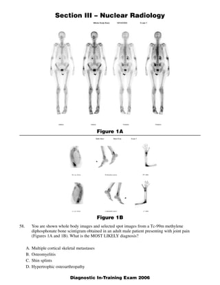
  • 1. Section III – Nuclear Radiology Figure 1A Figure 1B 58. You are shown whole body images and selected spot images from a Tc-99m methylene diphosphonate bone scintigram obtained in an adult male patient presenting with joint pain (Figures 1A and 1B). What is the MOST LIKELY diagnosis? A. Multiple cortical skeletal metastases B. Osteomyelitis C. Shin splints D. Hypertrophic osteoarthropathy 1 Diagnostic In-Training Exam 2006
  • 2. Section III – Nuclear Radiology Question 58 Rationales: A. Incorrect. Focal cortical metastases are uncommon, but do occur. However, the symmetrical metadi- aphyseal distribution of the lesions and involvement of both the proximal and distal appendicular skeleton are highly atypical for metastases. The clinical history of joint pain also points to other more likely etiologies for the findings present. B. Incorrect. Osteomyelitis may produce areas of increased cortical tracer uptake, secondary to perios- titis associated with the active infectious process. Abnormality is not usually limited to cortical areas alone, and is often more intense than the findings in this case. Furthermore, the bilaterally symmetrical distribution of the findings is highly atypical for osteomyelitis. C. Incorrect. Shin splints may also produce areas of increased uptake along the cortical surfaces of long bones, as seen in this case. However, the findings in shin splints are usually confined to the tib- iae, and most often involve primarily the diaphyseal regions, often with only low-level increased uptake. Furthermore, involvement of the upper extremities would not be expected with shin splints. D. Correct. The findings in this case are characteristic in appearance and location for secondary hyper- trophic osteoarthropathy (formerly known as hypertrophic pulmonary osteoarthropathy or HPO). This disorder is most often associated with intrathoracic lesions, such as primary lung neoplasms or mesothelioma. It can also occur in association with other intrathoracic lesions, including bronchiec- tasis, emphysema, lung infections, metastatic disease, etc. It may be seen in patients with congenital heart disease, inflammatory bowel disease and lymphoma. Patients may present with joint pain, clubbing and periosteal new bone formation on radiographs. The differential diagnosis also includes pachydermoperiostosis and thyroid acropachy. 2 American College of Radiology
  • 3. Section III – Nuclear Radiology Figure 2A 59. You are shown serial 5-minute anterior images and final right anterior oblique and right lateral images from a Tc-99m DISIDA hepatobiliary scan performed on a 55-year-old man with abdominal pain, fever and ascites, s/p paracentesis (Figures 2A and 2B). What is the MOST LIKELY diagnosis? A. Acute cholecystitis B. Bile leak C. Common bile duct obstruction D. Normal study 3 American College of Radiology
  • 4. Section III – Nuclear Radiology Figure 2B 4 Diagnostic In-Training Exam 2006
  • 5. Section III – Nuclear Radiology Question 59 Findings: There is prompt hepatic uptake, with early visualization of activity in the region of the gall- bladder fossa. Faint, amorphous activity is noted inferior to the liver on the right, beginning at 10 min- utes and better seen thereafter. In addition, there is accumulation of activity throughout the peritoneal cavity, beginning at 15 minutes post-injection, and progressively increasing throughout the study. There is also the appearance of abnormal linear activity along the inferior margin of the left lobe of the liver, beginning at 25-30 minutes into the study and progressively increasing in intensity. The right lateral image demonstrates activity spreading anterior to the liver, also consistent with intraperitoneal biliary leakage. Hepatic clearance is also moderately prolonged. Rationales: A. Incorrect. The findings are not consistent with acute cholecystitis. There is prompt visualization of the gallbladder as early as 5-10 minute post-injection, which essentially excludes acute cholecysti- tis. Furthermore, acute cholecystitis does not explain the presence of biliary leakage present in this case. Perforation of the gallbladder may occur in gangrenous cholecystitis, but that entity is virtual- ly always associated with cystic duct obstruction, which would result in non-visualization of the gallbladder as well. B. Correct. The findings in this case described above are consistent with a relatively large bile leak, most likely arising in the region of the gallbladder fossa. In this case, the findings may be secondary to trauma from paracentesis. C. Incorrect. There is prolonged hepatic clearance and non-visualization of the small bowel, both find- ings that occur in the presence of common bile duct obstruction. However, in common duct obstruc- tion, there is often complete non-visualization of the biliary tree, including the gallbladder, even in the absence of cholecystitis. In addition, common duct obstruction is not usually associated with bil- iary leakage, which is present in this case. D. Incorrect. This study is not normal. A significant degree of biliary leakage is demonstrated, as described above. Furthermore, the images also demonstrate prolonged hepatic clearance and non- visualization of the small bowel, both of which are also abnormal findings. 5 American College of Radiology
  • 6. Section III – Nuclear Radiology Figure 3 60. A 2-month-old male with marked hypertension is referred for captopril renography. You are shown serial 1-minute posterior pre- and post-captopril images (Figure 3). What is the MOST LIKELY diagnosis? A. Normal study B. Right renal artery stenosis C. Left renal artery stenosis D. Bilateral renal artery stenosis 6 American College of Radiology
  • 7. Section III – Nuclear Radiology Question 60 Findings: The baseline pre-captopril study demonstrates mildly decreased tracer uptake bilaterally, with normal excretion. The post-captopril images demonstrate significant bilateral deterioration in excretion, with marked cortical retention noted bilaterally. Rationales: A. Incorrect. Although initial (left) study appears symmetrically normal, there is clearly a bilateral delay in cortical clearance and excretion on the post-captopril study. B. Incorrect. In unilateral right renal artery stenosis, ACE-inhibitor should create an asymmetric delay in right renal washout, not the bilaterally delayed washout present in this case. C. Incorrect. In unilateral left renal artery stenosis, ACE-inhibitor should create an asymmetric delay in left renal washout, not the bilaterally delayed washout present in this case. D. Correct. The post-captopril study fails to demonstrate sequential right and left renal pelvis and bladder activity seen at midpoint of the baseline pre-captopril study. Administration of the ACE inhibitor has produced a symmetric delay in renal cortical clearance, manifested by marked bilateral cortical retention and non-visualization of the renal pelves and bladder. These findings are typical for bilateral ACE-inhibition of compensatory post-glomerular vascular constriction, with resultant delay in transcortical clearance, in this child with bilateral congenital renal artery stenosis. 7 Diagnostic In-Training Exam 2006
  • 8. Section III – Nuclear Radiology Figure 4 61. You are shown representative coronal, transaxial and sagittal images from an F-18 FDG (fluorodeoxyglucose) PET scan (Figure 4). What is the MOST LIKELY diagnosis? A. Lymphoma B. Bronchogenic carcinoma C. Esophageal carcinoma D. Normal variant 8 American College of Radiology
  • 9. Section III – Nuclear Radiology Question 61 Rationales: A. Incorrect. The abnormal uptake in this case is located in the posterior mediastinum, where adenopa- thy due to lymphoma may occur. However, the linear configuration of the activity is characteristic of esophageal activity, rather than the typical focal rounded appearance of adenopathy. Furthermore, no other sites of adenopathy are present. The findings are characteristic of an esophageal neoplasm, making squamous cell carcinoma or adenocarcinoma far more likely than lymphoma. B. Incorrect. As discussed above, the linear uptake located in the posterior mediastinum is characteris- tic in appearance for an esophageal neoplasm. There are no focal pulmonary nodules or foci of mediastinal or hilar adenopathy, as would be anticipated in the presence of bronchogenic carcinoma. C. Correct. The linear pattern of increased FDG uptake in the posterior mediastinum, in the expected location of the esophagus, is characteristic in appearance for an esophageal neoplasm, most likely representing squamous cell carcinoma of the esophagus. D. Incorrect. Mildly increased uptake near the gastroesophageal junction may be seen as a normal vari- ant, or in patients with gastroesophageal reflux. Mild diffuse esophageal uptake may also occur in esophagitis. The uptake in this case is far more intense than would be anticipated as a normal vari- ant, and the location of the activity remote from the gastroesophageal junction is not consistent with a normal variant. 9 Diagnostic In-Training Exam 2006
  • 10. Section III – Nuclear Radiology Figure 5 62. A 28 year-old HIV positive woman presents with headache, papilledema, and a ring-enhancing right thalamic mass on CT (not shown). You are shown a transaxial Tl-201 chloride image of the brain (Figure 5). What is the MOST LIKELY diagnosis? A. Lymphoma B. Cytomegalovirus infection C. Toxoplasmosis infection D. Normal study 10 American College of Radiology
  • 11. Section III – Nuclear Radiology Question 62 Findings: Transaxial Tl-201 chloride SPECT images of the brain demonstrate a focal area of increased tracer uptake near the midline, in the region of the CT lesion in the basal ganglia. Rationales: A. Correct. CNS lymphoma may produce a ring-enhancing lesion on CT and is thallium-avid. These findings are most consistent with CNS lymphoma arising in an immunocompromised host. B. Incorrect. CMV is not thallium-avid, as is the lesion in this case. C. Incorrect. Toxoplasmosis can produce cerebral ring-enhancing CT lesion, but it is not thallium-avid, as is the lesion in this case. D. Incorrect. The focal area of increased tracer uptake in the midline basal ganglia region represents a striking abnormality, which is not attributable to any normal finding. This is not a normal study. 11 Diagnostic In-Training Exam 2006
  • 12. Section III – Nuclear Radiology Figure 6 63. You are shown representative coronal, transaxial and sagittal tomographic radionuclide images (Figure 6). Which one of the following radiotracers was MOST LIKELY utilized for this study? A. Tc-99m methylene diphosphonate B. Tc-99m sulfur colloid C. F-18 fluorodeoxyglucose D. F-18 sodium fluoride 12 American College of Radiology
  • 13. Section III – Nuclear Radiology Question 63 Rationales: A. Incorrect. The normal biodistribution of Tc-99m methylene diphosphonate (MDP) includes the axial and appendicular skeleton, kidneys and bladder. The liver, spleen, mediastinum, and brain, which are visualized in this case, are not seen on a normal bone scintigram. B. Incorrect. The normal biodistribution of Tc-99m sulfur colloid includes intense liver and spleen activity. Less intense activity is identified in the central bone marrow (skull, ribs, sternum, vertebral bodies, pelvis, proximal humeri and femora). The most intense activity in this study is osseous. Moderate activity is seen within the spleen and low level activity in the liver, mediastinum and brain. This biodistribution is not typical for sulfur colloid. C. Correct. The normal biodistribution of F-18 fluorodeoxyglucose (FDG) is accumulation in the brain, myocardium, blood vessels, pharynx, liver, spleen, bone marrow, kidneys, ureters, urinary bladder, and GI tract. Intense marrow uptake is seen in this patient with lymphoma after administra- tion of granulocyte colony stimulating factor (G-CSF), which is given to support bone marrow func- tion following therapy. Normal marrow uptake is usually less intense than hepatic uptake. While this distribution is not normal, it is more characteristic of FDG than any of the other tracers listed. D. Incorrect. The normal biodistribution of F-18 sodium fluoride is osseous, with uptake dependent on regional blood flow and osteoblastic activity by chemisorption. Hydroxyl groups are exchanged to form fluoroapatite in the hydroxyapatite crystals. Because of the superior spatial resolution and three-dimensional localization afforded by PET imaging, there is a high sensitivity for the detection of metabolically active skeletal lesions using F-18 sodium fluoride. 13 Diagnostic In-Training Exam 2006
  • 14. Section III – Nuclear Radiology 64. Concerning subacute thyroiditis, serum thyroid hormone levels are elevated as the result of which one of the following? A. Increased thyroid hormone production B. Increased TSH secretion by the pituitary gland C. Release of pre-formed thyroid hormone into the circulation D. Iodine excess in the thyroid gland Question 64 Rationales: A. Incorrect. Thyroid hormone production is reduced in subacute thyroiditis. The elevated thyroid function tests and signs and symptoms of hyperthyroidism that occur early in the disorder are relat- ed to release of pre-formed thyroid hormone into the circulation from the inflamed thyroid gland. B. Incorrect. The increased thyroid hormone levels produced by the release of pre-formed hormone into the circulation results in a feedback inhibition of TSH secretion by the pituitary, resulting in decreased serum TSH levels. C. Correct. Subacute thyroiditis is a viral disorder, often following a recent upper respiratory infec- tion. The inflammatory response in the gland results in increased permeability and increased release of pre-formed thyroid hormone into the circulation from the colloid. The increased serum thyroid hormone levels, in turn, result in clinical evidence of hyperthyroidism, despite a low thyroid uptake. D. Incorrect. The pathophysiology of subacute thyroiditis does not involve abnormalties in iodine metabolism per se. The acute inflammatory response in this disorder is associated with decreased iodide uptake and organification during the early stage of the disease. 14 American College of Radiology
  • 15. Section III – Nuclear Radiology Gallium-67 citrate scintigraphy is preferred over In-111 leukocyte scintigraphy in which one of 65. the following entities? A. Abdominal abscess B. Infected joint prosthesis C. Disk space infection D. Inflammatory bowel disease Question 65 Rationales: A. Incorrect. While both radiopharmaceuticals are efficacious for the detection of abdominal abscesses, Indium-111 leukocyte imaging is often preferred, as the result of the absence of potentially confus- ing normal bowel activity, as occurs in Gallium-67 scintigraphy. This normal bowel uptake may lead to false positive gallium studies. B. Incorrect. Indium-111 leukocyte imaging is superior to gallium-67 scintigraphy in the evaluation of suspected infected joint prostheses, in part related to the bone seeking properties of gallium, leading to potential false positive gallium studies due to increased tracer localization secondary to increased bone turnover in the absence of infection. C. Correct. While sensitive for osteomyelitis, Indium-111 leukocyte scintigraphy has been found to be less sensitive than gallium-67 scintigraphy for the detection of disc space infection. D. Incorrect. Again, the absence of normal bowel localization makes In-111 leukocyte scintigraphy better suited to the assessment of active inflammatory bowel disease. In gallium-67 scintigraphy, normal bowel uptake, especially in the colon, can be incorrectly attributed to inflammatory bowel disease. 15 Diagnostic In-Training Exam 2006
  • 16. Section III – Nuclear Radiology 66. Concerning the presence of multiple focal “hot spots” on a Tc-99m macroaggregated albumin (MAA) scan, which one of the following is CORRECT? A. The study may need to be repeated on another day. B. The patient is at risk for the development of acute hypoxemia. C. A false positive study will result. D. The patient has multiple arteriovenous malformations (AVMs). Question 66 Rationales: A. Correct. The finding of focal “hot spots” on a Tc-99m MAA scan indicates the aggregation of the radiopharmaceutical into larger particles, which lodge in the pulmonary vascular bed. This artifact may be produced by drawing blood back into the syringe during injection or by failing to resuspend the particles prior to injection, in the event the dose is left sitting for a prolonged time after being drawn up. While it is not associated with any adverse effects in the patient, these foci of increased activity may obscure portions of the underlying lungs, resulting in the need to repeat the study after significant radioactive decay has occurred. B. Incorrect. While technically these foci do represent small, iatrogenic pulmonary emboli, they are virtually never associated with any clinically demonstrable adverse effects. In general, pulmonary perfusion imaging with Tc-99m MAA is associated with transient occlusion of less the 0.1% of the pulmonary capillary bed. Thus, this occurrence is unlikely to produce acute hypoxemia. C. Incorrect. While these “hot spots” may obscure underlying detail in evaluating pulmonary perfu- sion, they are not associated with artifactual perfusion defects that would produce a false positive study. D. Incorrect. Pulmonary AVMs are associated with right to left shunting, permitting Tc-99m MAA par- ticles to bypass the pulmonary capillary bed. Thus, AVMs would tend to produce focal perfusion defects, rather than focal areas of increased tracer localization. 16 American College of Radiology
  • 17. Section III – Nuclear Radiology Concerning the analysis of radionuclide gated blood pool (MUGA) studies, which one of the 67. following will result in an UNDERESTIMATION of the left ventricular ejection fraction? A. Placement of the background region of interest over the splenic blood pool activity B. Assignment of too small a systolic region of interest C. Use of a single region of interest for both the systolic and diastolic frames D. Inclusion of a portion of the left atrium in the diastolic region of interest Question 67 Distractors: SCORE ALL CHOICES AS CORRECT A. Placement of the background region of interest over the splenic blood pool activity B. Assignment of too small a systolic region of interest C. Use of a single region of interest for both the systolic and diastolic frames D. Inclusion of a portion of the left atrium in the diastolic region of interest Rationales: A. Placement of the background ROI over the spleen will result in excessive background subtraction. The relative effect of the extra background subtraction will be greater on the systolic ROI, which has fewer counts, and therefore will not “cancel out”. Thus, the denominator of the ejection fraction equation will be relatively reduced, resulting in an artifactually elevated, rather than reduced. B. Too small of a systolic region will result in exclusion of value counts from the systolic region, mak- ing the percentage change between systolic and diastolic counts appear to be larger than it actually is. Again, this error would result in an artifactually elevated calculated ejection fraction. C. This technique was initially used when the procedure was originally introduced. However, by using the same region for both measurements, the systolic region extends beyond the actual margins of the ventricle in end-systole, thus including background counts from adjacent structures, making the apparent ejection fraction artifactually too low. For this reason, the standard method of analysis at present requires assignment of separate diastolic and systolic regions of interest, in order to improve the accuracy of the measurement. D. Inclusion of a portion of the left atrium in the diastolic region of interest will have the effect of introducing additional counts into the region that are not valid ventricular counts. Thus, the apparent total end-diastolic counts will be inappropriately high, resulting in an apparent higher percentage of ventricular emptying and a falsely elevated ejection fraction calculation. 17 Diagnostic In-Training Exam 2006
  • 18. Section III – Nuclear Radiology Concerning radionuclide myocardial perfusion imaging, which one of the following is NOT 68. associated with an inferior wall perfusion defect on a stress Tc-99m sestamibi SPECT study? A. Inferior wall exercise-induced ischemia B. Prior inferior wall myocardial infarction C. Left bundle branch block D. Diaphragmatic attenuation artifact Question 68 Rationales: A. Incorrect. Inferior wall ischemia characteristically produces a perfusion defect in this region on stress myocardial perfusion images. In the case of reversible ischemia, the defect would be expected to resolve on a resting study. B. Incorrect. An area of prior myocardial infarction typically produces a “fixed” perfusion defect, which would be visible both on stress and resting images. Thus, from evaluation of stress images alone, it cannot be differentiated from a defect due to exercise-induced ischemia, as in item A. C. Correct. Left bundle branch block may be the result of myocardial ischemia or infarction, or may be an incidental finding. It may produce perfusion abnormalities on myocardial perfusion scintigra- phy in the absence of coronary artery disease. When it produces abnormalities, the most common finding is a reversible perfusion defect in the interventricular septum, not in the inferior wall. In patients with known left bundle branch block, it is preferable to perform a pharmacologic stress test, using dipyridamole or adenosine, rather than treadmill exercise in conjunction with the imaging, since this artifact is more commonly associated with the latter procedure. D. Incorrect. Diaphragmatic attenuation artifact commonly produces apparent defects in the inferior wall. These defects may or may not be present both on stress and resting images, and may be sus- pected by inspection of planar rotating images from the raw data set. This artifact most often occurs in male patients, and is more common in obese patients as well. 18 American College of Radiology
  • 19. Section III – Nuclear Radiology 69. Which is an appropriate use of F-18 fluorodeoxyglucose (FDG) PET imaging in breast carcinoma? A. Screening B. Initial staging C. Differentiating between a pulmonary metastasis and a primary lung carcinoma D. Treatment monitoring Question 69 Rationales: A. Incorrect. FDG PET imaging is not an appropriate or approved study for breast cancer screening. Screening is best done by self-examination and periodic mammography, which are more sensitive and cost-effective approaches to breast cancer screening. B. Incorrect. FDG PET imaging is less sensitive for the initial staging of breast cancer than lym- phoscintigraphy with sentinel lymph node biopsy. Very high sensitivity is provided by the latter approach, particularly when immunohistochemistry techniques are utilized. This approach to staging is rapidly becoming the standard of care for these patients. C. Incorrect. FDG PET imaging is not capable of differentiating between a solitary pulmonary metas- tasis and a primary lung tumor. In most cases, both lesions are associated with increased glucose metabolism and thus, increased FDG uptake. D. Correct. As is true for a number of neoplasms, FDG PET imaging is very sensitive and specific for assessing the response to therapy in breast carcinoma, whether performed after the completion of therapy (re-staging) or during the course of therapy (treatment monitoring). 19 Diagnostic In-Training Exam 2006
  • 20. Section III – Nuclear Radiology 70. A post-menopausal woman with osteoporosis undergoes dual-energy x-ray absorptiometry (DEXA) scanning, demonstrating marked osteopenia of the lumbar spine and hip, but normal bone density of the distal forearm. What is the BEST explanation for these findings? A. Inappropriate scanning of the dominant forearm rather than the non-dominant B. Insensitivity of forearm bone density measurement secondary to preponderance of cortical bone C. Underestimation of the bone density in the spine and hip secondary to arthritic changes D. Scan performed too distally in the forearm Question 70 Rationales: A. Incorrect. While it is true that it is preferable to scan the non-dominant forearm or hip in DEXA scanning, and scanning the dominant side could produce a higher bone density value, the differ- ences between the dominant and non-dominant sides are often minimal, and this is therefore not the best explanation for the findings. B. Correct. The bones of the extremities, such as the radius and ulna, are composed primarily of corti- cal bone, and contain relatively less trabecular bone than either the spine or hip. Quantitatively, the extremities account for the majority of the whole body bone mineral content. Thus, bone density measurements of the forearm are most valuable in patients with metabolic bone disease, or other conditions associated with decreases in total skeletal calcium content. Post-menapausal osteoporosis preferentially involves the trabecular bone initially, which is present in higher percentages in the vertebral bodies and femoral neck regions. Therefore, forearm measurements tend to be relatively insensitive for the early detection of post-menopausal osteoporosis. C. Incorrect. In fact, the opposite is true. The presence of arthritic changes is most often associated with falsely elevated bone density measurements, especially in the spine, secondary to increased bone density at sites of spurring or sclerosis associated with arthritic involvement. D. Incorrect. Again, the opposite is true. Moving from proximal to distal in the forearm, there is a pro- gressive increase in the relative trabecular bone content. Typically, bone density measurements of the forearm are performed in the distal third of the radius and ulna, in order to maximize the contri- bution of trabecular bone in the measurement. Scanning more distally may also be performed, to further increase the percentage of trabecular bone being evaluated. Thus, scanning more distally would tend to decrease the measured bone mineral density of the forearm. 20 American College of Radiology
  • 21. Section III – Nuclear Radiology 71. What is the most commonly cited threshold for the diagnosis of malignancy using standardized uptake value (SUV) on PET imaging for a solitary pulmonary nodule? A. 1.0 B. 1.5 C. 2.5 D. 3.0 Question 71 Rationales: A. Incorrect. The correct value is 2.5. B. Incorrect. The correct value is 2.5. C. Correct. Many malignant lesions will greatly exceed this value, and some lesions with SUV values < 2.5 are malignant, but 2.5 is the most commonly cited threshold for the diagnosis of malignancy using SUV analysis. D. Incorrect. The correct value is 2.5. 21 Diagnostic In-Training Exam 2006
  • 22. Section III – Nuclear Radiology Concerning infection imaging with In-111 labeled leukocytes, which one is CORRECT? 72. A. Uptake is dependent on regional blood flow. B. It is insensitive for the detection of inflammatory bowel disease. C. Transient pulmonary uptake clears within 15 minutes post-injection. D. It is more sensitive than Ga-67 citrate imaging for detection of Pneumocystis carinii pneumonia (PCP). Question 72 Rationales: A. Correct. While not the sole determinant of uptake, the uptake of In-111 labeled leukocytes is dependent upon regional blood flow. For example, a walled-off abscess without a direct blood sup- ply will not accumulate In-111 labeled leukocytes, and may appear as a photopenic defect. B. Incorrect. In-111 WBC imaging is very sensitive for active inflammatory bowel disease. It has advantages over Ga-67 citrate imaging in this clinical setting, as a result of the absence of normal bowel uptake of the tracer. C. Incorrect. Transient lung uptake can be seen 4 hours after injection or even longer, sometimes mak- ing the diagnosis of pulmonary infection difficult. D. Incorrect. In-111 WBC’s are less sensitive than Ga-67 citrate for detecting chest infections, such as PCP. As a result, Ga-67 citrate imaging is preferred in the clinical settings of suspected chest infec- tion or in immunocompromised patients presenting with fever of unknown origin. 22 American College of Radiology
  • 23. Section III – Nuclear Radiology 73. The Nuclear Regulatory Commission (NRC) mandates daily performance testing of the ioniza- tion chamber radioisotope dose calibrator for which one of the following? A. Geometry B. Constancy C. Linearity D. Accuracy Question 73 Rationales: A. Incorrect. Assessment of the effects of geometry is required at time of initial setup or after alter- ation/repair of well calibrator only. This insures that variations in radioactive dose volume or posi- tion in counting chamber will not produce aberrant dose determination. B. Correct. This daily mandated test measures instrument precision and is designed to show repro- ducible readings day after day on all clinical energy settings. This is essentially a mini-accuracy test that does not account for half-life of long-lived low, medium, and high energy sealed standards. More or less rigor is applied, depending on whether a single 137Cs source is counted in all standard energy settings (Tc99m, 201TI, 123I, 131I, etc.) and the same reading is compared day to day or a more elaborate daily count of multiple sealed sources (57Co, 133Ba, 137Cs) is obtained. No more than a 5% daily count rate variation is allowable. C. Incorrect. Sequential assay of count rates of the same radioisotope from low to high activity, usually by counting an initially high activity Tc-99m source as it decays over 48 hours. This multi-day study can’t be performed daily, and is usually performed at installation, quarterly thereafter and whenever the device undergoes repair. D. Incorrect. Designed to insure correct readings throughout the entire energy spectrum clinically encountered, this rigorous test requires reproduction of count rates with low, medium, and high energy sealed standard sources 57Ba, 137Cs. This elaborate test is performed at installation of the device, annually thereafter and whenever the device undergoes repair. 23 Diagnostic In-Training Exam 2006
  • 24. Section III – Nuclear Radiology 74. For the man-made radiation contributions to the background radiation in the United States, which of the following represents the MOST significant source of exposure to the U.S. population? A. Medical x-rays B. Radon C. High-altitude air travel D. Nuclear medicine Question 74 Rationales: A. Correct. Medical x-rays are the most significant source of man-made radiation sources. They con- tribute an annual effective dose of 0.39 mSv or 39 mrem to the U.S. population. B. Incorrect. Radon is a naturally occurring source of radiation. C. Incorrect. High-altitude air travel adds to an individual’s cosmic ray exposure, and is of very small quantity. D. Incorrect. Nuclear does not contribute as much as medical x-rays as a source of exposure to the U.S. population. They contribute an annual effective dose of 0.14 mSv or 14 mrem to the U.S. popula- tion. 24 American College of Radiology
  • 25. Section III – Nuclear Radiology Which one of the following sets of I-123 thyroid scintigraphy findings and history of radiation 75. exposure is associated with the LOWEST relative risk for thyroid carcinoma? A. Multiple cold nodules with previous head and neck irradiation B. Multiple cold nodules without prior head and neck irradiation C. Solitary cold nodule without prior head and neck irradiation D. Solitary cold nodule with previous head and neck irradiation Question 75 Rationales: A. Incorrect. This combination of scan findings and history is associated with the highest relative like- lihood of malignancy of all those listed, in the range of 40%. B. Correct. The finding of multiple cold nodules without prior radiation exposure is consistent with a non-specific multinodular goiter, and carries a risk of underlying malignancy of only ~ 5%. C. Incorrect. While the absence of prior head and neck irradiation reduces the likelihood of malignan- cy, the prevalence of malignancy in patients presenting with solitary cold thyroid nodules is still in the range of about 15-20% overall. D. Incorrect. The history of prior head and neck irradiation significantly increases the likelihood of malignancy in a patient with a solitary cold nodule, with the likelihood being somewhere in the range of 30-40%, slightly lower than for option A. 25 Diagnostic In-Training Exam 2006
  • 26. Section III – Nuclear Radiology Which one of the following is NOT a normal site of F-18 fluorodeoxyglucose (FDG) localization? 76. A. Salivary glands B. Gallbladder C. Colon D. Kidneys Question 76 Rationales: A. Incorrect. Symmetrical salivary gland uptake is a normal finding on FDG PET imaging. B. Correct. The gallbladder is not a normal site of FDG localization. Increased uptake in the gallblad- der suggests the presence of cholecystitis or a neoplastic process within the gallbladder. C. Incorrect. While variable in intensity and extent, colonic uptake of FDG is normal. D. Incorrect. Renal uptake is almost always visualized on FDG PET studies. Renal excretion into the collecting systems and bladder is also seen in the majority of cases. 26 American College of Radiology
  • 27. Section III – Nuclear Radiology 77. What is the basic principle underlying the C-14 urea breath test for Helicobacter pylori infection in patients with peptic ulcer disease? A. Absence of urease in mammalian cells B. Chemical breakdown of C-14 urea by gastric acid C. Formation of C-14 labeled glucose D. Renal excretion of C-14 urea absorbed from the stomach Question 77 Rationales: A. Correct. The basis of this study is that the Helicobacter pylori bacteria present in the stomach in patients with this infection contain the enzyme urease, necessary for the breakdown of urea. This metabolism of C-14 labeled urea results in the formation of C-14 labeled CO2 gas, which is then detected using a liquid scintillation counter. In the absence of the bacterial infection, the cells of the gastric mucosa, which lack the enzyme urease (like all mammalian tissue), are unable to break down the urea, and thus no C-14 labeled CO2 gas is formed, resulting in a negative study. B. Incorrect. The study has nothing to do with the presence or absence of gastric acid. Only the pres- ence of the enzyme urease, found in the Helicobacter pylori organisms, but not in the gastric cells, can break down the C-14 urea to form C-14 labeled CO2 gas. C. Incorrect. The physiology of the study is as described above. In no way is the formation of glucose or other aspects of carbohydrate metabolism involved. D. Incorrect. Again, the metabolism of C-14 labeled urea by bacterial urease is the basis of the study. Renal excretion is not involved, and no urine collections are performed. The study is performed by having the patient ingest the radiopharmaceutical, followed by collection of two breath samples, which are analyzed in a liquid scintillation counter for the presence of C-14 labeled CO2 gas. 27 Diagnostic In-Training Exam 2006
  • 28. Section III – Nuclear Radiology A patient with pernicious anemia had a normal Stage 1 Schilling Test. Which one of the 78. following could explain the result? A. Prior radioisotope study B. Incomplete urine collection C. Prior resection of terminal ileum D. Concurrent vitamin B-12 therapy Question 78 Rationales: A. Correct. The situation described is one where the test yields a false-negative result in a patient with pernicious anemia (as indicated in the history). Measurement of the excreted Cobalt-57 labeled vita- min B-12 is performed by counting the urine. Typical window settings used for counting are 50-200 keV for the 122 and 136-keV photons of Cobalt-57. The presence of other radioactive material in the urine that emits photons within the acceptance window will increase the measured counts, and can result in an inaccurate determination of the excretion of the radiolabeled vitamin B-12. B. Incorrect. The situation described is one where the test result is a false-negative. Incomplete urine collection could result in a low measured excretion, and a false positive (not a false negative) result C. Incorrect. The situation described is one where the test result is a false-negative. Prior resection of terminal ileum could result in a reduced absorption of the orally administered vitamin B-12, and thereby a low excretion D. Incorrect. The situation described is one where the test result is a false-negative. Prior vitamin B-12 therapy may result in a low measured excretion, and a false positive study. The patient should not receive parental vitamin B-12 for at least 3 days prior to the study. Biliary excretion of the previous- ly administered vitamin B-12 may decrease the fractional absorption of the test dose. If it does not get absorbed, it cannot get excreted into the urine, so measured excretion will be low. 28 American College of Radiology
  • 29. Section III – Nuclear Radiology 79. In nuclear medicine, what is the main difference between an intrinsic uniformity and extrinsic uniformity quality control test? A. The intrinsic test is performed without the collimator, and the extrinsic test is performed with the collimator. B. The intrinsic test uses Co-57, while the extrinsic test uses Tc-99m. C. The intrinsic test utilizes an internal electronic test mode of the gamma camera, while the extrin- sic test utilizes an external flood source. D. The intrinsic mode uses an internal calibration source within the gamma camera, while the extrinsic test utilizes an external flood source. Question 79 Rationales: A. Correct. The intrinsic uniformity or flood test is performed without the collimator and is an indica- tion of the uniformity of the camera itself. The extrinsic test is performed with the collimator on using a large flood source. B. Incorrect. Either source material may be used. Typically a syringe of Tc-99m at a distance several time larger than the camera crystal is used for the intrinsic test, and the extrinsic test is performed with a large water and Tc-99m filled flood source, or a solid Co-57 flood source. C. Incorrect. Internal electronic checks are different from the measured uniformity tests. D. Incorrect. There are no internal radiation sources to a gamma camera used for uniformity testing. 29 Diagnostic In-Training Exam 2006
  • 30. Section III – Nuclear Radiology 80. Concerning the presence of hydrolyzed reduced Tc-99m in a dose of Tc-99m MDP (methylene diphosphonate) administered intravenously for a bone scan, which is CORRECT? A. It results in thyroid visualization. B. It can be identified using a dose calibrator. C. It is more likely to occur in the presence of excess stannous ion. D. It occurs more commonly when multidose vials are used. Question 80 Rationales: A. Incorrect. Hydrolyzed reduced technetium-99m is a colloidal impurity that results in hepatic and reticuloendothelial visualization, not thyroid visualization, which is typical of the presence of free pertechnetate as an impurity. B. Incorrect. Only chromatography pre-imaging will detect this radiopharmaceutical impurity. C. Incorrect. On the contrary, Sn(II)ion is a reducing agent protecting MDP from hydrolysis. D. Correct. The introduction of air into a multidose MDP vial is the most frequent cause of this hydrolyzed technetium-99m contaminant. The more violations of the vial, the more likely air will be introduced. 30 American College of Radiology
  • 31. Section III – Nuclear Radiology 81. Concerning treatment of intractable pain from widespread metastatic bone lesions with Metastron® (Sr-89) and Quadramet® (Sm-153), which one is CORRECT? A. Both can be imaged using a gamma camera to assess the biodistribution of the therapeutic dose. B. The longer half-life of Metastron (50 days) versus Quadramet (1.9 days) provides a superior therapeutic effect. C. Because of the highly energetic beta particles produced by both agents, a lead syringe shield is employed during dose administration. D. Recovery from bone marrow toxicity is faster following Quadramet administration. Question 81 Rationales: A. Incorrect. Metastron is a pure beta emitter. The absence of an imagable gamma photon precludes verification of bone lesion uptake. By contrast, Sm-153 has an imagable gamma photon energy of 103 keV, permitting bone scintigraphy to be performed in conjunction with the therapeutic proce- dure. B. Incorrect. While it is true that the half-life of Metastron is significantly longer, resulting in more prolonged lesion irradiation, the clinical efficacy of both treatments are quite similar. C. Incorrect. Due to bremsstrahlung production of high energy photons when high atomic number material (eg. lead) is used for shielding, acrylics are the preferred material for handling of these materials. Materials with lower atomic numbers, such as plastic or acrylics make ideal shields. In addition, bremsstrahlung production is proportional to the atomic number, which is lower for these materials. D. Correct. The major limitation of both therapies is myelosuppression. Metastron causes 15-30% drops in the platelet and WBC counts from pre-injection values, and Quadramet, 40-50%. However, 8-12 weeks are required for full bone marrow recovery from Metastron, versus only 6-8 weeks for Quadramet. 31 Diagnostic In-Training Exam 2006
  • 32. Section III – Nuclear Radiology 82. Reduced occipital lobe glucose metabolism on F-18 FDG (fluorodeoxyglucose) cerebral PET imaging is MOST common in which one of the following progressive dementias? A. Alzheimer’s B. Pick’s C. Parkinson’s D. Lewy body Question 82 Rationales: A. Incorrect. Alzheimer’s dementia at the earliest stages is associated with temporoparietal and later frontal lobe FDG hypometabolism, with typical sparing of sensorimotor and visual cortex (occipital lobe). B. Incorrect. Pick’s disease is a degenerative dementia predominately involving frontal and temporal lobes. Frontal hypometabolism precedes development of temporal hypometabolism. The visual cor- tex is generally uninvolved. C. Incorrect. Parkinson’s dementia is a late manifestation of a neurodegenerative disease, primarily affecting the basal ganglia. There is occasional involvement of the occipital cortex, although tem- poroparietal hypometabolism pattern similar to that of Alzheimer’s, but with additional striatal hypometabolism, is a more common FDG pattern. D. Correct. Decreasing cognitive function accompanied by visual disturbance including hallucinations is common presentation in diffuse Lewy body disease (DLBD) which is becoming more widely rec- ognized and accounts for up to 20% of all autopsy confirmed dementias. Medical and lateral occipi- tal lobe FDG metabolism is more severely reduced in DLBD than other dementias. When identified on FDG PET images, cholinergic therapy has been useful in controlling the disease. 32 American College of Radiology
  • 33. Section III – Nuclear Radiology 83. Concerning the biodistribution of Indium-111 ibritumomab tiuxetan (Zevalin®) 48 hours following intravenous administration, which one is CORRECT? A. Persistent blood pool activity indicates the presence of a human anti-mouse antibody (HAMA) response. B. Absence of bone marrow activity indicates > 25% marrow infiltration by lymphoma. C. Renal activity less intense than hepatic is indicative of altered biodistribution. D. Hepatic activity more intense than bowel uptake is normal. Question 83 Rationales: A. Incorrect. The cardiac blood pool activity gradually decreases with time as Zevalin is distributed to the other organs and a small component is excreted. Persistent but decreased blood pool activity is normal at 48 hours. The development of a HAMA response occurs in < 2% of patients. More rapid clearance of the Zevalin antibody can occur with the development of a HAMA response, and hence, a shorter circulation time. B. Incorrect. The Zevalin therapeutic regime should not be given to patient’s with greater than or equal to 25% lymphoma marrow involvement. Altered biodistribution is suggested with rapid blood pool clearance and increased marrow uptake. C. Incorrect. Normal renal activity with Indium-111 Zevalin is generally manifested as faint activity (moderately low to very low activity), which is much less intense than hepatic uptake. Altered renal biodistribution is present if renal activity greater than liver is demonstrated on the posterior images. D. Correct. Bowel activity is common and normal. However, normal gastrointestinal biodistribution is activity that is less intense than liver and decreases over time (moderately low to very low intensi- ty). Bowel activity more intense than hepatic uptake is indicative of altered biodistribution. 33 Diagnostic In-Training Exam 2006