

Recomendados
Más contenido relacionado
La actualidad más candente
La actualidad más candente (20)
Similar a Thorax
Similar a Thorax (20)
Más de Rekha Pathak
Más de Rekha Pathak (20)
Último
Último (20)
Thorax
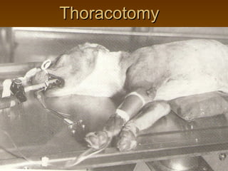
- 1. Thoracotomy
- 2. Approaches depends upon following factors
- 3. Factors Lesion type: DH, lobectomy, pericardiotomy, thoracocentesis Condition of the animal- whether an early diagnosed - withstand extensive sternotomy(splitting of sternum)/ Intercostal incision should be taken up
- 4. factors Shape and size of thorax Less capacious- more manipulation- sternotomy More capacious – more manipulation- intercostal is sufficient/ rib split/ rib resection
- 5. Techniques of thoracotomy Intercostal incision: • Cranial to the rib – intercostal vessels are located caudally • Extend the incision to desired length • A self retaining rib retractor is used for adequate exposure of the intrathoracic organs.
- 6. Techniques • Serratus ventralis dorsally and external abdominal obliqus ventrally – after incising the facia. Separate the fibres to expose external intercostal muscle.
- 7. Thoracotomy • During expiratory pause the intercostal m and pleura – incised- midway between the ribs
- 8. Closure Chromic catgut/nylon – cranial and caudal to incision- ribs opposed with towel clamps Adv: simple&quick Dis: insufficient- heart & great vessels - Rib fracture when held with rib retractor
- 10. Technique St. incision- over rib- reflect periosteum- lat. and medial
- 11. Rib resection Periosteal elevator- used to separate the periosteum medially and laterally
- 13. Closure Adv: -good Series of healing interrupted No gap sutures placed about 1 cm apart- lateral Disadv: lot of and medial skill periosteal Time consuming surfaces- cranial Weak point- and caudal edge absence of rib of incision
- 14. Split rib technique Expose the rib St. longitudinal incision- center- oscillating bone saw. Rib is sectioned- transversely at either ends- of primary incision
- 15. Split rib technique Adv: maximum exposure- without involvement of rib retractor Closure is simple and quick- interrupted stainless steel wire
- 16. Disadv: Dent formation along margins Sternum splitting incision (Median sternotomy) Required for extensive manipulation- cardiac defects and associated structures Animal on dorsal recumbency Skin incision- manubrium to xiphoid Sternum split- chisel/splitter/ electrical saw Don’t sever vessels – either side of midline
- 17. Closure Drill hole in sternabrae- suture with monofilament nylon Disadv: -Postoperative pain- discomfort- depth of respiration is affected -Inaccurate apposition -cardiac output is reduced due to increased CVP
- 18. Transabdominal Other approaches like transabdominal to repair DH- paracostal incision Heart worm disease: -dirofilaria immitis -Mosquitoes- vectors -Dog- Primary reservoir
- 20. Heart worm disease: -3rdstage larva- infective- 2-3 weeks- mosquito mouth parts -Penetrate skin- susceptible animal- 3m- immature worms – reach right side of heart- obtain full size- 15-35 cm – 5-6months- live for >5years- non- infective microfilariae
- 21. Heart worm disease: -adult worm – pulmonary trunk-less no. in rt. Ventricle- but found in rt. Atrium and caudal vena cava in heavily infested animal • -severity based on 3. No. of worms and location 4. Host immune response 5. Duration of infection
- 22. Heart worm disease Pathogenesis: Adult worm causes mechanical irritation of intima and pulmonary arterial walls- CHF Glomerulonephritis- immune complexes Pulmonary inflammation and edema Symptoms: coughing, exercise intolerance, dyspnoea, cyanosis,wt.loss despite good appetite hemoptysis, syncope, epistaxis and ascites
- 23. Diagnosis: Antigen detection test Right ventricular hypertrophy patterns are seen Detection of microfilariae in routine blood examination- failure to detect- doesnot rule out- presence of microfilariae in heart- go for concentration technique- count / ml of blood – 1000 MF=1 adult worm 5-10% dogs – adult worm- no detectable circulating MF- eosinophillia is suggestive
- 24. Mild cases- RG appearance- normal Angiocardiograms- linear filling defects – branches of pulmonary artery Moderate and severe infections- RG- dilatation of rt. Heart enlargement and dilatation of pulmonary trunk and its arteries
- 25. Splitting of second heart sound- suggestive of pulmonary hypertension- confirmed by direct cardiac catheterization –measurement of rt.ventricular/ pulmonary artery pressure Treatment : 1.Melarsomine dihydrochloride @ 2.5 mg/kg deep i.m. 2.Ivermectin
- 26. 3. Diethyl carbamazine @ 2.5 mg/ lb b.w.daily -10 days-1month 4. 6 weeks after-disappearance of clinical signs- dithiazanine iodide (dizan)2-3mg/lb orally- kill the microfilariae-7 days 5. Digoxin and diuretics are given in CHF
- 27. 3. Restrict exercise- to reduce thrombosis and endothelial damage 4. Class IV dirofilariasis – Caval syndrome- vena cavae syndrome -extreme infestation- sudden onset- collapse with haemoglobinaria and respiratory distress 5. Surgical removal –questionable- suggested that – dogs- with less than 50 worms- can tolerate chemotherapy
- 29. Puncture ventriculotomy: Apply a purse string suture- pass an alligator foreceps – in rt. Ventricle- repeatedly introduced and taken out while hemorrhage is controlled by purse- string suture
- 30. Through median sternotomy-from right atrium and orifice of tricuspid valve and caudal vena cava to save the life of the dog.
- 31. Rigid / flexible alligator forceps/ intravascular retrieval snare- via rt. Jugular vein with fluoroscopic guidance- pass the instrument untill worms are no longer retrieved Fluid therapy