Continuum biomechanics modeling of homologue proteins
1. Continuum Biomechanics Modeling of
Homologue Proteins
Jonathan Chang, Regine Labog, Sweta Ramachandran
Abstract nucleation and further growth of the actin
Actin and mreB possess homologous traits protofilament. Once ATP is incorporated into the
that interest scientists who believe that the actin filament, it hydrolyzes immediately and the
conservation of protein sequencing suggests a
ADP remains in the actin filament until it
common ancestor. After observing their similarities
in sequence and structure, we will use COMSOL to depolymerizes and an ATP is sent into the
discover whether or not their biomechanical nucleation site while the previous ADP exits. G-
properties account for actin's and mreB's differing actin creates filamentous actin (F-actin) when ATP,
biological functions. To do so, f-actin and mreB Mg, and K are present. However, above the critical
were modeled while taking into account f-actin's concentration of G-actin, the molecules polymerize.
122 degree twist and mreB parallel structure. These Below the critical concentration, the actin filaments
models revealed that mreB's maximum
depolymerize.
displacement was significantly lower than actin's.
This exponential displacement of actin is due to the
g-actin's angle when the force was applied and the Actin filaments possess polarity. The positive end
linear displacement of mreB was due to the parallel of G-actin is opposite the cleft holding the ATP
application of force. By apply forces on the two molecule, which is the negative end. Growth and
proteins, we see that the flexibility of actin is polymerization occurs more rapidly in the positive
necessary for actin which must handle multiple end. Though the intermolecular interactions of two
functions in a eukaryotic cell. However, in bacteria
actin molecules is weak, adding a third actin
where mreB's main function is to provide the
structure for bacteria, it requires a more rigid monomer stabilizes the overall complex. Once the
structural component. Our model accounts for the dimer becomes a trimer, the actin molecules adds
structures of the two proteins which will, in the more monomers and forms a nucleation site.
future, help in determining when the two proteins Adding actin filaments or key actin binding proteins
diverged from their common ancestor. elongates the actin molecules to form a long helical
polymer. After the growth period, the polymer
Introduction reaches an equilibrium phase where
Actin is a component of the cytoskeletal system
depolymerization controls the length of the polymer
allowing cell movement and cellular processes.
as new monomers are added.
Actin filaments are called microfilaments of thin
mreB
filaments that undergo constant rearrangement to
create movement. Actin is a globular protein with
Bacillus subtilis mreB is a bacterial, actin-like
the ATP binding site at the center of the molecule.
protein that has been shown to perform essential
G-actin is short for globular actin, a short
functions in cellular physiology. It affects cell
polypeptide chain made up of 375 amino acids. G-
growth, cell shape, chromosome segregation and
actin combines with other g-actin monomers to
polar localization of proteins, and localization as
create an actin filament. It serves as a site for
helical filaments under the cell membrane. MreB
2. performs dynamic, motor-like movements in the its designated 166 degree twist, the primary design
cells and extend along helical tracks in seconds. to incorporate. In COMSOL, we used a 3D,
structural, static model. Since we were more
MreB is a bacterial protein considered an actin interested in comparing the difference in response
homologue based on its similarities in tertiary to loads between actin and MreB, using a transient
structure and conservation in the active site's model was not of interest. With the static model,
peptide sequence. MreB has filaments located under one end was fixed and the other end was applied a
the cellular membrane to control the width of rod vertical/parallel load. By simplifying the model of
shaped bacteria F-actin to incorporate half the number of subunits
for clarity sake, calculated the precise positions and
Aside from tubulin, the other major component of direction vectors of the subunits was possible while
the eukaryotic cytoskeleton is F-actin (filamentous the overall structural design was not sacrificed.
actin), a relatively thin protein composed of two Edge gaps between subunits was modeled in
strands twisted around each other. Actin works in both actin and MreB since separation does naturally
cell motility, shape determination, phagocytosis, occur between subunits--the gaps were designed to
cytokinsesis, and rearragement of surface be as consistent as possible between the two
components. It is 43kDa bi-lobed protein that binds COMSOL models. Two forces that were
ATP in a cleft between the two lobes. The mreB determined through literature research to be the
gene is associated with prokaryotic cell shape usual load forces for these proteins was applied to
determination but not cell envelope synthesis. the non-fixed end: 100 pico-Newtons, and 100
Research on Bacillus subtilis showed that the large micro-Newtons. This led to interesting results
spirals encircling the cytoplasm under the cell wherein displacement along inside edge of both
membrane suggests that mreB forms filamentous proteins could be determined and outputted as a
structures in bacteria similar to the eukaryotic actin graph.
cytoskeleton. In vitro, purified mreB forms
polymers consisting of protofilaments of 51 Results
angstroms which is close to the spacing between the
subunits of filamentous actin which is 55
angstroms. The three-dimensional structure of actin
and mreB is also very similar. The striking
difference between mreB and actin is that the F-
actin twists around each other whereas mreB
protofilaments are straight.
Research Design and Methods
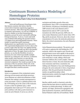
We used COMSOL to model the actin and MreB Figure 1a: COMSOL Diagram of Actin Protein,
based on the values determined through literature Front View
research; this includes density, Young's Modulus,
Poisson's ratio, and the dimensions of F-actin, as
well as the dimensions of its subunits. The values
we have determined are as follows: F-actin total
diameter 7 nm, length of interest 20 nm, subunit
diameter of 5.4nm, Young's Modulus of 44e6 Nm-2,
and a Poisson's ratio of 0.3. The length of interest
was determined to be the length at which it makes
3. Figure 1b: COMSOL Diagram of Actin Protein, Figure 4: COMSOL Diagram of MreB with
Front View, Meshed Force Applied, Boundary View
Figure 5: COMSOL Diagram of MreB with
Figure 2: COMSOL Diagram of MreB Protein, Force Applied, Streamline View
Front View
Figure 6: COMSOL Diagram of Actin with
Figure 3: COMSOL Diagram of Actin with Displacement Edge Outlined in Red
Force Applied, Boundary View
Figure 7: COMSOL Diagram of Total
4. Displacement Along Actin Edge, Load of 100
Micro-Newtons
Figure 11: COMSOL Diagram of Total
Displacement Along MreB Edge, Load of 100
Figure 8: COMSOL Diagram of Total Pico-Newtons
Displacement Along Actin Edge, Load of 100
Pico-Newtons Maximum
Protein
Displacement
Actin 1.8e-8 meters
4.614e-28
MreB
meters
Figure 12: Maximum Displacements with 100
Pico-Newtons Load
Figure 9: COMSOL Diagram of MreB with Discussion
Displacement Edge Outlined in Red Maximum displacement was measured and
analyzed for both actin and MreB. The
displacement curve of actin (Figures 7 and 8) is
exponential, which can be explained by the angle of
the subunit on which the force is applied due to the
helical conformation of the protein. In contrast, the
displacement of MreB (Figures 10 and 11) is linear
because the uniaxial force is applied in parallel to
the major axis of the MreB filaments. Based on our
results, it is apparent that the maximum
displacement of MreB (Figure 4) is significantly
smaller than that of actin (Figure 3). This can be
Figure 10: COMSOL Diagram of Total explained by the rotational twist in the F-actin
Displacement Along MreB Edge, Load of 100 conformation, which makes the protein less rigid.
Micro-Newtons Thus, it can be inferred that these homologue
proteins, which have similar amino acid sequences
and tertiary structures, play different roles in
eukaryotic and prokaryotic cells. Since actin must
handle multiple functions in a eukaryotic cell,
including mechanical support, cell motility, cargo
transport, and cytokinesis, flexibility and an ability
to change conformations efficiently may be an
essential characteristic for the protein. The larger
5. displacement that was observed supports this motif. MreC/D and other actin-like proteins for proper
However, the primary function of MreB in bacteria localization." BMC Cell Biology. PubMed central, 3 Mar.
is to provide the organism with a rigid, inter-cellular 2005. Web. 3 Dec. 2009.
backbone. Consequently, the smaller displacement <http://www.ncbi.nlm.nih.gov/pmc/articles/PMC555950/
observed in MreB upholds the notion that the >.
bacterial protein must be relatively inflexible and
stiff.
The models of actin and MreB that were
constructed represent the fundamental building
blocks of the two proteins. Only four subunits of the
protein were modeled, and in the future, a larger
number of subunits can be modeled to verify that
the proteins behave similarly at the subunit level
and as a complete protein. Moreover, the
interactions between the individual filaments, such
as hydrogen bonding and amino acid interactions,
were not considered. In order to account for these
interactions, the individual amino acids can be
modeled to determine if these interactions affect the
displacement of the protein as a whole. Once a
thorough model of actin is established, it would be
interesting to study the elongation of the actin
filament and the biomechanics that underlies the
propagation of the protein through the cytosol of a
cell.
Limitations:
did not take into account interactions
between the actin filaments
only modeled 4 subunits of the protein
Future Studies:
interactions between actin and other proteins
elongation of actin
References
1. Figge, Rainer M., Arun V. Divakaruni, and James W.
Gober. "MreB, the cell shape-determining bacterial actin
homologue, co-ordinates cell wall morphogenesis in
Caulobacter crescentus."Molecular Microbiology 2004:
1321-332. Blackwell Publishing Ltd. Web.
<http://www.biochemistry.ucla.edu/biochem/Faculty/Gob
er/PDF/1321.pdf>.
2. Van den Ent, Fusinita, Linda Amos, and Jan Löwe.
"Bacterial Ancestry of Actin and Tubulin."Current
Opinion in Microbiology 2001: 634-48. Elsevier Science
Ltd. Web. 3 Dec. 2009. <http://www2.mrc-
lmb.cam.ac.uk/groups/jyl/PDF/current%20opinion%20mi
cro%202001.pdf>.
3. Defeu, Joël, and Peter Graumann. "Bacillus subtilis actin-
like protein MreB influences the positioning of the
replication machinery and requires membrane proteins