Gutell 093.jphy.2005.41.0380
•
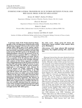
0 recomendaciones•460 vistas

This document discusses evidence of lateral transfer of a group IE intron between fungal and red algal small subunit rRNA genes. It finds that a group IE intron inserted at position 989 in the nuclear SSU rRNA gene of the red alga Hildenbrandia rubra is closely related to similar fungal IE introns, providing evidence the intron was laterally transferred rather than vertically inherited. Phylogenetic analysis of intron sequences and comparisons of intron secondary structures support a relationship between the red algal intron and fungal introns, making lateral transfer the most likely explanation for the intron's presence in H. rubra.

Recomendados
Más contenido relacionado
La actualidad más candente
La actualidad más candente (20)
Destacado
Destacado (18)
Similar a Gutell 093.jphy.2005.41.0380
Similar a Gutell 093.jphy.2005.41.0380 (20)
Más de Robin Gutell
Más de Robin Gutell (20)
Último
Último (20)
Gutell 093.jphy.2005.41.0380
- 1. EVIDENCE FOR LATERAL TRANSFER OF AN IE INTRON BETWEEN FUNGAL AND RED ALGAL SMALL SUBUNIT rRNA GENES1 Kirsten M. Mu¨ller2 , Darlene W. Ellenor Department of Biology, University of Waterloo, Waterloo, Ontario, N2L 3G1, Canada Alison R. Sherwood Department of Botany, University of Hawaii, Honolulu, Hawaii 96822, USA Robert G. Sheath Provost’s Office, California State University San Marcos, San Marcos, California 92096, USA and Jamie J. Cannone and Robin R. Gutell Institute of Cellular and Molecular Biology and the Section of Integrative Biology, University of Texas at Austin, Austin, Texas 78712, USA A previous study of the North American bioge- ography of the red algal genus Hildenbrandia noted the presence of group I introns in the nuclear small subunit (SSU) rRNA gene of the marine species H. rubra (Sommerf.) Menegh. Group IC1 introns have been previously reported at positions 516 and 1506 in the nuclear SSU RNA genes in the Bangiales and Hildenbrandiales. However, the presence of an un- classified intron at position 989 in a collection of H. rubra from British Columbia was noted. This in- tron is a member of the IE subclass and is the first report of this intron type in the red algae. Phyloge- netic analyses of the intron sequences revealed a close relationship between this IE intron inserted at position 989 and similar fungal IE introns in posi- tions 989 and 1199. The 989 IE introns formed a moderately to well-supported clade, whereas the 1199 IE introns are weakly supported. Unique structural helices in the P13 domain of the 989 and 1199 IE introns also point to a close relation- ship between these two clades and provide further evidence for the value of secondary structural char- acteristics in identifying homologous introns in ev- olutionarily divergent organisms. The absence of the 989 IE intron in all other red algal nuclear SSU rRNA genes suggests that it is unlikely that this in- tron was vertically inherited from the common an- cestor of the red algal and fungal lineages but rather is the result of lateral transfer between fungal and red algal nuclear SSU rRNA genes. Key index words: fungi; group IE intron; Hi- ldenbrandia; lateral transfer; small subunit rRNA gene; Rhodophyta Abbreviations: LSU, large subunit; MP, maximum parsimony; NJ, neighbor joining; SSU, small sub- unit All group I introns have the same chemical reactions to splice the intron out of the exon; at the reactive sites, group I introns are also similar in shape and confor- mation. Although a subset of these group I introns ex- cises from the exon without the presence of proteins, the remainder needs various proteins to facilitate their excision (Cech 1990). The core of the group I intron structure contains base pairings, helices, and loops that are common to all group I introns. In contrast, the structural elements beyond this core adopt various forms (Burke et al. 1987, Michel and Westhof 1990). These different structural arrangements have been categorized into 12 subclasses (IA1-IA3, IB1-IB4, IC1-IC3, ID, and IE), based on conserved primary and secondary structural elements (Michel and West- hof 1990, Suh et al. 1999). Introns interrupt many different genes, including the small subunit (SSU) and large subunit (LSU) nuclear and mitochondrial rRNA genes in fungi and protists and other nuclear, mi- tochondrial, and chloroplast genes of plants and photo- synthetic protists (Belfort 1991, Wilcox et al. 1992, Van Oppen et al. 1993, Bhattacharya et al. 1994, 1996, 2001, De Jonckheere 1994, Yamada et al. 1994, Hib- bett 1996, Johansen et al. 1996, Takashima and Nakase 1997, Suh et al. 1999, Cannone et al. 2002). Group IC1 introns have been reported to interrupt the nuclear SSU rRNA genes of several genera within the Rhodophyta. These introns have only been 1 Received 12 August 2003. Accepted 9 December 2004. 2 Author for correspondence: e-mail kmmuller@sciborg.uwaterloo. ca. 380 J. Phycol. 41, 380–390 (2005) r 2005 Phycological Society of America DOI: 10.1111/j.1529-8817.2005.03146.x
- 2. observed in positions 516 and 1506 (Escherichia coli numbering) of the bangialean genera Bangia and Porphyra (Stiller and Waaland 1993, Oliveira and Ragan 1994, Oliveira et al. 1995, Mu¨ller et al. 1998, 2001) as well as at position 1506 of the florideophyte Hildenbrandia rubra (Sommerfelt) Meneghini (Ragan et al. 1993, Sherwood and Sheath 1999). In their biogeographic and systematic study of the genus Hi- ldenbrandia in North America, Sherwood and Sheath (1999) noted the presence of an additional intron in position 989 in the nuclear SSU rRNA gene of Hi- ldenbrandia rubra collected from British Columbia, Canada. Our analysis of this intron in the 989 position revealed sequence and structural features characteris- tic of the IE subclass, first described by Suh et al. (1999) from an analysis of an intron in position 989 in the nuclear SSU rRNA of the ascomycete fungus Cryptendoxyla hypophloia Malloch et Cain. Suh et al. (1999) also characterized other intron sequences be- longing to the subclass IE that were previously noted in the Chlorophyta (green algae) (Kranz et al. 1995, Krienitz et al. 1996), numerous ascomycete and bas- idiomycete fungi (Suh and Sugiyama 1994, Haase et al. 1995), and the fungal endosymbiont of a lichen (DePriest and Been, 1992). Although group IE introns contain the structural elements P, Q, R, and S observed in all group I introns, the consensus sequences for these regions differ con- siderably from those of the other subclasses (Cech 1990, Suh et al. 1999). In addition, IE introns are shorter and contain a unique P13 domain thought to be involved in the tertiary interactions with the P9.1a domain necessary for excising the intron out of the rRNA gene (Cech 1990, Suh et al. 1999). This is the first report of a IE intron in a red alga; no IE introns have been reported in the more than 900 nuclear en- coded SSU or LSU rRNA gene sequences in the Rho- dophyta (all known orders) that are publicly available from GenBank (http://www.ncbi.nlm.nih.gov/) and the Comparative RNA (CRW) Site (Cannone et al. 2002, http://www.rna.icmb.utexas.edu). There are two possible explanations for the pres- ence of the IE intron in H. rubra. First, the 989 IE in- tron was present in the nuclear SSU rRNA gene of the common ancestor of the red algae and was vertically inherited by H. rubra and subsequently lost in all other red algal lineages. A second explanation is the lateral transfer of the 989 intron from another organism to position 989 in the nuclear SSU rRNA gene of H. rubra. Lateral transfer of introns between organ- isms as well as organelles has been noted previously and appears to be much more widespread than initially hypothesized (Turmel et al. 1995, Bhattacharya 1998, Nishida et al. 1998, Watanabe et al. 1998, Suh et al. 1999, Friedl et al. 2000, Bhattacharya et al. 2001). The present study addresses the origin of the IE intron in- serted at position 989 of the nuclear SSU rRNA gene of H. rubra using phylogenetic analyses of introns and SSU rRNA coding regions and structural analysis of the intron sequence. MATERIALS AND METHODS Sequences of the group I introns and the nuclear SSU rRNA genes used in the present study are listed in the Ap- pendix. The nuclear SSU rRNA genes including the IC1 and IE introns from H. rubra (BC2) from British Columbia (upper tide pool in mid section of rocky headland, Snickett Park, 0.14 km west of Ocean Avenue and Boulevard, Sechelt, B.C., Canada) were amplified and sequenced according to the proto- cols outlined in Sherwood and Sheath (1999), and the Gen- Bank accession numbers for these sequences are also noted in the Appendix. Sequence alignment and secondary structure models. The rRNA and intron sequences were aligned manually with the alignment editor ‘‘AE2’’ (developed by T. Macke, Larsen et al. 1993) that runs on the Solaris operating system on SUN Microsystems workstations (SUN Microsystems, Santa Clara, CA, USA). Nucleotides in the different rRNA and intron se- quences that map to the same locations in the secondary and tertiary structure models are aligned in the same column in the alignment with the alignment editor AE2. Regions of the rRNA and intron sequences with significant amounts of sim- ilarity can be aligned with only the nucleotide sequence in- formation. However, sequences with considerable variation can only be aligned accurately and confidently when other information such as secondary and/or tertiary structure in- formation is included. The rRNA and intron secondary struc- ture models were initially predicted with covariation analysis (Gutell et al. 1985, 1992, Michel and Westhof 1990). Ap- proximately 97%–98% of the 16S and 23S rRNA base pairs predicted with comparative analysis are present in the high- resolution crystal structures from the 30S and 50S ribosomal subunits (Gutell et al. 2002). The secondary structure diagrams for the H. rubra nuclear SSU rRNA gene were templated from the other Eukarya nu- clear SSU rRNA genes (Cannone et al. 2002), whereas the sec- ondary structure diagrams for the group IC1 and IE introns were determined from the original IC1 (Michel and Westhof 1990) and IE (Suh et al. 1999) structure models and the larger collection of group I intron structure models (Damberger and Gutell 1994, Cannone et al. 2002). The 18S rRNA and group I intron secondary structure diagrams were drawn with the in- teractive secondary structure program XRNA (developed by B. Weiser and H. Noller, University of California, Santa Cruz). This version of the program was written in C programming language to run under the Solaris operating system on SUN Microsystems workstations. However, this version of XRNA has been replaced with a platform-independent version devel- oped in Java (http://rna.ucsc.edu/rnacenter/). An alignment with representative group IE and IC1 se- quences that contain the major forms of IE and IC1 intron structure and sequence variation was prepared. These introns occur at several IE insertion sites, three in the SSU rRNA and four in the LSU rRNA. The four IC1 introns are evenly split, with two occurring in the SSU rRNA and two in the LSU rRNA. This alignment, with 51 sequences, was used for the structural and phylogenetic analyses in this study. Although there are more than 200 IE introns available in GenBank, phylogenetic analyses used representative sequences (based on secondary structure). Future analyses will analyze all IE introns; however, this will require considerable additional time. The alignment and secondary structure diagrams generated for this project are available online at http://www.rna.icmb.utexas.edu/ PHYLO/HILDINT/. A sampling of the phylogenetically and structurally diverse SSU rRNA and group I intron secondary structure models are available at the Comparative RNA website (http://www.rna.icmb.utexas.edu, Cannone et al. 2002). Test for mutational saturation and phylogenetic analyses. Be- fore performing the phylogenetic analyses with the intron LATERAL TRANSFER OF IE INTRON 381
- 3. sequences, the data were analyzed to determine whether they encoded a significant phylogenetic signal. To do this, the data set of 51 group IC1 and IE intron sequences was used to es- tablish the extent of superimposed substitutions. Uncorrect- ed distances were plotted against those corrected with the simple DNA substitution model HKY-85 (Hasegawa et al. 1985) to detect mutational saturation with respect to transi- tions and transversions (Daugbjerg and Andersen 1997, Lopez et al. 1999). Phylogenetic analyses of both the nucle- ar SSU rRNA gene and group I introns were carried out us- ing only well-aligned (homologous) regions of the sequences. The group I intron alignment consisted of 25 group IC1 and 26 IE intron sequences, resulting in an alignment with a total length of 2296 characters. The considerable length of this alignment is due to the inclusion of gaps to correctly align homologous regions. For example, the group IC1 intron in Porphyra spiralis var. amplifolia Oliveira Filho et Coll has a large insertion (approximately 430 nt) not observed in any of the remaining sequences in the alignment. Hence, gaps needed to be added to all other sequences to account for this nonhomologous region and to align homologous regions properly. The nonhomologous regions are considered au- tapomorphies and hence are excluded from phylogenetic analyses, which were based on a final alignment of 391 char- acters. This alignment was subjected to a pair-wise distance analysis using PAUP, version 4.0 beta 10 (Swofford 2003), and incorporating the HKY-85 model (Hasegawa et al. 1985) with equal rates of change across sites and a transition/transver- sion ratio of 2. The resulting distance matrix was used to build a neighbor-joining (NJ) tree. The data were then sub- jected to bootstrap resampling (1000 replicates). Modeltest, version 3.06 (Posada and Crandall 1998) was also used to examine 56 possible models of DNA substitution and identify the model that best fit the intron data set. The model selected was the general time-reversible model GTR þ I þ G (Rodrı´quez et al. 1990) that calculated the base frequencies (A 5 0.2268, C 5 0.2688, G 5 0.2999, T 5 0.2045) and the gamma distribution shape parameter (a 5 0.6025). This mod- el was used to calculate maximum likelihood trees and boot- strap values (1000 replicates) for groups identified by the HKY- 85 distance tree and provided support for different groups within the intron tree using an evolutionary model that was parameter rich. Parsimony analysis was also carried out using PAUP with a heuristic search under the constraints of random sequence addition (100 replicates), steepest descent, and tree- bisection-reconnection branch swapping, with bootstrap re- sampling (1000 replicates). Phylogenetic trees for the intron data sets were all mid-point rooted because of the absence of a suitable outgroup. PAUP was used to search for the best max- imum likelihood tree with and without constraint conditions, and likelihood values for all trees were then compared using the Kishino-Hasegawa test (Kishino and Hasegawa 1989) to test for significant (Po0.05) differences among them. Phylogenetic analyses of the nuclear SSU rRNA gene from a diverse group of eukaryotes were similar to those described above for the IE and IC1 introns. The nuclear SSU rRNA gene alignment consisted of 41 sequences, resulting in an alignment with a total length of 2639 characters, including alignment gaps. However, this alignment was reduced to 1660 characters because some sequences were not complete and nonhomolo- gous regions were excluded. Subsequently, this alignment was subjected to a pair-wise distance analysis, using the HKY-85 model with equal rates across sites and a transition/transversion ratio of 2. The resulting distance matrix was used to build a NJ tree. The data were then subjected to bootstrap resampling (1000 replicates). The Modeltest program was again used to identify the model that best fit this data set. The model selected was the general time-reversible model GTR þ I þ G that calcu- lated the base frequencies to be A 5 0.2652, C 5 0.2051, G 5 0.2683, T 5 0.2615. Bootstrap analysis (1000 replicates) under this model was also used to calculate support for the groups noted in the HKY-85 NJ tree. The nuclear SSU rRNA gene alignment was also analyzed using maximum parsimony (MP) under the same criteria noted previously for the intron alignment. The data were then subjected to bootstrap resam- pling (1000 replicates). The nuclear SSU rRNA gene sequenc- es from Homo sapiens Linnaeus and Xenopus laevis Daudin were used as outgroups for these analyses. RESULTS Analyses of the intron structures. The secondary structure of the intron at position 989 (E. coli num- FIG. 1. Comparative secondary structure models for the group IE intron at position 989 in the nuclear SSU rRNA gene of Hi- ldenbrandia rubra (BC2). The 50 and 30 splice junctions are marked with arrows. G:C and A:U base pairs are connected with short lines; G:U base pairs with ‘‘ Á ’’; other noncanonical (non-Watson Crick) base pairs with a ‘‘ .’’ The P2.1 domain is shaded (see Fig. 2). KIRSTEN M. MU¨LLER ET AL.382
- 4. bering) in the nuclear SSU rRNA gene of H. rubra (Fig. 1) is similar to the IE secondary structure diagrams available at the Comparative RNA website (Cannone et al. 2002, http://www.rna.icmb.utex- as.edu/). Group IE introns have only been observed in the rRNA genes. A total of 242 (as of July 2004) of these has been identified in the SSU rRNA genes, whereas 19 occur in the LSU rRNA genes. They are distributed among 10 unique rRNA insertion sites, 6 in the SSU rRNA gene and 4 in the LSU rRNA gene. Approximately 41% of the IE introns occur at SSU rRNA position 516, another 41% at SSU rRNA posi- tion 1199, 9% at position 989, and the remaining 9% occur at seven other different SSU and LSU rRNA positions. Of the 261 known IE introns, 84% (219) occur in fungi, with 91% of these being in the As- comycota, 7% in the Basidiomycota, and 2% in the Chytridiomycota. The remaining 16% (42) are dis- tributed among six phylogenetic groups, with most being in the Chlorophyta (Cannone et al. 2002). All 24 IE introns at position 989 (except for H. rubra) occur in the ascomycete fungi. Ninety-two of the 104 IE introns at position 1199 are also in ascomycete fungi, 10 are in basidiomycete fungi, and 2 are in Chytridiomycota fungi. A visual examination of the alignment and second- ary structure diagram of the IE intron in H. rubra and other IE introns reveals more similarity between the 989 SSU introns and most of the 1199 SSU rRNA in- trons than between the 989 introns and the remainder of the IE introns (not shown). In addition, the H. rubra IE intron shares more secondary structural character- istics with all the ascomycete fungi IE introns that in- terrupt the SSU rRNA at position 989 and the ascomycete and basidomycete fungi IE introns at po- sition 1199. The most striking characteristic is the set of three helices, P2.1a, P2.1b, and P2.1c, flanking the P13 helix in the 989 and 1199 SSU rRNA introns (Fig. 2, b–e), which differs considerably from that of the other IE introns (Fig. 2a, represented by Metarhizium an- isopliae (Metschnikoff) Sorokin). The first helix, P2.1a, is situated between the P2.1 helix and the third insert- ed helix, P2.1c, whereas the second inserted helix, P2.1b, caps this structural element. Helix P2.1a is con- served in length, with 7 bp in the 989 and 1199 IE structures (Fig. 2, b–e). The second inserted helix, FIG. 2. Gallery revealing similarities and differences in the P2.1 domains (shaded in Fig. 1) in secondary structure models of four group IE introns. Each helical region is labeled; only the 50 half of P13 is shown. (a) Metarhizium anisopliae var. anisopliae LSU 2066 (Fungi); (b) Cryptendoxyla hypophloia SSU 989 (Fungi); (c) Exophiala nigra SSU 989 (Fungi); (d) Hildenbrandia rubra SSU 989 (red algae); (e) alignment of the P2.1 domain of eight representative group IE introns. Helices are boxed and connected by lines; only the 50 half of P13 is shown. Parentheses enclose hairpin loops. Species names are abbreviated, and the intron positions in the rRNA genes are shown in parentheses. Crypt.hypo., Cryptendoxyla hypophloia; Cryph.paras., Cryphonectria parasitica; Gaeum.gram., Gaeumannomyces graminis var. tritici; Exo.nigra, Exophiala nigra; Hild.rubra., Hildenbrandia rubra; Metar.anis., Metarhizium anisopliae var. anisopliae; Scyta.dimi., Scytalidium dimidiatum; Skele.pseu., Skeletonema pseudocostatum; Tille.flav., Tilletiopsis flava. LATERAL TRANSFER OF IE INTRON 383
- 5. P2.1b, is also conserved in length, 4 bp (Fig. 2, b–e), with the 50 half of the helix typically consisting of purines (primarily G residues). The 50 half of the P13 helix follows the P2.1b helix and the 30 half of the P13 helix caps the P9.1a helix (Fig. 1). This helix ranges in length from 5 to 8 nt within the IE introns, although 7 bp is the typical length. Of the three inserted helices, P2.1c is the most variable in length, ranging from 15 to 36 bp. Thus far, these helices are only present in the IE introns inserted into the nuclear SSU rRNA gene at positions 989 and 1199. Thus, this structural element is common to the IE introns at position 989 and 1199 and distinguishes them from other IE introns. These ‘‘signature’’ secondary structural elements are homo- logous among the 989 IE introns in Hildenbrandia and various fungi. Phylogenetic analyses. Before conducting phyloge- netic analyses, the intron data were assessed for mu- tational saturation to ensure they encoded significant signal. In this graphical analysis, curvature of the line at higher uncorrected distances indicates mutational saturation (Moritz et al. 1992). For transversions cur- vature was observed at a distance of 0.2, indicating a minor level of mutational saturation, whereas for transitions it was 0.15, thereby indicating an even lower level of mutational saturation (not shown). This suggests that group I introns encode a significant amount of phylogenetic signal that can be used to estimate evolutionary relationships. Parsimony analysis of 246 phylogenetically inform- ative characters of the group IC1 and IE introns re- sulted in 108 most-parsimonious trees (not shown) with a length of 1749 and a consistency index of 0.35. These 108 trees differed in topology because of unresolved relationships among clades of introns from different insertion sites as well as relationships among intron sequences within an insertion site. Despite the low resolution, MP of the group IC1 and IE intron se- quences is similar in some respects to the distance analyses and hence only the NJ tree is shown, with parsimony bootstrap values (Fig. 3). Maximum likeli- hood trees (not shown) were computed for the best tree without constraints as well as the best tree with an enforced or constrained topology (forcing the IE in- tron in Hildenbrandia as a sister group to the IE introns in position 989 and forcing the IE intron in Hi- ldenbrandia to group with the IC1 introns in position 1506 in H. rubra). Maximum likelihood analyses with- out the constrained topology resulted in two trees with a likelihood value of 4123.77. These placed the 989 IE in H. rubra basal to the other 989 IE introns in the fungi, as in the first constrained topology noted above, and the likelihood values for the constrained analysis were therefore identical to the nonenforced topology. However, maximum likelihood analysis (likelihood val- ue 5 4223.99) of the enforced topology of the IE in- tron in H. rubra and the 1506 introns in the same species were significantly different from the noncon- strained likelihood tree. The Kishino-Hasegawa test showed a significant difference (Po0.05) between the two trees, with the nonconstrained tree being the favored hypothesis. Figure 3 depicts the NJ tree derived from the dis- tance analysis of a representative set of group IE and IC1 introns from nuclear SSU and LSU rRNA genes used in the present study. The IC1 introns form a moderately to weakly supported clade (83% bootstrap [HKY-85], 67% bootstrap [GTR þ I þ G], ando50% bootstrap [MP]) and the IE introns form a separate weakly supported clade (64% bootstrap [HKY-85], o50% bootstrap [GTR þ I þ G], and o50% bootstrap [MP]). The introns within both of the subgroups (IE and IC1) appear to form moderately to well-supported clades based on the insertion position within the SSU rRNA or LSU rRNA genes. For example, the IC1 in- trons inserted at position 943 of the SSU rRNA gene form a clade that is well supported by HKY-85 boot- strap analysis (100%) and parsimony bootstrap (96%), although it is less so with GTR þ I þ G bootstrap anal- ysis (76%). Similarly, the IE intron in H. rubra (BC2), which is inserted at position 989 of the nuclear SSU rRNA gene, forms a moderate to well-supported clade (100% bootstrap support [HKY-85], 64% bootstrap support [GTR þ I þ G], and 99% bootstrap support [MP]) with the IE introns inserted at position 989 in the ascomycete fungi Exophiala nigra (Issatschenko) Haase et de Hoog, Cryptendoxyla hypophloia, Cordyceps coccidiicola Kobayasi, and Xylaria polymorpha (Persoon) Greville (Fig. 3). Within this clade, the H. rubra (BC2) intron is most closely associated with the 989 intron in E. nigra with weak support (60% [HKY-85] and 72% [MP]). The IE introns inserted at positions 989 and 1199 of the nuclear SSU rRNA gene form a clade moderately supported by HKY-85 bootstrap analysis only (77%). Interestingly, all the IE introns at position 1199 occur in the nuclear SSU rRNA genes of different fungal lineages. Although the IE intron in H. rubra is strongly associated with other IE introns inserted at position 989 (see above), it is clearly distinct from the well-supported clade of H. rubra IC1 introns at position 1506 of the nuclear SSU rRNA genes (92% bootstrap [HKY-85] and 63% bootstrap [GTR þ I þ G]) (Fig. 3). Phylogenetic analysis of the nuclear SSU rRNA genes in red algae and other eukaryotes (Fig. 4) shows two distinct and well-supported clades. The Rho- dophyta, Plasmodiophorida, Alveolata, Chlorophyta, and vascular plants (Viridiplantae) form one moder- ately supported clade (72% bootstrap support [HKY- 85], 71% bootstrap support [GTR þ I þ G], and 75% bootstrap support [MP]) that is a sister group to the FIG. 3. Neighbor-joining tree constructed using corrected distances (HKY-85 model) of IE and IC1 introns inserted in the nuclear rRNA genes of different eukaryotes. Numbers above branches represent distance bootstrap values (1000 replicates) using the HKY-85 model (first number), distance bootstrap val- ues using the GTR þ I þ G model (second number), and boot- strap values using maximum parsimony (third number). Branches with an asterisk or lacking a value had less than 50% support in that analysis. The insertion site in the LSU or SSU rRNA genes are indicated to the right of the taxa. KIRSTEN M. MU¨LLER ET AL.384
- 6. LATERAL TRANSFER OF IE INTRON 385
- 7. solid fungal clade (100% bootstrap in HKY-85, GTR þ I þ G, and MP) (Fig. 4). Recent and more ex- tensive phylogenetic analyses suggest that the red al- gae share a most recent common ancestry with green algae and land plants (Burger et al. 1999, Moreira et al. 2000). These analyses of the host nuclear SSU rRNA gene phylogeny suggest it is unlikely that this intron was vertically inherited from the common an- cestor of the red algae and fungi because these lineages only share a very distant evolutionary relationship, and this would require multiple losses of the intron over many distinct phylogenetic groups. This is further supported by the limited distribution of the intron in each of the host nuclear SSU rRNA gene lineages. DISCUSSION The secondary structure and phylogenetic analyses of the intron sequences in the present study strongly suggest that the group IE intron inserted at 989 in the nuclear SSU rRNA gene of H. rubra is homologous to the fungal IE introns inserted at position 989 in the SSU rRNA gene. In addition, our analysis revealed a close relationship between the IE introns inserted at positions 989 and 1199. The IE introns at these two positions form moderate to well-supported clades that are closely associated in all phylogenetic analyses and contain structural signatures in the P2.1 domain. Similar ‘‘signature’’ structural characteristics have been noted in other group I introns and are indicative of homology. For example, Mu¨ller et al. (2001) noted secondary structure elements characteristic of the group IC1 intron inserted at position 516 and 1506 (nuclear SSU rRNA gene) in members of the Bangiales. The P5b domain contains a signature bifurcated helix that distinguishes the 516 IC1 introns in the Bangiales from all other group IC1 introns. The conserved P8 domain was variable in length within the 1506 IC1 in- tron in the Bangiales, which also differentiated these introns from those in the 516 position in the Bangiales and from all other IC1 introns (Mu¨ller et al. 2001). Mu¨ller et al. (2001) concluded that these structural and sequence signatures provided further evidence that the IC1 introns in positions 516 and 1506 of the nuclear SSU rRNA gene in the Bangiales were probably the result of a single lateral transfer event and subsequent vertical inheritance. Bhattacharya et al. (2001) also not- ed the bifurcated helix in the P5b domain of a group IC1 intron inserted at position 516 (nuclear SSU rRNA gene) of the alga Aureoumbra lagunensis D. A. Stockwell, DeYoe, Hargraves et P. W. Johnson (Pelagophyceae). Based on this secondary structure element and phylo- genetic analyses, they concluded that the 516 IC1 intr- ons in the Bangiales and Aureoumbra are specifically related, although their host cells are not (Bhattacharya et al. 2001). Mu¨ller et al. (2001) postulated that these structural signatures might provide a means for deter- mining lateral transfer events of group I introns across a wide phylogenetic range. In addition, introns that occur at the same insertion site within the rRNA gene are usually more similar to one another than they are to introns at other positions in the rRNA genes (Tan 1997). Hence, introns at the same insertion site are thought to have a common an- cestry. These introns may be traced back to one or more events in which they were inserted into the rRNA and vertically inherited or lost (Bhattacharya et al. 1994, Tan 1997, Mu¨ller et al. 2001). For example, Bhattacharya et al. (1994) noted that bootstrap analy- ses yielded little support for the single origin of all group I introns at different insertion sites within rRNA in the green algal order Zygnematales. Mu¨ller et al. (2001) noted that the group IC1 introns inserted at positions 516 and 1506 (nuclear SSU rRNA gene) within the Bangiales formed two separate yet distinct clades. In the protist Acanthamoeba, high sequence di- vergence among introns in four different insertion po- sitions in the rRNA genes suggests that the intron acquisition occurred independently at these four sites after divergence of the taxa within the tree (Schroeder- Diedrich et al. 1998). High sequence similarity be- tween introns would be consistent with descent from an ancestral intron that was initially acquired and ver- tically inherited (Schroeder-Diedrich et al. 1998). Hence, because of similarities in sequences and speci- fic structural elements in the IE introns inserted at po- sitions 989 and 1199, these two intron families may be specifically related. It has been postulated that lateral transfer and vertical inheritance have contributed extensively to the evolution of group I introns (Sogin and Edman 1989, Tan 1997). The distribution of group I introns is 1) not uniform across the nuclear, chloroplast, and mitochondrial gen- omes; 2) present in a wide range of phylogenetically distant taxa; and 3) within many different exons. Hence, group I introns appear to be very successful at invading and maintaining their sequences among a diverse group of eukaryotic organisms (Burke 1988, Dujon 1989, Bhattacharya et al. 1994, 2001, Turmel et al. 1995, Tan 1997, Bhattacharya 1998, Nishida et al. 1998, Watanabe et al. 1998, Suh et al. 1999, Friedl et al. 2000, Mu¨ller et al. 2001, Cannone et al. 2002). However, group I introns are relatively rare in the bacterial phylogenetic group. Most of these, all be- longing to the IC3 subgroup, occur in select tRNA genes of the cyanobacteria (Xu et al. 1990, Rudi et al. 2002). In addition, group I introns occur in three bac- terial rRNAs: Simkania negevensis K. D. Everett, R. M. Bush et A. A. Andersen (Everett et al. 1999); Coxiella burnetii (Derrick) Philip (Seshadri et al. 2003); and Thermatoga naphthophila Y. Takahata, M. Nishijima, T. FIG. 4. Neighbor-joining tree constructed using corrected distances (HKY-85 model) of the nuclear SSU rRNA gene se- quences from different eukaryotes. Numbers above branches represent distance bootstrap values (1000 replicates) using the HKY-85 model (first number), distance bootstrap values using the GTR þ I þ G model (second number), and bootstrap values using maximum parsimony (third number). Branches with an asterisk or lacking a value had less than 50% support in that analysis. KIRSTEN M. MU¨LLER ET AL.386
- 8. LATERAL TRANSFER OF IE INTRON 387
- 9. Hoaki et T. Maruyama (Nesb and Doolittle 2003). In sharp contrast, there are approximately 1025 docu- mented introns that are associated with approximately 2000 known fungal nuclear encoded nuclear SSU rRNA genes (Cannone et al. 2002). The mechanism of lateral transfer between organisms is an important process that is still poorly known (Woodson 1996, Friedl et al. 2000). It has been suggested that the lat- eral transfer of group I introns between distantly re- lated organisms may be facilitated by viruses, because group I introns that have high sequence similarity oc- cur at the same position in the rRNA genes (Yamada et al. 1994, Bhattacharya et al. 1996, Nishida et al. 1998, Nozaki et al. 1998, Friedl et al. 2000). Close cell-to-cell contact occurring in a mutualistic or para- sitic relationship may enable the viral-mediated trans- fer of group I introns to occur between distantly related organisms. For example, Neuveglise et al. (1997) suggested that most fungi with rRNA genes containing introns are mutualistic or symbiotic so the presence of introns can be used to identify these par- ticular fungi. In addition, Friedl et al. (2000), in a study of group I introns in the lichen-forming chlorophyte, Trebouxia, suggested that the close cell-to-cell contact during lichenization has facilitated the transfer of group I introns at least three times between the two symbionts. Besides lichens, there are numerous associations between algae and fungi (mycophycobioses), including symbioses, parasitisms, and endophyses (Kohlmeyer and Kohlmeyer 1979, Sherwood and Sheath 1999) that could facilitate the lateral transfer of group I in- trons. For example, Kohlmeyer and Hawkes (1983) reported an obligate symbiotic union between the as- comycete Mycophycias apophlaeae (Kohlmeyer) Kohlm- eyer et Volkmann-Kohlmeyer and the red alga Apophlaea sp., which according to current systematic understanding is a member of the Hildenbrandiales (Saunders and Bailey 1999). In addition, endophytic fungi have been isolated from H. rubra, but because no fruiting bodies have been observed, a positive identi- fication has not been made (unpublished data). Thus, the precedent of lateral gene transfer in organisms liv- ing in close proximity with these recent observations about H. rubra and other members of the Hilden- brandiales make lateral intron transfer between a fun- gus and a red alga a possibility. Based on the close phylogenetic relationships and the signature structural characteristics among the group IE introns inserted in the nuclear SSU rRNA gene at positions 989 and 1199, we suggest that the 989 IE intron in H. rubra originated from a lateral transfer from an ascomycete fungus; it was not vertically inherited through the Rhodophyta since the 989 intron is absent from the large number (4600) of Rhodophyte nuclear SSU rRNA genes sequenced. This research was supported by CFI, OIT, and NSERC (RGP 238619) grants to K. M. M., NSERC grant RGP 0183503 to R. G. S., NSF grant MCB-0110252 and NIH grants GM 48207 and GM067317 to R. R. G, and NSERC PGSA/PGSB to A. R. S. Technical assistance in DNA sequencing by Angela Holliss is gratefully acknowledged. Belfort, M. 1991. Self-splicing introns in prokaryotes: migrant fos- sils? Cell 64:9–11. Bhattacharya, D. 1998. The origin and evolution of protist group I introns. Protist 149:113–22. Bhattacharya, D., Cannone, J. J. Gutell, R. R. 2001. Group I intron lateral transfer between red and brown algal ribosomal RNA. Curr. Genet. 40:82–90. Bhattacharya, D., Friedl, T. Damberger, S. 1996. Nuclear-encod- ed rDNA group I introns: origin and phylogenetic relation- ships of insertion site lineages in the green algae. Mol. Biol. Evol. 13:978–89. Bhattacharya, D., Surek, B., Rusing, M., Damberger, S. Me- lkonian, M. 1994. Group I introns are inherited through com- mon ancestry in the nuclear-encoded rRNA of Zygnematales (Charophyceae). Proc. Natl. Acad. Sci. USA 91:9916–20. Burger, G., Saint-Louis, D., Gray, M. W. Lang, B. F. 1999. Com- plete sequence of the mitochondrial DNA of the red alga Porphyra purpurea: cyanobacterial introns and shared ancestry of red and green algae. Plant Cell 11:1675–94. Burke, J. M. 1988. Molecular genetics of group I introns: RNA structures and protein factors required for splicing-a review. Gene 73:259–71. Burke, J. M., Belfort, M., Cech, T. R., Davies, R. W., Schweyen, R. J., Shub, D. A., Szostak, J. W. Tabak, H. F. 1987. Structural conventions for group I introns. Nucleic Acids Res. 15:7217–21. Cannone, J. J., Subranmanian, S., Schnare, M. N., Collett, J. R., D’Souza, L. M., Du, Y., Feng, B., Lin, N., Madabusi, L. V., Mu¨ller, K. M., Pande, N., Shang, Z., Yu, N. Gutell, R. R. 2002. The Comparative RNA Web (CRW) site: an online da- tabase of comparative sequence and structure information for ribosomal, intron and other RNAs. Biomed. Central. 3:2. Cech, T. R. 1990. Self-splicing of group I introns. Annu. Rev. Bio- chem. 59:543–68. Damberger, S. H. Gutell, R. R. 1994. A comparative database of group I intron structures. Nucleic Acids Res. 22:3508–10. Daugbjerg, N. Andersen, R. A. 1997. Phylogenetic analyses of the rbcL sequences from haptophytes and heterokont algae suggest their chloroplasts are unrelated. Mol. Biol. Evol. 14:1242–51. De Jonckheere, J. F. 1994. Evidence for the ancestral origin of group I introns in the SSU rDNA of Naegleria spp. J. Eukaryot. Microbiol. 41:457–63. DePriest, P. T. Been, M. D. 1992. Numerous group I introns with variable distributions in the ribosomal DNA of a lichen fungus. J. Mol. Biol. 228:315–21. Dujon, B. 1989. Group I introns as mobile genetic elements: facts and mechanistic speculations. A review. Gene 82:91–114. Everett, K. D. E., Kahane, S., Bush, R. M. Friedman, M. G. 1999. An unspliced group I intron in 23S rRNA links Chlamydiales, Chloroplasts and Mitochondria. J. Bacteriol. 181:4734–40. Friedl, T., Besendahl, A., Pfeiffer, P. Bhattacharya, D. 2000. The distribution of group I introns in lichen algae suggests that lichenization facilitates intron lateral transfer. Mol. Phylogenet. Evol. 14:342–52. Gutell, R. R., Lee, J. Cannone, J. J. 2002. The accuracy of the ribosomal RNA comparative structure models. Curr. Opin. Struc. Biol. 12:301–10. Gutell, R. R., Power, A., Hertz, G., Putz, E. Stormo, G. 1992. Identifying constraints on the higher-order structure of RNA: continued development and application of comparative sequence analysis methods. Nucleic Acids Res. 20:5785–95. Gutell, R. R., Weiser, B., Woese, C. R. Noller, H. F. 1985. Com- parative anatomy of 16S-like ribosomal RNA. Prog. Nucleic Acids. Res. Mol. Biol. 32:155–216. Haase, G., Sonntag, L., Van De Peer, Y., Vijthof, J. M., Podbielski, A. Melzerkrick, B. 1995. Phylogenetic analysis of the black yeast species using nuclear small subunit rRNA gene sequ- ences. Anton. Leeuwenh. 68:19–33. KIRSTEN M. MU¨LLER ET AL.388
- 10. Hasegawa, M., Kishino, H. Yano, T. 1985. Dating of the human- ape splitting by a molecular clock of mitochondrial DNA. J. Mol. Evol. 22:160–74. Hibbett, D. S. 1996. Phylogenetic evidence for horizontal trans- mission of group I introns in the nuclear ribosomal DNA of mushroom-forming fungi. Mol. Biol. Evol. 13:903–17. Johansen, S., Muscarella, D. E. Vogt, V. M. 1996. Insertion el- ements in ribosomal DNA. In Zimmermann, R. A. Dahlberg, A. E. [Eds.] Ribosomal RNA. Structure, Evolution, Processing, and Function in Protein Biosynthesis. CRC Press, New York, pp. 89–110. Kishino, H. Hasegawa, M. 1989. Evaluation of the maximum likelihood estimate of the evolutionary tree topologies from DNA sequence data, and the branching order of in Homino- idea. J. Mol. Evol. 29:170–9. Kohlmeyer, J. Hawkes, M. W. 1983. A suspected case of my- cophycobiosis between Mycosphaerella apophlaeae (ascomycetes) and Apophlaea spp. (Rhodophyta). J. Phycol. 19:257–60. Kohlmeyer, J. Kohlmeyer, E. 1979. Marine Mycology. The Higher Fungi. Academic Press, New York, 690 pp. Kranz, H. D., Miks, D., Siegler, M. L., Capesius, I., Sensen, C. W. Huss, V. A. R. 1995. The origin of land plants: phylogenetic relationships among Charophytes, Bryophytes, and vascular plants inferred from complete small-subunit ribosomal RNA gene sequences. J. Mol. Evol. 41:74–84. Krienitz, L., Huss, V. A. R. Huemmer, C. 1996. Picoplanktonic Choricystis-species (Chlorococcales, Chlorophyta) and the mis- conception of ‘‘Nannochloris-like algae’’. Phycologia 35:332–41. Larsen, N., Olsen, G. J., Maidak, B. L., McCaughey, M. J., Over- beek, R., Macke, T. J., Marsh, T. L. Woese, C. R. 1993. The ribosomal database project. Nucleic Acids Res. 21:3021–3. Lopez, P., Forterre, P. Philippe, H. 1999. The root of the tree of life in the light of the covarion model. J. Mol. Evol. 49:496–508. Michel, F. Westhof, F. 1990. Modelling of the three-dimensional architecture of group I catalytic introns based on comparative sequence analysis. J. Mol. Biol. 216:585–610. Moreira, D., Le Guyader, H. Philippe, H. 2000. The origin of red algae and the evolution of chloroplasts. Nature 405:69–72. Moritz, C., Schneider, C. J. Wake, D. B. 1992. Evolutionary re- lationships within the Ensatina eschscholtzii complex confirm the ring species interpretation. Syst. Biol. 41:273–91. Mu¨ller, K. M., Cannone, J. J., Gutell, R. R. Sheath, R. G. 2001. A structural and phylogenetic analysis of the group IC1 in- trons in the order Bangiales (Rhodophyta). Mol. Biol. Evol. 18:1654–67. Mu¨ller, K. M., Sheath, R. G., Vis, M. L., Crease, T. J. Cole, K. M. 1998. Biogeography and systematics of Bangia (Bangiales, Rhodophyta) based on the RuBisCO spacer, rbcL gene and 18S rRNA gene sequences and morphometric analyses. I. North America. Phycologia 37:195–207. Nesb, C. L. Doolittle, W. F. 2003. Active self-splicing group I introns in 23S rRNA genes of hyperthermophilic bacteria, de- rived from introns in eukaryotic organelles. Proc. Natl. Acad. Sci. USA 100:10806–11. Neuveglise, C., Brygoo, Y. Riba, G. 1997. 28S rDNA group-I introns: a powerful tool for identifying strains of Beauveria brongniartii. Mol. Ecol. 6:373–81. Nishida, K., Suzuki, S., Kimura, Y., Normura, N., Fujie, M. Ya- mada, T. 1998. Group I introns found in Chlorella viruses: biological implications. Virology 15:319–26. Nozaki, H., Ohta, N., Yamada, T. Takano, H. 1998. Character- ization of rbcL group IA introns from two colonial volvocalean species (Chlorophyceae). Plant Mol. Biol. 37:77–85. Oliveira, M. C., Kurniawan, J., Bird, C. J., Rice, E. L., Murphy, C. A., Singh, R. K., Gutell, R. R. Ragan, M. A. 1995. A pre- liminary investigation of the order Bangiales (Bang- iophycidae, Rhodophyta) based on sequences of nuclear small-subunit ribosomal RNA genes. Phycol. Res. 43:71–9. Oliveira, M. C. Ragan, M. A. 1994. Variant forms of a group I intron in nuclear small-subunit rRNA genes of the marine red alga Porphyra spiralis var. amplifolia. Mol. Biol. Evol. 11:195–207. Posada, D. Crandall, K. A. 1998. Modeltest: testing the model of DNA substitution. Bioinformatics 14:817–8. Ragan, M. A., Bird, C. J., Rice, E. L. Singh, R. K. 1993. The nuclear 18S ribosomal RNA gene of the red alga Hildenbrandia rubra contains a group I intron. Nucleic Acids Res. 16:3898–9. Rodriguez, F., Oliver, J. F., Marı´n, A. Medina, J. R. 1990. The general stochastic model of nucleotide substitution. J. Theor. Biol. 142:485–501. Rudi, K., Fossheim, T. Jacobsen, K. S. 2002. Nested evolution of a tRNA (Leu) (UAA) group I intron by both horizontal transfer and recombination of the entire tRNA locus. J. Bacteriol. 184:666–71. Saunders, G. W. Bailey, J. G. 1999. Molecular systematic analyses indicate that the enigmatic Apophlaea is a member of the Hil- denbrandiales (Rhodophyta, Florideophycidae). J. Phycol. 35:171–5. Schroeder-Diedrich, J. M., Fuerst, P. A. Byers, T. J. 1998. Group I introns with unusual sequences at three sites in nuclear rRNA genes of Acanthamoeba lenticulata. Curr. Genet. 34:71–8. Seshadri, R., Paulsen, I. T., Eisen, J. A., Read, T. D., Nelson, K. E., Nelson, W. C., Ward, N. L., Tettelin, H., Davidsen, T. M., Beanan, M. J., DeBoy, R. T., Daugherty, S. C., Brinkac, L. M., Madupu, R., Dodson, R. J., Khouri, H. M., Lee, K. H., Carty, H. A., Scanlan, D., Heinzen, R. A., Thompson, H. A., Samuel, J. E., Fraser, C. M. Heidelberg, J. F. 2003. Complete genome sequence of the Q-fever pathogen Coxiella burnetii. Proc. Natl. Acad. Sci. USA 100:5455–60. Sherwood, A. R. Sheath, R. G. 1999. Biogeography and system- atics of Hildenbrandia (Rhodophyta, Hildenbrandiales) in North America: inferences from morphometrics and rbcL and 18S rRNA gene sequence analyses. Eur. J. Phycol. 34:523–32. Sogin, M. L. Edman, J. C. 1989. A self-splicing intron in the small subunit rRNA gene of Pneumocystis carnii. Nucleic Acids Res. 17:5349–59. Stiller, J. W. Waaland, J. R. 1993. Molecular analysis reveals cryptic diversity in Porphyra (Rhodophyta). J. Phycol. 29:506–17. Suh, S. O., Jones, K. G. Blackwell, M. 1999. A group I intron in the nuclear small subunit rRNA gene of Cryptendoxyla hypo- phloia, an ascomycetous fungus: evidence for a new major class of group I introns. J. Mol. Evol. 48:493–500. Suh, S. O. Sugiyama, J. 1994. Phylogenetic placement of the basidiomycetous yeasts Kondoa malvinella and Rhodosporidium dacryoidum, and the anamorphic yeast Sympodiomycopsis pap- hiopedili by means of 18S rRNA gene sequence analysis. Mycoscience 35:367–75. Swofford, D. L. 2003. PAUP*. Phylogenetic Analysis Using Parsimony (*and Other Methods). Version 4. Sinauer Associates, Sunder- land, Massachusetts. Takashima, M. Nakase, T. 1997. A phylogenetic analysis of three group I introns found in the nuclear small subunit ribosomal RNA gene of the ballistoconidiogenous anamorphic yeast-like fungus Tilletiopsis flava. Genes Genet. Syst. 72:205–14. Tan, M. K. 1997. Origin and inheritance of group I introns in 26S rRNA genes of Gaeumannomyces graminis. J. Mol. Evol. 44:637–45. Turmel, M., Coˆte´, V., Otis, C., Mercier, J. P., Gray, M. W., Lonergan, K. M. Lemieux, C. 1995. Evolutionary transfer of ORF- containing group I introns between different subcellular com- partments (chloroplast and mitochondrion). Mol. Biol. Evol. 12:533–45. Van Oppen, M. J. H., Olsen, J. L. Stam, W. T. 1993. Evidence for independent acquisition of group I introns in green algae. Mol. Biol. Evol. 10:1317–26. Watanabe, K. I., Ehara, M., Inagaki, Y. Ohama, T. 1998. Dis- tinctive origins of group I introns in the COXI genes of three green algae. Gene 213:1–7. Wilcox, L. W., Lewis, L. A., Fuerst, P. A. Floyd, G. L. 1992. Group I introns within the nuclear-encoded small subunit rRNA gene of three green algae. Mol. Biol. Evol. 9:1103–18. Woodson, S. 1996. Splicing and evolution of introns in ribosomal RNA. In Green, R. Schroeder, R. [Eds.] Ribosomal RNA and Group I Introns. Landes, Texas, pp. 199–219. LATERAL TRANSFER OF IE INTRON 389
- 11. Xu, M. Q., Kathe, S. D., Goodrich-Blair, H., Nierzwicki-Bauer, S. A. Shub, D. A. 1990. Bacterial origin of a chloroplast intron: conserved self-splicing group I introns in cyanobacteria. Science 250:1566–70. Yamada, T., Tamura, K., Aimi, T. Songsri, T. 1994. Self-splicing group I introns in eukaryotic viruses. Nucleic Acids Res. 22:2532–7. APPENDIX Nuclear SSU rRNA. Arxula terrestris AB000663; Au- douinella arcuata AF079786; Bangia fuscopurpurea AF342745; Batrachospermum gelatinosum AF026045; Beauveria bassiana AB079125; Bensingtonia sakaguchii AB001746; Bensingtonia yamatoana AF101826; Co- mpsopogon coeruleus AF342748; Cordyceps capitata AB027318; Cordyceps coccidiicola AB031195; Cordyceps inegoensis AB027322; Cordyceps kanzashiana AB027325; Cryphonectria parasitica L42441; Cry- phonectria radicalis L42442; Dendryphiopsis atra AF053731; Graphiola phoenicis D63928; Graphium pen- icillioides AB007681; Hildenbrandia rubra AF076995; Hildenbrandia rubra (AK1) AF108399; Hildenbrandia rubra (CA1) AF108401; Hildenbrandia rubra (CT1) AF108408; Hildenbrandia rubra (MA1) AF108409; Hi- ldenbrandia rubra (NS1) AF108412; Hildenbrandia ru- bra (BC2) to be submitted; Homo sapiens M10098; Nadsoniella nigra X91896; Nectria ochroleuca AB012952; Oryza sativa X00755; Oxytricha nova M14601; Plasmodiophora brassicae U18981; Porphyra spiralis var. amplifolia L26177; Rhodella maculata U21217; Rhodochaete parvula AF139462; Scenedesmus costatus AB037090; Scytalidium dimidiatum AF258604; Stichococcus bacillaris AB055864; Tilletiopsis flava AB045703; Trichocoma paradoxa AB003944; Xenopus laevis X04025; Xylaria polymorpha AB014043; Zea mays K02202. IC1 introns. Chlorophyta: Stichococcus bacillaris AB055864 (SSU 1506). Fungi: Arthrobotrys cladodes U51945 (SSU 1506); Beauveria bassiana AB079125 (SSU 943); Bensingtonia yamatoana AF101826 (SSU 1506); Bionectria ochroleuca AB012952 (SSU 943); Cenococcum geophilum Z48521 (SSU 1506); Cordyceps coccidiicola AB031195 (SSU 943); Cordyceps kanzashi- ana AB044639 (LSU 2563); Cordyceps prolifica AB044640 (LSU 1921); Graphium penicillioides AB007681 (SSU 943); Metarhizium anisopliae AF197122 (LSU 1921), AF197124 (LSU 2449); Till- etiopsis flava AB045703 (SSU 1506); Venturia inaequalis AF065831 (SSU 1506); Xylaria polymorpha AB014043 (SSU 943). Rhodophyta: Bangia fuscopurpurea AF342745 (SSU 1506); Hildenbrandia rubra (1) L19345 (SSU 1506); Hildenbrandia rubra (2) AF076995 (SSU 1506); Hildenbrandia rubra (AK1) AY571898 (SSU 1506); Hildenbrandia rubra (CA1) AY571897 (SSU 1506); Hildenbrandia rubra (CT1) AY571896 (SSU 1506); Hildenbrandia rubra (MA1) AY571894 (SSU 1506); Hildenbrandia rubra (NF1) AY571899 (SSU 1506); Hildenbrandia rubra (NS1) AY571895 (SSU 1506); Porphyra spiralis var. amplifolia; L26177 (SSU 1506). IE introns. Chlorophyta: Stichococcus bacillaris AB055864 (SSU 516). Fungi: Beauveria bassiana AB079125 (SSU 1199); Bensingtonia sakaguchii AB001746 (SSU 1199); Bionectria ochroleuca AB012952 (SSU 1199); Candida albicans X74272 (LSU 1923); Candida dubliniensis AF405231 (LSU 1923); Cordyceps capitata AB027318 (SSU 516); Cord- yceps coccidiicola AB031195 (SSU 989); Cordyceps in- egoensis AB027322 (SSU 1199); Cordyceps prolifica AB044640 (LSU 2066); Cryphonectria parasitica L42441 (SSU 1199); Cryptendoxyla hypophloia AF015912 (SSU 989); Exophiala nigra X91897 (SSU 989); Fusarium solani f. robiniae AF150489 (SSU 1199); Gaeumannomyces graminis U17160 (LSU 2563); Met- arhizium anisopliae AF197121 (LSU 2066); Roccella canariensis AF110343 (SSU 516); Rotaliella elatiana X78521 (LSU 1926); Scytalidium dimidiatum AF258604 (SSU 1199); Tilletiopsis flava AB045703 (SSU 516); Trichocoma paradoxa AB003944 (SSU 516); Xylaria polymorpha AB014043 (SSU 989). Rho- dophyta: Hildenbrandia rubra (BC2) AY571893 (SSU 989). Stramenopile: Skeletonema pseudocostatum Y11512 (LSU 1926). Plasmodiophorida: Plasm- odiophora brassicae U18981 (SSU 516). KIRSTEN M. MU¨LLER ET AL.390