
Recomendados
Más contenido relacionado
La actualidad más candente
La actualidad más candente (20)
Destacado
Similar a Lecture 7 glycosylation in cell culture
Similar a Lecture 7 glycosylation in cell culture (20)
Más de Sarah Aira Santos
Más de Sarah Aira Santos (15)
Lecture 7 glycosylation in cell culture
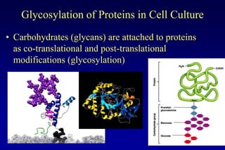
- 1. Glycosylation of Proteins in Cell Culture • Carbohydrates (glycans) are attached to proteins as co-translational and post-translational modifications (glycosylation)
- 2. Many biological molecules are Glycoproteins Glycosylation affects the functional qualities of the protein. We must know how the structure relates to the function to create effective biotherapeutics.
- 3. Overview 1. Why is glycosylation important in biotechnology. 2. Purpose of glycosylation in general. 3. Types of glycans: N-linked, O-linked. 4. Variation in glycosylation between cell types 5. Synthesis of N-linked glycans 6. Cell culture conditions that affect glycosylation
- 4. Understanding glycosylation in biological drugs is important for two main reasons: • Glycan can affect many of the protein properties: pharmacokinetics (uptake and length of time in the body), bioactivity, secretion, in vivo clearance, solubility, recognition, and antigenicity • Quantitative and qualitative aspects of glycosylation affected by production process in culture, including cell line, method of culture, extracellular environment, and protein itself
- 5. Why is the biotech industry concerned about Glycosylation? • Batch to batch variability of glycosylation patterns affect product quality • Too much variation in glycosylation leads to discarding of product • Regulatory agencies (eg FDA, Health Canada) have regulations for amount of acceptable variability in glycosylation –deviations can lead to redoing clinical trials • Change in glycosylation can lead to another company claiming a new patent • Adverse reactions in patients to non-human glycosylation
- 6. beta-interferon Immunoglobulin (monoclonal antibodies, Mabs) Protein N-glycan sites Erythropoietin (EPO) gp120 25 huCD36 9 huICAM-1 (CD54) 8 hu-tPA 3 hu-Epo 3 hu-IFN gamma 3 rhu ant-IL-8 (IgG) 2 hu-CSF 2 hu-IFN beta 1
- 7. Approved Monoclonal Ab’s (2008)
- 8. Monoclonal Antibodies in Cancer Treatment Majidi et al 2009
- 9. The list of glycosylated biopharmaceuticals in rapidly growing. Proper glycosylation is essential for the function of these biotherapeutics. Kawasaki et al 2008
- 10. FAQs about glycosylation • 50% of eukaryotic proteins are glycosylated • N-linked (Asn) and O-linked (Ser/Thr) glycosylation • N-linked glycosylation is the more complex • 65% of sequons (attachment sites) are occupied • Macroheterogeneity = variation in occupancy of sequons (eg. one site vs two site occupied) • Microheterogeneity = variation in structures of glycans (eg. Biantennary vs triantennary at site)
- 11. Why is Glycosylation Important in Biotherapeutics? Carbohydrate structures can affect the properties of the glycoprotein, including: → pharmacokinetics → bioactivity → secretion → in vivo clearance → solubility → recognition → antigenicity
- 12. General Function of Glycosylation • N-linked glycosylation prevalent in eukaryotes but not as common in prokaryotes • Function of glycans not well defined for many GP’s: – May be to aid protein folding and transport process – Prevent self adhesion of molecule (eg. beta-interferon) – Oligosaccharide can limit the approach of other macromolecules • Eg. Inhibit digestion of glycoprotein by proteases (eg. high concentration on cell surface) – Regulatory roles • Eg. Notch: cell surface signalling receptor – important for proper cell fate determination (O-glycosylated)
- 13. Glycosylation is Important in Development: Cell fate choices dependent on the Notch receptor require appropriate glycan expression A- Aberrant wing morphology results from mutation in glycosylation of the Notch receptor B- normal neural development, C-mutant with altered glycosylation Essentials of Glycobiology Second Edition Chapter 24, Figure 3
- 14. Purpose of Glycosylation (cont’d) • Activation of secondary pathways: – Eg. IgG (monoclonal antibodies) • Differences in glycan structure can change the way the antibody elicits a response when it binds to the antigen Fv region which binds the antigen IgG oligosaccharide affects the conformation Fab region which of the Fab region and binds effector affects how the antibody molecules and binds to other molecules cells which results in an immunue response
- 15. Glycosylated vs Nonglycosylated • Glycosylation can add up to 100% mass to a protein • Example: EPO – erythropoetin 39 kDa 18 kDa
- 16. Glycosylation of protein in cell culture • Mammalian vs prokaryotes, lower eukaryotes – Mammalian cells perform post-translational modifications and achieve a product close to that produced in vivo – Most Prokaryotes lack glycosylation machinery (exception: Campylobacter, N-linked glycosylation) – Yeast, insect, and plant cells produce different glycan structures • glycan processing in golgi differs from mammalian cells
- 17. Organisms Differ in Glycosylation • Bacteria are incapable of glycosylating recombinant mammalian proteins • Yeast have the tendency to hyper-mannosylate • Plant and Insect produced glycoproteins tend to have α 1,3- Peptide linked fucose and xylose residues N-acetylglucosamine Mannose Galactose • CHO cells are most commonly used for recombinant protein Fucose production N-glycolylneuraminic acid – Close to human glycosylation N-acetylneuraminic acid Xylose • Important to use Mammalian cells for glycoprotein production Transgenic Transgenic Bacteria Yeast Insect Plants Animal Cells Human
- 18. Two Primary types of glycosylation are differentiated by the type of linkage to the protein Serine Asparagine
- 19. Differences in Oligosacchride Structures in N-linked or O-linked Glycans N-acetylglucosamine Mannose Galactose N-acetylneuraminic acid Fucose N-acetylgalactosamine N-linked or O-linked oligosaccharide chains on proteins can have many different patterns of sugar residues at the same sequon. This is called Microheterogeneity.
- 20. O-glycans • Glycan is bound via an O-glycosidic bond of GalNAc to a Ser/Thr (O- glycosylation) • Classified as one of 8 core structures • Any Ser/Thr residue is a potential site for O-glycosylation, no consensus sequence identified • Addition of the glycan occurs on a fully folded protein
- 21. Core Structures Can Have Many Additions (Rose and Voynow 2006)
- 22. O-linked Glycans • Huge variety of structures: from very short to very long chains • Are important in mucins (major component of mucus), with very long chains • Are often found altered in cancer cells • Important in blood cell types (A, B, etc)
- 23. Mucins
- 24. N-linked Glycosylation • N-linked precursor added to most proteins in RER membranes • Only Asn in Asn-X-Ser/Thr become glycosylated • Core region survives extensive oligosaccharide trimming in Golgi Figure 12-51 (Alberts)
- 25. Presence of Sequon (Asn-X-Ser/Thr) does not guarantee glycosylation 1. Spatial arrangement of the peptide during translation process may expose or hide the tripeptide sequence 2. Glycosylation depends on X: (sequon Asn-X-Ser/Thr) glycosylation high when X = Ser, Phe, intermediate for Leu, Glu, very low for Asp, Trp, and Pro 3. Availability and correct assembly of precursors (eg. nucleotide sugars) 4. Level of expression of the oligosaccharyltransferase enzyme(s) 5. Disulfide bond formation within protein (makes site inaccessible to precursor addition)
- 26. 3 Types of N-linked Glycans Core region Complex n-Linked Glycan: Core with Terminal Can be heterogeneous -3 terminal branches “Sequon” -2 or 4 also common High Mannose N-linked Glycan: •Not trimmed to core and more mannose are added on •2 to 6 Additional mannose added onto core Hybrid N-Linked Glycan: Hybrid of high mannose and complex One Mannose Branch One GlcNAc and Gal branch
- 27. Protein Glycosylation in RER Polypeptide enters ER lumen Proteins and lipid-glycan are generated separately then glycan transferred on to the protein structure from the lipid. 9 Mannose Oligosaccharyl transferase enzyme Man- Man transfers precursor Man oligosaccharide from Man- Man dolichol to Asn GlcNAc-GlcNAc-Man Man-Man-Man-Glc-Glc-Glc 2 N- 3 glucose Acetylglucosamine Figure 12-52 (Alberts)
- 28. Production of the Lipid-Glycan The molecule is flipped from the ER membrane to the ER lumen. Cytoplasm Lumen Sugar residues are added sequentially to the Additional sugars are added via dolichol lipid to give a Man5-Glc3 structure (using phosphate. Finally, the oligosaccharide nucleotides sugars. (14 residues) is transferred to a specific Asn in the lumen (Man9-Glc3)
- 29. Dolichol Cycle -synthesis of the sugar chain on the lipid, dolichol FLIPPASE ENZYME Flips oligosaccharide to internal lumen of ER membrane Oligosaccharide is transferred from dolichol- phosphate to the protein at a sequon (Asn-X-Thr/Ser)
- 30. The Processing Reactions: the introduction of structure variation in the glycan Begins after the glycan is added to the protein. l n k j m jProcessing begins – removal of glucoses ER Lumen kMannosidase I removes 1 mannose lGolgi mannosidase I removes 3 mannose Golgi Lumen mN-acetylglucosamine transferase I adds GlcNAc nMannosidase II to removes 2 mannose
- 31. Role of N-linked Glycosylation in Protein folding If the glycoprotein is not correctly folded, glucose will be readded and sent back through the calnexin cycle To the GOLGI for processing and modification of the glycan and protein -Binds glycoprotein to help with folding -Recognizes glucose residues and glucosidase cleaves off
- 32. N-Linked Glycosylation PathwayOligomannose Asn Xaa Ser/Thr Type Dol Endoplasmic Reticulum P a-Glc I a-Glc II a-Glc II a-Man I P NH2 Oligosaccharide transferase Man Glc Golgi Glc Glc SialT GalT GnTII Man II GnTI FucT Complex type Man Hybrid Processing Reactions type
- 33. Fig 11 Production of tri- and tetra-antennary structures GnT IV GnT V Asn GnT V Asn GnT IV Asn M3Gn2 M3Gn4 Asn M3Gn3
- 34. Fig 12 Reaction network for N-linked glycosylation Leads to great diversity in structures M9 (From Umana and Bailey, 1997) 4x ManI 1-4 M5 GTI 5 GalT13 M Gn GnTIII 20 27 M5 GnGn b GalT M M5 GnG 5 5 GalT 6 GnTIII 28 GnGnbG M4 GnG 14 M4 Gn 21 M4 GnGnb GalT M4 GalT 7 GnTIII GnGnbG M3 GnG 29 22 M3 GnGnb GalT M3 15 M3 Gn 8 30 GnGnbG 16 GnTIII M3 Gn2G GalT M Gn 23 M Gn Gnb GalT M3 Gn2GnbG 3 2 3 2 GnTIV10 9 GnTV 18GalT 17 GalT M3 Gn3G M3 Gn3 M3 Gn3’ M3 Gn3’G 12 GnTIII GnTIII GnTV 25 b GnTIV 24 GalT M3 Gn3Gn 11 M3 Gn3 ’Gnb M3 Gn3’Gnb M3 Gn4 31 GalT GalT GnTIII 32 19 26 GalT M3 Gn3Gn bG M3 Gn4G M3 b b 33 M3 Gn4Gn G
- 35. Cell-associated factors that affect product glycosylation in cell culture • host cell line - complement of processing enzymes • mode of culture - suspension/ attached - batch/ continuous • specific protein productivity - changes rate of transit through Golgi • extracellular degradative enzymes - release of sialidases by cells
- 36. Factors affecting protein glycosylation (N-linked) 1. Host cell • glycan structures on the same proteins can vary between species and even different tissues • due to: – differences in relative activities of glycan processing enzymes (glycosidases and glycosyltransferases) – differences in the monosaccharide precursors
- 37. CHO and BHK • Structure of sialic acid from CHO and BHK differ from human sialic acid (also in rodents, pigs, sheep, cows, and new world monkeys) – NGNA – N-glycoyl-neuraminic acid (humans don’t produce this) – NANA – N-acetyl-neuraminic acid (most common sialic acid) • Presence of a2,3 terminal sialic acid addition compared to a2,6 terminal sialic acid (in humans) • Absence of a functional a1,3 fucosyltransferase • Absence of N-acetylglucosaminyltransferase III (Gn TIII) – differences do not lead to immunogenic responses to glycoproteins – no adverse physiological effect due to structural differences
- 38. Hamster vs Mouse cells • Mouse cells express: a1,3 galactosyltransferase: generating Gala1,3-Galb1,4-GlcNAc (not found in humans) – gene is present in CHO and BHK but not expressed • Limits use of murine cells in therapeutic glycoprotein production
- 39. 2. Culture environment • Specific conditions of the culture can affect glycosylation independently of the cell line • During the process of a batch culture, nutrient consumption and product accumulation can change the culture environment – gradually decreasing the extent of protein glycosylation • may lead to variable glycoform heterogeneity and batch-to-batch variation
- 40. Adherent Cells Suspension 3. Mode of culture • adaptation from anchorage dependent growth to suspension culture may also affect the glycsosylation process • presence or absence of serum also has a significant affect on glycosylation – presence of hormones and growth factors, high activities of sialidase and fucosidase
- 41. 4. Protein productivity • differences in growth rate, specific productivity, and cell density among the bioreactors may cause variation in the pattern of N-linked glycan structures • rate of protein expression may also affect glycosylation
- 42. 5. Glucose availability • glucose limitation results in incomplete protein glycosylation – synthesis of abnormal dolichyl precursor oligosaccharides – sequences that are normally glycosylated remain empty 6. Ammonia • accumulated ammonia is inhibitory to cell growth and to protein glycosylation – increase in pH of the normally acidic distal golgi – increase in the UDP-GNAc pool (reduces sialylation) 7. pH • maximum glycosylation of a protein occurs between pH 6.9-8.2
- 43. 8. Oxygen limitations • Limiting nutrient because of it’s low solubility in media 1. reduced dissolved oxygen (DO) may lead to reduction in UDP-Gal – reduced oxidative phosphorylation of UDP-Gal – reduced UDP-Gal transport from the cytosol to the golgi 2. formation of premature disulfide bonds in the nascent protein
- 44. Effect of Dissolved Oxygen on Sialylation of EPO 100 % sialylated structues 95 90 85 80 75 70 65 60 3% 10% 50% 100% 200% DO concentration (% air saturation)
- 45. 9. Growth factors, vitamins and hormones • up- and down-regulation of specific glycosyltransferases in conjunction with hormonal induction of cell differentiation • changes due to induction or repression or induction of the enzymes involved in protein glycosylation
- 46. 10. Extracellular degradation of glycoproteins • glycosidases may be released to the extracellular environment by secretion or by cell lysis • activity of glycosidases depends on medium pH, temperature, residence time of glycoprotein, and level of extracellular activity
Notas del editor
- The oligosaccharides have many diverse functions on glycoproteins, and defined functionality for some has only recently been determined. Much more research is required. Glycoproteins – associated with the cell membrane with the carbohydrate is sticking out – GlycoproteinFunction – interaction with other moleculesParetoif receptor that interacts with cell growth factorsAssists to cell-to-cell communicationModifies protein functionModulates antibody function- facilitates binding to viruses and bacteria
- Recombinant glycoprotein – needs to be similar to the product in clinical trials – batch to batch variabilityPlatforms – consistent type of production systemEPO – needs to treat anemia and those that are in kidney dialysisAthletes who take EPO are caught because of the glycan structure differs in the drug compared to the natural one in the bodyDeviations – dramatic changes in glycosylation (percentage of difference) – can lead to redoing clinical trials (3rd stage) Clinical trials – w/ small group – later stages of a disease2nd stage – larger group than the first clinical trial – testing different dosage3rd stage – thousands of people – costs multimillion dollars needs to decide whether its worth to go into 3rd stage.Certain percentage of people have adverse reactions (antigenic reactions)
- Recombinant glycoproteins have differences in the number of oligosacchrides attached. The site of attachment and type of oligosaccharide is specific for the glycoprotein.Glycosylation sites are protein specific Beta-interferon – one carbohydrate chain that is bound to the same siteEPO – have 3 glycosylation sitesMabs – glycosylation occurs between the two heavy chains in the Fc region of the antibody, and sometimes is also glycosylated in the tip of the Fab regionFab – antigen binding sitesFc – effector - binds to receptors – gets rid of antigen
- Monoclonal antibodies (Mabs) are a large portion of the new biotherapeutics being developed. All monoclonal antibodies are glycoproteins. Mabs can be used in a wide variety of treatments, eg. cancer, asthma, inflammatory diseases.
- Huge amount of research into generating monoclonals to treat cancer. Many are in use, but are not very good and are often used in combination with chemotherapy. Some are used to treat several types of cancer. Mab – aren’t good enough on their own, need to be combined with other treatments such as chemotherapy- due to glycosylation patterns
- There are many other types of biotherapeutics which are glycoproteins, that treat many different types of diseases. They are a wide variety of applications, and they all work in different ways. For many of them, proper glycosylation is essential for their function.
- N- linked seemed to be more important than O-linkedSequons – glycan attachment sitesMacrohetergeneity (larger picture) - # of glycosylation sites occupied by glycansMicroheterogeneity – variations of the glycan structure at a particular siteBoth are good to consider in quality of the proteinNeed to control both factors
- Mucins have very long chains of glycans (oligosaccharides), which allows high hydration and the “slippery” character of the molecules.
- Another series of enzymes leads to more branching and the production of triantennary and tetra-antennary structures.
- This figure just shows the great amount of diversity that can be generated in the processing reactions.
- The concentration of dissolved oxygen in the bioreactor has an effect on the amount of sialic acid that is added at the terminal end of the oligosaccharide. Sialic acid, because of its charge, can have a big effect on the function of the molecule.
- Glycosidases secreted by the cell can affect glycosylation of the recombinant protein after it is secreted from the cell. Monitoring of glycosidase activity can be done to check for levels over the culture production.