
Recomendados
Recomendados
Más contenido relacionado
La actualidad más candente
La actualidad más candente (20)
Similar a Cystic liver lesions - An ultrasound perspective
Similar a Cystic liver lesions - An ultrasound perspective (20)
Más de Samir Haffar
Más de Samir Haffar (20)
Último
Último (20)
Cystic liver lesions - An ultrasound perspective
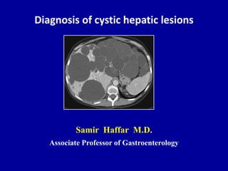
- 1. Diagnosis of cystic hepatic lesions Samir Haffar M.D. Associate Professor of Gastroenterology
- 2. Cystic lesions of the liver • Simple hepatic cyst • Hydatid cyst of the liver • Congenital fibrocystic liver diseases • Miscellaneous cystic lesions of the liver Murray KF & Larson AM. Fibrocystic diseases of the Liver. Springer Science + Business Media, Springer, 2010.
- 3. Simple hepatic cyst Congenital origin • Incidence 2.5 – 10% in general population by US Increases dramatically in > 60 years • Symptoms Rare – cyst > 5 cm especially in women Obstructive effect: pain, jaundice, PHT • Complications Hemorrhage – rupture – infection • Diagnosis US highly sensitive & specific Hemorrhagic cyst: CEUS – CECT • CA19-9 Usually normal or slightly elevated • Treatment Conservative, sclerotherapy, fenestration Shaked O et al. Clin Gastroenterol Hepatol 2011 ; 9 : 547 – 562.
- 4. Typical hepatic cyst Lesions such as this require no further imaging evaluation Tchelepi H et al. Ultrasound Quarterly 2004 ; 20 : 155 – 169. Anechoic lesion Thin wall Well-defined border Lateral refraction shadow Posterior acoustic enhancement
- 5. Mobile echoes in large liver cysts Naganuma H et al. J Clin Ultrasound 2010 ; 38 : 475 – 479. No intra-cystic artifact PRF: 4 kHzPRF: 6 kHz Deep location PRF: 5.5 kHz Superficial location Intracystic range-ambiguity artifacts Erroneous display of returning echoes from heart Depth according to PRF – Disappear at a low PR
- 6. Atypical liver cyst Dietrich CF et al. Ultrasound of the liver, European course book, EFSUMB. http://www.efsumb.org/ecb/ Conventional ultrasound Pseudo-solid pattern Small diameter - Slice thickness artefact Contrast enhanced ultrasound CEUS proves the cyst
- 7. Simple hepatic cyst Murray KF & Larson AM. Fibrocystic diseases of the Liver. Springer Science + Business Media, Springer, 2010. Non CECT Non- enhancement of cyst wall or contents CECTUltrasound Typical features
- 8. Simple liver cyst complicated by hemorrhage Rumack CM et al. Diagnostic ultrasound. Elsevier Mosby, St. Louis, Missouri, 3rd edition, 2005. Well-defined mass with uniform low-level internal echoes misinterpreted as solid mass Ultrasound Nonenhancing low-density mass consistent with cyst CECT scan
- 9. Diagrammatic representation of metacestode of Echinococcus granulosus Manual on Echinococcosis in humans & animals: a public health problem of global concern. Eds: J. Eckert, M.A.Gemmell, F.-X. Meslin & Z.S. Pawłowski. Paris, France 2001. Adventitial layer Host tissue Laminated layer Germinal layer Daughter cyst Capsule with protoscolex
- 10. WHO-IWGE classification Sonographic findings relating to viability of parasite Active InactiveTransitional WHO-IWGE: World Health Organization Informal Working Group on Echinococcosis WHO, IWGE. Acta Trop 2003 ; 85 (2) : 253 – 61. CL (cystic lesion) added: Parasitic nature not determined on US
- 11. WHO-IWGE classification International consensus classification 5 types on the basis of US appearances CE I Unilocular simple cysts CE3 CE2 CE4 CE 5 WHO-IWGE: World Health Organization Informal Working Group on Echinococcosis WHO Informal Working Group. Acta Trop 2003 ; 85 : 253 – 261. Floating membrane (water lily sign) Multivesicular multiseptated cyst Heterogeneous degenerative contents Thick calcified wall Active Transitional Active Inactive Inactive
- 12. Hydatid cyst of the liver CE 1 Turgut AT et al. Ultrasound Quarterly 2008 ; 24 : 17 – 29. Hydatid sand Pathognomonic Double-line sign PathognomonicSimple hepatic cyst or hydatid cyst Cystic lesion (CL) Snow flake sign
- 13. Sonographic scoring system with routine serology* to differentiate hepatic from hydatid cysts • No wall sign • Irregular borders • Irregular shape • No wall calcifications • Females • Stable US appearance at follow-up • Septa: one or two septa – irregular shape * Serology: IHA (indirect hemagglutination assays) – ELISA IgG Grisolia A et al. J Ultrasound 2009 ; 12 : 75 – 79.
- 14. Septate hepatic cyst Grisolia A et al. J Ultrasound 2009 ; 12 : 75 – 79. Slightly irregular septum
- 15. Hydatid cyst of the liver CE 2 – Daughter cysts “honeycomb-like” or „„rosette-like‟‟ structure Pathognomonic Czermak BV et al. Abdom Imaging 2008 ; 33 : 133 – 143.
- 16. Liver hydatid cyst CE 3 – Water lily sign Turgut AT et al. Ultrasound Quarterly 2008 ; 24 : 17 – 29. Lutz HT et al. Manual of diagnostic ultrasound in infectious tropical diseases. Springer, 2006. Focal splitting of cyst wall Extended splitting of cyst wall „„floating membrane‟‟
- 17. Water lily
- 18. Czermak BV et al. Abdom Imaging 2008 ; 33 : 133 – 143. „„ball of wool sign‟‟ Hypoechoic, hyperechoic, or intermediate pattern Liver hydatid cyst CE 4
- 19. Liver hydatid cyst CE 5 Well-defined hyperechoic wall Calcified dead hydatid cyst Turgut AT et al. Ultrasound Quarterly 2008 ; 24 :17 – 29.
- 20. US in operated cases for hydatid cyst Recurrence or pseudocyst? Follow-up US • Partial involusion in follow-up images • Follow-up should be performed every 6 months 12 months to distinguish recurrence from pseudocyst Pseudocyst might persist for many years Ozturk A et al. Eur J Radiol 2005 : 56 : 91 – 96. Pseudocyst • Cavities smaller than original cyst • Irregular & thick borders
- 21. US findings in operated cases for hydatid cyst 77 patients – Mean age 38 years – Recurrence 11% Ozturk A et al. Eur J Radiol 2005 : 56 : 91 – 96. Hypoechoic Thick & irregular wall Pseudocyst Anechoic Thick & irregular wall Pseudocyst Recurrence Anechoic Unilocular hydatid cyst
- 22. Congenital fibrocystic liver diseases Ductal plate malformation • Biliary hamartomas • Peribiliary cysts • Choledochal cysts • Autosomal dominant polycystic kidney disease (ADPKD) • Autosomal recessive polycystic kidney disease (ARPKD) • Polycystic liver disease (PCLD) • Other ciliopathies Bardet-Biedl syndrome Joubert syndrome Meckel-Gruber syndrome Jeune syndrome
- 23. Normal ductal plate development Single-layered ductal plate Double-layered ductal plates Primary resorption Slit-like primitive BD Extensive resorption Fine BD around PV Insufficient resorption Dilated BD & abnormal branching “central dot sign” Abnormal development Turkbey B et al. Pediatr Radiol 2009 ; 39 : 100 – 111.
- 24. Structure & function of primary cilium Hair-like projection from plasma membrane into extracellular space Murray KF & Larson AM. Fibrocystic diseases of the Liver. Springer, NY, 2010. Intraflagellar transport (IFT) proteins bi-directionally Mechanosensory, chemosensory, & osmosensory functions
- 25. Association of fibrocystic diseases of the liver Series of 51 patients Combination of CHF & Caroli’s disease most striking Biliary hamartomas reported in 10 patients Summerfield JA et al. J Hepatol 1986 ; 2 : 141.
- 26. • Cystic dilated bile ducts < 10 mm in diameter • Usually distributed equally throughout the liver • 5 % in autopsy series, 0.5% in needle biopsy • Liver biopsy: dilated or branching BD lined by single layer of cuboidal cells embedded in collagenous stroma • Misdiagnosed as multiple liver metastases on imaging • MRI is the best imaging modality Biliary hamartomas Von Meyenburg complexes Drenth JPH et al. Best Prac Res Clin Gastroenterol 2010 ; 24 : 573 – 584.
- 27. Biliary hamartomas Von Meyenburg complexes Cystic space with fibrous stroma lined by layer of biliary epithelium Bile plugs within dilated bile ducts Brancatelli G et al. RadioGraphics 2005 ; 25 : 659 – 670.
- 28. Rumack CM et al. Diagnostic ultrasound. Elsevier Mosby, St. Louis, Missouri, 3rd edition, 2005. Bright echogenic foci with "ringdown" artifact Single small hypoechoic liver mass Biliary hamartomas Von Meyenburg complexes
- 29. Biliary hamartomas Von Meyenburg complexes Smaller than 10 mm Disconnected from biliary tree White onT2-weighted images Gadolinium-enhanced T2-weighted MRI Drenth JPH et al. Best Prac Res Clin Gastroenterol 2010 ; 24 : 573 – 584.
- 30. Peribiliary cysts Chronic liver disease specifically with PHT • Dilated peribiliary glands along portal veins & bile ducts Located in hepatic hilum & larger portal tract No communicate with biliary tree Few millimeters to 10 mm in size – larger cysts reported • Usually clinical benign condition Focal biliary obstruction as result of larger cysts • US, CT & MRI: rounded anechoic structures along portal tract May be confounded with dilated bile ducts Drenth JPH et al. Best Prac Res Clin Gastroenterol 2010 ; 24 : 573 – 584.
- 31. Peribiliary cysts Fewer than 60 reported cases Brancatelli G et al. Eur J Radiol 2007 ; 61 : 57 – 69. T2-weighted MRI High intensity cysts lying along left PV “string of beads” T1-weighted MRI Low intensity cysts lying along left PV Peribiliary cysts at hilum High intensity areas in RL MRCP
- 32. Choledochal cysts Todani’s classification Type II CBD diverticulum Type III Choledococele Type IV IH & EH Todani T et al. Am J Surg 1977 ; 134 : 263 – 9. Savader SJ et al. Am J Roentgenol 1991 ; 156 : 327. Type I Most frequent 78% Type V Caroli‟s disease 1 %
- 33. Choledochal cyst type I Large cystic mass in RUQ connected to biliary tract Levy AD et al. Curr Probl Diagn Radiol 2003 ; 32 : 233 – 263. Ultrasound Classic triad: abdominal pain, jaundice, palpable mass (rare)
- 34. Choledochal cyst type I Brancatelli G et al. RadioGraphics 2005 ; 25 : 659 – 670. Cystic mass due to dilatation of distal portion of CBD Contrast-enhanced CT Fusiform dilatation of CBD Dilatation of IHBT Reformatted image
- 35. Mild fusiform dilatation of common hepatic duct Marked fusiform dilatation of common bile duct Levy AD et al. Curr Probl Diagn Radiol 2003 ; 32 : 233 – 263. Choledochal cyst type I MRCP
- 36. Fusiform dilatation of extra-hepatic bile ducts Aberrant entry of CBD at side of pancreatic duct Brancatelli G et al. RadioGraphics 2005 ; 25 : 659 – 670. PTC Choledochal cyst type I
- 37. Normal pancreatico-biliary junction Levy AD et al. Curr Probl Diagn Radiol 2003 ; 32 : 233 – 263. Common channel for pancreatic & common bile duct Separate channels for pancreatic & common bile duct
- 38. Common channel of pancreatic & biliary ducts Forbes A & Murray-Lyon IM. Gut Supplement 1991, S 116 – S 122. Long common channel of pancreatic & biliary ducts Normal common channel of pancreatic & biliary ducts
- 39. Anomalous pancreatico-biliary junction With choledochal cyst 65 – 95% by ERCP Without choledochal cyst Carcinoma of gallbladder & biliary tract Gallbladder adenomyomatosis Cholelithiasis Pancreatitis Levy AD et al. Curr Probl Diagn Radiol 2003 ; 32 : 233 – 263. No further evaluation “forme fruste choledochal cyst”
- 40. Choledochal cyst with cholangiocarcinoma 50-year-old woman with 1 month history of abdominal pain Levy AD et al. Curr Probl Diagn Radiol 2003 ; 32 : 233 – 263. 2 cm enhancing polypoid mass from wall of choledochal cyst Dominant filling-defect: adenoca Other filling defects: sludge A series of 48 patients reported incidence of 12.5%
- 41. Levy AD et al. Curr Probl Diagn Radiol 2003 ; 32 : 233 – 263. Diverticulum connected to CBD by thin neck Choledochal cyst type II Biliary diverticula ERCP
- 42. Choledochal cyst type III / Choledochocele Coined by Wheeler in 1940 – Similarity to ureterocele Saccular dilation of intramural segment of distal CBD Very small risk of carcinoma (4 reported cases) Levy AD et al. Curr Probl Diagn Radiol 2003 ; 32 : 233 – 263.
- 43. Choledochocele CBD W Singh M & Coyle W. Gastroenterology 2010 ; 138 : e3 – e4. EUS
- 44. Scholz classification of choledochocele Scholz FJ et al. Radiology 1976 ; 118 : 25 – 8. Levy AD et al. Curr Probl Diagn Radiol 2003 ; 32 : 233 – 263. Type A Cystic dilation of ampulla Type B Diverticulum extending from ampulla
- 45. Caroli J et al. Semin Hop Paris 1958 ; 34 : 128 – 35. http://emedicine.medscape.com Choledochal cyst type V Caroli’s disease Described by Caroli & coworkers in 1958
- 46. Caroli’s disease & Caroli’s syndrome Caroli’s disease Caroli’s syndrome (more common) Segmental dilatation of large intra-hepatic biliary ducts Segmental dilatation of small intra-hepatic biliary ducts & congenital hepatic fibrosis Drenth JPH et al. Best Prac Res Clin Gastroenterol 2010 ; 24 : 573 – 584.
- 47. Caroli’s disease / Ultrasound Lefere M et al. Eur J Gastroenterol Hepatol 2011 ; 23 : 578 – 585. Cystic dilation of intra-hepatic bile ducts communicating with normal bile duct
- 48. Caroli’s disease / Central dot sign Akhan O et al. Eur J Radiol 2007 ; 61 : 18 – 24. PV branch protruding into lumen of dilated bile duct Cystic ductal dilatation Characteristic central dot sign T1-weighted MRI Cystic ductal dilatation Characteristic central dot sign CECT scan
- 49. Caroli’s disease / Central dot sign Turkbey B et al. Pediatr Radiol 2009 ; 39 : 100 – 111. Dilated IHBD with central dot sign Coarsened liver consistent with fibrosis Flow within central dot sign representing portal venous flow 7-year-old boy with Caroli syndrome High-resolution US image with 6-MHz probe
- 50. Caroli’s disease / PTC Multiple saccular dilatations of intra-hepatic bile ducts Mostly at periphery of the liver Brancatelli G et al. RadioGraphics 2005 ; 25 : 659 – 670.
- 51. Complications of Caroli’s disease • Cholangitis Frequency vary greatly • Liver abscess • Lithiasis Intra & extra-hepatic lithiasis • Cholangiocarcinoma 2.5 – 15% • Amyoidosis Yonem O & Bayraktar Y. World J Gastroenterol 2007 ; 13 : 1930 – 1933.
- 52. Biliary stones in Caroli’s disease Lefere M et al. Eur J Gastroenterol Hepatol 2011 ; 23 : 578 – 585. Brancatelli G et al. RadioGraphics 2005 ; 25 : 659 – 670. Ultrasound Saccular IHBD dilatation containing stone Dilated bile ducts with multiple stones Unenhanced CT scan
- 53. Adenocarcinoma in Caroli disease ERCP Levy AD et al. Curr Probl Diagn Radiol 2003 ; 32 : 233 – 263. Irregular narrowing of left hepatic duct at site of adenocarcinoma Dilated ducts peripheral to neoplasm containing lithiasis
- 54. Autosomal Dominant Polycystic Kidney Disease Systemic disorder • Polycystic liver disease Most common • Other cysts Splenic cysts Pancreatic cysts Seminal vesicle cysts Arachnoid cysts • Intra-cerebral aneurysms 8% • Cardiac valve disease Mitral valve prolapse: 25% Drenth JPH et al. Best Prac Res Clin Gastroenterol 2010 ; 24 : 573 – 584.
- 55. Autosomal dominant polycystic kidney disease Ultrasound Weber TM. Ultrasound Clin 2006 ; 1 : 15 – 24. 15.6 cm Cysts of variable size with bilaterally enlarged kidneys
- 56. Screening of ADPKD Renal US & DNA analysis in 319 patients at risk Ravine D et all. Lancet 1994 ; 343 : 824 – 827. Nicolau C et al. Radiology 1999 ; 213 : 273 – 276. Person at risk & younger than 30 years Two cysts in one kidney or one cyst in each kidney Sen: 95% (ADPKD-1) – 65% (ADPKD-2) Person at risk & aged 30 – 59 years Two cysts in each kidney (Sen: 100%) Person at risk & aged 60 years or older Four cysts in each kidney (Sen: 100%)
- 57. Autosomal Recessive Polycystic Kidney Disease Pediatric disease – 1/20 000 live births • Late pregnancy or at birth Oligohydramnios Massively enlarged kidneys Pulmonary hypoplasia Potter facies Contracted limbs & clubfeet • Congenital hepatic fibrosis Almost all patients – PHT • Other ductal plate malformations Drenth JPH et al. Best Prac Res Clin Gastroenterol 2010 ; 24 : 573 – 584.
- 58. Fibrocystin in cilia of bile ducts of liver & collecting ducts of kidney Turkbey B et al. Pediatr Radiol 2009 ; 39 : 100 – 111. Defect of this protein leads to cyst formation in liver & kidney
- 59. Turkbey B et al. Pediatr Radiol 2009 ; 39 : 100 – 111. Partial medullary involvement Complete medullary involvement Complete medullary involvement < 50% partial cortical involvement Complete medullary involvement with >50% cortical involvement Autosomal Recessive Polycystic Kidney Disease
- 60. Autosomal Recessive Polycystic Kidney Disease Turkbey B et al. Pediatr Radiol 2009 ; 39 : 100 – 111. Mildly enlarged kidney Preserved cortex Significant echogenic renal medulla Conventional 3.5-MHz probe Complete medullary involvement No cortical involvement Schematic drawing
- 61. Autosomal Recessive Polycystic Kidney Disease Turkbey B et al. Pediatr Radiol 2009 ; 39 : 100 – 111. Weber TM. Ultrasound Clin 2006 ; 1 : 15 – 24. Enlarged echogenic kidney Poor corticomedullary differentiation RK: 10 cm Conventional 3.5-MHz probe Complete medullary involvement >50% cortical involvement Schematic drawing
- 62. Autosomal Recessive Polycystic Kidney Disease 26 patients – HR linear array transducers Traubici J et al. Am J Roentgenol 2005 ; 184 : 1630 – 1633. Dilated tubules Hyperechoic foci some with ring-down artifact Macroscopic cysts
- 63. Autosomal Recessive Polycystic Kidney Disease Multiple small cysts Salt & pepper pattern 27 years old woman Tuma J et al. Genitourinary ultrasound, European course book, EFSUMB. http://www.efsumb.org/ecb/
- 64. Congenital hepatic fibrosis Affecting hepatobiliary &renal systems • Congenital inherited in autosomal recessive fashion • ARPKD with less renal involvement & longer survival • Onset usually in adolescence or young adulthood Early childhood to 5th or 6th decade • Liver biopsy: High specificity – Low sensibility Periportal fibrosis & irregular proliferating bile ducts Akhan O et al. Eur J Radiol 2007 ; 61 : 18 – 24. Difficulty in clinical & pathologic diagnosis
- 65. Congenital hepatic fibrosis Unknown incidence & prevalence Portal tract expanded by fibrosis Several BD some with curvilinear profiles (ductal plate malformation) Murray KF & Larson AM. Fibrocystic diseases of the Liver. Springer Science + Business Media, Springer, 2010.
- 66. Imaging of congenital hepatic fibrosis • Distinctive morphologic findings Atrophic right lobe Cirrhosis/CHF Hypertrophic caudate lobe Cirrhosis/CHF Hypertrophic left lateral segment Cirrhosis/CHF Normal or hypertrophic left medial segment CHF • Enlarged hepatic artery • Large regenerative nodules • Alterations of echotexture & homogeneity Brancatelli G et al. RadioGraphics 2005 ; 25 : 659 – 670.
- 67. Hypertrophic left medial segment Hypertrophic left lateral segment Splenomegaly CECT scan – Portal phase Congenital hepatic fibrosis Zeitoun D et al. Radiology 2004 ; 231 : 109 – 116.
- 68. Turkbey B et al. Pediatr Radiol 2009 ; 39 : 100 – 111. Congenital hepatic fibrosis 35-year-old man with CHF/ARPKD Multiple vessels around PV resembling cavernoma Patent portal vein “Cavernoma-like periportal vessels”
- 69. Akhan O et al. Eur J Radiol 2007 ; 61 : 18 – 24. Congenital hepatic fibrosis Regenerative nodules Dilatation of intra-hepatic bile ducts US image ultrasound
- 70. Congenital hepatic fibrosis Turkbey B et al. Pediatr Radiol 2009 ; 39 : 100 – 111. Echogenic liver compared with RK Secondary to fibrosis Significantly coarsened parenchymal echotexture High-resolution US (6-MHz)Conventional US (4-MHz) 10-year-old boy with CHF/ARPKD
- 71. Congenital hepatic fibrosis Turkbey B et al. Pediatr Radiol 2009 ; 39 : 100 – 111. 10-year-old boy with CHF/ARPKD Moderately echogenic liver parenchyma Thickened periportal space consistent with fibrosis “Periportal cuffing”
- 72. Autosomal Dominant Polycystic Liver Disease ADPLD PRKCSH: Protein kinase substrate 80K-H Onori P et al. Dig Liver Dis 2010 ; 42 : 261 – 271.
- 73. Types of PCLD Drenth JPH et al. Hepatology 2010 ; 52 : 223 – 2230. One or few dominant cysts Type I Multiple cysts limited to one part of liver Type II Cysts through several segments of liver Some segments relatively free from cysts Type III Extensive polycystic liver Hardly recognizable liver parenchyma Type IV
- 74. Polycystic Liver Disease (PCLD) Drenth JPH et al. Best Prac Res Clin Gastroenterol 2010 ; 24 : 573 – 584. Diagnosis made when > 20 liver cysts are present Or > 4 liver cysts in member of family with known PCLD
- 75. Polycystic Liver Disease (PCLD) Murray KF & Larson AM. Fibrocystic diseases of the Liver. Springer Science + Business Media, Springer, 2010. Numerous cysts, many of them calcified Most calcifications not completely circular
- 76. Treatment of PCLD Drenth JPH et al. Hepatology 2010 ; 52 : 223 – 2230. Best treated by Percutaneous aspiration & alcohol sclerosis Symptomatic dominant cyst
- 77. Treatment of PCLD Drenth JPH et al. Hepatology 2010 ; 52 : 223 – 2230. Best treated by Combined right hepatectomy & fenestration Severe PCLD Relative sparing of left lobe
- 78. Autosomal Recessive Polycystic Liver Disease ARPLD PKHD1: Polycystic Kidney & Hepatic Disease 1 Onori P et al. Dig Liver Dis 2010 ; 42 : 261 – 271.
- 79. Turkbey B et al. Pediatr Radiol 2009 ; 39 : 100 – 111. Numerous anechoic lesions Consistent with hepatic cysts 39-year-old woman with CHF Autosomal Recessive Polycystic Liver Disease ARPLD
- 80. Miscellaneous cystic lesions of the liver • Ciliated hepatic foregut cyst • Peliosis hepatis • Primary neoplasm: biliary cystadenoma – hemangioma – HCC • Cystic metastasis • Hepatic abscess: pyogenic – amebic • Intrahepatic pancreatic pseudocyst • Endometriosis • Hematoma & biloma Murray KF & Larson AM. Fibrocystic diseases of the Liver. Springer Science + Business Media, Springer, 2010.
- 81. Ciliated hepatic foregut cyst Sharma S et al. Dig Dis Sci 2008 ; 53 : 2818 – 2821. T2 MRI: hyperintense cyst Hypodense non-enhanced lesionHypoechoic cystic mass Columnar epithelium with cilia
- 82. Conditions associated with peliosis hepatis Azathioprine 6-Thioguanine AIDS/Bartonella infection Tuberculosis Myeloproliferative diseases Leukemia Lymphoma Multiple myeloma Macroglobulinemia Anabolic steroids Oral contraceptives Arsenic Thoratrast Vinyl chloride Yamada’s textbook of gastroenterology, Blackwell Publishing, 5th Edition, 2009.
- 83. Peliosis hepatis From Greek pelios (purple) Kleinig P et al. Clinical Radiology 2003 ; 58 : 995 – 998. US: hypoechoic lesion in background of liver steatosis CEUS: transient “fast surge” central echo-enhancement
- 84. Biliary cystadenoma Often incorrectly diagnosed as simple cysts Tchelepi H et al. Ultrasound Quarterly 2004 ; 20 : 155 – 169. Irregular cystic mass with thick septations Useful cystic fluid analysis for CA 19-9 & CEA Excision to prevent recurrence & malignant transformation (25%)
- 85. Biliary cystadenoma < 200 reported cases – women in 85% 56-year-old asymptomatic woman – CECT scan Multilocular cyst in porta hepatis Enhancing septae Multilocular cyst in porta hepatis Mural calcification Levy AD et al. Curr Probl Diagn Radiol 2003 ; 32 : 233 – 263.
- 86. Biliary cystadenoma Dietrich CF et al. Ultrasound of the liver, European course book, EFSUMB. http://www.efsumb.org/ecb/ Gray-scale ultrasound Hepatic cyst CEUS Intra-cystic thick septation
- 87. Large heterogeneous hemangioma (rare) Partially or entirely cystic mass Nonenhanced CT scan Hypoattenuating lesion with markedly hypoattenuating center Contrast-enhanced CT scan Typical enhancement pattern of hemangioma Vilgrain V et al. RadioGraphics 2000 ; 20 : 379 – 397.
- 88. Cystic hepato-cellular carcinoma (rare) Partially or entirely cystic mass Teoh AYB et al. World J Surg 2006 ; 30 : 1560 – 1566. Internal necrosis: Rapid growth, stemic & locoregional treatment Signs or complications of liver cirrhosis, ↑α fetoprotein
- 89. Cyst-like appearance of hepatic metastases • Hypervascular metastasis with rapid growth & necrosis Neuroendocrine tumors Sarcoma Melanoma Certain subtypes of lung & breast carcinoma • Mucinous adenocarcinoma Colorectal carcinoma Ovarian carcinoma Mortelé KJ et al. RadioGraphics 2001; 21 : 895 – 910.
- 90. Cystic metastases from breast cancer Lencioni R, Cioni D & Bartolozzi C. Focal Liver Lesions. Springer-Verlag, Berlin, 2005. Cyst-like hypoattenuating lesion without peripheral enhancement Portal phase CECT scan Arterial phase CECT scan Peripheral rim-enhancement Typical of metastatic lesion Known primary tumor, multiple lesions, rim enhancement
- 91. Cystic metastasis from ovarian cancer Portal venous phase CECT scan Mortele KJ et al. RadioGraphics 2001; 21 : 895 – 910. Ovarian metastases spread by peritoneal seeding Elliptical cystic lesion on surface of liver
- 92. Pyogenic hepatic abscess Thick-walled cystic lesion Peripheral enhancement “double target” sign Lencioni R, Cioni D & Bartolozzi C. Focal Liver Lesions. Springer-Verlag, Berlin Heidelberg , 2005. Portal venous-phase CECT scan
- 93. Pyogenic liver abscess Gas within lesion Rumack CM et al. Diagnostic ultrasound. Elsevier Mosby, St. Louis, Missouri, 3rd edition, 2005. Poorly defined hypoechoic mass with multiple gas bubbles Liver mass with extensive gas content
- 94. Amebic hepatic abscess Feldman M et al. Sleisenger and Fordtran's Gastrointestinal & Liver Disease. Saunders Elsevier, 9th edition, 2010. “Double target” sign Serologic tests (ELISA & IHA): Sen & Sp 95%
- 95. Intra-hepatic pancreatic pseudocyst Systematic review – 23 reported cases Guesmi F et al. Tunis Med. 2009 ; 87 : 801 – 4. Acute severe pancreatitis 3 weeks earlier Cystic lesion of left lobe – Thin capsule Pancreatitis , left liver lobe, thin capsule, signs of pancreatitis
- 96. Endometrial cyst of liver Extremely rare – 7 reported cases Cystic lesion in right lobe Ultrasound T2-weighted MRI High intensity signal in right lobe Rao M et al. Gut 2006 ; 55 : 742. Cyst aspirated under CT guidance Endometrial glandular & stromal elements
- 97. Hepatic hematoma Subcapsular hemorrhage after US-guided liver biopsy T2-weighted MRI High signal intensity & internal debris Mortelé KJ et al. RadioGraphics 2001 ; 21 : 895 – 910. T1-weighted MRI High signal intensity Paramagnetic effect of methemoglobin Liver trauma, known HCC or adenoma
- 98. Hepatic bilomas Three intra-hepatic collections compatible with bilomas 21-year-old man with biliary leakage after severe accident Portal phase CECT scan Mortele KJ et al. RadioGraphics 2001; 21 : 895 – 910. Trauma or biliary surgery – No septa capsules or calcifications
- 99. Conclusion • Clinical context is of utmost importance • Laboratory tests could be very beneficial in some cases • Imaging modalities (US, CEUS, EUS, CT, MRI) alone can made the correct diagnosis in the majority of cases • Liver biopsy when diagnosis remains elusive
- 100. Thank You
Notas del editor
- Biliarycystadenoma or cystadenocarcinomahighe levels of serum CA19-9 are observed
- Simple hepatic cysts are benign developmental lesions that do not communicate with the biliary tree. The current theory regarding the origin of true hepatic cysts is that they originate from hamartomatous tissue. Hepatic cysts are commonand are presumed to be present in 2.5% of the population. They are more often discovered in women and are almost always asymptomatic. Simple hepatic cysts can be solitary or multiple, with the latter being the more typical scenario. At histopathologic analysis, true hepatic cysts contain serous fluid and are lined by a nearly imperceptible wall consisting of cuboidal epithelium, identical to that of bile ducts, and a thin underlying rim of fibrous stroma.
- With the frequent use of ultrasonography and CT scan, simple hepatic cyst are now detected in 2.5-5% of the population.Only 15-16% of such cysts are symptomatic. Symptomatic cysts are found more commonly in women who are over 50 years of age.
- Laminated: مضغوطةAdventitial: الغلالة البرانيةProto-scolex: طليعة الرؤيس
- Three categories: active, transitional, and inactive.Five types: CE1 to CE5.
- Water lily: زنبق الماء - النيلوفرThere are more than 15 classification schemes for liver hydatid cysts.The liver is the most common site of hydatid disease involvement, and most cysts are located in the right lobe.a CL (cystic lesion) type has been added in the WHO classification that was not included in Gharbi’s classification, for those cysts whose parasitic nature cannot be determined based on solely on the results of the US examination.There are 3 types of cyst rupture: contained, communicating, or direct.
- Water lily: زنبق الماء - النيلوفر
- Studies on the postoperative appearance of the cyst are limited in number.Recurrence rate 8 – 10 %.Postoperative collection of fluid and blood in the lesion area may be misinterpreted as a pseudocyst.The parasite residing in the liver grows up to 1 cm in the first 6 months and 2–3 cm annually depending on the immune status of the host and the pathogenity of the organism.
- Studies on the postoperative appearance of the cyst are limited in numberLate ultrasonographic findings in cases operated for hydatid cyst of the liverEuropean Journal of Radiology 56 (2005) 91–96. Purpose: The aim of this study was to evaluate and present the images due to surgical intervention and to recurrences in patients who had been operated for hydatid cyst of the liver at least 12 months prior to the imaging process.Material and methods: A total of 77 patients (46 females, 31 males) with a mean age of 38 years (10–60 years) who had undergone surgical intervention for hydatid cyst of the liver were included in this study. The type and the number of operations were determined by reviewing previous medical records of the patients. Recurrence findings and postoperative images were examined by ultrasonography in all patients.Results: Of the 77 patients, 68 had undergone surgical operation for hydatid cyst of the liver for once, six cases for twice, one patient for three and another patient for four times. Ultrasonographic examination was considered normal in 9 (11.6%) patients. The most frequent finding in the remaining patients was hypoechoic (n = 6) and anechoic (n = 14) images with a hyperechoic periphery within the operation area. While a coarse heterogenous area was visualized in 12 cases (15.5%), a sole hypoechoic image was present in 10 patients. Recurrence was detected in 9 (11.6%) patients of whom 7 were asymptomatic. While daughter cysts were detected in two recurrent cases; the remaining were unilocular cysts. An omentum image extending to the operation area was detected in 11 patients. Calcification was present in 14 patients, whereas four cases had less common findings of anechoic tubular structures adjacent to the operation area.Conclusion: While the liver may seem normal by ultrasonography in the late postoperative period in patients, who had been operated forhydatid cyst of the liver, various images may also be present. These images may be misinterpreted as recurrence or other pathologies. Thus, the radiologist should be familiar with the postoperative ultrasonographic findings of hydatid cyst and should not misinterpret the image of anechoic fluid as recurrence. When in doubt, ultrasonographic follow-up is essential. An early postoperative ultrasonographic examination may be the key point in precluding a misdiagnosis.
- Motile and sensory functions expressed on almost all vertebrate cells and show remarkable conservation from protozoa to humans.In the liver, cholangiocyte cilia are positioned to detect changes in bile flow, composition,and osmolality and have been shown to possess mechanosensory, chemosensory, and osmosensory functions.
- Congenital bile duct malformations due to the failure of embryonic involution
- The prevalence of biliaryhamartomas has been estimated up to 5–6 % in autopsy series, and 0.6% in needle biopsy series.Cystic dilated bile ducts smaller than 10 mm in diameter & it is probably the result of ductal plate malformation.VMC are often confused with metastatic cancer and reports describe single, multiple, or-most often innumerable well-defined solid nodules usually less than 1 cm in diameter. Nodules are usually uniformly hypoechoic and less commonly hyperechoic on sonographyand hypodense on contrast-enhancedCT scan. Bright echogenic foci in the liver with distal "ringdown" artifact without obvious mass effect are also documented on sonograms on patients with VMC. We believe that these echogenic foci could be related to the presence of tiny cysts beyond the resolution of the ultrasound equipment. VMC are usually isolated and insignificant observations. VMC may occur with other congenital disorders such as congenital hepatic fibrosis or polycystic kidney or liver disease. Association of VMC with cholangiocarcinoma has been suggested.
- The best imaging modality probably is MRI as biliaryhamartomas usually appear hypointense on T1-weighted images with corresponding bright white lesions high signal intensity on T2-weighted images.Small (<1 cm) – Often multiple – Easily mistaken for metastasesVon Meyenburg complexes (also known as biliaryhamartomas) are composed of a cluster of proliferated bile ducts embedded in a fibrous stroma. These small (usually <1 cm) benign lesions are detected incidentally in 1% to 5% of reported autopsies and are often multiple. They are of no clinical significance except that they can be easily mistaken for metastases on CT and ultrasonography. Von Meyenburg complexes are usually uniformly hypoechoic on ultrasonography but may contain echogenic foci with ringdown artifact related tothe presence of cholesterol crystals in the dilated tubules.Von Meyenburg complexes usually can be differentiated from metastases at MRI with very high signal on T2-weighted images. No enhancement or only a rim of enhancement is visible on post-gadolinium images. This rim enhancement is thought to be caused by compressed liver parenchyma surrounding the biliaryhamartoma.
- Peribiliary cysts are multiple small cystic dilatations of the intrahepatic extramural peribiliary glands. They are located exclusively in the hepatic hilum and within the larger portal tract. To date, fewer than 60 cases of multiple hepatic peribiliary cysts have been reported in the literature, but the prevalence appears to be higher and has been estimated at 0.26% in all autopsy series. The pathogenesis is incompletely understood and it is currently unclear whether this condition has a significant genetic component or not. Peribiliary cysts can be found in cases with cirrhosis, specifically in those patients with portal hypertension. It has been suggested that circulatory disturbances may be involved in the pathogenesis of these cysts.Peribiliary cysts are adjacent to, but did not communicate with the biliary tree and are lined with columnar or cuboidal epithelium. Peribiliary cysts are usually a clinically benign condition; however, focal biliary obstruction can arise as a result of larger cysts. Long term follow-up has demonstrated that they can increase in size and number over time.On ultrasound, rounded or tubular anechoic structures can be observed along the portal tracts, findings that may be confounded with dilated bile ducts. Likewise cystic structures central in the liver can be seen along the portal tracts of the liver at CT and MRI.It is key to make an accurate diagnosis of peribiliary cyst because complications secondary to peribiliary cysts, such as obstructive jaundice, can arise, and may avoid unnecessary workup and therapy.
- Type IV (15%)Type IVa Multiple dilatations of the intrahepatic and extrahepaticbiliary tree.Type IVb Multiple dilatations restricted to the extrahepatic bile duct.Reported rates range from one case per two million live births to one case per 100,000–150,000 live births in Western countries. The etiology of choledochal cysts has still to be elucidated. Consistent with other fibrocystic disorders it can be hypothesised that it results from ductal plate malformation of the extrahepatic bile ducts. An alternative possibility might be a persistent reflux of pancreatic enzymes into the common bile duct secondary to an anomalous pancreaticobiliary junction and obstruction of the distal common bile duct.
- A very large proportion of (and possibly all) patients with choledochal cyst have an unusually long common channel between the junction of the common bile duct and the main pancreatic duct and their joint outflow into the duodenum. The increased intraductal pressure associated with this anomaly predisposes to reflux and may also be associated with ectasia of the common channel or indeed with pancreatitis.
- Prevalence rates for cholangiocarcinoma vary widely among different case series (0–28%) depending on size of the series and length of follow-up. A larger series of 48 patients reported an incidence of 12.5%.
- Given their rarity, diverticular cysts are the most difficult to classify and for which to propose an underlying etiology. They are characterized by localized pathological dilation in an otherwise normal bile duct. They are connected to the normal bile duct by a well-defined neck. Diverticula have been reported to extend off of the extrahepatic bile ducts as well as the right and left hepatic ducts. They may occur at any age and no gender predilection as been reported. They may be single or multiple.
- Very small risk of carcinomain association with choledochoceleTo our knowledge, there have been four cases reported in the literature to date.
- The “dot” is a portal or arterial branch at the periphery or center of a cyst or within a pseudo-septum running through the cyst.
- Central dot sign not specific for Caroli’s disease.It has been described in some cysts without communication with the biliary tract. Its existence is correlated with the presence of ductal plate malformation.Reference:Luciani A. GastroenterolClinBiol 2005 ; 29 : 870 – 874.
- Parfrey PS et al. N Engl J Med 1990 ; 323 : 1085 – 90.US, because of high sensitivity and low cost, has become the primary method of diagnosing ADPKD and following the cysts.US screening for ADPKD typically begins between ages 10 & 15 years, (false negatives in 14% of patients younger than 30 years). Bear and colleagues developed criteria that are widely used to diagnose ADPKD. In adults who have a family history of ADPKD, the presence of at least three cysts in both kidneys, with at least one cyst in each kidney, is a positive finding.
- Renal US & DNA analysis for ADPKD were performed in 319 patients who were at risk. PKD1: Encodes polycystin-1 – Short arm of chromosome 16 – Account for 85 – 90 % of population with ADPKD.PKD2: encodes polycystin-2 – Long arm of chromosome 4 – Account for 10 – 15% of population with ADPKD.In some other families, no linkage to either PKD1 or PKD2 has been reported.
- Patients with two truncating mutations generally display a severe phenotype with perior neonatal death, whereas patients surviving the neonatal period usually carry at least one missense mutation.
- Mutations in the polycystic kidney and hepatic disease 1 gene (PKHD1) on chromosome 6p21.1-p12 were recently identified as the genetic cause of autosomal recessive polycystic kidney disease/CHF. This gene is responsible primarily for the expression of the protein named polyductin or fibrocystin and the defect of this protein leads to cyst formations in different organ systems including liver, pancreas and kidney.
- Unlike in autosomal dominant polycystic kidney disease:1- Cysts are rarely complicated by hemorrhage or infection2- Kidneys markedly enlarged before birth & continue to grow in the first 2–3 years of life Thereafter, kidney growth plateaus or even decreases. Kidneys associated with ADPKD demonstrate progressive growth throughout life as the size & number of macrocysts increase.
- It is a disease primarily of ectatic tubules and fibrosis.It is a hereditary form of cystic kidney disease that has an estimated incidence of one in 20,000 live birthsOrigins of the foci are uncertain: 1- Lucaya et al. suggested that they may represent urine stagnation within dilated tubules including calcium precipitation secondary to diminished citrate excretion 2- An alternative explanation is that the multiple sharp interfaces between dilated collecting ducts causes artefactualechogenic foci. However, the precise nature of the echogenic foci remains to be determined.
- With advancing clinical course and development of larger renal cysts accompanied by interstitial fibrosis, the kidney structure of ARPKD may increasingly resemble that of ADPKD.
- Congenital hepatic fibrosis is a dynamic disorder that is characterized histologically by a variable degree of periportal fibrosis.The term autosomal recessive polycystic disease is used for those cases in which renal involvement is the predominant feature.It can occur alone but almost always occurs in association with autosomal recessive polycystic-kidney disease.Congenital hepatic fibrosis associated with Caroli disease (dilatation of intrahepatic bile ducts) is termed Caroli syndrome.Four clinical forms of congenital hepatic fibrosis have been described: portal hypertensive, cholangitic, mixed portal hypertensive-cholangitic, and latent forms.There is a progression in the extent of liver fibrosis over time, with occasional evolution into true cirrhosis of the liver. In most patients, the first manifestations of the disease are signs or symptoms related to portal hypertension, especially splenomegaly and varices, often with spontaneous gastrointestinal bleeding.The timing of the onset of signs and symptoms is variable, ranging from early childhood to the 5th or 6th decade of life, although most cases are diagnosed in adolescents or young adults.Liver function test results may remain normal or be only modestly elevated.In patients with congenital hepatic fibrosis, there are some distinctive hepatic morphologic findings (along with associated abnormalities).Atrophy of the right lobe and hypertrophy of the left lateral segment and caudate lobe are common both in patients with congenital hepatic fibrosis and in those with advanced viral or alcoholic cirrhosis.
- The gallbladder separates the right lobe from the left medial segment.
- The gallbladder separates the right lobe from the left medial segment.
- these cavernous transformation-like periportal vessels probably represent a developmental duplication of hepatic vascular branching that is part of CHF and is not an acquired pathology since it is usually present before the onset of PH and is not associated with occlusion of theportal vein.
- Fenestration is a technique that combines aspiration and surgical deroofing of the cyst in asingle procedure. Surgical access has the advantage that multiple cysts can be treated at once during the procedure.
- Fenestration is a technique that combines aspiration and surgical deroofing of the cyst in asingle procedure. Surgical access has the advantage that multiple cysts can be treated at once during the procedure.
- Its precise pathogenesis is unknown, and many possible pathologic processes have been proposed, including detached outpouching of hepatic diverticulum or adjacent enteric foregut. Thus, histologic findings of CHFC are basically similar to those of a brochogenic or esophageal cyst. Ciliated foregut cysts are relatively rare and usually seen at the level of the mediastinum and esophagus; hepatic foregut cysts are very rare. Reports of US findings of CHFC have been relatively scarce and divergent, ranging from completely echo free to echo rich (pseudosolid pattern).The chief differential diagnosis in CHFCs that present with complex cysts or containing echogenic material in the lumen is either cystadenoma or cystadenocarcinoma. The essential differentiating feature in the latter includes the multilocular nature and sometimes the presence of intramural nodules.Further confirmation of the diagnosis may be possible by FNAC, which has been tried in a total of 17 CHFC cases and achieves a positive predictive value of 76%. The characteristic feature on FNAC is the presence of the ciliated pseudostratified tall columnar epithelial cells suspended in a mucoid background.Surgical removal of CHFC should be avoided because it is benign and has no malignant potential.The lesion appears as a lined, ciliated, pseudostratified, mucin-secreting, columnar epithelium.
- Schoenlank first described peliosishepatis in 1916. It is a benign disorder defined by the presence of irregularly shaped cystic blood filled cavities in the liver. Although the term was originally applied to a macroscopic appearance (from Greek pelios, meaning dusky/purple), it is now defined microscopically. Peliosis can affect organs other than the liver, especially the spleen, lymph nodes and, less commonly, bone marrow, lungs, pleura, kidneys, adrenals, stomach and ileum.The aetiology of peliosishepatis is unknown, however a number of causes have been postulated. These include outflow obstruction of blood flow at sinusoidal/centrilobular level, hepatocellular necrosis, a direct lesion of the sinusoidal barrier or a toxic effect on the sinusoidal endothelium.Peliosishepatis is associated with drugs (anabolic, oestrogenic or adrenocortical steroids, azathioprine and tamoxifen), wasting syndromes (neoplasm, especially hepatocellular carcinoma and tuberculosis), toxins (polyvinyl chloride, arsenic and thorium oxide) and infection in acquired immunodeficiency syndrome patients. In 20–50% of cases no associated condition is identified.The clinical presentation is variable. The disease may be asymptomatic and focal liver lesions discovered by chance on hepatic imaging or there may be hepatomegaly, abnormal liver function tests, and rarely, complications such as liver failure, portal hypertension or intra-abdominal haemorrhage. Imaging findings in peliosishepatis are variable depending on differing pathological patterns of disease and the presence of concomitant hepatic steatosis. Conventional grey-scale ultrasound shows homogeneous, hypoechoic lesions in patients with fatty liver, hyperechoic lesions in patients with a normal liver and heterogeneous lesions if complicated by haemorrhage. After enhancement with Levovist (a galactose and palmitic acid compound) transient “fast surge” echo enhancement was seen centrally in some lesions, with no centripetal enhancement, a potential differentiating feature from hepatic haemangiomata.The differential diagnoses for transient central enhancement includes high-flow haemangioma, focal nodular hyperplasia and malignant tumours with arteriovenous shunts as described by Wermke.
- In the largest series, tumor size averaged greater than 10 cm.Most HBCAs are multiloculated with internal septations. Other common features are focal calcification, nodularity, and septal thickening.
- Cystadenomas are uncommon tumors that are often incorrectly diagnosed as simple cysts.Less than 200 cases of cystadenomas and a little more than half as many cystadenocarcinomas have been reported in the literature. More than 85% of cystadenomas are reported in women.Preoperative imaging that demonstrates the presence of internal septations highly suggests the diagnosis of cystadenoma.
- Most hepatic metastases are solid, but some have a complete or partially cystic appearance. In general, two different pathologic mechanisms can explain the cystlike appearance of hepatic metastases. First, hypervascular metastatic tumors with rapid growth may lead to necrosis and cystic degeneration. This mechanism is frequently demonstrated in metastases from neuroendocrine tumors, sarcoma, melanoma, and certain subtypes of lung and breast carcinoma. Second, cystic metastases may also be seen with mucinousadenocarcinomas, such as colorectal or ovarian carcinoma.
- Fever, sepsis Presence of air, double target sign, enhancing wall
- Presence of air within a lesion, although uncommon, is diagnostic of a gas-forming organism if there is no history of instrumentationor rupture into a hollow viscus. The shape and location (air fluid level) should enable correct diagnosis.
- Increased peripheral rim enhancement, which is secondary to increased capillary permeability in the surrounding liver parenchyma (the “double target” sign). Because of hemorrhage into the cavities, the abscesses are sometimes filled with a chocolate-colored, pasty material known as “anchovy paste” . Secondary bacterial infection may make these abscesses purulent. In many patients, it is difficult to differentiate amebic abscesses from pyogenic abscesses, but epidemiologic & clinical information, in conjunction with positive amebic titers, may suggest the diagnosis. Available serologic tests include an ELISA and indirect hemagglutination, cellulose acetate precipitin, counterimmunoelectrophoresis, immunofluorescent antibody, and rapid latex agglutination tests. Serologic test results must be interpreted in the clinical context because serum antibody levels may remain elevated for years after recovery or cure. The sensitivity of these tests is approximately 95%, and the specificity is more than 95%. False-negative results may occur within the first 10 days of infection. PCR-based tests to detect amebic DNA and an ELISA to detect amebic antigens in serum are available in the research setting.
- Tunis Med. 2009 Dec;87(12):801-4.Pancreatic pseudocysts located in the liver: a systematic review of the literature.Guesmi F, Zoghlami A, Saidi Y, Najeh N, Dziri C.Service de ChirurgieGénérale, Centre de Traumnatologie et des GrandsBrûles, Ben Arous.AbstractBACKGROUND: Pancreatic pseudocysts (PC) are a common complication of both acute and chronic pancreatitis. Most pancreatic pseudocysts are located within the head and the body of the pancreas, but 20% of them are extrapancreatic (pleura, mediastinum, pelvis and spleen). The location of a pseudocyst in the liver is an exceptional event, only thirty three cases are reported in the literature.AIM: This article aimed to report a new case of PC located in the liver combined with a systematic review of reported cases published in peer-reviewed journals.METHODS: A new case of PC located in the liver was reported. An extensive electronic search of the relevant literature since 1990 was carried out using Medline. We retained only the articles reporting one or several cases. When the article was unavailable, we considered the relevant abstracts which should report clinical patterns and therapeutic modalities. Reviews of the literature, systematic reviews, letters to editors and incomplete abstracts were excluded. A descriptive analysis of the collected sample including our case was performed. Morphological, therapeutic and outcome variables were reported.RESULTS: The analysis of 22 cases reported in the literature and our observation provided the following data: 17 men and 6 women with a mean age of 51 +/- 3.2 years. Seventeen patients presented an acute pancreatitis, complicating a chronic pancreatitis in seven cases, alcoholic in six cases, biliary in three cases and traumatic in one case. Six patients presented a chronic pancreatitis. The PC was located in the left lobe of the liver in 12 cases, in the right lobe in 6 cases and interested the two lobes in 5 cases. The lesion was unique in 8 patients and multiple in 15 patients. Fifteen patients were treated by Ultrasound or CT guided percutaneous drainage. Four patients were managed surgically. Three patients had no specific treatment. One patient was successfully treated by endoscopic transpapillary drainage. The evolution was favourable for all patients except in three patients who died.CONCLUSIONS: Pancreatic pseudocyst located in the liver is an exceptional event, commonly following acute pancreatitis, rising in older male, involving the left lobe of the liver and treated by percutaneous drainage.
- MRI imaging more suitable than CT for detection & characterization of hemorrhage.Magnetic: مغناطيسي