SlideShare una empresa de Scribd logo
1 de 172
Descargar para leer sin conexión
Imaging in maxillofacial
trauma patient Shivani gaba
JR-III,OMFS
1
PAST: Cut, then see!
2
Preoperative imaging
Intraoperative execution
PRESENT : See, then cut!
3
FUTURE: Combine, see, minimally cut!
image guidance Augmented reality
4
Introduction
The skeletal anatomy of maxillofacial region is the most complex in the body. Injuries of
this region range from isolated injuries involving only one or two osseous components to
complex facial injuries involving the entire facial skeleton.
Diagnostic imaging has traditionally played central role in providing information
essential in initial diagnosis and treatment of these injuries.
Until 1980,diagnostic imaging of face consisted exclusively of plain films and panoramic
studies .
The introduction of CT in late 1970s and early 1980s represented a major advancement .
This was followed by softwares capable of generating 3-D reformatted images.
The late 1980s and 1990s witnessed significant advancement capable of performing
high speed helical or spiral CT , which can scan acutely traumatized patients in a matter of
seconds . Spiral CTs represent state of the art imaging for the patients with severe
maxillofacial injuries.
5
Objectives
Normal biomechanics of maxillofacial skeleton-IMPORTANCE
Imaging modalities and techniques
When to do imaging
Diagnostic imaging of maxillofacial injuries
 Plain radiography-READ AND REMINDERS
For upper &middle third
Lower third
 CT SCANS -NORMAL
Recognition and interpretation of facial injuries
Advanced imaging
6
Normal biomechanics of maxillofacial skeleton
The radiographic evaluation of facial trauma must begin with a basic understanding of osseous
anatomy of the skull and midface.
Knowledge of areas of inherent biomechanical weakness within the facial skeleton is essential
.Understanding the biomechanics of craniomaxillofacial trauma gives insight in understanding the
pattern of injury. A helpful approach in assessing imagining studies in these injuries is to divide the
craniofacial skeleton into FOUR principal components:
Ovoid shaped cranium
Pyramid shape midface
Tripod shaped zygoma
Ring shape mandible
7
The buttress system of face is
formed by strong frontal, maxillary,
zygomatic ,sphenoid and mandible
bones and their attachments to one
another. The central midface
contains many fragile bones that
could easily crumble when
subjected to strong forces. These
fragile bones are surrounded by
thicker bones of the facial buttress
system lending them some strength
and stability. These buttress
represent the best available
understanding of the mechanical
support of face as they determine
how an impact is distributed over
the face
Buttress system of face
8
PLAIN RADIOGRAPHS
Foundation of day to day imaging.
May be sufficient to provide a radiological diagnosis, or may need to be complimented by CT.
 General rule (anywhere in the body) is that fractures should be imaged in at least two planes, preferably at right
angles to one another.
Face is often divided into upper, middle, and lower thirds, and the middle third is further divided into central and
lateral components.
COMPUTED TOMOGRAPHY
Advantage of providing multiplanar thin slice images of the
facial skeleton, overcoming the problem of superimposition
of structures that inevitably occurs on plain radiographs.
Generally, a slice of 2-4mm thickness is adequate in
assessing facial trauma.
In almost every case , axial slice images are obtained.
Coronal images are particularly helpful in orbital
examination . Coronal imaging has the benefit of visualizing
horizontal struts or plates of bone, which in standard axial
slices lie parallel to the slice plane and therefore are poorly
visualized.
3-D datasets have become increasingly requested since the
advent of navigational surgical techniques. With the advent
of digital image storage & Picture Archiving and
Communication System(PACS), CT images are now routinely
assessed on the computer rather than on the cut film of old.
Imaging modalities and techniques
Conebeam Computed Tomography
(CBCT)
 Relatively new CT technique that uses a cone
shaped beam of radiation rather than the
standard fan shaped beam.
 Allow for low dose scanning of small areas of
the body.
Current application centers on
dentomaxillofacial imaging.
Limited by poor soft tissue resolution .
CBCT scanners are much smaller and less
expensive
CBCT does, however, offer a viable alternative
to MDCT.
9
OTHER MODALITIES
A number of other modalities, such as
 angiography
 dacrocystography
 sialography
Arthrography
 Angiography may be required in the acute phase,
and sialography has a place in the evaluation of
suspected parotid duct transaction.
Others are usually reserved for assessing the
delayed sequelae.
MAGNETIC RESONANCE IMAGING (MRI)
Uses the manipulation of H- atoms within the body to form images. Approximately two thirds of the total hydrogen
atoms in the human body are located in water or fat molecules. MRI uses a strong electromagnetic field to line up
the nuclei of these hydrogen atoms in a single plane. Tissues with plentiful hydrogen atoms, such as CSF or fat
produce high (bright) signals, and those with little hydrogen such as bone,produce low(dark) signal.
So,because of the low number of hydrogen atoms in bone, the demonstration of fine bone detail in the face by MRI
remains inferior to CT.
MRI does have growing application in a number of specific areas. Most importantly is its ability to detect CSF fistulae
and leaks, post traumatic meningoceles.
It has increasing application in the orbit for injuries to the globe and optic nerve sheath complex, acute soft tissue
injury in the TMJ.
ULTRASONOGRAPHY
Limited use in assessment of bony trauma of the facial
skeleton,
 Well defined role in assessment of soft tissue injury and
fluid collections. It is the modality of choice for assessment
of the globe, for assessing intraocular foreign bodies.
Imaging modalities and techniques
10
When the patient is stabilized Clinically (Airway, Breathing, Circulation -
stable)
Initial goal is to preserve life - then later restore the form and function of the
face
Radiographically
For cervical spine clearance
When to Do Imaging of the Face?
11
When to Do Imaging of the Face?-CT head
12
•Can be obtained to screen for facial injury if CT is not immediately available
•Multiple plain film projections are relative to ‘canthomeatal line’; an imaginary
line drawn from outer canthus to external auditory meatus
• Proper positioning (of patient’s head), alignment of xray beam is critical for
evaluation because facial skeletal anatomy is complex
Remember: plain film is a 2D image of a 3D object
Overlapping structures significantly obscure anatomic detail. This problem is
solved by standard views (to minimize overlap, allow visualization of important
structures, familiarity for interpretation)
 Rule of symmetry: two sides of the face are quite symmetrical
 Symmetry is usual, and asymmetry is suspect
Multiplicity: fractures of facial bones are frequently multiple.
Do not stop looking for others when see one
Plain Film Radiography
13
Of these views, the most consistently helpful view in facial trauma is the Waters
view. It tends to show all of the major facial structures at least as well and often
better than other radiographic views of the face
Mandible series
Orthopanthogram
Oblique view
PA mandible
 Towne’s view
Facial series
Water’s view (PA view with angulation)
Caldwell view (PA view)
Towne’s view
Lateral view
Base view
Plain Film Radiography
14
Water’s view (occipitomental view-37⁰) Described by-waters and waldron
15
16
17
McGrigor & Campbell lines
•A PATTERN OF FOUR LINES that the eye should follow in OM.
•USING THESE LINES allows one to examine all those parts of face where
fractures and others signs are most likely to be found and reduces the
chances of missing a fracture.
Fourth line-cross mandibular ramus and
bite line
Fifth- across inferior border of mandible.
18
Clinical-search pattern and reminders !
19
20
'Tripod' fractures
Trauma to the zygoma may result in impaction of the whole bone into the maxillary antrum with fracture to
the orbital floor and lateral wall of the maxillary antrum.
The displaced zygoma is detached from the maxillary bone, the inferior orbital rim, the frontal bone at the
zygomatico-frontal suture, and from the zygomatic arch. The result is said to liken a 'tripod', but in reality
these fractures are often more complex than is appreciated on plain X-ray. 'Quadripod' would perhaps be a
more accurate term as four fractures may be visible
21
22
Orbital 'blowout' fractures
Trauma to the orbit may lead to increased pressure in the orbit such that the thin bone of the
orbital floor bursts. This manifests as the 'teardrop' sign which is due to herniation of orbital
contents into the maxillary antrum.
23
Orbital emphysema
Occasionally a 'tripod' or 'blowout' fracture will cause a leak of air from the maxillary
antrum into the orbit. This can have the appearance of a dark 'eyebrow'.
24
Fracture mimics
X-ray appearances can easily be misinterpreted unless a systematic approach is
used to look for the common fracture patterns. Any suspected injury should be
correlated to the clinical features. Overlying structures such as sutures should not
be interpreted as fractures.
25
PA Projections
PA skull view
Axial PA skull-caldwell view(10-15⁰ angulation)
26
Caldwell view(occipito-frontal): Same as straight PA. PP projected
onto the floor showing SOF and sphenoid wings
27
28
Frontal projection (Caldwell view)
1. Zygomaticofrontal suture
2. Orbital process of frontal bone
3. Anterior orbital roof
4. Upper rim of orbit
5. Frontal sinus
6. Lamina papyracea
7. Posterior orbital floor
8. Posterior lacrimal crest
9. Anterior orbital wall
10. Frontal process of maxilla
11. Lateral nasal wall
12. Lateral maxillary wall
13. Hard palate
14. Perpendicular ethmoid plate and
vomer
15. Superior orbital fissure
16. Oblique orbital line
17. Orbital process of zygoma
29
Towne’s view (anteroposterior projection)
30
31
Image receptor and patient placement:
Image receptor is positioned II to the patient’s midsaggital
plane .The side of interest placed towards image receptor.
Position of central X-ray beam:
The central beam is perpendicular to mid saggital plane
and film and centered over EAM.
Lateral view skull
32
33
Clinical search patterns and reminders!
34
Clinical search patterns and reminders!
35
36
Cervical spine –normal anatomy
Clinical considerations are particularly important in the context of Cervical spine (C-spine) injury. This is because
normal C-spine X-rays cannot exclude significant injury, and because a missed C-spine fracture can lead to death,
or life long neurological deficit.Imaging should not delay resuscitation.Further imaging with CT or MRI (not
discussed) is often appropriate in the context of a high risk injury, neurological deficit, limited clinical examination,
or where there are unclear X-ray findings
37
Cervical spine –normal anatomy
38
Atlantoaxial dislocation
Hangman's fracture
39
Submento-vertex view(jug handle view)
Image receptor and patient placement:
Image receptor II to transverse plane of patient , and perpen . to coronal plane.
Neck is extended as far backward as possible.
Position of central X-ray beam:
Perpen. to image receptor .directed from below mandible toward the vertex of
skull.
40
41
Mandible fractures
The mandible can be considered as an anatomical ring of bone, stabilised at each
end at the temporomandibular joints. A break of the ring in one place will usually be
accompanied by further break in the ring elsewhere. If you see one fracture, look for
a second fracture, or a dislocation of the temporomandibular joint.
Standard views
Orthopantomogram (OPG) and Mandible views.
Both views are necessary because fractures are often only seen on one image. the
mandible views include PA/AP mandible, lateral oblique mandible , towne’s view
,reverse towne’s view
Routine diagnosis of this type of fracture should include radiographs taken in two
planes at 90° to each other; the minimum requirement is a PA view and a panoramic
view.
Mandible series
42
43
44
45
46
47
48
49
Lateral oblique mandible
Indications:
Mandibular body ,
angle , ramus
condyle ,coronoid
fractures
50
51
Possible false-positive radiographic finding of a mandibular
fracture. The anterior margin of C2 often simulates a
fracture of a condyle. Here, although the margin is too
obvious to be mistaken for a fracture, the orientation of the
anterior cortex of C2, which overlaps the left condyle, is
demonstrated.
Left angle fracture on a left oblique radiographic image
52
Left oblique radiographic image shows a left angle
fracture in a patient with a dislodged molar tooth
Panoramic radiographic image of the left angle fracture.
Note that the back left molar is now horizontal.
53
The coronoid is best seen on an oblique radiographic view.
54
Mandibular view-PA mandible
55
Posterior anterior radiograph clearly
demonstrating bilateral nondisplaced
mandibular subcondylar and displaced
comminuted para-symphyseal fractures (white
arrows). While providing adequate
visualization of the mandible, this view should
be supplemented with additional projections
(eg, Waters) to assess midfacial injury.
56
Radiograph of a comminuted fracture of the
body and symphysis caused by a gunshot
wound.
57
Reverse towne’s view(tilted PA mandible)
Indications:
Fracture of condylar head & neck,
intracapsular fracture
Best seen:
Posterior view of both
condylar head and neck
58
Towne’s view(AP projection)
59
Towne’s (oblique PA) X-ray taken at 90° to
show displacement of left condylar
process fracture. Vertical shortening of
the left mandible is noted along with the
right anterior body fracture
60
Conventional TMJ views
Transcranial view
Lateral aspect of:
Glenoid fossa
Articular eminence
Joint space
Condylar head
61
TRANSPHARYNGEAL
VIEW/Infracranial/McQueen Dell
Lateral view:
Condylar head & neck
Articular surface
62
Transorbital (zimmer
projection)
Entire latero-medial articulating
surfaces
Medial displacement of fractured
condyle
Fracture of neck of condyle
-Disadvantage : if the patient cannot
open wide, areas of the joint articulating
surfaces will be obscured because of
mutual superimposition
63
In radiographic evaluation of plain films there are five key structures that must be
evaluated in a systemic and precise fashion. Theses are zygoma ,orbits ,nasal
structures , paranasal sinus and mandible
Radiographic evaluation of plain films-some facts!
64
The four ‘S’ described by delbalse et al .are good reminder of what to look for on radiograph
Symmetry
Sharpness
Sinus
Soft tissue
Symmetry: With exception of frontal sinus, the face is reasonably symmetrical in most people. Any
asymmetry that is noticed should draw examiner attention to that area for further evaluation.
Sharpness: Refers to accentuated sharpness brought about by # fragment being rotated or displaced
into tangency with x-ray beam ,which cause them to appear accentuated on the image.
e.g. trapdoor sign.
Sinus : Almost all of the midface # involve one or more paranasal sinus.
Soft tissue: swelling , foreign bodies, and emphysema may all be apparent and can help in evaluation of
patient.
Radiographic evaluation of plain films-some facts!
65
The fact that a # of single site may occur as a part of several different fractures types or patterns
is important. Failure to appreciate this fact can lead to error (clinically as well as Radiographically)
One should not be too rigid about such patterns and classifications , because hybrid and mixed
forms are more common than pure forms. Particularly in panfacial type injuries ,facial skeleton
may split like an eggshell into dozens of fragments, making any attempt to ordered classification
fruitless.
Concomitant injuries pattern also occur ,and identification of one should alert the examiner to
check whether the other is present . e.g. association of depressed zygomatic arch fracture with
coronoid process fracture, Lefort-I with palatal ,parasymphysis with opposite condylar neck or
body.
Radiographic evaluation of plain films-some facts!
66
1.Sepration sign: cortical defect/crack
Fry et al.-radiographic appearance of fracture lines may vary in width acc.to
Amount of destruction
Amount of displacement
Angle of projection of x-ray
Stage of repair
Always inspect the margins:
• In fresh fractures : well defined and sharp!
• Rounding off indicates either that its not a fracture or that infection or nonunion is occurring.
The width :
Can increase slightly due to healing process.
More extensive widening: displacement/intervening bone loss/infection(delayed)
If displacement : wide gaps indicate periosteal tearing and in calvaria , dural tearing.
Always inspect lamina dura of tooth socket and developing tooth buds. In children, a fracture may be
visible only as a lucency crossing the wall of tooth bud.
Radiographic evaluation of plain films-some facts!
Direct radiographic signs of fracture
All signs of injury may be considered as being abnormal densities ,or as being abnormalities
of position.
67
Pitfalls:
Neurovascular channels produce lucent lines as do sutures -cause confusion
On CT .-knowledge of anatomy is important
Clue : margins of these structure tent to be sclerotic unlike those of fresh fractures
7.Widening of PDL :This is important but often overlooked clue to fracture. In absence of disease
and partial extrusion ,a wider PDL on one side than other should raise possibility that a mandibular
fracture passes into it.
2.Sutural diastasis : sometimes difficult to detect the mild degrees of separation
3.Overlap sign : when there is displacement at fracture site such that two fragment overlap either
wholly or in part.-band of increased opacity.
4.Disappearing fragment sign:absence of bone from expected position.eg. In CT –the empty glenoid
fossa,implying mandibular condylar neck fracture or dislocation
5.Abnormal angulation:curvature may be seen in children with greenstick fracture or fracture of
antral walls,in zygomatic arches(esp.outward bowing).it may be the only clue due to injury,or it may
be an obvious feature of otherwise readily recognizable injury.
6.Step deformity: displacement of bone in plane at right angle to x-ray beam ,gives rise to sharp step
in the countour of the outer margins of cortex.w
Radiographic evaluation of plain films-some facts!
68
4.Changes to occlusal plane : best assessed clinically .e.g. open anterior bite may suggest one
of lefort or condylar neck fractures.
2.Paranasal sinus opacifiaction:
In a trauma patient, an opacified sinus must be assumed to be secondary to trauma until
proven otherwise
Radiographic evaluation of plain films-some facts!
Indirect radiographic signs:
1.Soft tissue swelling: a common and fairly nonspecific sign that is often present with no
underlying fracture. But if localized, it may direct attention to particular area of face on
radiograph.
e.g.-cheeks-zygoma.
It may also affect airway and should always be inspected on any lateral view and CT images,
bearing in mind that free blood may give rise to similar opacifications.
3.Air in soft tisuues:
Soft tissue emphysema: mostly created by communication via a fracture with maxillary sinus.
Pitfalls: air lateral to maxillary sinus may be mimicked by lateral extension of sphenoid sinus.
Facial trauma should always be discouraged from nose blowing ,close mouth sneezing as
increased air pressure can cause large amounts of air into soft tissues-increasing risk of
infection.
Intraorbital air: should always be differentiated from deep sulcus of upper eyelid in an elderly
or enopthalmic patient, can be caused by floor or medial fractures or delayed manifestation
of orbitoantral fistula, particularly after repair of blowout fractures.
69
Inspect clinically first-ALWAYS!!
SUSPECT
STABLE-SEND FOR RADIOGRAPHS BASED ON SUSPICIAN
&PATTERNS
Identify fractures, fragment displacement and rotation,
stable bone for use in surgical repair
 PLAN ACCORDINGLY
Imaging approach plain films
70
71
ADVANCED IMAGING IN
MAXILLOFACIAL TRAUMA SHIVANI GABA
JR-III,OMFS
Objectives
 CT SCANS -NORMAL
Recognition and interpretation of facial injuries
Advanced imaging
ENDOSCOPY
CBCT
CAS
CAS APPLICATION -CASES
72
Computed Tomography (CT)
Preferred modality for imaging of the face
More sensitive for fracture detection
 Show significant soft tissue injury, especially the globe
 Easier to perform, quicker than complete views of plain film radiographs
 Pre-surgical planning for complex injuries
Disadvantage of CT
CT can miss subtle tooth fracture along the axial plane, additional orthopanthogram
may be helpful to detect tooth fracture
73
AXIAL SECTION
74
AXIAL SECTION
Frontal sinuses within the frontal bone. It also shows the unique construction of the cranial bones with diploe sandwiched between
two layers of compact bone.
75
76
Note that the septum between them is not quite in the median plane. The
posterior wall of the frontal sinuses is only a thin bony lamella in close vicinity to
the brain.
The left orbita is opened and the levator palpebrae muscle is cut. The right orbital roof
is the floor of the frontal sinus.
AXIAL SECTION- at midorbit level
77
78
The posterior ethmoid cells directly border the sphenoid sinus. Note the carotid artery, pituitary
and cavernous sinus dorsal to the sphenoid sinus and the rectus medialis muscle just lateral to
the posterior ethmoid cells
Axial section-level at just below roof of maxillary sinus
79
Axial section -lower level of the maxillary sinuses.
80
Axial section –at level of alveolus and palatal bone
81
Axial section-shows the mandible, along with the alveolar process of
the maxillary bones where the teeth insert.
82
Disarticulated mandible
with AXIAL CT planes
illustrated
AXIAL CT planes-MANDIBLE
83
Disarticulated mandible
with AXIAL CT planes
illustrated
AXIAL CT planes-MANDIBLE
84
85
Coronal section
Coronal sections-CT
86
Coronal sections-CT
87
Coronal sections-CT
88
Coronal sections-CT
89
90
Saggital sections
Saggital section-CT
91
CT-Orthopantomogram
92
Orbit-soft tissue window
93
3-D CT Scans face-normal anatomy
94
3-D CT Scans face-normal anatomy
95
 Although 2D axial and coronal CT is more accurate and more sensitive than 3D
reformatting, numerous studies have explored the utility of 3D imaging.
 Three-dimensional images are created from the original 2D slices; therefore, there is no
new information in the images, and artifacts may be produced in the reformation process.
 Nonetheless, reconstructed 3D images may assist in the visualization of large comminuted,
displaced, and complex fractures , particularly in regard to the midface and the
zygomatico-maxillary complex. These 3D scans enable clinicians to better assess the
localization of bone fragments and their direction of displacement. 3D CT permits isolation
of affected sections of the facial skeleton and enables perspectives that may not readily be
generated or appreciated by the clinician in his or her ‘‘mind’s eye.’’
 3D images provide only information regarding bony architecture; fat and muscle
entrapment, encephaloceles, hematomas, and associated injuries must be assessed
radiographically through 2D CT manipulation of soft-tissue windows.
 With this in mind, it is useful to think of 3D imaging as a complementary study that can add
important information to multiplanar imaging the face as it recreates the surgeon’s
complex mental process of visualizing fractures in operative planning.
 Continuing advances in computer software algorithms and improved precision in the
acquisition of radiographic data will make 3D reformatted CT imaging a necessary
complement to traditional 2D CT imaging in the management of complex facial trauma
96
Mandibular fractures
Pitfalls of plain imaging
Recognition of fractures-face!
Upper and mid face
Nasal fractures
Nasoethmoid
Zygomatic
•Tripod fractures
•Isolated zygoma
Orbital
Maxillary fractures
• Maxillary sagittal fracture
• Alveolar process fracture
• LeFort fractures
Lower face
97
M/C isolated fractures
R/G-water’s, bilateral lateral nasal views
CT- axial views >coronal
Nasal FRACTURES
Nasal fractures. Waters view shows a deviated nasal septum,
quadrangular cartilage displaced from the maxillary crest,
and a nasal root deviated to the right
Nasal fractures. Axial CT scan demonstrates a nasal
fracture with root deviation to the right. Note that the
fracture involves the septum as well and that the
septum is severely deviated.
Nasal fractures. Coronal CT scan demonstrates a nasal
fracture with root deviation to the left. Note that a
portion of the nasal bones is involved
98
These fractures involve the central upper
midface, disrupting the confluence of the
medial maxillary buttress with the upper
transverse maxillary buttress as well as their
posterior extensions along the medial orbital
wall and floor. Fractures of the NOE complex
can be one of the most difficult fracture
patterns to accurately repair.
Drawings show the Manson classification of NOE
fractures. A type I fracture involves a large bone fragment.
A type II fracture involves comminution of the bone
fragments. A type III fracture is defined by avulsion of the
medial canthal ligament from its osseous insertion.
Right-sided Manson type I fracture and a left-sided Manson type II
fracture (among other fractures). In the type I fracture, large bone
fragment (black arrow) with the attachment of the medial canthus.
The fragment has been pulled laterally and inferiorly. In the type II
fracture, the medial canthal attachment is on a small bone fragment,
which is not visualized in this plane(medial vertical buttress (white
arrow).
Naso-orbitoethmoid (NOE)
Medial vertical maxillary buttresses (*) are posteriorly and laterally
displaced due to disruption of the upper transverse maxillary buttress.
The medial canthi of the lids are attached to the rims of the lacrimal
fossae (black arrows). The sagittal extension of the medial maxillary
buttress along the medial orbital wall has also been disrupted,
resulting in exophthalmos from the “blow-in” fractures (white arrow)
and loss of nasal dorsal projection.
99
NOE fracture pearls are as follows:
(a) NOE fractures are distinguished from simple nasal fractures by posterior disruption of the
medial canthal region, the ethmoids, and the medial orbital
walls.
(b) Clinically, the most obvious deformity is loss of nasal projection in profile and apparent
increased distance between the inner corners of the eyes.
(c) NOE fractures can be classified by the degree of injury to the region where the medial
canthus attaches around the lacrimal fossa.
(d) Although the frontal sinus may not be directly injured, if the nasofrontal ducts are disrupted,
then frontal sinus surgery is needed to prevent a mucocele in the future.
(a) Coronal CT image shows the normal anatomy in the
region of the nasofrontal ducts (*).
(b) Coronal CT image of a patient with NOE fractures
shows comminution of the ethmoid region and likely
disruption of the nasofrontal ducts (*).
100
ZYGOMATIC FRACTURES
TRIPOD
TRIPOD FRACTURES
R/G-WATERS>CALDWELL,SMV,TOWNS (ADD)
CT-SCAN-3-D CT> AXIAL>CORONAL
The two “cornerstone” zygomaticomaxillary
complexes (ZMCs) are surgically important in
establishing orbital volume and serving as a
reference for reduction of maxillary
fractures.
101
ZMC fracture-a displaced fracture of the left zygoma.
The rotational deformity of the zygoma is demonstrated by angulation
of the lateral orbital wall at the zygomaticosphenoid suture. The lateral
displacement (black arrow) of the lateral vertical buttress (*) has
resulted in increased orbital volume and enophthalmos
ZMC fracture pearls are as follows:
(a) The ZMC relates to the temporal bone, maxilla,
frontal bone, and skull base and is therefore a
quadripod structure.
(b) Displaced ZMC fractures often increase orbital
volume by angulation of the lateral orbital wall
at the zygomaticosphenoid suture or blow-out
of the orbital floor.
(c) The zygomatic arch establishes both facial width
and profile. Surgical exposure is indicated if it is
severely comminuted or angulated.
102
Right-sided depressed zygomatic arch fracture
Fracture of the zygomatic arch with medial displacement
of fragments can often impinge on the coronoid process of
the mandible and cause trismus.CT scan shows coronoid
impingement by zygomatic arch fracture
Zygomatic arch fractures
M/c isolated fracture of zygoma
Best views: slightly underpenetrated SMV ,Waters’.
CT-Axial sections
Comminuted right zygomatic arch fracture demonstrated in
standard two-dimensional coronal image and an obliquely
angled three-dimensional reconstruction better
demonstrating displacement of the fracture fragments
103
Orbital floor fractures can occur in isolation(blow out
–third M/C isolated midface), but they are also
commonly associated with posterior propagation of
ZMC and Le Fort II fractures (orbital floor fractures)
and NOE fractures (medial orbital wall fractures)
For orbital floor defects(blow out),
Best views: waters’ and caldwell
CT-CORONAL>AXIAL
Attention to the shape and position of the inferior
rectus muscle on coronal CT scans can provide
information regarding the damage to the fascial sling
of the globe.
Coronal CT image -the inferior rectus remains flattened in
cross-sectional appearance, indicating that the fascial
support of the globe is likely intact
. The inferior rectus (arrow) is displaced inferiorly into the maxillary sinus.
Note that the cross-sectional appearance of the muscle has changed from
ovoid to circular. The fascial support has been disrupted, and the muscle is
entrapped.
Coronal nonenhanced CT image shows a left orbital floor
fracture in another patient. The inferior rectus (arrow) is
displaced inferiorly into the maxillary sinus. Note that
the cross-sectional appearance of the muscle has
changed from ovoid to circular. The fascial support has
been disrupted, and the muscle is entrapped.
“trap-door” fracture of the left orbital floor in a pediatric patient. The orbital
floor disrupted by the impact but then sprang back into place, trapping the
inferior rectus (arrow) within the maxillary sinus. Because the bone has returned
to its anatomic location, the diagnosis could easily be missed unless attention is
paid to the locations of the extraocular muscles. Trapdoor fractures require
emergent treatment.
Orbital Fractures
104
Medial orbital wall fractures have a strong
association with diplopia due to loss of the posterior-
medial bulge of the orbit or mechanical entrapment
of the medial rectus muscle . Both blow-in and blow-
out patterns can result in muscle entrapment
Axial CT image -posteromedial bulge of the left lamina
papyracea has collapsed toward the midline. This collapse has
increased the orbital volume and resulted in enophthalmos,
with retroposition of the ipsilateral globe (small arrow)
relative to the coronal plane (white line). The affected medial
rectus (large arrow) has lost the typical flattened profile seen
in the uninjured contralateral orbit (*) and herniated into the
fracture site.
Axial CT image shows the lamina papyracea impinging
on the medial rectus (arrow). This result is evident
from the angulation of the muscle in the orbit
105
Orbital apex fractures are fortunately rare; however,
they are emergent surgical cases if there is radiologic
and clinical evidence of optic nerve impingement
Axial CT image shows the lamina papyracea
impinging on the medial rectus (arrow). This result
is evident from the angulation of the muscle in the
orbit
Axial nonenhanced CT image shows impingement of the orbital apex
secondary to a sphenoid–skull base fracture. The orbital apex fracture
was associated with loss of vision secondary to compression of the
optic nerve by a fragment of the sphenoid (arrow) with inward
angulation of the lateral orbital wall (white line). The fracture was
emergently reduced, and the patient regained her vision. Orbital roof fracture in an adult. Coronal CT image
shows an isolated blow-in fracture of the left orbital
roof (arrow). The associated exophthalmos and dural
tears were treated with an intracranial approach.106
Orbital fracture pearls are as follows:
(a) Orbital fractures can occur in isolation or with other fracture patterns.
(b) The position and shape of the medial and inferior rectus muscles can indicate whether
entrapment and clinical diplopia are likely.
(c) Pediatric trapdoor orbital fractures are a surgical emergency.
(d) The size of the orbital floor defect can be underestimated in severely impacted ZMC
fractures.
(e) Medial orbital wall blow-out fractures cause enophthalmos if the posterior-medial
orbital bulge is lost.
(f) Orbital apex compression with clinical decreasing vision is a surgical emergency
107
– Maxillary sagittal fracture (maxillary sinus
fracture)
– Palate fracture
– Alveolar process fracture
– LeFort fractures
• LeFort I fracture
• LeFort II fracture
• LeFort III fracture
• Combination (bilateral, hemi)
Maxillary fractures
Maxillary sagittal fracture
Fracture of a maxilla in sagittal plane, involving
anteriorlateral wall of a maxillary sinus (LeFort
fractures represent bilateral maxillary
fractures)
– Due to direct blow to either right or left
midface
There is a sagittal plane fracture of the left
maxillary sinus (red arrow) with hemosinus (H)
108
Isolated alveolar process fracture
– Fracture of any portion of the alveolar process
– Clinically evident by malalignment and
displacement of teeth
– Even on CT, fracture may be subtle and easily
overlooked
– Further imaging may be needed when the
diagnosis is made
•BEST VIEWS: IOPA/OPG
• Chest radiograph (look for aspirated teeth)
Red arrows show the comminuted fractures of the maxillary
alveolar process on the right side. These fractures are
considered ‘open’ as they are connected to the oral cavity.
109
Thèse fractures involve separation of all or a
portion of the maxilla from the skull base. For this
to occur, the posterior vertical maxillary buttress at
the junction of the posterior maxillary sinus with
the pterygoid plates of the sphenoid must be
disrupted.
This can occur either through the posterior walls
of the sinus or through the plates themselves, as
seen at axial CT.
Common Le Fort fracture patterns. The Le Fort I pattern involves
fractures through the inferior portions of the medial and lateral
maxillary buttresses. The Le Fort II pattern involves fractures
through the zygomaticomaxillary and frontomaxillary sutures. The
Le Fort III pattern involves complete craniofacial dissociation
Le Fort fractures
110
LeFort I fracture
Transverse (horizontal) fracture of inferior maxillae,
involving maxillary sinuses (all except superior walls),
lateral margin of nasal fossa, nasal septum and
pterygoid plates
(transmaxillary fracture)
•Le Fort I pattern involves fractures through the inferior
portions of the medial and lateral maxillary buttresses.
•The Le Fort I fracture is the only one that involves the
anterolateral margin of the nasal fossa just above the
maxillary alveolar process.
•If the anterolateral margin of the nasal fossa is intact, a Le
Fort I fracture is excluded.
•Transverse injury that crosses the floor of the nose,
pyriform aperture, canine fossa, and lateral wall from the
maxilla, resulting in separation of the palate from the
maxilla.
pterygomaxillary disjunction. Axial CT image shows a
right-sided unilateral pterygomaxillary disjunction
(white arrows), which has resulted in separation of the
posterior vertical maxillary buttress (*) from the rest
of the maxilla; this appearance is indicative of a Le Fort
fracture. The contralateral pterygomaxillary junction is
intact because the fracture exited in the form of a
parasagittal palate fracture (black arrow).
(b) Axial CT image shows comminuted bilateral pterygomaxillary
disjunctions (white arrows) with a sagittal palate fracture (black
arrow). There is a complete posterior maxillary fracture with
disruption of both posterior vertical maxillary buttresses (*), resulting
in separation of the maxilla from its attachment to the skull base.
111
•Le Fort II fracture involves fractures through the
zygomaticomaxillary and frontomaxillary sutures.
The Le Fort II fracture is the only one that involves
the inferior orbital rim. If the inferior orbital rim
is intact, a Le Fort II fracture is excluded.
•It crosses the nasal bones on the ascending process
of the maxilla and lacrimal bone and crosses the
orbital rim .Only the Le Fort II fracture violates the
orbital rim. Le Fort II fracture extends posteriorly to
the pterygoid plates at the base of the skull. A Le
Fort I fracture is characterized by a low septal
fracture, whereas a Le Fort II fracture results in a
high septal fracture.
Coronal CT image shows right Le Fort I and bilateral Le
Fort II fractures. The right medial maxillary buttress is
fractured inferiorly -Le Fort I pattern; it is also fractured
superiorly (left black arrow), thus contributing to the
bilateral Le Fort II pattern. The left-sided Le Fort II medial
fracture (right black arrow) is not at the level of the
nasofrontal junction; however, it is distinguished from the
Le Fort I pattern in that a portion of the orbit is included in
the mobile segment.
LeFort II fracture
112
Le Fort III fracture is the only one that involves the
zygomatic arch . Crosses the frontal process of the
maxilla, the lacrimal bone, the lamina papyracea,
and the orbital floor. This fracture often involves the
posterior plate of the ethmoid.
Le Fort III fracture
Blue arrows define LeFort II
fracture. Red arrows define
theLeFort III fracture. Bilateral
pterygoid plate fractures are
present (not shown).
113
Coronal CT image shows bilateral Le Fort I, II, and III fractures. The
lateral and medial maxillary buttresses (white lines) are fractured
inferiorly and superiorly (junctions of white lines and black lines). To
confirm the diagnosis, pterygomaxillary disjunction and fractures of
the zygomatic arches would need to be observed on axial images.
Le Fort fracture pearls are as follows:
(a) All Le Fort fractures require disruption of the
pterygoids from the posterior maxilla, as seen at
axial imaging.
(b) Any combination of Le Fort I, II, and III patterns
can occur.
(c) A sagittal or parasagittal hard palate fracture
with a Le Fort pattern will result in a widened
maxillary arch.
(d) Displaced unilateral Le Fort fractures are possible
only with a sagittal or parasagittal palate fracture.
114
Axial and coronal two-dimensional images demonstrate extensive comminuted fractures involving the medial and
lateral walls of the maxillary sinuses as well as the bilateral zygomatic arches. The three-dimensional
reconstruction demonstrates the overall relationship of the multiple fractures to the remaining intact skeleton and
complements the information gathered in two-dimensional views.
115
Mandibular Fractures
Relevant anatomy
– Mandible is a ring or arc bone which is usually difficult to break in one location. Approximately
half of mandible fracture occurs in multiple locations.
• Search for a second fracture after initial fracture has been identified (usually at contralateral
side)
– In symphyseal, parasymphyseal fractures: Digastric, geniohyoid and genioglossus muscles pull the
symphysis downward posteriorly
– In angle fracture: 3 muscles attaching to the ramus of mandible (masseter, temporalis and medial
pterygoid) pull the proximal fragment upward and medially
Imaging recommendation
– Plain film mandible series (PA, lateral, Towne’s and bilateral oblique) show nearly all fractures BUT
may be difficult to obtain in multitrauma patients
– Panoramic radiography (orthopantogram)
• Need patient in upright position
• Not optimal as a single radiograph to detect mandibular fracture
• Better to look for subtle tooth fracture
– CT
• Show all mandibular fractures AND other facial fractures (coexistence 6-10%), as well as position
and alignment of fragments
• Display associated soft tissue injuries
• Easy to perform in multitrauma patient
116
117
118
119
ramus
120
121
122
123
124
Three-dimensional reconstructions of mildly displaced left angle and right
parasymphysial mandibular fractures
Two-dimensional axial image and three-dimensional reconstructions of a displaced left parasymphysial fracture. The 3-
D images more easily demonstrate the amount of displacement throughout the fracture and the spatial relationship of
the fracture fragments.
125
Three-dimensional manipulation demonstrating lingual view of a nondisplaced left mandibular angle fracture
extending through an impacted third molar. 126
Imaging Approach CT
Clear sinus sign (= all sinuses and mastoid are clear
of fluid)
– Nasal bone fractures
– Isolated zygomatic arch fractures
– Orbital roof/lateral wall fractures
– Mandible fractures
• Hemosinuses
– Pterygoid plate fracture present probable
LeFort fracture
• With fracture of lateral margin of nasal fossa =
LeFort I
• With fracture of inferior orbital rim = LeFort II
• With fracture of zygomatic arch = LeFort III
– Maxillary wall fractures
– Orbital floors, NOE region fractures
– ZMC fractures
127
 Treat lifethreatening injury first (ABC of trauma)
CT is more accurate, faster to do than plain film and can be performed at the
same time as trauma head CT
Emergency in face injury
-Airway compromise due to severe soft tissue swelling,
-fracture or obstructed foreign body
- Life threatening hemorrhage can be from nasal injury
- Facial fractures that compromise vision
- Orbital apex fracture may injure optic nerve, requiring urgent Rx
- Entrapment of intraocular muscle requires urgent Rx
Checklist for Facial Radiograph/CT
128
Checklist for Facial Radiograph/CT
 Facial structures are quite symmetrical
Do not stop searching when see one abnormality
Significant (but can be subtle) fractures
- Fracture involves the optic foramen which can cause permanent visual loss if not treated
promptly
- Fracture of the posterior wall of frontal sinus requires neurosurgical evaluation and may require
antibiotics prophylaxis
-Fracture/dislocation of the TMJ usually missed on initial survey. It can cause significant disability
if left untreated
- Look for significant soft tissue injuries
- Globe rupture, hemorrhage
129
Advanced imaging
Computer assisted maxillofacial surgery
Intraoperative imaging
Intraoperative navigation
Virtual model reconstruction
Endoscopic assisted fracture treatment
Cone beam CT
130
virtual surgery and computer-aided design and manufacturing can be used by the
craniomaxillofacial surgeon to create tremendously accurate postoperative results and
provide confidence with even the most complex three-dimensional bony reconstructions.
With advancements in software technology and three-dimensional printing, the ability to plan
and execute precise bony reconstruction has become a reality. With this technology, guides
can be made to ensure exact bony repositioning or replacement. These guides can help guide
cutting of the bone and can act as splints to precisely reposition the bone and direct plate
placement. With use of these computer-aided design and manufacturing guides and the
addition of guidance technology, the position of the bone can be guaranteed intraoperatively.
J Craniofac Surg. 2012 Jan;23(1):288-93. doi: 10.1097/SCS.0b013e318241ba92.
Computer-aided design and manufacturing in craniomaxillofacial surgery: the new state of the
art.
Levine JP, Patel A, Saadeh PB, Hirsch DL
131
Recent advances in computer-modeling software
allow reconstruction of facial symmetry in a virtual
environment
Applications for endoscopic treatment of facial
trauma include subcondylar fractures of the
mandible,[orbital blow-out fractures
(OBFs),frontal sinus fractures, and zygomatic
fractures
Advantages of endoscopic repair include the
following:
More accurate fracture visualization
Small external incisions
Reduced soft tissue dissection
The potential for visualization around
corners
The possibility of reduced duration of
hospital stays
Improved teaching opportunities (since the
procedure can be visualized on a television
monitor)
Disadvantages of endoscopic repair
include the following:
A current lack of dedicated
instrumentation
A moderate learning curve for the
techniques
A narrow field of view
A limited ability for bimanual
instrumentation without an assistant
Indications for endoscopic repair are
generally related to fracture location, size,
degree of comminution, and the
surgeon's abilities.
Endoscopic assisted fracture treatment
132
Photograph of the sublabial exposure required
for endoscopic repair of orbital blow-out
fractures. A 1- X 2-cm antrostomy has been
performed. The arrow shows the endoscope
notch placed in the inferior portion of the
antrostomy. This gives the assistant surgeon
tactile feedback to hold the scope steady.
Reconstruction of isolated orbital floor fractures through an endoscopic approach
appears to be safe and effective. High-level evidence prospectively comparing
endoscopic and external approaches, however, is lacking
Endoscopic assisted fracture treatment
133
Endoscopic assisted fracture treatment
134
Cone-beam computed tomography (CBCT) systems have been designed for imaging
hard tissues of the maxillofacial region. CBCT is capable of providing sub-millimetre
resolution in images of high diagnostic quality, with short scanning times (10–70
seconds) and radiation dosages reportedly up to 15 times lower than those of
conventional CT scans. Increasing availability of this technology provides the oral
surgeon with an imaging modality capable of providing a 3-dimensional representation
of the maxillofacial skeleton with minimal distortion.
Cone Beam CT was first introduced in the
imaging of the dental and maxillofacial region
in 1997
There are a number of drawbacks of CBCT
technology over that of medical-grade CT
scans, such as increased susceptibility to
movement artifacts (in first generation
machines) and to the lack of appropriate bone
density determination.
Cone Beam CT
135
CBCT image of a 13-year-old male patient. (A) Facial view shows buccal cortical plate
fracture (arrow). (B) Lingual view shows no fracture to lingual plate. (C) Fracture is visible
on panoramic radiograph, with no distinction possible if the fracture is buccal, lingual, or
both.
Cone Beam CT
136
When using CBCT, as compared to CT and conventional radiograph, information about
dentoalveolar fractures is more detailed. This makes CBCT uniquely useful in alveolar
fracture diagnosis.
All possible two-dimensional views taken with conventional radiography can be
created from a single CBCT scan, which can take less than 10 seconds. One possible
reconstruction is the conventional dental panoramic image .A single CBCT following
a traumatic event quickly captures a significant amount of useful patient information
for diagnosis
Cone Beam CT
Because of the low radiation dose, CBCT can only provide bony detail and is unable
to provide images of the soft tissues
Most of this research remains preliminary; further prospective and outcomes-based
research is required to make informed recommendations on the appropriate use of
CBCT in dentomaxillofacial imaging
CONE BEAM COMPUTED TOMOGRAPHY IN DENTISTRY: WHAT DENTAL EDUCATORS AND LEARNERS SHOULD KNOW J
Dent Educ 2012 76: 1437-1442
137
Computer assisted maxillofacial surgery
138
139/46
Computer-based technologies
Preoperative planning
robotics
Intraoperative navigation
3-D visualization
The ultimate goal is to accurately simulate surgical interventions on virtual patient models in
view of better preparation, improved surgical outcome, and shorter operation time. Therefore
Computer Assisted Surgery (CAS) is likely to become a new paradigm of health care.
It is most helpful for maxillofacial procedures that require precise postoperative
symmetry with limited intraoperative visualization.
If the surgeon would consider obtaining CT to check implant placement or symmetry,
then intraoperative imaging could also be considered. It will provide the surgeon with
required anatomical information in the operating room and allow for a revision if
necessary.
Modalities : CT,CBCT
Both are rapid(<2 min. /scan) and give adequate bone resolution. Due to better
handling ,lower costs, and less x-ray exposures ,intraop imaging by CBCT has won
recognition.
The C-arm machine provides 3-D recon without any repositioning of patient . The
acquired data can easily be imported into the existing planning software of the
navigation device .With use of image fusion ,the preoperative data can be compared
with intraoperative image data. This shifts the postoperative control into the
operating room ,decreasing complications and costs.
INTRAOPERATIVE IMAGING
140
141
142
Intraoperative navigation
143
Preoperative computed tomography/MRI is performed.
The data is transferred to the computer in the operating room via the local network
.
 The computer software restructures the computed tomography images and displays
them on the screen in different perspectives (axial, coronal, sagittal).
A 3-D model of the patient’s anatomy is built
A ‘‘map’’ relating CT data to physical points (that is, the patient’s anatomy) is derived
from a process named ‘‘co-registration’’ , the surgeon selects particular points
(‘‘landmarks’’) from the computed tomography images. He then selects corresponding
points from the patient’s external anatomy (specific anatomical points around the skull)by
means of an optical probe.
By analysing the relationship between these points the computer software can build a
map, which matches each point in the computed tomography data with its corresponding
point on the patient’s anatomy. Green and yellow spheres/circles illustrate the regions
within which the localisation error deviates 1 mm or less (green) and 2 mm or less
(yellow).
Intraoperative navigation video
144
Whenever the surgeon selects a point from the patient’s anatomy (for eg.lateral
orbital wall)using the tip of one of the optical probes, the computer uses the
above ‘‘map’’ to identify the corresponding point on the computed tomography
images. The point is then displayed on the monitor within all the different image
planes .The surgeon then knows exactly where the tip of the probe is located in
the patient’s anatomy and can perform safe surgery.
Intraoperative navigation
145
Optimal use of new technologies to safely plan and carry out surgical interventions
requires operative planning system that can be consulted by the surgeon during
preoperative planning intraoperative intervention , and postoperative evaluation.
During preoperative planning ,the first task is to define the operative aim. For e.g.
the bone segments to be moved must be specified and virtually cut, moved and
positioned crating a virtual reduction of segments or bony reconstruction
Virtual recostruction
The virtual reconstruction ,intraoperative imaging and navigation ,when used in
their best ways lead to high standard of intraoperative quality control.
146
147
148
149
150
151
152
Complexorbital wall fracture---Implant preparation, approach and
insertion
153
154
155
156
157
Self inflicted gunshot
158
159
160
161
162
163
164
165
166
167
168
169
170
171
THANKYOU!
172

Más contenido relacionado

La actualidad más candente

Maxillofacial radiology
Maxillofacial radiologyMaxillofacial radiology
Maxillofacial radiologyRince Mohammed
 
Oral and Maxillofacial Radiology
Oral and Maxillofacial RadiologyOral and Maxillofacial Radiology
Oral and Maxillofacial RadiologyTahmasub Tayyab
 
Radiology in oral & maxillofacial surgery
Radiology in oral & maxillofacial surgeryRadiology in oral & maxillofacial surgery
Radiology in oral & maxillofacial surgeryDr pappu kumar yadav
 
Mandibular Condylar fractures & its Management
Mandibular Condylar fractures & its ManagementMandibular Condylar fractures & its Management
Mandibular Condylar fractures & its ManagementMehul Hirani
 
Orbital fractures
Orbital fracturesOrbital fractures
Orbital fracturesanchalag8
 
How To Read Facial Bone X-Rays By Peter Andre Soltau -Jan2015
How To Read Facial Bone X-Rays By Peter Andre Soltau -Jan2015How To Read Facial Bone X-Rays By Peter Andre Soltau -Jan2015
How To Read Facial Bone X-Rays By Peter Andre Soltau -Jan2015Dr. Peter Andre Soltau
 
Imaging Of Facial Trauma Part 1
Imaging Of Facial Trauma Part 1Imaging Of Facial Trauma Part 1
Imaging Of Facial Trauma Part 1Rathachai Kaewlai
 
Diagnostic imaging in head and neck pathology
Diagnostic imaging in head and neck pathologyDiagnostic imaging in head and neck pathology
Diagnostic imaging in head and neck pathologyHayat Youssef
 
Orbit Floor Fx Slides
Orbit Floor Fx SlidesOrbit Floor Fx Slides
Orbit Floor Fx Slidesshabeel pn
 
Radiographic evaluation of midface fracture
Radiographic evaluation of midface fractureRadiographic evaluation of midface fracture
Radiographic evaluation of midface fracturejyoti sharma
 
Extra-oral Radiology Techniques II
Extra-oral Radiology Techniques IIExtra-oral Radiology Techniques II
Extra-oral Radiology Techniques IIIAU Dent
 
Orbital anatomy and orbital fracture/oral surgery courses by indian dental ac...
Orbital anatomy and orbital fracture/oral surgery courses by indian dental ac...Orbital anatomy and orbital fracture/oral surgery courses by indian dental ac...
Orbital anatomy and orbital fracture/oral surgery courses by indian dental ac...Indian dental academy
 
Zmc fractures and management
Zmc fractures and managementZmc fractures and management
Zmc fractures and managementBharath omfs
 
Orbital Fracture & Management
Orbital Fracture & ManagementOrbital Fracture & Management
Orbital Fracture & ManagementDr. Akash Bhatt
 

La actualidad más candente (20)

Maxillofacial radiology
Maxillofacial radiologyMaxillofacial radiology
Maxillofacial radiology
 
Oral and Maxillofacial Radiology
Oral and Maxillofacial RadiologyOral and Maxillofacial Radiology
Oral and Maxillofacial Radiology
 
Radiology in oral & maxillofacial surgery
Radiology in oral & maxillofacial surgeryRadiology in oral & maxillofacial surgery
Radiology in oral & maxillofacial surgery
 
Mandibular Condylar fractures & its Management
Mandibular Condylar fractures & its ManagementMandibular Condylar fractures & its Management
Mandibular Condylar fractures & its Management
 
Orbital fractures
Orbital fracturesOrbital fractures
Orbital fractures
 
How To Read Facial Bone X-Rays By Peter Andre Soltau -Jan2015
How To Read Facial Bone X-Rays By Peter Andre Soltau -Jan2015How To Read Facial Bone X-Rays By Peter Andre Soltau -Jan2015
How To Read Facial Bone X-Rays By Peter Andre Soltau -Jan2015
 
20160925 ser dr.rathachai kaewlai
20160925 ser dr.rathachai kaewlai20160925 ser dr.rathachai kaewlai
20160925 ser dr.rathachai kaewlai
 
Imaging Of Facial Trauma Part 2
Imaging Of Facial Trauma Part 2Imaging Of Facial Trauma Part 2
Imaging Of Facial Trauma Part 2
 
Imaging Of Facial Trauma Part 1
Imaging Of Facial Trauma Part 1Imaging Of Facial Trauma Part 1
Imaging Of Facial Trauma Part 1
 
Diagnostic imaging in head and neck pathology
Diagnostic imaging in head and neck pathologyDiagnostic imaging in head and neck pathology
Diagnostic imaging in head and neck pathology
 
Orbit Floor Fx Slides
Orbit Floor Fx SlidesOrbit Floor Fx Slides
Orbit Floor Fx Slides
 
Imagine modalities in Maxillofacial trauma patients.pptx
Imagine modalities in Maxillofacial trauma patients.pptxImagine modalities in Maxillofacial trauma patients.pptx
Imagine modalities in Maxillofacial trauma patients.pptx
 
Radiographic evaluation of midface fracture
Radiographic evaluation of midface fractureRadiographic evaluation of midface fracture
Radiographic evaluation of midface fracture
 
Imaging Of Facial Trauma Part 3
Imaging Of Facial Trauma Part 3Imaging Of Facial Trauma Part 3
Imaging Of Facial Trauma Part 3
 
Extra-oral Radiology Techniques II
Extra-oral Radiology Techniques IIExtra-oral Radiology Techniques II
Extra-oral Radiology Techniques II
 
Orbital anatomy and orbital fracture/oral surgery courses by indian dental ac...
Orbital anatomy and orbital fracture/oral surgery courses by indian dental ac...Orbital anatomy and orbital fracture/oral surgery courses by indian dental ac...
Orbital anatomy and orbital fracture/oral surgery courses by indian dental ac...
 
Orbital fracture
Orbital fractureOrbital fracture
Orbital fracture
 
Zmc fractures and management
Zmc fractures and managementZmc fractures and management
Zmc fractures and management
 
Orbital Fracture & Management
Orbital Fracture & ManagementOrbital Fracture & Management
Orbital Fracture & Management
 
Panaromic radiography
Panaromic radiographyPanaromic radiography
Panaromic radiography
 

Similar a conventional radiography in maxillofacial trauma

Imaging of spinal trauma
Imaging of spinal traumaImaging of spinal trauma
Imaging of spinal traumaVishal Sankpal
 
imagingofspinaltrauMA
imagingofspinaltrauMAimagingofspinaltrauMA
imagingofspinaltrauMASandra710258
 
Approach to problem fractures
Approach to problem fracturesApproach to problem fractures
Approach to problem fracturesairwave12
 
diagnosticimaginginoromaxillofacialsurgery-141214161540-conversion-gate02.pptx
diagnosticimaginginoromaxillofacialsurgery-141214161540-conversion-gate02.pptxdiagnosticimaginginoromaxillofacialsurgery-141214161540-conversion-gate02.pptx
diagnosticimaginginoromaxillofacialsurgery-141214161540-conversion-gate02.pptxSamarElgendi
 
Evaluation and management of cervical spine injury
Evaluation and management of cervical spine injuryEvaluation and management of cervical spine injury
Evaluation and management of cervical spine injuryLove2jaipal
 
Recent advances in imaging of scoliosis final
Recent advances in imaging of scoliosis finalRecent advances in imaging of scoliosis final
Recent advances in imaging of scoliosis finalSelf-employed
 
CVJ Anatomy
CVJ AnatomyCVJ Anatomy
CVJ Anatomyheman4ss
 
Principles and practice of corneal astigmatic surgery
Principles and practice of corneal astigmatic surgeryPrinciples and practice of corneal astigmatic surgery
Principles and practice of corneal astigmatic surgeryLaurence Sullivan
 
Mri in corellation to surgery
Mri in corellation to surgeryMri in corellation to surgery
Mri in corellation to surgeryShoulder Library
 
Zygomatic complex fractures
Zygomatic complex fracturesZygomatic complex fractures
Zygomatic complex fracturesAhmed Adawy
 
approach to radiology of spinal cord.pptx
approach to radiology of spinal cord.pptxapproach to radiology of spinal cord.pptx
approach to radiology of spinal cord.pptxshaitansingh8
 
Radiographic Features of Temporomandibular Joint and Disorders
Radiographic Features of Temporomandibular Joint and DisordersRadiographic Features of Temporomandibular Joint and Disorders
Radiographic Features of Temporomandibular Joint and DisordersHadi Munib
 

Similar a conventional radiography in maxillofacial trauma (20)

Imaging of spinal trauma
Imaging of spinal traumaImaging of spinal trauma
Imaging of spinal trauma
 
imagingofspinaltrauMA
imagingofspinaltrauMAimagingofspinaltrauMA
imagingofspinaltrauMA
 
MRI kolano cz.1
MRI kolano cz.1MRI kolano cz.1
MRI kolano cz.1
 
Approach to problem fractures
Approach to problem fracturesApproach to problem fractures
Approach to problem fractures
 
Ariunaa spine trauma
Ariunaa spine traumaAriunaa spine trauma
Ariunaa spine trauma
 
Zomc Fracture
Zomc FractureZomc Fracture
Zomc Fracture
 
diagnosticimaginginoromaxillofacialsurgery-141214161540-conversion-gate02.pptx
diagnosticimaginginoromaxillofacialsurgery-141214161540-conversion-gate02.pptxdiagnosticimaginginoromaxillofacialsurgery-141214161540-conversion-gate02.pptx
diagnosticimaginginoromaxillofacialsurgery-141214161540-conversion-gate02.pptx
 
Evaluation and management of cervical spine injury
Evaluation and management of cervical spine injuryEvaluation and management of cervical spine injury
Evaluation and management of cervical spine injury
 
Recent advances in imaging of scoliosis final
Recent advances in imaging of scoliosis finalRecent advances in imaging of scoliosis final
Recent advances in imaging of scoliosis final
 
CVJ Anatomy
CVJ AnatomyCVJ Anatomy
CVJ Anatomy
 
Principles and practice of corneal astigmatic surgery
Principles and practice of corneal astigmatic surgeryPrinciples and practice of corneal astigmatic surgery
Principles and practice of corneal astigmatic surgery
 
Clavicle fracture
Clavicle fractureClavicle fracture
Clavicle fracture
 
Cone beam
Cone beamCone beam
Cone beam
 
Imaging in periodontics
Imaging in periodonticsImaging in periodontics
Imaging in periodontics
 
Imaging in sports injury
Imaging in sports injuryImaging in sports injury
Imaging in sports injury
 
Mri in corellation to surgery
Mri in corellation to surgeryMri in corellation to surgery
Mri in corellation to surgery
 
Zygomatic complex fractures
Zygomatic complex fracturesZygomatic complex fractures
Zygomatic complex fractures
 
How to read ct
How to read ctHow to read ct
How to read ct
 
approach to radiology of spinal cord.pptx
approach to radiology of spinal cord.pptxapproach to radiology of spinal cord.pptx
approach to radiology of spinal cord.pptx
 
Radiographic Features of Temporomandibular Joint and Disorders
Radiographic Features of Temporomandibular Joint and DisordersRadiographic Features of Temporomandibular Joint and Disorders
Radiographic Features of Temporomandibular Joint and Disorders
 

Último

COVID-19 (NOVEL CORONA VIRUS DISEASE PANDEMIC ).pptx
COVID-19  (NOVEL CORONA  VIRUS DISEASE PANDEMIC ).pptxCOVID-19  (NOVEL CORONA  VIRUS DISEASE PANDEMIC ).pptx
COVID-19 (NOVEL CORONA VIRUS DISEASE PANDEMIC ).pptxBibekananda shah
 
Measurement of Radiation and Dosimetric Procedure.pptx
Measurement of Radiation and Dosimetric Procedure.pptxMeasurement of Radiation and Dosimetric Procedure.pptx
Measurement of Radiation and Dosimetric Procedure.pptxDr. Dheeraj Kumar
 
World-Health-Day-2024-My-Health-My-Right.pptx
World-Health-Day-2024-My-Health-My-Right.pptxWorld-Health-Day-2024-My-Health-My-Right.pptx
World-Health-Day-2024-My-Health-My-Right.pptxEx WHO/USAID
 
April 2024 ONCOLOGY CARTOON by DR KANHU CHARAN PATRO
April 2024 ONCOLOGY CARTOON by  DR KANHU CHARAN PATROApril 2024 ONCOLOGY CARTOON by  DR KANHU CHARAN PATRO
April 2024 ONCOLOGY CARTOON by DR KANHU CHARAN PATROKanhu Charan
 
Culture and Health Disorders Social change.pptx
Culture and Health Disorders Social change.pptxCulture and Health Disorders Social change.pptx
Culture and Health Disorders Social change.pptxDr. Dheeraj Kumar
 
Valproic Acid. (VPA). Antiseizure medication
Valproic Acid.  (VPA). Antiseizure medicationValproic Acid.  (VPA). Antiseizure medication
Valproic Acid. (VPA). Antiseizure medicationMohamadAlhes
 
Monoclonal antibody production by hybridoma technology
Monoclonal antibody production by hybridoma technologyMonoclonal antibody production by hybridoma technology
Monoclonal antibody production by hybridoma technologyHasnat Tariq
 
PHYSIOTHERAPY IN HEART TRANSPLANTATION..
PHYSIOTHERAPY IN HEART TRANSPLANTATION..PHYSIOTHERAPY IN HEART TRANSPLANTATION..
PHYSIOTHERAPY IN HEART TRANSPLANTATION..AneriPatwari
 
Presentation on Parasympathetic Nervous System
Presentation on Parasympathetic Nervous SystemPresentation on Parasympathetic Nervous System
Presentation on Parasympathetic Nervous SystemPrerana Jadhav
 
Music Therapy's Impact in Palliative Care| IAPCON2024| Dr. Tara Rajendran
Music Therapy's Impact in Palliative Care| IAPCON2024| Dr. Tara RajendranMusic Therapy's Impact in Palliative Care| IAPCON2024| Dr. Tara Rajendran
Music Therapy's Impact in Palliative Care| IAPCON2024| Dr. Tara RajendranTara Rajendran
 
Lippincott Microcards_ Microbiology Flash Cards-LWW (2015).pdf
Lippincott Microcards_ Microbiology Flash Cards-LWW (2015).pdfLippincott Microcards_ Microbiology Flash Cards-LWW (2015).pdf
Lippincott Microcards_ Microbiology Flash Cards-LWW (2015).pdfSreeja Cherukuru
 
MedDRA-A-Comprehensive-Guide-to-Standardized-Medical-Terminology.pdf
MedDRA-A-Comprehensive-Guide-to-Standardized-Medical-Terminology.pdfMedDRA-A-Comprehensive-Guide-to-Standardized-Medical-Terminology.pdf
MedDRA-A-Comprehensive-Guide-to-Standardized-Medical-Terminology.pdfSasikiranMarri
 
Introduction to Sports Injuries by- Dr. Anjali Rai
Introduction to Sports Injuries by- Dr. Anjali RaiIntroduction to Sports Injuries by- Dr. Anjali Rai
Introduction to Sports Injuries by- Dr. Anjali RaiGoogle
 
Radiation Dosimetry Parameters and Isodose Curves.pptx
Radiation Dosimetry Parameters and Isodose Curves.pptxRadiation Dosimetry Parameters and Isodose Curves.pptx
Radiation Dosimetry Parameters and Isodose Curves.pptxDr. Dheeraj Kumar
 
systemic bacteriology (7)............pptx
systemic bacteriology (7)............pptxsystemic bacteriology (7)............pptx
systemic bacteriology (7)............pptxEyobAlemu11
 
Informed Consent Empowering Healthcare Decision-Making.pptx
Informed Consent Empowering Healthcare Decision-Making.pptxInformed Consent Empowering Healthcare Decision-Making.pptx
Informed Consent Empowering Healthcare Decision-Making.pptxSasikiranMarri
 
Clinical Pharmacotherapy of Scabies Disease
Clinical Pharmacotherapy of Scabies DiseaseClinical Pharmacotherapy of Scabies Disease
Clinical Pharmacotherapy of Scabies DiseaseSreenivasa Reddy Thalla
 
Presentació "Real-Life VR Integration for Mild Cognitive Impairment Rehabilit...
Presentació "Real-Life VR Integration for Mild Cognitive Impairment Rehabilit...Presentació "Real-Life VR Integration for Mild Cognitive Impairment Rehabilit...
Presentació "Real-Life VR Integration for Mild Cognitive Impairment Rehabilit...Badalona Serveis Assistencials
 
Primary headache and facial pain. (2024)
Primary headache and facial pain. (2024)Primary headache and facial pain. (2024)
Primary headache and facial pain. (2024)Mohamed Rizk Khodair
 
LUNG TUMORS AND ITS CLASSIFICATIONS.pdf
LUNG TUMORS AND ITS  CLASSIFICATIONS.pdfLUNG TUMORS AND ITS  CLASSIFICATIONS.pdf
LUNG TUMORS AND ITS CLASSIFICATIONS.pdfDolisha Warbi
 

Último (20)

COVID-19 (NOVEL CORONA VIRUS DISEASE PANDEMIC ).pptx
COVID-19  (NOVEL CORONA  VIRUS DISEASE PANDEMIC ).pptxCOVID-19  (NOVEL CORONA  VIRUS DISEASE PANDEMIC ).pptx
COVID-19 (NOVEL CORONA VIRUS DISEASE PANDEMIC ).pptx
 
Measurement of Radiation and Dosimetric Procedure.pptx
Measurement of Radiation and Dosimetric Procedure.pptxMeasurement of Radiation and Dosimetric Procedure.pptx
Measurement of Radiation and Dosimetric Procedure.pptx
 
World-Health-Day-2024-My-Health-My-Right.pptx
World-Health-Day-2024-My-Health-My-Right.pptxWorld-Health-Day-2024-My-Health-My-Right.pptx
World-Health-Day-2024-My-Health-My-Right.pptx
 
April 2024 ONCOLOGY CARTOON by DR KANHU CHARAN PATRO
April 2024 ONCOLOGY CARTOON by  DR KANHU CHARAN PATROApril 2024 ONCOLOGY CARTOON by  DR KANHU CHARAN PATRO
April 2024 ONCOLOGY CARTOON by DR KANHU CHARAN PATRO
 
Culture and Health Disorders Social change.pptx
Culture and Health Disorders Social change.pptxCulture and Health Disorders Social change.pptx
Culture and Health Disorders Social change.pptx
 
Valproic Acid. (VPA). Antiseizure medication
Valproic Acid.  (VPA). Antiseizure medicationValproic Acid.  (VPA). Antiseizure medication
Valproic Acid. (VPA). Antiseizure medication
 
Monoclonal antibody production by hybridoma technology
Monoclonal antibody production by hybridoma technologyMonoclonal antibody production by hybridoma technology
Monoclonal antibody production by hybridoma technology
 
PHYSIOTHERAPY IN HEART TRANSPLANTATION..
PHYSIOTHERAPY IN HEART TRANSPLANTATION..PHYSIOTHERAPY IN HEART TRANSPLANTATION..
PHYSIOTHERAPY IN HEART TRANSPLANTATION..
 
Presentation on Parasympathetic Nervous System
Presentation on Parasympathetic Nervous SystemPresentation on Parasympathetic Nervous System
Presentation on Parasympathetic Nervous System
 
Music Therapy's Impact in Palliative Care| IAPCON2024| Dr. Tara Rajendran
Music Therapy's Impact in Palliative Care| IAPCON2024| Dr. Tara RajendranMusic Therapy's Impact in Palliative Care| IAPCON2024| Dr. Tara Rajendran
Music Therapy's Impact in Palliative Care| IAPCON2024| Dr. Tara Rajendran
 
Lippincott Microcards_ Microbiology Flash Cards-LWW (2015).pdf
Lippincott Microcards_ Microbiology Flash Cards-LWW (2015).pdfLippincott Microcards_ Microbiology Flash Cards-LWW (2015).pdf
Lippincott Microcards_ Microbiology Flash Cards-LWW (2015).pdf
 
MedDRA-A-Comprehensive-Guide-to-Standardized-Medical-Terminology.pdf
MedDRA-A-Comprehensive-Guide-to-Standardized-Medical-Terminology.pdfMedDRA-A-Comprehensive-Guide-to-Standardized-Medical-Terminology.pdf
MedDRA-A-Comprehensive-Guide-to-Standardized-Medical-Terminology.pdf
 
Introduction to Sports Injuries by- Dr. Anjali Rai
Introduction to Sports Injuries by- Dr. Anjali RaiIntroduction to Sports Injuries by- Dr. Anjali Rai
Introduction to Sports Injuries by- Dr. Anjali Rai
 
Radiation Dosimetry Parameters and Isodose Curves.pptx
Radiation Dosimetry Parameters and Isodose Curves.pptxRadiation Dosimetry Parameters and Isodose Curves.pptx
Radiation Dosimetry Parameters and Isodose Curves.pptx
 
systemic bacteriology (7)............pptx
systemic bacteriology (7)............pptxsystemic bacteriology (7)............pptx
systemic bacteriology (7)............pptx
 
Informed Consent Empowering Healthcare Decision-Making.pptx
Informed Consent Empowering Healthcare Decision-Making.pptxInformed Consent Empowering Healthcare Decision-Making.pptx
Informed Consent Empowering Healthcare Decision-Making.pptx
 
Clinical Pharmacotherapy of Scabies Disease
Clinical Pharmacotherapy of Scabies DiseaseClinical Pharmacotherapy of Scabies Disease
Clinical Pharmacotherapy of Scabies Disease
 
Presentació "Real-Life VR Integration for Mild Cognitive Impairment Rehabilit...
Presentació "Real-Life VR Integration for Mild Cognitive Impairment Rehabilit...Presentació "Real-Life VR Integration for Mild Cognitive Impairment Rehabilit...
Presentació "Real-Life VR Integration for Mild Cognitive Impairment Rehabilit...
 
Primary headache and facial pain. (2024)
Primary headache and facial pain. (2024)Primary headache and facial pain. (2024)
Primary headache and facial pain. (2024)
 
LUNG TUMORS AND ITS CLASSIFICATIONS.pdf
LUNG TUMORS AND ITS  CLASSIFICATIONS.pdfLUNG TUMORS AND ITS  CLASSIFICATIONS.pdf
LUNG TUMORS AND ITS CLASSIFICATIONS.pdf
 

conventional radiography in maxillofacial trauma

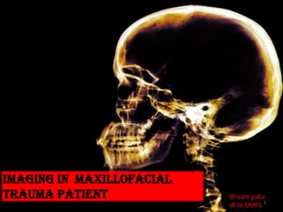
  • 1. Imaging in maxillofacial trauma patient Shivani gaba JR-III,OMFS 1
  • 4. FUTURE: Combine, see, minimally cut! image guidance Augmented reality 4
  • 5. Introduction The skeletal anatomy of maxillofacial region is the most complex in the body. Injuries of this region range from isolated injuries involving only one or two osseous components to complex facial injuries involving the entire facial skeleton. Diagnostic imaging has traditionally played central role in providing information essential in initial diagnosis and treatment of these injuries. Until 1980,diagnostic imaging of face consisted exclusively of plain films and panoramic studies . The introduction of CT in late 1970s and early 1980s represented a major advancement . This was followed by softwares capable of generating 3-D reformatted images. The late 1980s and 1990s witnessed significant advancement capable of performing high speed helical or spiral CT , which can scan acutely traumatized patients in a matter of seconds . Spiral CTs represent state of the art imaging for the patients with severe maxillofacial injuries. 5
  • 6. Objectives Normal biomechanics of maxillofacial skeleton-IMPORTANCE Imaging modalities and techniques When to do imaging Diagnostic imaging of maxillofacial injuries  Plain radiography-READ AND REMINDERS For upper &middle third Lower third  CT SCANS -NORMAL Recognition and interpretation of facial injuries Advanced imaging 6
  • 7. Normal biomechanics of maxillofacial skeleton The radiographic evaluation of facial trauma must begin with a basic understanding of osseous anatomy of the skull and midface. Knowledge of areas of inherent biomechanical weakness within the facial skeleton is essential .Understanding the biomechanics of craniomaxillofacial trauma gives insight in understanding the pattern of injury. A helpful approach in assessing imagining studies in these injuries is to divide the craniofacial skeleton into FOUR principal components: Ovoid shaped cranium Pyramid shape midface Tripod shaped zygoma Ring shape mandible 7
  • 8. The buttress system of face is formed by strong frontal, maxillary, zygomatic ,sphenoid and mandible bones and their attachments to one another. The central midface contains many fragile bones that could easily crumble when subjected to strong forces. These fragile bones are surrounded by thicker bones of the facial buttress system lending them some strength and stability. These buttress represent the best available understanding of the mechanical support of face as they determine how an impact is distributed over the face Buttress system of face 8
  • 9. PLAIN RADIOGRAPHS Foundation of day to day imaging. May be sufficient to provide a radiological diagnosis, or may need to be complimented by CT.  General rule (anywhere in the body) is that fractures should be imaged in at least two planes, preferably at right angles to one another. Face is often divided into upper, middle, and lower thirds, and the middle third is further divided into central and lateral components. COMPUTED TOMOGRAPHY Advantage of providing multiplanar thin slice images of the facial skeleton, overcoming the problem of superimposition of structures that inevitably occurs on plain radiographs. Generally, a slice of 2-4mm thickness is adequate in assessing facial trauma. In almost every case , axial slice images are obtained. Coronal images are particularly helpful in orbital examination . Coronal imaging has the benefit of visualizing horizontal struts or plates of bone, which in standard axial slices lie parallel to the slice plane and therefore are poorly visualized. 3-D datasets have become increasingly requested since the advent of navigational surgical techniques. With the advent of digital image storage & Picture Archiving and Communication System(PACS), CT images are now routinely assessed on the computer rather than on the cut film of old. Imaging modalities and techniques Conebeam Computed Tomography (CBCT)  Relatively new CT technique that uses a cone shaped beam of radiation rather than the standard fan shaped beam.  Allow for low dose scanning of small areas of the body. Current application centers on dentomaxillofacial imaging. Limited by poor soft tissue resolution . CBCT scanners are much smaller and less expensive CBCT does, however, offer a viable alternative to MDCT. 9
  • 10. OTHER MODALITIES A number of other modalities, such as  angiography  dacrocystography  sialography Arthrography  Angiography may be required in the acute phase, and sialography has a place in the evaluation of suspected parotid duct transaction. Others are usually reserved for assessing the delayed sequelae. MAGNETIC RESONANCE IMAGING (MRI) Uses the manipulation of H- atoms within the body to form images. Approximately two thirds of the total hydrogen atoms in the human body are located in water or fat molecules. MRI uses a strong electromagnetic field to line up the nuclei of these hydrogen atoms in a single plane. Tissues with plentiful hydrogen atoms, such as CSF or fat produce high (bright) signals, and those with little hydrogen such as bone,produce low(dark) signal. So,because of the low number of hydrogen atoms in bone, the demonstration of fine bone detail in the face by MRI remains inferior to CT. MRI does have growing application in a number of specific areas. Most importantly is its ability to detect CSF fistulae and leaks, post traumatic meningoceles. It has increasing application in the orbit for injuries to the globe and optic nerve sheath complex, acute soft tissue injury in the TMJ. ULTRASONOGRAPHY Limited use in assessment of bony trauma of the facial skeleton,  Well defined role in assessment of soft tissue injury and fluid collections. It is the modality of choice for assessment of the globe, for assessing intraocular foreign bodies. Imaging modalities and techniques 10
  • 11. When the patient is stabilized Clinically (Airway, Breathing, Circulation - stable) Initial goal is to preserve life - then later restore the form and function of the face Radiographically For cervical spine clearance When to Do Imaging of the Face? 11
  • 12. When to Do Imaging of the Face?-CT head 12
  • 13. •Can be obtained to screen for facial injury if CT is not immediately available •Multiple plain film projections are relative to ‘canthomeatal line’; an imaginary line drawn from outer canthus to external auditory meatus • Proper positioning (of patient’s head), alignment of xray beam is critical for evaluation because facial skeletal anatomy is complex Remember: plain film is a 2D image of a 3D object Overlapping structures significantly obscure anatomic detail. This problem is solved by standard views (to minimize overlap, allow visualization of important structures, familiarity for interpretation)  Rule of symmetry: two sides of the face are quite symmetrical  Symmetry is usual, and asymmetry is suspect Multiplicity: fractures of facial bones are frequently multiple. Do not stop looking for others when see one Plain Film Radiography 13
  • 14. Of these views, the most consistently helpful view in facial trauma is the Waters view. It tends to show all of the major facial structures at least as well and often better than other radiographic views of the face Mandible series Orthopanthogram Oblique view PA mandible  Towne’s view Facial series Water’s view (PA view with angulation) Caldwell view (PA view) Towne’s view Lateral view Base view Plain Film Radiography 14
  • 15. Water’s view (occipitomental view-37⁰) Described by-waters and waldron 15
  • 16. 16
  • 17. 17
  • 18. McGrigor & Campbell lines •A PATTERN OF FOUR LINES that the eye should follow in OM. •USING THESE LINES allows one to examine all those parts of face where fractures and others signs are most likely to be found and reduces the chances of missing a fracture. Fourth line-cross mandibular ramus and bite line Fifth- across inferior border of mandible. 18
  • 19. Clinical-search pattern and reminders ! 19
  • 20. 20
  • 21. 'Tripod' fractures Trauma to the zygoma may result in impaction of the whole bone into the maxillary antrum with fracture to the orbital floor and lateral wall of the maxillary antrum. The displaced zygoma is detached from the maxillary bone, the inferior orbital rim, the frontal bone at the zygomatico-frontal suture, and from the zygomatic arch. The result is said to liken a 'tripod', but in reality these fractures are often more complex than is appreciated on plain X-ray. 'Quadripod' would perhaps be a more accurate term as four fractures may be visible 21
  • 22. 22
  • 23. Orbital 'blowout' fractures Trauma to the orbit may lead to increased pressure in the orbit such that the thin bone of the orbital floor bursts. This manifests as the 'teardrop' sign which is due to herniation of orbital contents into the maxillary antrum. 23
  • 24. Orbital emphysema Occasionally a 'tripod' or 'blowout' fracture will cause a leak of air from the maxillary antrum into the orbit. This can have the appearance of a dark 'eyebrow'. 24
  • 25. Fracture mimics X-ray appearances can easily be misinterpreted unless a systematic approach is used to look for the common fracture patterns. Any suspected injury should be correlated to the clinical features. Overlying structures such as sutures should not be interpreted as fractures. 25
  • 26. PA Projections PA skull view Axial PA skull-caldwell view(10-15⁰ angulation) 26
  • 27. Caldwell view(occipito-frontal): Same as straight PA. PP projected onto the floor showing SOF and sphenoid wings 27
  • 28. 28
  • 29. Frontal projection (Caldwell view) 1. Zygomaticofrontal suture 2. Orbital process of frontal bone 3. Anterior orbital roof 4. Upper rim of orbit 5. Frontal sinus 6. Lamina papyracea 7. Posterior orbital floor 8. Posterior lacrimal crest 9. Anterior orbital wall 10. Frontal process of maxilla 11. Lateral nasal wall 12. Lateral maxillary wall 13. Hard palate 14. Perpendicular ethmoid plate and vomer 15. Superior orbital fissure 16. Oblique orbital line 17. Orbital process of zygoma 29
  • 31. 31
  • 32. Image receptor and patient placement: Image receptor is positioned II to the patient’s midsaggital plane .The side of interest placed towards image receptor. Position of central X-ray beam: The central beam is perpendicular to mid saggital plane and film and centered over EAM. Lateral view skull 32
  • 33. 33
  • 34. Clinical search patterns and reminders! 34
  • 35. Clinical search patterns and reminders! 35
  • 36. 36
  • 37. Cervical spine –normal anatomy Clinical considerations are particularly important in the context of Cervical spine (C-spine) injury. This is because normal C-spine X-rays cannot exclude significant injury, and because a missed C-spine fracture can lead to death, or life long neurological deficit.Imaging should not delay resuscitation.Further imaging with CT or MRI (not discussed) is often appropriate in the context of a high risk injury, neurological deficit, limited clinical examination, or where there are unclear X-ray findings 37
  • 40. Submento-vertex view(jug handle view) Image receptor and patient placement: Image receptor II to transverse plane of patient , and perpen . to coronal plane. Neck is extended as far backward as possible. Position of central X-ray beam: Perpen. to image receptor .directed from below mandible toward the vertex of skull. 40
  • 41. 41
  • 42. Mandible fractures The mandible can be considered as an anatomical ring of bone, stabilised at each end at the temporomandibular joints. A break of the ring in one place will usually be accompanied by further break in the ring elsewhere. If you see one fracture, look for a second fracture, or a dislocation of the temporomandibular joint. Standard views Orthopantomogram (OPG) and Mandible views. Both views are necessary because fractures are often only seen on one image. the mandible views include PA/AP mandible, lateral oblique mandible , towne’s view ,reverse towne’s view Routine diagnosis of this type of fracture should include radiographs taken in two planes at 90° to each other; the minimum requirement is a PA view and a panoramic view. Mandible series 42
  • 43. 43
  • 44. 44
  • 45. 45
  • 46. 46
  • 47. 47
  • 48. 48
  • 49. 49
  • 50. Lateral oblique mandible Indications: Mandibular body , angle , ramus condyle ,coronoid fractures 50
  • 51. 51
  • 52. Possible false-positive radiographic finding of a mandibular fracture. The anterior margin of C2 often simulates a fracture of a condyle. Here, although the margin is too obvious to be mistaken for a fracture, the orientation of the anterior cortex of C2, which overlaps the left condyle, is demonstrated. Left angle fracture on a left oblique radiographic image 52
  • 53. Left oblique radiographic image shows a left angle fracture in a patient with a dislodged molar tooth Panoramic radiographic image of the left angle fracture. Note that the back left molar is now horizontal. 53
  • 54. The coronoid is best seen on an oblique radiographic view. 54
  • 56. Posterior anterior radiograph clearly demonstrating bilateral nondisplaced mandibular subcondylar and displaced comminuted para-symphyseal fractures (white arrows). While providing adequate visualization of the mandible, this view should be supplemented with additional projections (eg, Waters) to assess midfacial injury. 56
  • 57. Radiograph of a comminuted fracture of the body and symphysis caused by a gunshot wound. 57
  • 58. Reverse towne’s view(tilted PA mandible) Indications: Fracture of condylar head & neck, intracapsular fracture Best seen: Posterior view of both condylar head and neck 58
  • 60. Towne’s (oblique PA) X-ray taken at 90° to show displacement of left condylar process fracture. Vertical shortening of the left mandible is noted along with the right anterior body fracture 60
  • 61. Conventional TMJ views Transcranial view Lateral aspect of: Glenoid fossa Articular eminence Joint space Condylar head 61
  • 63. Transorbital (zimmer projection) Entire latero-medial articulating surfaces Medial displacement of fractured condyle Fracture of neck of condyle -Disadvantage : if the patient cannot open wide, areas of the joint articulating surfaces will be obscured because of mutual superimposition 63
  • 64. In radiographic evaluation of plain films there are five key structures that must be evaluated in a systemic and precise fashion. Theses are zygoma ,orbits ,nasal structures , paranasal sinus and mandible Radiographic evaluation of plain films-some facts! 64
  • 65. The four ‘S’ described by delbalse et al .are good reminder of what to look for on radiograph Symmetry Sharpness Sinus Soft tissue Symmetry: With exception of frontal sinus, the face is reasonably symmetrical in most people. Any asymmetry that is noticed should draw examiner attention to that area for further evaluation. Sharpness: Refers to accentuated sharpness brought about by # fragment being rotated or displaced into tangency with x-ray beam ,which cause them to appear accentuated on the image. e.g. trapdoor sign. Sinus : Almost all of the midface # involve one or more paranasal sinus. Soft tissue: swelling , foreign bodies, and emphysema may all be apparent and can help in evaluation of patient. Radiographic evaluation of plain films-some facts! 65
  • 66. The fact that a # of single site may occur as a part of several different fractures types or patterns is important. Failure to appreciate this fact can lead to error (clinically as well as Radiographically) One should not be too rigid about such patterns and classifications , because hybrid and mixed forms are more common than pure forms. Particularly in panfacial type injuries ,facial skeleton may split like an eggshell into dozens of fragments, making any attempt to ordered classification fruitless. Concomitant injuries pattern also occur ,and identification of one should alert the examiner to check whether the other is present . e.g. association of depressed zygomatic arch fracture with coronoid process fracture, Lefort-I with palatal ,parasymphysis with opposite condylar neck or body. Radiographic evaluation of plain films-some facts! 66
  • 67. 1.Sepration sign: cortical defect/crack Fry et al.-radiographic appearance of fracture lines may vary in width acc.to Amount of destruction Amount of displacement Angle of projection of x-ray Stage of repair Always inspect the margins: • In fresh fractures : well defined and sharp! • Rounding off indicates either that its not a fracture or that infection or nonunion is occurring. The width : Can increase slightly due to healing process. More extensive widening: displacement/intervening bone loss/infection(delayed) If displacement : wide gaps indicate periosteal tearing and in calvaria , dural tearing. Always inspect lamina dura of tooth socket and developing tooth buds. In children, a fracture may be visible only as a lucency crossing the wall of tooth bud. Radiographic evaluation of plain films-some facts! Direct radiographic signs of fracture All signs of injury may be considered as being abnormal densities ,or as being abnormalities of position. 67
  • 68. Pitfalls: Neurovascular channels produce lucent lines as do sutures -cause confusion On CT .-knowledge of anatomy is important Clue : margins of these structure tent to be sclerotic unlike those of fresh fractures 7.Widening of PDL :This is important but often overlooked clue to fracture. In absence of disease and partial extrusion ,a wider PDL on one side than other should raise possibility that a mandibular fracture passes into it. 2.Sutural diastasis : sometimes difficult to detect the mild degrees of separation 3.Overlap sign : when there is displacement at fracture site such that two fragment overlap either wholly or in part.-band of increased opacity. 4.Disappearing fragment sign:absence of bone from expected position.eg. In CT –the empty glenoid fossa,implying mandibular condylar neck fracture or dislocation 5.Abnormal angulation:curvature may be seen in children with greenstick fracture or fracture of antral walls,in zygomatic arches(esp.outward bowing).it may be the only clue due to injury,or it may be an obvious feature of otherwise readily recognizable injury. 6.Step deformity: displacement of bone in plane at right angle to x-ray beam ,gives rise to sharp step in the countour of the outer margins of cortex.w Radiographic evaluation of plain films-some facts! 68
  • 69. 4.Changes to occlusal plane : best assessed clinically .e.g. open anterior bite may suggest one of lefort or condylar neck fractures. 2.Paranasal sinus opacifiaction: In a trauma patient, an opacified sinus must be assumed to be secondary to trauma until proven otherwise Radiographic evaluation of plain films-some facts! Indirect radiographic signs: 1.Soft tissue swelling: a common and fairly nonspecific sign that is often present with no underlying fracture. But if localized, it may direct attention to particular area of face on radiograph. e.g.-cheeks-zygoma. It may also affect airway and should always be inspected on any lateral view and CT images, bearing in mind that free blood may give rise to similar opacifications. 3.Air in soft tisuues: Soft tissue emphysema: mostly created by communication via a fracture with maxillary sinus. Pitfalls: air lateral to maxillary sinus may be mimicked by lateral extension of sphenoid sinus. Facial trauma should always be discouraged from nose blowing ,close mouth sneezing as increased air pressure can cause large amounts of air into soft tissues-increasing risk of infection. Intraorbital air: should always be differentiated from deep sulcus of upper eyelid in an elderly or enopthalmic patient, can be caused by floor or medial fractures or delayed manifestation of orbitoantral fistula, particularly after repair of blowout fractures. 69
  • 70. Inspect clinically first-ALWAYS!! SUSPECT STABLE-SEND FOR RADIOGRAPHS BASED ON SUSPICIAN &PATTERNS Identify fractures, fragment displacement and rotation, stable bone for use in surgical repair  PLAN ACCORDINGLY Imaging approach plain films 70
  • 71. 71 ADVANCED IMAGING IN MAXILLOFACIAL TRAUMA SHIVANI GABA JR-III,OMFS
  • 72. Objectives  CT SCANS -NORMAL Recognition and interpretation of facial injuries Advanced imaging ENDOSCOPY CBCT CAS CAS APPLICATION -CASES 72
  • 73. Computed Tomography (CT) Preferred modality for imaging of the face More sensitive for fracture detection  Show significant soft tissue injury, especially the globe  Easier to perform, quicker than complete views of plain film radiographs  Pre-surgical planning for complex injuries Disadvantage of CT CT can miss subtle tooth fracture along the axial plane, additional orthopanthogram may be helpful to detect tooth fracture 73
  • 75. AXIAL SECTION Frontal sinuses within the frontal bone. It also shows the unique construction of the cranial bones with diploe sandwiched between two layers of compact bone. 75
  • 76. 76 Note that the septum between them is not quite in the median plane. The posterior wall of the frontal sinuses is only a thin bony lamella in close vicinity to the brain. The left orbita is opened and the levator palpebrae muscle is cut. The right orbital roof is the floor of the frontal sinus.
  • 77. AXIAL SECTION- at midorbit level 77
  • 78. 78 The posterior ethmoid cells directly border the sphenoid sinus. Note the carotid artery, pituitary and cavernous sinus dorsal to the sphenoid sinus and the rectus medialis muscle just lateral to the posterior ethmoid cells
  • 79. Axial section-level at just below roof of maxillary sinus 79
  • 80. Axial section -lower level of the maxillary sinuses. 80
  • 81. Axial section –at level of alveolus and palatal bone 81
  • 82. Axial section-shows the mandible, along with the alveolar process of the maxillary bones where the teeth insert. 82
  • 83. Disarticulated mandible with AXIAL CT planes illustrated AXIAL CT planes-MANDIBLE 83
  • 84. Disarticulated mandible with AXIAL CT planes illustrated AXIAL CT planes-MANDIBLE 84
  • 94. 3-D CT Scans face-normal anatomy 94
  • 95. 3-D CT Scans face-normal anatomy 95
  • 96.  Although 2D axial and coronal CT is more accurate and more sensitive than 3D reformatting, numerous studies have explored the utility of 3D imaging.  Three-dimensional images are created from the original 2D slices; therefore, there is no new information in the images, and artifacts may be produced in the reformation process.  Nonetheless, reconstructed 3D images may assist in the visualization of large comminuted, displaced, and complex fractures , particularly in regard to the midface and the zygomatico-maxillary complex. These 3D scans enable clinicians to better assess the localization of bone fragments and their direction of displacement. 3D CT permits isolation of affected sections of the facial skeleton and enables perspectives that may not readily be generated or appreciated by the clinician in his or her ‘‘mind’s eye.’’  3D images provide only information regarding bony architecture; fat and muscle entrapment, encephaloceles, hematomas, and associated injuries must be assessed radiographically through 2D CT manipulation of soft-tissue windows.  With this in mind, it is useful to think of 3D imaging as a complementary study that can add important information to multiplanar imaging the face as it recreates the surgeon’s complex mental process of visualizing fractures in operative planning.  Continuing advances in computer software algorithms and improved precision in the acquisition of radiographic data will make 3D reformatted CT imaging a necessary complement to traditional 2D CT imaging in the management of complex facial trauma 96
  • 97. Mandibular fractures Pitfalls of plain imaging Recognition of fractures-face! Upper and mid face Nasal fractures Nasoethmoid Zygomatic •Tripod fractures •Isolated zygoma Orbital Maxillary fractures • Maxillary sagittal fracture • Alveolar process fracture • LeFort fractures Lower face 97
  • 98. M/C isolated fractures R/G-water’s, bilateral lateral nasal views CT- axial views >coronal Nasal FRACTURES Nasal fractures. Waters view shows a deviated nasal septum, quadrangular cartilage displaced from the maxillary crest, and a nasal root deviated to the right Nasal fractures. Axial CT scan demonstrates a nasal fracture with root deviation to the right. Note that the fracture involves the septum as well and that the septum is severely deviated. Nasal fractures. Coronal CT scan demonstrates a nasal fracture with root deviation to the left. Note that a portion of the nasal bones is involved 98
  • 99. These fractures involve the central upper midface, disrupting the confluence of the medial maxillary buttress with the upper transverse maxillary buttress as well as their posterior extensions along the medial orbital wall and floor. Fractures of the NOE complex can be one of the most difficult fracture patterns to accurately repair. Drawings show the Manson classification of NOE fractures. A type I fracture involves a large bone fragment. A type II fracture involves comminution of the bone fragments. A type III fracture is defined by avulsion of the medial canthal ligament from its osseous insertion. Right-sided Manson type I fracture and a left-sided Manson type II fracture (among other fractures). In the type I fracture, large bone fragment (black arrow) with the attachment of the medial canthus. The fragment has been pulled laterally and inferiorly. In the type II fracture, the medial canthal attachment is on a small bone fragment, which is not visualized in this plane(medial vertical buttress (white arrow). Naso-orbitoethmoid (NOE) Medial vertical maxillary buttresses (*) are posteriorly and laterally displaced due to disruption of the upper transverse maxillary buttress. The medial canthi of the lids are attached to the rims of the lacrimal fossae (black arrows). The sagittal extension of the medial maxillary buttress along the medial orbital wall has also been disrupted, resulting in exophthalmos from the “blow-in” fractures (white arrow) and loss of nasal dorsal projection. 99
  • 100. NOE fracture pearls are as follows: (a) NOE fractures are distinguished from simple nasal fractures by posterior disruption of the medial canthal region, the ethmoids, and the medial orbital walls. (b) Clinically, the most obvious deformity is loss of nasal projection in profile and apparent increased distance between the inner corners of the eyes. (c) NOE fractures can be classified by the degree of injury to the region where the medial canthus attaches around the lacrimal fossa. (d) Although the frontal sinus may not be directly injured, if the nasofrontal ducts are disrupted, then frontal sinus surgery is needed to prevent a mucocele in the future. (a) Coronal CT image shows the normal anatomy in the region of the nasofrontal ducts (*). (b) Coronal CT image of a patient with NOE fractures shows comminution of the ethmoid region and likely disruption of the nasofrontal ducts (*). 100
  • 101. ZYGOMATIC FRACTURES TRIPOD TRIPOD FRACTURES R/G-WATERS>CALDWELL,SMV,TOWNS (ADD) CT-SCAN-3-D CT> AXIAL>CORONAL The two “cornerstone” zygomaticomaxillary complexes (ZMCs) are surgically important in establishing orbital volume and serving as a reference for reduction of maxillary fractures. 101
  • 102. ZMC fracture-a displaced fracture of the left zygoma. The rotational deformity of the zygoma is demonstrated by angulation of the lateral orbital wall at the zygomaticosphenoid suture. The lateral displacement (black arrow) of the lateral vertical buttress (*) has resulted in increased orbital volume and enophthalmos ZMC fracture pearls are as follows: (a) The ZMC relates to the temporal bone, maxilla, frontal bone, and skull base and is therefore a quadripod structure. (b) Displaced ZMC fractures often increase orbital volume by angulation of the lateral orbital wall at the zygomaticosphenoid suture or blow-out of the orbital floor. (c) The zygomatic arch establishes both facial width and profile. Surgical exposure is indicated if it is severely comminuted or angulated. 102
  • 103. Right-sided depressed zygomatic arch fracture Fracture of the zygomatic arch with medial displacement of fragments can often impinge on the coronoid process of the mandible and cause trismus.CT scan shows coronoid impingement by zygomatic arch fracture Zygomatic arch fractures M/c isolated fracture of zygoma Best views: slightly underpenetrated SMV ,Waters’. CT-Axial sections Comminuted right zygomatic arch fracture demonstrated in standard two-dimensional coronal image and an obliquely angled three-dimensional reconstruction better demonstrating displacement of the fracture fragments 103
  • 104. Orbital floor fractures can occur in isolation(blow out –third M/C isolated midface), but they are also commonly associated with posterior propagation of ZMC and Le Fort II fractures (orbital floor fractures) and NOE fractures (medial orbital wall fractures) For orbital floor defects(blow out), Best views: waters’ and caldwell CT-CORONAL>AXIAL Attention to the shape and position of the inferior rectus muscle on coronal CT scans can provide information regarding the damage to the fascial sling of the globe. Coronal CT image -the inferior rectus remains flattened in cross-sectional appearance, indicating that the fascial support of the globe is likely intact . The inferior rectus (arrow) is displaced inferiorly into the maxillary sinus. Note that the cross-sectional appearance of the muscle has changed from ovoid to circular. The fascial support has been disrupted, and the muscle is entrapped. Coronal nonenhanced CT image shows a left orbital floor fracture in another patient. The inferior rectus (arrow) is displaced inferiorly into the maxillary sinus. Note that the cross-sectional appearance of the muscle has changed from ovoid to circular. The fascial support has been disrupted, and the muscle is entrapped. “trap-door” fracture of the left orbital floor in a pediatric patient. The orbital floor disrupted by the impact but then sprang back into place, trapping the inferior rectus (arrow) within the maxillary sinus. Because the bone has returned to its anatomic location, the diagnosis could easily be missed unless attention is paid to the locations of the extraocular muscles. Trapdoor fractures require emergent treatment. Orbital Fractures 104
  • 105. Medial orbital wall fractures have a strong association with diplopia due to loss of the posterior- medial bulge of the orbit or mechanical entrapment of the medial rectus muscle . Both blow-in and blow- out patterns can result in muscle entrapment Axial CT image -posteromedial bulge of the left lamina papyracea has collapsed toward the midline. This collapse has increased the orbital volume and resulted in enophthalmos, with retroposition of the ipsilateral globe (small arrow) relative to the coronal plane (white line). The affected medial rectus (large arrow) has lost the typical flattened profile seen in the uninjured contralateral orbit (*) and herniated into the fracture site. Axial CT image shows the lamina papyracea impinging on the medial rectus (arrow). This result is evident from the angulation of the muscle in the orbit 105
  • 106. Orbital apex fractures are fortunately rare; however, they are emergent surgical cases if there is radiologic and clinical evidence of optic nerve impingement Axial CT image shows the lamina papyracea impinging on the medial rectus (arrow). This result is evident from the angulation of the muscle in the orbit Axial nonenhanced CT image shows impingement of the orbital apex secondary to a sphenoid–skull base fracture. The orbital apex fracture was associated with loss of vision secondary to compression of the optic nerve by a fragment of the sphenoid (arrow) with inward angulation of the lateral orbital wall (white line). The fracture was emergently reduced, and the patient regained her vision. Orbital roof fracture in an adult. Coronal CT image shows an isolated blow-in fracture of the left orbital roof (arrow). The associated exophthalmos and dural tears were treated with an intracranial approach.106
  • 107. Orbital fracture pearls are as follows: (a) Orbital fractures can occur in isolation or with other fracture patterns. (b) The position and shape of the medial and inferior rectus muscles can indicate whether entrapment and clinical diplopia are likely. (c) Pediatric trapdoor orbital fractures are a surgical emergency. (d) The size of the orbital floor defect can be underestimated in severely impacted ZMC fractures. (e) Medial orbital wall blow-out fractures cause enophthalmos if the posterior-medial orbital bulge is lost. (f) Orbital apex compression with clinical decreasing vision is a surgical emergency 107
  • 108. – Maxillary sagittal fracture (maxillary sinus fracture) – Palate fracture – Alveolar process fracture – LeFort fractures • LeFort I fracture • LeFort II fracture • LeFort III fracture • Combination (bilateral, hemi) Maxillary fractures Maxillary sagittal fracture Fracture of a maxilla in sagittal plane, involving anteriorlateral wall of a maxillary sinus (LeFort fractures represent bilateral maxillary fractures) – Due to direct blow to either right or left midface There is a sagittal plane fracture of the left maxillary sinus (red arrow) with hemosinus (H) 108
  • 109. Isolated alveolar process fracture – Fracture of any portion of the alveolar process – Clinically evident by malalignment and displacement of teeth – Even on CT, fracture may be subtle and easily overlooked – Further imaging may be needed when the diagnosis is made •BEST VIEWS: IOPA/OPG • Chest radiograph (look for aspirated teeth) Red arrows show the comminuted fractures of the maxillary alveolar process on the right side. These fractures are considered ‘open’ as they are connected to the oral cavity. 109
  • 110. Thèse fractures involve separation of all or a portion of the maxilla from the skull base. For this to occur, the posterior vertical maxillary buttress at the junction of the posterior maxillary sinus with the pterygoid plates of the sphenoid must be disrupted. This can occur either through the posterior walls of the sinus or through the plates themselves, as seen at axial CT. Common Le Fort fracture patterns. The Le Fort I pattern involves fractures through the inferior portions of the medial and lateral maxillary buttresses. The Le Fort II pattern involves fractures through the zygomaticomaxillary and frontomaxillary sutures. The Le Fort III pattern involves complete craniofacial dissociation Le Fort fractures 110
  • 111. LeFort I fracture Transverse (horizontal) fracture of inferior maxillae, involving maxillary sinuses (all except superior walls), lateral margin of nasal fossa, nasal septum and pterygoid plates (transmaxillary fracture) •Le Fort I pattern involves fractures through the inferior portions of the medial and lateral maxillary buttresses. •The Le Fort I fracture is the only one that involves the anterolateral margin of the nasal fossa just above the maxillary alveolar process. •If the anterolateral margin of the nasal fossa is intact, a Le Fort I fracture is excluded. •Transverse injury that crosses the floor of the nose, pyriform aperture, canine fossa, and lateral wall from the maxilla, resulting in separation of the palate from the maxilla. pterygomaxillary disjunction. Axial CT image shows a right-sided unilateral pterygomaxillary disjunction (white arrows), which has resulted in separation of the posterior vertical maxillary buttress (*) from the rest of the maxilla; this appearance is indicative of a Le Fort fracture. The contralateral pterygomaxillary junction is intact because the fracture exited in the form of a parasagittal palate fracture (black arrow). (b) Axial CT image shows comminuted bilateral pterygomaxillary disjunctions (white arrows) with a sagittal palate fracture (black arrow). There is a complete posterior maxillary fracture with disruption of both posterior vertical maxillary buttresses (*), resulting in separation of the maxilla from its attachment to the skull base. 111
  • 112. •Le Fort II fracture involves fractures through the zygomaticomaxillary and frontomaxillary sutures. The Le Fort II fracture is the only one that involves the inferior orbital rim. If the inferior orbital rim is intact, a Le Fort II fracture is excluded. •It crosses the nasal bones on the ascending process of the maxilla and lacrimal bone and crosses the orbital rim .Only the Le Fort II fracture violates the orbital rim. Le Fort II fracture extends posteriorly to the pterygoid plates at the base of the skull. A Le Fort I fracture is characterized by a low septal fracture, whereas a Le Fort II fracture results in a high septal fracture. Coronal CT image shows right Le Fort I and bilateral Le Fort II fractures. The right medial maxillary buttress is fractured inferiorly -Le Fort I pattern; it is also fractured superiorly (left black arrow), thus contributing to the bilateral Le Fort II pattern. The left-sided Le Fort II medial fracture (right black arrow) is not at the level of the nasofrontal junction; however, it is distinguished from the Le Fort I pattern in that a portion of the orbit is included in the mobile segment. LeFort II fracture 112
  • 113. Le Fort III fracture is the only one that involves the zygomatic arch . Crosses the frontal process of the maxilla, the lacrimal bone, the lamina papyracea, and the orbital floor. This fracture often involves the posterior plate of the ethmoid. Le Fort III fracture Blue arrows define LeFort II fracture. Red arrows define theLeFort III fracture. Bilateral pterygoid plate fractures are present (not shown). 113
  • 114. Coronal CT image shows bilateral Le Fort I, II, and III fractures. The lateral and medial maxillary buttresses (white lines) are fractured inferiorly and superiorly (junctions of white lines and black lines). To confirm the diagnosis, pterygomaxillary disjunction and fractures of the zygomatic arches would need to be observed on axial images. Le Fort fracture pearls are as follows: (a) All Le Fort fractures require disruption of the pterygoids from the posterior maxilla, as seen at axial imaging. (b) Any combination of Le Fort I, II, and III patterns can occur. (c) A sagittal or parasagittal hard palate fracture with a Le Fort pattern will result in a widened maxillary arch. (d) Displaced unilateral Le Fort fractures are possible only with a sagittal or parasagittal palate fracture. 114
  • 115. Axial and coronal two-dimensional images demonstrate extensive comminuted fractures involving the medial and lateral walls of the maxillary sinuses as well as the bilateral zygomatic arches. The three-dimensional reconstruction demonstrates the overall relationship of the multiple fractures to the remaining intact skeleton and complements the information gathered in two-dimensional views. 115
  • 116. Mandibular Fractures Relevant anatomy – Mandible is a ring or arc bone which is usually difficult to break in one location. Approximately half of mandible fracture occurs in multiple locations. • Search for a second fracture after initial fracture has been identified (usually at contralateral side) – In symphyseal, parasymphyseal fractures: Digastric, geniohyoid and genioglossus muscles pull the symphysis downward posteriorly – In angle fracture: 3 muscles attaching to the ramus of mandible (masseter, temporalis and medial pterygoid) pull the proximal fragment upward and medially Imaging recommendation – Plain film mandible series (PA, lateral, Towne’s and bilateral oblique) show nearly all fractures BUT may be difficult to obtain in multitrauma patients – Panoramic radiography (orthopantogram) • Need patient in upright position • Not optimal as a single radiograph to detect mandibular fracture • Better to look for subtle tooth fracture – CT • Show all mandibular fractures AND other facial fractures (coexistence 6-10%), as well as position and alignment of fragments • Display associated soft tissue injuries • Easy to perform in multitrauma patient 116
  • 117. 117
  • 118. 118
  • 119. 119
  • 121. 121
  • 122. 122
  • 123. 123
  • 124. 124
  • 125. Three-dimensional reconstructions of mildly displaced left angle and right parasymphysial mandibular fractures Two-dimensional axial image and three-dimensional reconstructions of a displaced left parasymphysial fracture. The 3- D images more easily demonstrate the amount of displacement throughout the fracture and the spatial relationship of the fracture fragments. 125
  • 126. Three-dimensional manipulation demonstrating lingual view of a nondisplaced left mandibular angle fracture extending through an impacted third molar. 126
  • 127. Imaging Approach CT Clear sinus sign (= all sinuses and mastoid are clear of fluid) – Nasal bone fractures – Isolated zygomatic arch fractures – Orbital roof/lateral wall fractures – Mandible fractures • Hemosinuses – Pterygoid plate fracture present probable LeFort fracture • With fracture of lateral margin of nasal fossa = LeFort I • With fracture of inferior orbital rim = LeFort II • With fracture of zygomatic arch = LeFort III – Maxillary wall fractures – Orbital floors, NOE region fractures – ZMC fractures 127
  • 128.  Treat lifethreatening injury first (ABC of trauma) CT is more accurate, faster to do than plain film and can be performed at the same time as trauma head CT Emergency in face injury -Airway compromise due to severe soft tissue swelling, -fracture or obstructed foreign body - Life threatening hemorrhage can be from nasal injury - Facial fractures that compromise vision - Orbital apex fracture may injure optic nerve, requiring urgent Rx - Entrapment of intraocular muscle requires urgent Rx Checklist for Facial Radiograph/CT 128
  • 129. Checklist for Facial Radiograph/CT  Facial structures are quite symmetrical Do not stop searching when see one abnormality Significant (but can be subtle) fractures - Fracture involves the optic foramen which can cause permanent visual loss if not treated promptly - Fracture of the posterior wall of frontal sinus requires neurosurgical evaluation and may require antibiotics prophylaxis -Fracture/dislocation of the TMJ usually missed on initial survey. It can cause significant disability if left untreated - Look for significant soft tissue injuries - Globe rupture, hemorrhage 129
  • 130. Advanced imaging Computer assisted maxillofacial surgery Intraoperative imaging Intraoperative navigation Virtual model reconstruction Endoscopic assisted fracture treatment Cone beam CT 130
  • 131. virtual surgery and computer-aided design and manufacturing can be used by the craniomaxillofacial surgeon to create tremendously accurate postoperative results and provide confidence with even the most complex three-dimensional bony reconstructions. With advancements in software technology and three-dimensional printing, the ability to plan and execute precise bony reconstruction has become a reality. With this technology, guides can be made to ensure exact bony repositioning or replacement. These guides can help guide cutting of the bone and can act as splints to precisely reposition the bone and direct plate placement. With use of these computer-aided design and manufacturing guides and the addition of guidance technology, the position of the bone can be guaranteed intraoperatively. J Craniofac Surg. 2012 Jan;23(1):288-93. doi: 10.1097/SCS.0b013e318241ba92. Computer-aided design and manufacturing in craniomaxillofacial surgery: the new state of the art. Levine JP, Patel A, Saadeh PB, Hirsch DL 131 Recent advances in computer-modeling software allow reconstruction of facial symmetry in a virtual environment
  • 132. Applications for endoscopic treatment of facial trauma include subcondylar fractures of the mandible,[orbital blow-out fractures (OBFs),frontal sinus fractures, and zygomatic fractures Advantages of endoscopic repair include the following: More accurate fracture visualization Small external incisions Reduced soft tissue dissection The potential for visualization around corners The possibility of reduced duration of hospital stays Improved teaching opportunities (since the procedure can be visualized on a television monitor) Disadvantages of endoscopic repair include the following: A current lack of dedicated instrumentation A moderate learning curve for the techniques A narrow field of view A limited ability for bimanual instrumentation without an assistant Indications for endoscopic repair are generally related to fracture location, size, degree of comminution, and the surgeon's abilities. Endoscopic assisted fracture treatment 132
  • 133. Photograph of the sublabial exposure required for endoscopic repair of orbital blow-out fractures. A 1- X 2-cm antrostomy has been performed. The arrow shows the endoscope notch placed in the inferior portion of the antrostomy. This gives the assistant surgeon tactile feedback to hold the scope steady. Reconstruction of isolated orbital floor fractures through an endoscopic approach appears to be safe and effective. High-level evidence prospectively comparing endoscopic and external approaches, however, is lacking Endoscopic assisted fracture treatment 133
  • 135. Cone-beam computed tomography (CBCT) systems have been designed for imaging hard tissues of the maxillofacial region. CBCT is capable of providing sub-millimetre resolution in images of high diagnostic quality, with short scanning times (10–70 seconds) and radiation dosages reportedly up to 15 times lower than those of conventional CT scans. Increasing availability of this technology provides the oral surgeon with an imaging modality capable of providing a 3-dimensional representation of the maxillofacial skeleton with minimal distortion. Cone Beam CT was first introduced in the imaging of the dental and maxillofacial region in 1997 There are a number of drawbacks of CBCT technology over that of medical-grade CT scans, such as increased susceptibility to movement artifacts (in first generation machines) and to the lack of appropriate bone density determination. Cone Beam CT 135
  • 136. CBCT image of a 13-year-old male patient. (A) Facial view shows buccal cortical plate fracture (arrow). (B) Lingual view shows no fracture to lingual plate. (C) Fracture is visible on panoramic radiograph, with no distinction possible if the fracture is buccal, lingual, or both. Cone Beam CT 136
  • 137. When using CBCT, as compared to CT and conventional radiograph, information about dentoalveolar fractures is more detailed. This makes CBCT uniquely useful in alveolar fracture diagnosis. All possible two-dimensional views taken with conventional radiography can be created from a single CBCT scan, which can take less than 10 seconds. One possible reconstruction is the conventional dental panoramic image .A single CBCT following a traumatic event quickly captures a significant amount of useful patient information for diagnosis Cone Beam CT Because of the low radiation dose, CBCT can only provide bony detail and is unable to provide images of the soft tissues Most of this research remains preliminary; further prospective and outcomes-based research is required to make informed recommendations on the appropriate use of CBCT in dentomaxillofacial imaging CONE BEAM COMPUTED TOMOGRAPHY IN DENTISTRY: WHAT DENTAL EDUCATORS AND LEARNERS SHOULD KNOW J Dent Educ 2012 76: 1437-1442 137
  • 139. 139/46 Computer-based technologies Preoperative planning robotics Intraoperative navigation 3-D visualization The ultimate goal is to accurately simulate surgical interventions on virtual patient models in view of better preparation, improved surgical outcome, and shorter operation time. Therefore Computer Assisted Surgery (CAS) is likely to become a new paradigm of health care.
  • 140. It is most helpful for maxillofacial procedures that require precise postoperative symmetry with limited intraoperative visualization. If the surgeon would consider obtaining CT to check implant placement or symmetry, then intraoperative imaging could also be considered. It will provide the surgeon with required anatomical information in the operating room and allow for a revision if necessary. Modalities : CT,CBCT Both are rapid(<2 min. /scan) and give adequate bone resolution. Due to better handling ,lower costs, and less x-ray exposures ,intraop imaging by CBCT has won recognition. The C-arm machine provides 3-D recon without any repositioning of patient . The acquired data can easily be imported into the existing planning software of the navigation device .With use of image fusion ,the preoperative data can be compared with intraoperative image data. This shifts the postoperative control into the operating room ,decreasing complications and costs. INTRAOPERATIVE IMAGING 140
  • 141. 141
  • 142. 142
  • 144. Preoperative computed tomography/MRI is performed. The data is transferred to the computer in the operating room via the local network .  The computer software restructures the computed tomography images and displays them on the screen in different perspectives (axial, coronal, sagittal). A 3-D model of the patient’s anatomy is built A ‘‘map’’ relating CT data to physical points (that is, the patient’s anatomy) is derived from a process named ‘‘co-registration’’ , the surgeon selects particular points (‘‘landmarks’’) from the computed tomography images. He then selects corresponding points from the patient’s external anatomy (specific anatomical points around the skull)by means of an optical probe. By analysing the relationship between these points the computer software can build a map, which matches each point in the computed tomography data with its corresponding point on the patient’s anatomy. Green and yellow spheres/circles illustrate the regions within which the localisation error deviates 1 mm or less (green) and 2 mm or less (yellow). Intraoperative navigation video 144
  • 145. Whenever the surgeon selects a point from the patient’s anatomy (for eg.lateral orbital wall)using the tip of one of the optical probes, the computer uses the above ‘‘map’’ to identify the corresponding point on the computed tomography images. The point is then displayed on the monitor within all the different image planes .The surgeon then knows exactly where the tip of the probe is located in the patient’s anatomy and can perform safe surgery. Intraoperative navigation 145
  • 146. Optimal use of new technologies to safely plan and carry out surgical interventions requires operative planning system that can be consulted by the surgeon during preoperative planning intraoperative intervention , and postoperative evaluation. During preoperative planning ,the first task is to define the operative aim. For e.g. the bone segments to be moved must be specified and virtually cut, moved and positioned crating a virtual reduction of segments or bony reconstruction Virtual recostruction The virtual reconstruction ,intraoperative imaging and navigation ,when used in their best ways lead to high standard of intraoperative quality control. 146
  • 147. 147
  • 148. 148
  • 149. 149
  • 150. 150
  • 151. 151
  • 152. 152
  • 153. Complexorbital wall fracture---Implant preparation, approach and insertion 153
  • 154. 154
  • 155. 155
  • 156. 156
  • 157. 157
  • 159. 159
  • 160. 160
  • 161. 161
  • 162. 162
  • 163. 163
  • 164. 164
  • 165. 165
  • 166. 166
  • 167. 167
  • 168. 168
  • 169. 169
  • 170. 170
  • 172. 172