
Recomendados
Recomendados
Más contenido relacionado
Destacado
Destacado (20)
Similar a 13709313 carbon-nanotubes
Similar a 13709313 carbon-nanotubes (20)
Más de siavoshani
Último
Último (20)
13709313 carbon-nanotubes
- 5. Elsevier Journals of Related Interest Applied Superconductivity Carbon Journal of Physics and Chemistry of Solids Nanostructured Materials Polyhedron Solid State Communications Tetrahedron Tetrahedron Letters
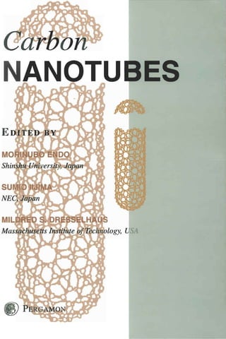
- 6. CARBON NANOTUBES Edited by MORINUBO END0 Shinshu University, Japan SUM10 IIJIMA NEC, Japan MILDRED S. DRESSELHAUS Massachusetts Institute of Technology, USA PERGAMON
- 7. U.K. Elsevier Science Ltd, The Boulevard, Langford Lane, Kidlington, Oxford OX5 lGB, U.K. U.S.A. Elsevier Science Inc., 660 White Plains Road, Tarrytown, New York 10591-5153, U.S.A. JAPAN Elsevier Science Japan, Tsunashima Building Annex, 3-20-12 Yushima Bunko-ku, Tokyo 113, Japan Copyright 0 1996 Elsevier Science Limited All Rights Reserved. No part of this publication may be reproduced, stored in a retrieval system or transmitted in any form or by any means, electronic, electrostatic, magnetic tape, mechanical, photocopying, recording or otherwise, without permission in writing from the publisher First edition 1996 Library of Congress Cataloging in Pulication Data A catalog record for this book is available from the Library of Congress British Library Cataloguing in Publication Data A catalogue record for this book is available in the British Library ISBN 008 0426824 Reprinted from: Carbon, Vol. 33, Nos 1, 2, 7, 12 Printed and bound in Great Britain by BPC Wheatons Ltd, Exeter
- 8. CONTENTS M. ENDO, S. IIJIMA and M. S. DRESSELHAUS: Editorial , . . . . . . . . . . . . . . . . . . . . . . vii M. S. DRESSELHAUS: Preface: Carbon nanotubes . . . . , . . . . , . . . . . . . . . . . . . . . . . . . . . . . ix M. ENDO, K. TAKEUCHI, K. KOBORI, K. TAKAHASHI, H. W. KROTO and A. SARKAR: Pyrolytic carbon nanotubes from vapor-grown carbon fibers.. . . . . . . 1 D. T. COLBERT and R. E. SMALLEY: Electric effects in nanotube growth.. . . . . . * . . 11 V. IVANOY, A. FONSECA, J. B. NAGY, A. LUCAS, P. LAMBIN, D. BERNAERTS and X. B. ZHANG: Catalytic production and purification of nanotubes having fullerene- scale diameters . . . . . . . . . . . . . . . . . . . . . . . . . . . . . . . . . . . . . . . . . . . . . . . . . . . . . . . . . . . . . 15 M. S. DRESSELHAUS, G. DRESSELHAUS and R. SAITO: Physics of carbon nanotubes 27 J. W. MINTMIRE and C. T. WHITE: Electronic and structural properties of carbon nanotubes .................................................................. 37 C.-H. KIANG, W. A. GODDARD 1 1 R. BEYERS and D. S. BETHUNE: Carbon 1, nanotubes with single-layer walls . . . . . . . . , . . . . . . . . . . . . . . . . . . . . . . . . . . . . . . . . . . . . 47 R. SETTON: Carbon nanotubes: I. Geometrical considerations . . . . . . . . . . . . . . . . . . . . . . 59 K. SATTLER: Scanning tunneling microscopy of carbon nanotubes and nanocones . . . . . 65 T. W. EBBESEN and T. TAKADA: Topological and SP3 defect structures in nanotubes 71 S. IHARA and S. ITOH: Helically coiled and torodial cage forms of graphitic carbon . . 77 A. FONSECA, K. HERNADI, J. B. NAGY, P. H. LAMBIN and A. A. LUCAS: Model structure of perfectly graphitizable coiled carbon nanotubes . . . . . . . . . . . . . . . . . . . . . 87 A. SARKAR, H. W. KROTO and M. ENDO: Hemi-toroidal networks in pyrolytic carbon nanotubes . . . . . . . . . . . . . . . . . . . . , . . . . . . . . . . . . . . . . . . . . . . . . . . . . . . . . . . . . . . . . . . . . . 105 X. K. WANG, X. W. LIN, S. N. SONG, V. P. DRAVID, J. B. KETTERSON and R. P. H. CHANG: Properties of buckytubes and derivatives.. . . . . . . . . . . . . . . . . . . . 111 J.-P. ISSI, L. LANGER, J. HEREMANS and C. H. OLK: Electronic properties of carbon nanotubes: Experimental results . . . . . . . . . . . . . . . . . . . . . . . . . . , . . . . . . . . . . . . . . . , . . . 121 P. C. EKLUND, J. M. HOLDEN and R. A. JISHI: Vibrational modes of carbon nanotubes: Spectroscopy and theory. . . . . . . . . . . . . . . . . . . . . .. . . . . . . . . . . . . . . . . . . . . . . . . . . . . . . 129 R. S. RUOFF and D. C. KORENTS: Mechanical and thermal properties of carbon nanotubes . . .. . . . . . . . . . . . . . . . . . . . . . . . . . . . . . . . . . . . . . . . . . . . . . . . . .. . . . . . . . . . .. . 143 J. IF. DESPRES, E. DAGUERRE and K. LAFDI: Flexibility of graphene layers in carbon nanotubes. . . . . . . . . . . . . . . . . . . . . . . . . . . . . . . . . . . . . . . . . . . . . . . . . . . . . . . . . . . . . . . . . . 1.49 V
- 9. Y. SAITO: Nanoparticles and filled nanocapsules ................................... 153 D. UGARTE: Onion-like graphitic particles ........................................ 163 U. ZIMMERMAN. N. MALINOWSKI. A . BURKHARDT and T. P. MARTIN: Metal- coated fullerenes ............................................................ 169 Subject Index ................................................................... 181 Author Index ................................................................... 183 vi
- 10. EDITORIAL Carbon nanotubes have been studied extensively in allotropes. Readers can then understand the fasci- relation to fullerenes, and together with fullerenes nation of graphene sheets when they are rolled into have opened a new science and technology field on a nanometer size tubular form from a flat network nano scale materials. This book aims to cover recent corresponding to conventional graphite. This book research and development in this area, and so provide also contains complementary reviews on carbon a convenient reference tool for all researchers in this nanoparticles such as carbon nano-capsules, onion- field. It is a.lso hoped that this book can serve to like graphite particles and metal-coated fullerenes. stimulate future work on carbon nanotubes. We hope this book will contribute to the dissemina- Carbon nanotubes have the same range of diameters tion of present understanding of the subject and to as fullerenes, and are expected to show various kinds future developments in the science and technology of of size effects in their structures and properties. carbon nanotubes and fullerenes, and of carbon Carbon nanotubes are one-dimensional materials and science more generally. fullerenes are zero-dimensional, which brings differ- ent effects to bear on their structures as well as on The editors thank all authors who contributed so their properties. A whole range of issues from the many excellent papers covering all aspects of carbon preparation, structure, properties and observation of nanotubes and the related fields. We are indebted to quantum effects in carbon nanotubes in comparison the Editor-in-Chief of Carbon, Professor Peter A. with 0-D fullerenes are discussed in this book. Thrower, for his suggestion and kind efforts, and also to Dr V. Kiruvanayagam for her kind cooperation In order to review the wide research area of carbon related to this book. nanotubes this book focuses on recent intensive Morinobu Endo work published in Carbon. The papers are written Sumio Iijima from the viewpoint that carbon nanotubes, as well Mildred S. Dresselhaus as fullerenes, are the most interesting new carbon Editors vii
- 12. PREFACE Since the start of this decade (the 1990's), fullerene efficiency and smaller nanotubes are discussed in research has blossomed in many different directions, the article by Ivanov and coworkers. The quantum and has attracted a great deal of attention to Carbon aspects of carbon nanotubes, stemming from their Science. It was therefore natural to assemble, under small diameters, which contain only a small number the guest editorship of Professor Harry Kroto, one of carbon atoms (< lo2), lead to remarkable sym- of the earliest books on the subject of fullerenes 111, metries and electronic structure, as described in the a book that has had a significant impact on the articles by Dresselhaus, Dresselhaus and Saito and subsequent developments of the fullerene field. by Mintmire and White. Because of the simplicity Stemming from the success of the first volume, it is of the single-wall nanotube, theoretical work has now appropriate to assemble a follow-on volume on focussed almost exclusively on single-wall nanotubes. Carbon Nanotubes. It is furthermore fitting that The remarkable electronic properties predicted for Dr Sumio Iijima and Professor Morinobu Endo serve carbon nanotubes are their ability to be either con- as the Guest Editors of this volume, because they are ducting or to have semiconductor behavior, depending the researchers who are most responsible for opening on purely geometrical factors, namely the diameter up the field of carbon nanotubes. Though the field and chirality of the nanotubes. The existence of is still young and rapidly developing, this is a very conducting nanotubes thus relates directly to the appropriate time to publish a book on the very active symmetry-imposed band degeneracy and zero-gap topic of carbon nanotubes. semiconductor behavior for electrons in a two-dirnen- The goal of this book is thus to assess progress in sional single layer of graphite (called a graphene the field, to identify fruitful new research directions, sheet). The existence of finite gap semiconducting to summarize the substantial progress that has thus behavior arises from quantum effects connected with far been made with theoretical studies, and to clarify the small number of wavevectors associated with some unusual features of carbon-based materials that the circumferential direction of the nanotubes. The are relevant to the interpretation of experiments on article by Kiang et al. reviews the present status of the carbon nanotubes that are now being so actively synthesis of single-wall nanotubes and the theoretical pursued. A second goal of this book is thus to implications of these single-wall nanotubes. The geo- stimulate further progress in research on carbon metrical considerations governing the closure, helicity nanotubes and related materials. and interlayer distance of successive layers in multi- 'The birth of the field of carbon nanotubes is layer carbon nanotubes are discussed in the paper by marked by the publication by Iijima of the observation Setton. of multi-walled nanotubes with outer diameters as Study of the structure of carbon nanotubes and small as 55 A, and inner diameters as small as 23 A, their common defects is well summarized in the and a nanotube consisting of only two coaxial review by Sattler, who was able to obtain scanning cyknders [2]. This paper was important in making tunneling microscopy (STM) images of carbon nan- the connection between carbon fullerenes, which are otube surfaces with atomic resolution. A discussion quantum dots, with carbon nanotubes, which are of common defects found in carbon nanotubes, quantum wires. Furthermore this seminal paper [2] including topological, rehybridization and bonding has stimulated extensive theoretical and experimental defects is presented by Ebbesen and Takada. The research for the past five years and has led to the review by Ihara and Itoh of the many helical and creation of a rapidly developing research field. toroidal forms of carbon nanostructures that may be The direct linking of carbon nanotubes to graphite realized provides insight into the potential breadth and the continuity in synthesis, structure and proper- of this field. The joining of two dissimilar nanotubes ties between carbon nanotubes and vapor grown is considered in the article by Fonseca et al., where carbon fibers is reviewed by the present leaders of this these concepts are also applied to more complex area, Professor M. Endo, H. Kroto, and co-workers. structures such as tori and coiled nanotubes. The role Further insight into the growth mechanism is pre- of semi-toroidal networks in linking the inner and sented in the article by Colbert and Smalley. New outer walls of a double-walled carbon nanotube is synthesis methods leading to enhanced production discussed in the paper by Sarkar et al. ix
- 13. X Preface From an experimental point of view, definitive The final section of the volume contains three measurements on the properties of individual carbon complementary review articles on carbon nano- nanotubes, characterized with regard to diameter and particles. The first by Y. Saito reviews the state of chiral angle, have proven to be very difficult to carry knowledge about carbon cages encapsulating metal out. Thus, most of the experimental data available and carbide phases. The structure of onion-like thus far relate to multi-wall carbon nanotubes and to graphite particles, the spherical analog of the cylin- bundles of nanotubes. Thus, limited experimental drical carbon nanotubes, is reviewed by D. Ugarte, information is available regarding quantum effects the dominant researcher in this area. The volume for carbon nanotubes in the one-dimensional limit. concludes with a review of metal-coated fullerenes by A review of structural, transport, and susceptibility T. P. Martin and co-workers, who pioneered studies measurements on carbon nanotubes and related on this topic. materials is given by Wang et al., where the inter- The guest editors have assembled an excellent set relation between structure and properties is empha- of reviews and research articles covering all aspects of sized. Special attention is drawn in the article by the field of carbon nanotubes. The reviews are pre- Issi et al. to quantum effects in carbon nanotubes, as sented in a clear and concise form by many of the observed in scanning tunneling spectroscopy, trans- leading researchers in the field. It is hoped that this port studies and magnetic susceptibility measure- collection of review articles provides a convenient ments. The vibrational modes of carbon nanotubes reference for the present status of research on carbon is reviewed in the article by Eklund et al. from both nanotubes, and serves to stimulate future work in the a theoretical standpoint and a summary of spec- field. troscopy studies, while the mechanical that thermal M. S. DRESSELHAUS properties of carbon nanotubes are reviewed in the article by Ruoff and Lorents. The brief report by REFERENCES Despres et al. provides further evidence for the 1. H. W. Kroto, Carbon 30, 1139 (1992). flexibility of graphene layers in carbon nanotubes. 2. S . Iijima, Nature (London) 354, 56 (1991).
- 14. PYROLYTIC CARBON NANOTUBES FROM VAPOR-GROWN CARBON FIBERS ENDO,'Kmn TAKEUCHI,' MORINOBU KIYOHARU KOBORI,'KATSUSHI TAKAHASHI, I HAROLD K R O T O ,and A. SARKAR' W. ~ 'Faculty of Engineering, Shinshu University, 500 Wakasato, Nagano 380, Japan 'School of Chemistry and Molecular Sciences, University of Sussex, Brighton BNl SQJ, U.K. (Received 21 November 1994; accepted 10 February 1995) Abstract-The structure of as-grown and heat-treated pyrolytic carbon nanotubes (PCNTs) produced by hydrocarbon pyrolysis are discussed on the basis of a possible growth process. The structures are com- pared with those of nanotubes obtained by the arc method (ACNT, arc-formed carbon nanotubes). PCNTs, with and without secondary pyrolytic deposition (which results in diameter increase) are found to form during pyrolysis of benzene at temperatures ca. 1060°C under hydrogen. PCNTs after heat treatment at above 2800°C under argon exhibit have improved stability and can be studied by high-resolution trans- mission electron microscopy (HRTEM). The microstructures of PCNTs closely resemble those of vapor- grown carbon fibers (VGCFs). Some VGCFs that have micro-sized diameters appear to have nanotube inner cross-sections that have different mechanical properties from those of the outer pyrolytic sections. PCNTs initially appear to grow as ultra-thin graphene tubes with central hollow cores (diameter ca. 2 nm or more) and catalytic particles are not observed at the tip of these tubes. The secondary pyrolytic depo- sition, which results in characteristic thickening by addition of extra cylindrical carbon layers, appears to occur simultaneously with nanotube lengthening growth. After heat treatment, HRTEM studies indicate clearly that the hollow cores are closed at the ends of polygonized hemi-spherical carbon caps. The most commonly observed cone angle at the tip is generally ca. 20", which implies the presence of five pentago- nal disclinations clustered near the tip of the hexagonal network. A structural model is proposed for PCNTs observed to have spindle-like shape and conical caps at both ends. Evidence is presented for the forma- tion, during heat treatment, of hemi-toroidal rims linking adjacent concentric walls in PCNTs. A possi- ble growth mechanism for PCNTs, in which the tip of the tube is the active reaction site, is proposed. Key Words-Carbon nanotubes, vapor-grown carbon fibers, high-resolution transmission electron micro- scope, graphite structure, nanotube growth mechanism, toroidal network. 1. INTRODUCTION been proposed involving both open-ended1131 and Since Iijima's original report[l], carbon nanotubes closed-cap[l 1,121 mechanisms for the primary tubules. Whether either of these mechanisms or some other oc- have been recognized as fascinating materials with curs remains to be determined. nanometer dimensions promising exciting new areas It is interesting to compare the formation process of carbon chemistry and physics. From the viewpoint of fibrous forms of carbon with larger micron diam- of fullerene science they also are interesting because eters and carbon nanotubes with nanometer diameters they are forms of giant fuIlerenes[2]. The nanotubes from the viewpoint of "one-dimensional)) carbon struc- prepared in a dc arc discharge using graphite elec- tures as shown in Fig. 1. The first class consists of trodes at temperatures greater than 3000°C under graphite whiskers and ACNTs produced by arc meth- helium were first reported by Iijima[l] and later by ods, whereas the second encompasses vapor-grown car- Ebbesen and Ajyayan[3]. Similar tubes, which we call bon fibers and PCNTs produced by pyrolytic processes. pyrolytic carbon nanotubes (PCNTs), are produced A third possibIe class would be polymer-based nano- by pyrolyzing hydrocarbons (e.g., benzene at ca. tubes and fibers such as PAN-based carbon fibers, 110OoC)[4-9]. PCNTs can also be prepared using the which have yet to be formed with nanometer dimen- same equipment as that used for the production of sions. In the present paper we compare and discuss the so called vapor-grown carbon fibers (VGCFs)[lOJ.The structures of PCNTs and VGCFs. VGCFs are micron diameter fibers with circular cross- sections and central hollow cores with diameters ca. a few tens of nanometers. The graphitic networks are 2. VAPOR-GROWN CARBON FIBERS AND arranged in concentric cylinders. The intrinsic struc- PYROLYTIC CARBON NANOTUBES tures are rather like that of the annual growth of trees. The structure of VGCFs, especially those with hollow Vapor-grown carbon fibers have been prepared by cores, are very similar to the structure of arc-formed catalyzed carbonization of aromatic carbon species carbon nanotubes (ACNTs). Both types of nanotubes, using ultra-fine metal particles, such as iron. The par- the ACNTs and the present PCNTs, appear to be ticles, with diameters less than 10 nm may be dispersed essentially Russian Doll-like sets of elongated giant on a substrate (substrate method), o r allowed to float ful,lerenes[ll,12]. Possible growth processes have in the reaction chamber (fluidized method). Both 1
- 15. 2 M. ENDO al. et Fig. 1. Comparative preparation methods for micrometer size fibrous carbon and carbon nanotubes as one-dimensional forms of carbon. methods give similar structures, in which ultra-fine catalytic particles are encapsulated in the tubule tips (Fig. 2). Continued pyrolytic deposition occurs on the initially formed thin carbon fibers causing thickening (ca. 10 pm diameter, Fig. 3a). Substrate catalyzed fi- bers tend to be thicker and the floating technique pro- duces thinner fibers (ca. 1 pm diameter). This is due to the shorter reaction time that occurs in the fluid- ized method (Fig. 3b). Later floating catalytic meth- ods are useful for large-scale fiber production and, thus, VGCFs should offer a most cost-effective means Fig. 3. Vapor-grown carbon fibers obtained by substrate of producing discontinuous carbon fibers. These method with diameter ca. 10 pm (a) and those by floating cat- VGCFs offer great promise as valuable functional car- alyst method (b) (inserted, low magnification). bon filler materials and should also be useful in car- bon fiber-reinforced plastic (CFRP) production. As seen in Fig. 3b even in the “as-grown” state, carbon 3. PREPARATION OF VGCFs AND PCNTs particles are eliminated by controlling the reaction conditions. This promises the possibility of producing The PCNTs in this study were prepared using pure ACNTs without the need for separating spheroidal the same apparatus[9] as that employed to produce carbon particles. Hitherto, large amounts of carbon VGCFs by the substrate method[l0,15]. Benzene va- particles have always been a byproduct of nanotube por was introduced, together with hydrogen, into a ce- production and, so far, they have only been eliminated ramic reaction tube in which the substrate consisted by selective oxidation[l4]. This has led to the loss of of a centrally placed artificial graphite rod. The tem- significant amounts of nanotubes - ca. 99%. perature of the furnace was maintained in the 1000°C range. The partial pressure of benzene was adjusted to be much lower than that generally used for the preparation of VGCFs[lO,lS] and, after one hour decomposition, the furnace was allowed to attain room temperature and the hydrogen was replaced by argon. After taking out the substrate, its surface was scratched with a toothpick to collect the minute fibers. Subsequently, the nanotubes and nanoscale fibers were heat treated in a carbon resistance furnace un- der argon at temperatures in the range 2500-3000°C for ca. 10-15 minutes. These as-grown and sequen- tially heat-treated PCNTs were set on an electron mi- croscope grid for observation directly by HRTEM at 400kV acceleration voltage. It has been observed that occasionally nanometer scale VGCFs and PCNTs coexist during the early Fig. 2. Vapour-grown carbon fiber showing relatively early stages of VGCF processing (Fig. 4). The former tend stage of growth; at the tip the seeded Fe catalytic particle is to have rather large hollow cores, thick tube walls and encapsulated. well-organized graphite layers. On the other hand,
- 16. Pyrolytic carbon nanotubes from vapor-grown carbon fibers 3 a t b Fig. 5 . Heat-treated pyrolytic carbon nanotube and enlarged one (inserted), without deposited carbon. This results in well-organized multi-walled concentric graphite tubules. The interlayer spacing (0.34 nm) is slightly wider on average than in the case of thick VGCFs treated at similar temperatures. This small in- crease might be due to the high degree of curvature of the narrow diameter nanotubes which appears to pre- vent perfect 3-dimensional stacking of the graphitic layers[ 16,171. PCNTs and VGCFs are distinguishable Fig. 4. Coexisting vapour-grown carbon fiber, with thicker diameter and hollow core, and carbon nanotubes, with thin- by the sizes of the well-graphitized domains; cross- ner hollow core, (as-grown samples). sections indicate that the former are characterized by single domains, whereas the latter tend to exhibit mul- tiple domain areas that are small relative to this cross- PCNTs tend to have very thin walls consisting of only sectional area. However, the innermost part of some a few graphitic cylinders. Some sections of the outer VGCFs (e.g., the example shown in Fig. 5 ) may often surfaces of the thin PCNTs are bare, whereas other consist of a few well-structured concentric nanotubes. sections are covered with amorphous carbon depos- Theoretical studies suggest that this “single grain” as- its (as is arrowed region in Fig. 4a). TEM images of pect of the cross-sections of nanotubes might give rise the tips of the PCNTs show no evidence of electron to quantum effects. Thus, if large scale real-space beam opaque metal particles as is generally observed super-cell concepts are relevant, then Brillouin zone- for VGCF tips[lO,l5]. The large size of the cores and foiding techniques may be applied to the description the presence of opaque particles at the tip of VGCFs of dispersion relations for electron and phonon dy- suggests possible differences between the growth namics in these pseudo one-dimensional systems. mechanism for PCNTs and standard VGCFs[7-91. A primary nanotube at a very early stage of thick- The yield of PCNTs increases as the temperature and ening by pyrolytic carbon deposition is depicted in the benzene partial pressure are reduced below the op- Figs. 6a-c; these samples were: (a) as-grown and (b), timum for VGCF production (i.e., temperature ca. (c) heat treated at 2500°C. The pyrolytic coatings 1000°-11500C). The latter conditions could be effec- shown are characteristic features of PCNTs produced tive in the prevention or the minimization of carbon by the present method. The deposition of extra car- deposition on the primary formed nanotubules. bon layers appears to occur more or less simultane- ously with nanotube longitudinal growth, resulting in spindle-shaped morphologies. Extended periods of py- 4. STRUCTURES OF PCNTs rolysis result in tubes that can attain diameters in the Part of a typical PCNT (ca. 2.4 nm diameter) af- micron range (e.g., similar to conventional (thick) ter heat treatment at 2800°C for 15 minutes is shown VGCFs[lO]. Fig. 6c depicts a 002 dark-field image, in Fig. 5. It consists of a long concentric graphite tube showing the highly ordered central core and the outer with interlayer spacings ca. 0.34 nm-very similar in inhomogeneously deposited polycrystalline material morphology to ACNTs[ 1,3]. These tubes may be very (bright spots). It is worthwhile to note that even the long, as long as 100 nm or more. It would, thus, ap- very thin walls consisting of several layers are thick pear that PCNTs, after heat treatment at high temper- enough to register 002 diffraction images though they atures, become graphitic nanotubes similar to ACNTs. are weaker than images from deposited crystallites on The heat treatment has the effect of crystallizing the the tube. secondary deposited layers, which are usually com- Fig. 7a,b depicts PCNTs with relatively large diam- posed of rather poorly organized turbostratic carbon. eters (ca. 10 nm) that appear to be sufficiently tough
- 17. 4 M. ENDOet al. Fig. 7. Bent and twisted PCNT (heat treated at 2500T). Fig. 6 . PCNTs with partially deposited carbon layers (arrow indicates the bare PCNT), (a) as-grown, (b) partially exposed nanotube and (c) 002 dark-field image showing small crys- tallites on the tube and wall of the tube heat treated at 2500°C. and flexible to bend, twist, or kink without fractur- ing. The basic structural features and the associated mechanical behavior of the PCNTs are, thus, very dif- ferent from those of conventional PAN-based fibers as well as VGCFs, which tend to be fragile and easily broken when bent or twisted. The bendings may occur at propitious points in the graphene tube network[l8]. Fig. 8a,b shows two typical types of PCNT tip morphologies. The caps and also intercompartment di- aphragms occur at the tips. In general, these consist of 2-3 concentric layers with average interlayer spac- ing of ca. 0.38 nm. This spacing is somewhat larger than that of the stackings along the radial direction, presumably (as discussed previously) because of sharp curvature effects. As indicated in Fig. 9, the conical shapes have rather symmetric cone-like shells. The an- gle, ca. 20°, is in good agreement with that expected for a cone constructed from hexagonal graphene sheets containing pentagonal disclinations -as is Fig. 9e. Ge and Sattler[l9] have reported nanoscale conical carbon materials with infrastructure explain- able on the basis of fullerene concepts. STM measure- Fig. 8. The tip of PCNTs with continuous hollow core (a) ments show that nanocones, made by deposition of and the cone-like shape (b) (T indicates the toroidal struc- very hot carbon on HOPG surfaces, often tend to ture shown in detail in Fig. 11).
- 18. Pyrolytic carbon nanotubes from vapor-grown carbon fibers (c) e =60.0° (d) e =38.9' (e) e =i9.2' e = 180- (360/ n )cos-' [l - (n/6)] [" I (n : number of pentagons) Fig. 9. The possible tip structure with cone shape, in which Growth direction the pentagons are included. As a function of the number of 4 pentagons, the cone shape changes. The shaded one with 19.2" tip angle is the most frequently observed in PCNTs. have an opening angle of ca. 20". Such caps may, however, be of five possible opening angles (e.g., from 112.9" to 19.2") depending on the number of pentag- onal disclinations clustered at the tip of the cone, as indicated in Fig. 9[8]. Hexagons in individual tube walls are, in general, arranged in a helical disposition with variable pitches. It is worth noting that the small- est angle (19.2') that can involve five pentagons is most frequently observed in such samples. It is fre- chiralsaucture quently observed that PCNTs exhibit a spindle-shaped structure at the tube head, as shown in Fig. 8b. Fig. 10. Growth mechanism proposed for the helical nanotubes (a) and helicity (b), and the model that gives the bridge and laminated tip structure (c). 5. GROWTH MODEL OF PCNTs In the case of the PCNTs considered here, the growth temperature is much lower than that for small closed cage fullerenes. Based on the observation ACNTs, and no electric fields, which might influence of open-ended tubes, Iijima et a1.[13] have discussed the growth of ACNTs, are present. It is possible that a plausible alternative way in which such tubules might different growth mechanisms apply to PCNT and possibly grow. The closed cap growth mechanism ef- ACNT growth and this should be taken into consid- fectively involves the addition of extended chains of eration. As mentioned previously, one plausible mech- sp carbon atoms to the periphery of the asymmetric anism for nanotube growth involves the insertion of 6-pentagon cap, of the kind whose Schlegel diagram small carbon species C,, n = 1,2,3 . . .) into a closed ( is depicted in Fig. loa, and results in a hexagonal fullerene cap (Fig. loa-c)[ll]. Such a mechanism is re- graphene cylinder wall in which the added atoms are lated to the processes that Ulmer et a1.[20] and McE1- arranged in a helical disposition[9,1 I] similar to that vaney et a1.[21] have discovered for the growth of observed first by Iijima[l].
- 19. 6 M. ENDOet al. It is proposed that during the growth of primary tubule cores, carbon atoms, diameters, and longer lin- ear clusters are continuously incorporated into the ac- tive sites, which almost certainly lie in the vicinity of the pentagons in the end caps, effectively creating he- lical arrays of consecutive hexagons in the tube wall as shown in Fig. 10a,b[9,11]. Sequential addition of 2 carbon atoms at a time to the wall of the helix re- sults in a cap that is indistinguishable other than by rotation[ll,l2]. Thus, if carbon is ingested into the cap and wholesale rearrangement occurs to allow the new atoms to “knit” smoothly into’ the wall, the cap can be considered as effectively fluid and to move in a screw-like motion leaving the base of the wall stationary- though growing by insertion of an essen- tially uniform thread of carbon atoms to generate a helical array of hexagons in the wall. The example shown in Fig. 10a results in a cylinder that has a di- ameter (ca. 1 nm) and a 22-carbon atom repeat cycle and a single hexagon screw pitch -the smallest arche- typal (isolated pentagon) example of a graphene nano- tube helix. Though this model generates a tubule that is rather smaller than is usually the case for the PCNTs observed in this study (the simplest of which have di- ameters > 2-3 nm), the results are of general semi- quantitative validity. Figure 10b,c shows the growth mechanism diagrammatically from a side view. When the tip is covered by further deposition of aromatic layers, it is possible that a templating effect occurs to form the new secondary surface involving pentagons in the hexagonal network. Such a process would ex- plain the laminated or stacked-cup-like morphology observed. In the case of single-walled nanotubes, it has been recognized recently that transition metal particles play a role in the initial filament growth process[23]. ACNTs and PCNTs have many similarities but, as the vapor- growth method for PCNTs allows greater control of the growth process, it promises to facilitate applica- tions more readily and is thus becoming the preferred Fig. 11. The sealed tip of a PCNT heat treated at 2800°C method of production. with a toroidal structure (T) and, (b) molecular graphics im- ages of archetypal flattened toroidal model at different orien- tations and the corresponding simulated TEM images. 6. CHARACTERISTIC TOROIDAL AND SPINDLE-LIKE STRUCTURES OF PCNTS In Fig. 1l a is shown an HRTEM image of part of the basis of archetypal double-walled nanotubes[24]. the end of a PCNTs. The initial material consisted of As the orientation changes, we note that the HRTEM a single-walled nanotube upon which bi-conical interference pattern associated with the rim changes spindle-like growth can be seen at the tip. Originally, from a line to an ellipse and the loop structures at the this tip showed no apparent structure in the HRTEM apices remain relatively distinct. The oval patterns in image at the as-grown state, suggesting that it might the observed and simulated HRTEM image (Fig. 1lb) consist largely of some form of “amorphous” carbon. are consistent with one another. For this preliminary After a second stage of heat treatment at 280O0C, the investigation a symmetric (rather than helical) wall amorphous sheaths graphitize to a very large degree, configuration was used for simplicity. Hemi-toroidal producing multi-walled graphite nanotubes that tend connection of the inner and outer tubes with helical to be sealed off with caps at points where the spindle- structured walls requires somewhat more complicated like formations are the thinnest. The sealed-off end re- dispositions of the 5/6/7 rings in the lip region. The gion of one such PCNT with a hemi-toroidal shape is general validity of the conclusions drawn here are, shown in Fig. 1la. however, not affected. Initial studies of the problem In Fig. 1l b are depicted sets of molecular graph- indicated that linking between the inner and outer ics images of flattened toroidal structures which are walls is not, in general, a hindered process.
- 20. Pyrolytic carbon nanotubes from vapor-grown carbon fibers The toroidal structures show interesting changes in morphology as they become larger-at least at the lip. The hypothetical small toroidal structure shown in Fig. 1 l b is actually quite smooth and has an essen- tially rounded structure[24]. As the structures become larger, the strain tends to focus in the regions near the pentagons and heptagons, and this results in more prominent localized cusps and saddle points. Rather elegant toroidal structures with Dnhand Dndsymme- try are produced, depending on whether the various paired heptagodpentagon sets which lie at opposite ends of the tube are aligned or are offset. In general, they probably lie is fairly randomly disposed positions. Chiral structures can be produced by off-setting the pentagons and heptagons. In the D5dstructure shown in Fig. 11 which was developed for the basic study, the walls are fluted between the heptagons at opposite ends of the inner tube and the pentagons of the outer wall rim[l7]. It is interesting to note that in the com- puter images the localized cusping leads to variations in the smoothness of the image generated by the rim, though it still appears to be quite elliptical when viewed at an angle[ 171. The observed image appears to exhibit variations that are consistent with the local- B ized cusps as the model predicts. c k In this study, we note that epitaxial graphitization Spinale-shapemodel is achieved by heat treatment of the apparently mainly Fig. 12. As-grown PCNTs with partially thickened spindle amorphous material which surrounds a single-walled shape (a) and the proposed structural model for spindle par- nanotube[ 171. As well as bulk graphitization, localized ticles including 12 pentagons in hexagon cage (b). hemi-toroidal structures that connect adjacent walls have been identified and appear to be fairly common in this type of material. This type of infrastructure ber of pentagons as required by variants of Euler’s may be important as it suggests that double walls may Law. Hypothetical structural models for these spin- form fairly readily. Indeed, the observations suggest dles are depicted in Fig. 12. It is possible that simi- that pure carbon rim-sealed structures may be readily lar two-stage growth processes occur in the case of produced by heat treatment, suggesting that the future ACNTs but, in general, the secondary growth appears fabrication of stabilized double-walled nanoscale to be intrinsically highly epitaxial. This may be be- graphite tubes in which dangling bonds have been cause in the ACNT growth case only carbon atoms are eliminated is a feasible objective. It will be interesting involved and there are fewer (non-graphitizing) alter- to prove the relative reactivities of these structures for native accretion pathways available. It is likely that their possible future applications in nanoscale devices epitaxial growth control factors will be rather weak (e.g., as quantum wire supports). Although the cur- when secondary deposition is very fast, and so thin vatures of the rims appear to be quite tight, it is clear layers may result in poorly ordered graphitic structure from the abundance of loop images observed, that the in the thicker sections. It appears that graphitization occurrence of such turnovers between concentric cylin- of this secondary deposit that occurs upon heat treat- ders with a gap spacing close to the standard graphite ment may be partly responsible for the fine structure interlayer spacing is relatively common. Interestingly, such as compartmentalization, as well as basic tip the edges of the toroidal structures appear to be readily morphology[ 171. visible and this has allowed us to confirm the relation- ship between opposing loops. Bulges in the loops of 7. VGCFs DERIVED FROM NANOTUBES the kind observed are simulated theoretically[ 171. Once one layer has formed (the primary nanotube In Fig. 13 is shown the 002 lattice images of an “as- core), further secondary layers appear to deposit with formed” very thin VGCF. The innermost core diam- various degrees of epitaxial coherence. When inhomo- eter (ca. 20 nm as indicated by arrows) has two layers; geneous deposition occurs in PCNTs, the thickening it is rather straight and appears to be the primary has a characteristic spindle shape, which may be a nanotube. The outer carbon layers, with diameters ca. consequence of non-carbon impurities which impede 3-4 nm, are quite uniformly stacked parallel to the graphitization (see below)- this is not the case for central core with 0.35 nm spacing. From the difference ACNTs were growth takes place in an essentially all- in structure as well as the special features in the me- carbon atmosphere, except, of course, for the rare gas. chanical strength (as in Fig. 7) it might appear possi- These spindles probably include the appropriate num- ble that the two intrinsically different types of material
- 21. 8 et M. ENDO al. of ca. 10 nm (white arrow), observed by field emission scanning electron microscopy (FE-SEM)[25]. It is, thus, suggested that at least some of the VGCFs start as nanotube cores, which act as a substrate for sub- sequent thickening by deposition of secondary pyro- lytic carbon material, as in the catalytically primarily grown hollow fiber. In Fig. 14b is also shown the TEM image corresponding to the extruded nanotube from a very thin fiber. It is clearly observed that the exposed nanotube is continuing into the fiber as a central hol- low core, as indicated by the white arrow in the figure. It is interesting that, as indicated before (in Fig. 14a), the core is more flexible than the pyrolytic part, which is more fragile. Fig. 13. HRTEM image of an as-grown thick PCNT. 002 lattice image demonstrates the innermost hollow core (core 8 CONCLUSION . diam. 2.13 nm) presumably corresponding to the “as-formed” Pyrolytic carbon nanotubes (PCNTs), which grow nanotube. The straight and continuous innermost two fringes similar to Fig. 5 are seen (arrow). during hydrocarbon pyrolysis, appear to have struc- tures similar to those obtained by arddischarge tech- niques using graphite electrodes (ACNTs). The PCNTs tend to exhibit a characteristic thickening feature due involved might be separated by pulverizing the VGCF to secondary pyrolytic carbon deposition. Various tip material. morphologies are observed, but the one most fre- In Fig. 14a, a ca. 10 wm diameter VGCF that has quently seen has a 20” opening angle, suggesting that, been broken in liquid nitrogen is depicted, revealing in general, the graphene conical tips possess a cluster the cylindrical graphitic nanotube core with diameter of five pentagons that may be actively involved in tube growth. PCNTs with spindle-like shapes and that have conical caps at both ends are also observed, for which a structural model is proposed. The spindle-like struc- tures observed for the secondary growth thickening that occurs in PCNTs may be a consequence of the lower carbon content present in the growth atmo- sphere than occurs in the case of ACNT growth. Pos- sible structural models for these spindles have been discussed. The longitudinal growth of nanotubes ap- pears to occur at the hemi-spherical active tips and this process has been discussed on the basis of a closed cap mechanism[9,11]. The PCNTs are interesting, not only from the viewpoint of the fundamental perspective that they are very interesting giant fullerene structures, but also because they promise to be applications in novel strategically important materials in the near fu- ture. PCNT production appears, at this time, more readily susceptible to process control than is ACNT production and, thus, their possible value as fillers in advanced composites is under investigation. Acknowledgements-Japanese authors are indebted to M. S. Dresselhaus and G. Dresselhaus of MIT and to A. Oberlin of Laboratoire Marcel Mathieu (CNRS) for their useful dis- cussions and suggestions. HWK thanks D. R. M. Walton for help and the Royal Society and the SERC (UK) for support. Part of the work by ME is supported by a grant-in-aid for scientific research in priority area “carbon cluster” from the Ministry. of Education, Science and Culture, Japan. Fig. 14. PCNTs (white arrow) appeared after breakage of VGCF, (a) FE-SEM image of broken VGCF, cut in liquid ni- REFERENCES trogen and (b) HRTEM image showing the broken part ob- served in very thin VGCF. The nanotube is clearly observed 1 . S. Iijima, Nature 354, 56 (1991). and this indicates that thin VGCF grow from nanometer core 2. H. W. Kroto, J. R. Heath, S. C .O’Brien, R. F. Curl, and by thickening. R. E. Samlley, Nature 318, 162 (1985).
- 22. Pyrolytic carbon nanotubes from vapor-grown carbon fibers 9 3. T. W. Ehhesen and P. M. Ajayan, Nature 358, 220 15. M. S. Dresselhaus, G Dresselhaus, K. Sugihara, I. L. ( 1 992). Spain, H. A. Goldberg, In Graphite Fibers and Fila- 4. M. Endo, H. Fijiwara, and E. Fukunaga, 18th Meeting ments, (edited by M . Cardona) pp. 244-286. Berlin, Japanese Carbon Society, (1991) p. 34. Springer. 5 . M. Endo, H. Fujiwara, and E. Fukunaga, 2nd C60 Sym- 16. J. S. Speck, M. Endo, and M. S. Dresselhaus, J. Crys- posium in Japan, (1992) p. 101. tal Growth 94, 834 (1989). 6. M. Endo, K. Takeuchi, S. Igarashi, and K. Kobori, 19th 17.. A. Sarkar, H. W. Kroto, and M. Endo (in preparation). Meeting Japanese Carbon Society, (1992) p. 192. 18. H. Hiura, T. W. Ebbesen, J. Fujita, K. Tanigaki, and 7. M. Endo, K. Takeuchi, S. Igarashi, K. Kobori, and M. T. Takada, Nature 367, 148 (1994). Shiraishi, Mat. Res. SOC. Spring Meet (1993) p.S2.2. 19. M. Ge and K. Sattler, Mat. Res. SOC. Spring Meet. S1.3, 8 . M. Endo, K. Takeuchi, S. Igarashi, K. Kobori, M. 360 (1993). Shiraishi, and H. W. Kroto, Mat. Res. SOC.FallMeet. 20. G. Ulmer, E. E. B. Cambel, R. Kuhnle, H. G. Busmann, 62.1 (1994). and 1. V . Hertel, Chem. Phys. Letts. 182, 114 (1991). 9. M. Endo, K. Takeuchi, S. Igarashi, K. Kobori, M. 21. S. W. McElvaney, M. N. Ross, N. S. Goroff, and E Shiraishi, and H. W. Kroto, J. Phys. Chem. Solids 54, Diederich, Science 259, 1594 (1993). 1841 (1993). 22. R. Saito, G . Dresselhaus, M. Fujita, and M. S. Dressel- 10. M. Endo, Chemtech 18, 568 (1988). haus, 4th NEC Symp. Phys. Chem. Nanometer Scale 11. M. Endo and H. W. Kroto, J. Phys. Chem. 96, 6941 Mats. (1992). (1992). 23. S. Iijima, Gordon Conference on the Chemistry of Hy- 12. I-I. W. KrOtQ, K. Prassides, R. Taylor, D. R. M. Wal- drocarbon Resources, Hawaii (1994). ton, and bd. Endo, International Conference Solid State 24. A. Sarker, H. W. Kroto, and M. Endo (to he published). Devices and Materials of The Japan Society of Applied 25. M. Endo, K. Takeuchi, K. Kobori, K. Takahashi, and Physics (1993), p. 104. H. W. Kroto (in preparation). 13. S. Iijima, Mat. Sci. Eng. B19, 172 (1993). 14. P. M. Ajayan, T. W. Ebbesen, T. Ichihashi, S. Iijirna, K. Tanigaki, and H. Hiura, Nature 362, 522 (1993).
- 24. ELECTRIC EFFECTS IN NANOTUBE GROWTH DANIEL COLBERT RICHARD SMALLEY T. and E. Rice Quantum Institute and Departments of Chemistry and Physics, MS 100, Rice University, Houston, TX 77251-1892, U.S.A. (Received 3 April 1995; accepted 7 April 1995) Abstract-We present experimental evidence that strongly supports the hypothesis that the electric field of the arc plasma is essential for nanotube growth in the arc by stabilizing the open tip structure against closure. By controlling the temperature and bias voltage applied to a single nanotube mounted on a mac- roscopic electrode, we find that the nanotube tip closes when heated to a temperature similar to that in the arc unless an electric field is applied. We have also developed a more refined awareness of “open” tips in which adatoms bridge between edge atoms of adjacent layers, thereby lowering the exothermicity in going from the open to the perfect dome-closed tip. Whereas realistic fields appear to be insufficient by themselves to stabilize an open tip with its edges completely exposed, the field-induced energy lower- ing of a tip having adatom spot-welds can, and indeed in the arc does, make the open tip stable relative to the closed one. Key Words-Nanotubes, electric field, arc plasma 1. INTRODUCTION the electric field concentrates, and never in the soots condensed from the carbon vapor exiting the arcing As recounted throughout this special issue, significant region, suggest a vital role for the electric field. Fur- advances in illuminating various aspects of nanotube thermore, the field strength at the nanotube tips is very growth have been made[l,2] since Iijima’s eventful large, due both to the way the plasma concentrates discovery in 1991;[3] these advances are crucial to most of the potential drop in a very short distance gaining control over nanotube synthesis, yield, and above the cathode, and to the concentrating effects of properties such as length, number of layers, and he- the field at the tips of objects as small as nanotubes. licity. The carbon arc method Iijima used remains the The field may be on the order of the strength required principle method of producing bulk amounts of qual- to break carbon-carbon bonds, and could thus dra- ity nanotubes, and provides key clues for their growth matically effect the tip structure. there and elsewhere. The bounty of nanotubes depos- In the remaining sections of this paper, we describe ited on the cathode (Ebbesen and Ajayan have found the experimental results leading to confirmation of the that up to 50% of the deposited carbon is tubular[4]) stabilizing role of the electric field in arc nanotube is particularly puzzling when one confronts the evi- growth. These include: relating the plasma structure dence of UgarteI.51 that tubular objects are energeti- to the morphology of the cathode deposit, which re- cally less stable than spheroidal onions. vealed that the integral role of nanotubes in sustain- It is largely accepted that nanotube growth occurs ing the arc plasma is their field emission of electrons at an appreciable rate only at open tips. With this con- into the plasma; studying the field emission character- straint, the mystery over tube growth in the arc redou- istics of isolated, individual arc-grown nanotubes; and bles when one realizes that the cathode temperature the discovery of a novel production of nanotubes that (-3000°C) is well above that required to anneal car- significantly alters the image of the “open” tip that the bon vapor to spheroidal closed shells (fullerenes and arc electric field keeps from closing. onions) with great efficiency. The impetus to close is, just as for spheroidal fullerenes, elimination of the dangling bonds that unavoidably exist in any open 2. NANOTUBES AS FIELD EMITTERS structure by incorporation of pentagons into the hex- agonal lattice. Thus, a central question in the growth Defects in arc-grown nanotubes place limitations of nanotubes in the arc is: How do they stay open? on their utility. Since defects appear to arise predom- One of us (RES) suggested over two years ago161 inantly due to sintering of adjacent nanotubes in the that the resolution to this question lies in the electric high temperature of the arc, it seemed sensible to try field inherent to the arc plasma. As argued then, nei- to reduce the extent of sintering by cooling the cath- ther thermal nor concentration gradients are close to ode better[2]. The most vivid assay for the extent of the magnitudes required to influence tip annealing, sintering is the oxidative heat purification treatment and trace impurities such as hydrogen, which might of Ebbesen and coworkers[7], in which amorphous keep the tip open, should have almost no chemisorption carbon and shorter nanoparticles are etched away be- residence time at 3000°C. The fact that well-formed fore nanotubes are substantially shortened. Since, as nanotubes are found only in the cathode deposit, where we proposed, most of the nanoparticle impurities orig- 11
- 25. 12 D. T.COLBERT R. E. SMALLEY and inated as broken fragments of sintered nanotubes, the bias was controlled relative to an opposing electrode, amount of remaining material reflects the degree of and if desired, reactive gases could be introduced. sintering. Two classes of emission behavior were found. An Our examinations of oxygen-purified deposits led inactivated state, in which the emission current in- to construction of a model of nanotube growth in the creased upon laser heating at a fixed potential bias, arc in which the nanotubes play an active role in sus- was consistent with well understood thermionic field taining the arc plasma, rather than simply being a emission models. Figure l a displays the emission cur- passive product[2]. Imaging unpurified nanotube-rich rent as the laser beam is blocked and unblocked, re- arc deposit from the top by scanning electron micros- vealing a 300-fold thermal enhancement upon heating. copy (SEM) revealed a roughly hexagonal lattice of Etching the nanotube tip with oxygen while the tube 50-micron diameter circles spaced -50 microns apart. was laser heated to 1500°C and held at -75 V bias After oxidative treatment the circular regions were seen produced an activated state with exactly the opposite to have etched away, leaving a hole. More strikingly, behavior, shown in Fig. 2b; the emission current in- when the deposit was etched after being cleaved ver- creased by nearly two orders of magnitude when the tically to expose the inside of the deposit, SEM imag- laser beam was blocked! Once we eliminated the pos- ing showed that columns the diameter of the circles sibility that species chemisorbed on the tip might be had been etched all the way from the top to the bot- responsible for this behavior, the explanation had to tom of the deposit, leaving only the intervening mate- invoke a structure built only of carbon whose sharp- rial. Prior SEM images of the column material (zone 1) ness would concentrate the field, thus enhancing the showed that the nanotubes there were highly aligned emission current. As a result of these studies[9], a dra- in the direction of the electric field (also the direction matic and unexpected picture has emerged of the of deposit growth), whereas nanotubes in the sur- nanotube as field emitter, in which the emitting source rounding region (zone 2) lay in tangles, unaligned with is an atomic wire composed of a single chain of car- the field[2]. Since zone 1 nanotubes tend to be in much bon atoms that has been unraveled from the tip by the greater contact with one another, they are far more force of the applied electric field (see Fig. 2). These susceptible to sintering than those in zone 2, resulting carbon wires can be pulled out from the end of the in the observed preferential oxidative etch of zone 1. nanotube only once the ragged edges of the nanotube These observations consummated in a growth layers have been exposed. Laser irradiation causes the model that confers on the millions of aligned zone 1 chains to be clipped from the open tube ends, result- nanotubes the role of field emitters, a role they play ing in low emission when the laser beam is unblocked, so effectively that they are the dominant source of but fresh ones are pulled out once the laser is blocked. electron injection into the plasma. In response, the This unraveling behavior is reversible and reproducible. plasma structure, in which current flow becomes con- centrated above zone 1, enhances and sustains the 4. THE STRUCTURE OF AN OPEN NANOTUBE TIP growth of the field emission source-that is, zone 1 nanotubes. A convection cell is set up in order to al- A portion of our ongoing work focusing on sphe- low the inert helium gas, which is swept down by col- roidal fderenes, particularly metallofullerenes, utilized lisions with carbon ions toward zone 1, to return to the same method of production as was originally used the plasma. The helium flow carries unreacted carbon in the discovery of fullerenes, the laser-vaporization feedstock out of zone 1, where it can add to the grow- method, except for the modification of placing the ing zone 2 nanotubes. In the model, it is the size and flow tube in an oven to create better annealing con- spacing of these convection cells in the plasma that de- ditions for fullerene formation. Since we knew that at termine the spacing of the zone l columns in a hex- the typical 1200°C oven temperature, carbon clusters agonal lattice. readily condensed and annealed to spheroidal fuller- enes (in yields close to 40%), we were astonished to find, upon transmission electron micrographic exam- 3. FIELD EMISSION FROM AN ATOMIC WIRE ination of the collected soots, multiwalled nanotubes Realization of the critical importance played by with few or no defects up to 300 nm long[lO]! How, emission in our arc growth model added impetus to we asked ourselves, was it possible for a nanotube investigations already underway to characterize nano- precursor to remain open under conditions known to tube field emission behavior in a more controlled man- favor its closing, especially considering the absence of ner. We had begun working with individual nanotubes extrinsic agents such as a strong electric field, metal in the hope of using them as seed crystals for con- particles, or impurities to hold the tip open for growth trolled, continuous growth (this remains an active and elongation? goal). This required developing techniques for harvest- The only conclusion we find tenable is that an in- ing nanotubes from arc deposits, and attaching them trinsic factor of the nanotube was stabilizing it against with good mechanical and electrical connection to closure, specifically, the bonding of carbon atoms to macroscopic manipulators[2,8,9]. The resulting nano- edge atoms of adjacent layers, as illustrated in Fig. 2. electrode was then placed in a vacuum chamber in Tight-binding calculations[l 1J indicate that such sites which the nanotube tip could be heated by applica- are energetically preferred over direct addition to the tion of Ar+-laser light (514.5 nm) while the potential hexagonal lattice of a single layer by as much as 1.5 eV
- 26. Electric effects in nanotube growth 13 Laser On Laser Off 1 0 0 25 50 75 100 Time (sec) Time (sec) Fig. 1. Field emission data from a mounted nanotube. An activated nanotube emits a higher current when heated by the laser than when the laser beam is blocked (a). When activated by exposing the nanotube to oxygen while heating the tip, this behavior is reversed, and the emission current increases dramatically when the laser is blocked. The activated state can also be achieved by laser heating while maintaining a bias voltage of -75 V. Note that the scale of the two plots is different; the activated current is always higher than the inactivated current. As discussed in the text, these data led to the conclusion that the emitting feature is a chain of carbon atoms pulled from a single layer of the nanotube-an atomic wire. per adatom. We also knew at this time that the electric field of the arc was not by itself sufficient to stabilize an open tip having no spot-welds against closure[l2], so we now regard these adatom "spot-welds'' as a nec- essary ingredient to explain growth of nanotubes in the oven laser-vaporization method as well as in the arc, and probably other existing methods of nanotube production. 5. ELECTRIC FIELD STABILIZATION OF AN OPEN NANOTUBE TIP The proposal that the essential feature of arc growth was the high electric field that concentrates at the growing nanotube tip prompted ab initio structure calculations[ 12,131 to assess this hypothesis quantita- tively. These calculations, which were performed for single-walled nanotubes in high applied electric fields, showed that field-induced lowering of the open tip en- ergy is not sufficient to make the open conformation more stable than the closed tip at any field less than 10 V/A. Whereas single-walled objects certainly an- Fig. 2. A graphic of a nanotube showing a pulled-out atomic neal and at 12000c form 'pheroidal t' wire and several stabilizing spot-welds. Only two layers have fullerenes[l4,151, open multiwalled species have other been shown for clarity, although typical multiwalled nano- alternatives, and thus may be auite different in this tubes have 10-15 layers. The spot-weld adatoms shown be- respect. In particular, for multiwalled species, adatom tween layers stabilize the open tip conformation against closure. The atomic wire shown was previously part of the spot-welds may be sufficiently stabilizing to allow hexagonal lattice of the inner layer. It is prevented from pull- growth and before succumbing to the ing out further by the spot-weld at its base. conformation.
- 27. 14 D. T . COLBERT R. E. SMALLEY and In the absence of an electric field, the dome-closed With expanding knowledge about the ways nano- conformation must be the most stable tip structure, tubes form and behave, and as their special properties even when spot-welds are considered, since only the are increasingly probed, the time is fast approaching perfectly dome-closed tip has no dangling bonds (Le., when nanotubes can be put to novel uses. Their size it is a true hemifullerene). At the 3000°C temperature and electrical properties suggest their use as nano- of the arc, the rate of tip annealing should be so fast probes, for instance, as nanoelectrodes for probing the that it is sure to find its most stable structure (i.e., to chemistry of living cells on the nanometer scale. The close as a dome). Clear evidence of this facile closure atomic wire may be an unrivaled cold field emission is the fact that virtually all nanotubes found in the arc source of coherent electrons. Such potential uses of- deposit are dome-closed. (Even stronger evidence is fer the prospect of opening up new worlds of investi- the observation of only dome-closed nanotubes made gation into previously unapproachable domains. at 1200°C by the oven laser vaporization method.) Such considerations constituted the original motiva- Acknowledgements-This work was supported by the Office tion for the electric field hypothesis. of Naval Research, the National Science Foundation, the Armed with these results, a direct test of the hy- Robert A. Welch Foundation, and used equipment designed pothesis using a single mounted nanotube in our vac- for study of fullerene-encapsulated catalysts supported by the uum apparatus was sensible. A dome-closed nanotube Department of Energy, Division of Chemical Sciences. harvested from the arc deposit gave inactivated state behavior at -75 V bias. Maintaining the bias voltage at -75 V, the nanotube was irradiated for about 30 sec- REFERENCES onds with sufficient intensity to sublime some carbon from the tip (-3000°C). Now the nanotube exhibited 1. T. W. Ebbesen, Ann. Rev. Mat. Sei. 24, 235 (1994); S. Iijima, P. M. Ajayan, and T. Ichihashi, Phys. Rev. Lett. typical activated emission behavior, indicating an open 69, 3100 (1992); Y. Saito, T . Yoshikawa, M. Inagaki, M. tip from which long carbon chains were pulled, con- Tomita, and T. Hayashi, Chem. Phys. Lett. 204, 277 stituting the emitting structures described in section 3 (1993). above. When the nanotube was reheated at 0 V bias, 2. D. T . Colbert et a/., Science 266, 1218 (1994). 3. S. Iijima, Nature 354, 56 (1991). the tip was re-closed. Subsequent heating to 3000°C 4. T. W. Ebbesen and P. M. Ajayan, Nature 358, 220 at -75 V bias re-opened the tip. These results can only (1992). be explained by the electric field’s providing the nec- 5 . D. Ugarte, Chem. Phys. Lett. 198,596 (1992); D. Ugarte, essary stabilization to keep the tip open. Nature 359, 707 (1992). The structure calculations on single-walled tubes 6. R. E. Srnalley, Mat Sci. Eng. B19, 1 (1993). 7. T. W. Ebbesen, P. M. Ajayan, H. Hiura, and K. show that the stabilizing effect of the field is at most Tanigaki, Nature 367, 519 (1994). about 10% of that required to lower the energy of the 8. A. G. Rinzler, J. H. Hafner, P. Nikolaev, D. T. Colbert, open below that of the closed tip, before reaching un- and R. E. Srnalley, MRS Proceedings 359 (1995). realistically strong field strengths. However, with our 9. A. G. Rinzler et al., in preparation. enhanced understanding of the structure of nanotube 10. T. Guo et al., submitted for publication. 11. J. Jund, S. G. Kim, and D. Tomanek, in preparation; tips, much of the energy lowering is achieved by the C. Xu and G. E. Scuseria, in preparation. adatom spot-welds (not included in the calculations), 12. L. Lou, P. Nordlander, and R. E. Srnalley, Phys. Rev. leaving less of an energy gap for the electric field ef- Lett. (in press). fect to bridge. We emphasize that spot-welds alone 13. A. Maiti, C. J. Brabec, C. M. Roland, and J. Bernholc, Phys. Rev. Lett. 13, 2468 (1994). cannot be sufficient to render the open tip stable rel- 14. R. E. Haufler et al., Mat. Res. Symp. Proc. 206, 621 ative to the dome-closed one, since the latter structure (1991). is the only known way to eliminate all the energetically 15. R. E. Srnalley, Acct. Chem. Res. 25, 98 (1992). costly dangling bonds. An electric field is necessary.
- 28. CATALYTIC PRODUCTION AND PURIFICATION OF NANOTUBULES HAVING FULLERENE-SCALE DIAMETERS V. I[vANov,~** A. FONSECA," J. B.NAGY,"+LUCAS," LAMBIN," BERNAERTS~ A. P. D. and X. B. ZHANG~ "Institute for Studies of Interface Science, FacultCs Universitaires Notre Dame de la Paix, 61 rue de Bruxelles, B-5000 Namur, Belgium bEMAT,University of Antwerp (RUCA),Groenenborgerlaan 171, B-2020 Antwerp, Belgium (Received 25 July 1994; accepted in revisedform 13 March 1995) Abstract-Carbon nanotubules were produced in a large amount by catalytic decomposition of acetylene in the presence of various supported transition metal catalysts. The influence of different parameters such as the nature of the support, the size of active metal particles and the reaction conditions on the formation of nanotubules was studied. The process was optimized towards the production of nanotubules having the same diameters as the fullerene tubules obtained from the arc-hscharge method. The separation of tubules from the substrate, their purification and opening were also investigated. Key Words-Nanotubules, fullerenes, catalysis. 1. INTRODUCTION production. The synthesis of the nanotubules of vari- ous diameters, length and structure as dependent on The catalytic growth of graphitic carbon nanofibers the parameters of the method is studied in detail. The during the decomposition of hydrocarbons in the elimination of amorphous carbon is also investigated. presence of either supported or unsupported metals, has been widely studied over the last years[ 1-61. The main goal of these studies was to avoid the 2. EXPERIMENTAL formation of "filamentous" carbon, which strongly The catalytic decomposition of acetylene was car- poisons the catalyst. More recently, carbon tubules ried out in a flow reactor at atmospheric pressure. A of nanodiameter were found to be a byproduct of ceramic boat containing 20-100 mg of the catalyst arc-discharge production of fullerenes [7]. Their cal- was placed in a quartz tube (inner diameter 4-10 mm, culated unique properties such as high mechanical length 60-100 cm). The reaction mixture of 2.5-10% strength[ 81, their capillary properties [91 and their CzH2 (Alphagaz, 99.6%) in N, (Alphagaz, 99.99%) remarkable electronic structure [ 10-121 suggest a was passed over the catalyst bed at a rate of wide range of potential uses in the future. The catalyti- 0.15-0.59 mol C2H2g-lh-' for several hours at tem- cally produced filaments can be assumed to be ana- peratures in the range 773-1073 K. logous to the nanotubules obtained from arc- The catalysts were prepared by the following discharge and hence to possess similar properties [51, methods. Graphite supported samples containing they can also be used as models of fullerene nano- 0.5-10 wt% of metal were prepared by impregnation tubes. Moreover, advantages over arc-discharge fibers of natural graphite flakes (Johnson-Matthey, 99.5%) include a much larger length (up to 50pm) and a with the solutions of the metal salts in the appropriate relatively low price because of simpler preparation. concentrations: Fe or Co oxalate (Johnson-Matthey), Unfortunately, carbon filaments usually obtained in Ni or Cu acetate (Merck). Catalysts deposited on catalytic processes are rather thick, the thickness SiO, were obtained by porous impregnation of being related to the size of the active metal particles. silica gel (with pores of 9 nm, ,S 600 m2g1, Janssen The graphite layers of as-made fibres contain many Chimica) with aqueous solutions of Fe(IJ1) or Co(I1) defects. These filaments are strongly covered with nitrates in the appropriate amounts to obtain 2.5 amorphous carbon, which is a product of the thermal wt% of metal or by ion-exchange-precipitation of the decomposition of hydrocarbons [ 131. The catalytic same silica gel with 0.015 M solution of Co(I1) nitrate formation of thin nanotubes was previously (Merck) following a procedure described in Ref. [ 151. reported[ 141. In this paper we present the detailed The catalyst prepared by the latter method had 2.1 description of the catalytic deposition of carbon on wt% of Co. All samples were dried overnight at 403 K various well-dispersed metal catalysts. The process and then calcined for 2 hours at 173 K in flowing has been optimized towards the large scale nanotubes nitrogen and reduced in a flow of 10% H, in Nz at 773 K for 8 hours. *To whom all correspondence should be addressed. +Permanent address: Laboratory of Organic Catalysis, Zeolite-supported Co catalyst was synthesized Chemistry Department, Moscow State University, 119899, by solid-state ion exchange using the procedure Moscow, Russia. described by Kucherov and SlinkinC16, 171. COO 15
- 29. 16 V.IVANOV et al. was mixed in an agath morter with HY zeolite. The ite sheets on the latter catalysts. The tubular filaments product was pressed, crushed, dried overnight at on Fe- and Co-graphite sometimes possessed well- 403 K and calcined in air for 1 hour at 793 K, then crystalline graphite layers. In the same growth batch for 1 hour at 1073 K and after cooling for 30 minutes we also observed a large amount of non-hollow in flowing nitrogen, the catalyst was reduced in a filaments with a structure similar to that observed flow of 10% H2 in N2 for 3 hours at 673K. The on Cu and Ni catalysts. concentration of COO was calculated in order to In general, encapsulated metal particles were obtain 8 wt% of Co in the zeolite. observed . on all graphite-supported catalysts. The list of studied catalysts and some characteris- According to Ref. [4] it can be the result of a rather tics are given in Table 1. weak metal-graphite interaction. We mention the The samples were examined before and after catal- existence of two types of encapsulated metal particles: ysis by SEM (Philips XL 20) and HREM by both a those enclosed in filaments (Fig. 1) and those encap- JEOL 200 CX operating at 200 kV and a JEOL 4000 sulated by graphite. It is interesting to note that EX operating at 40OkV. The specimens for TEM graphite layers were parallel to the surface of the were either directly glued on copper grids or dispersed encapsulated particles. in acetone by ultrasound, then dropped on the holey As was found in Ref. [131, the method of catalytic carbon grids. decomposition of acetylene on graphite-supported 'H-NMR studies were performed on a Bruker catalysts provides the formation of very long (50 pm) MSL-400 spectrometer operating in the Fourier tubes. We also observed the formation of filaments transform mode, using a static multinuclei probehead up to 60pm length on Fe- and Co-graphite. In all operating at 400.13 MHz. A pulse length of 1 ps is cases these long tubules were rather thick. The thick- used for the IH 90"flip angle and the repetition time ness varied from 40 to 100 nm. Note that the disper- used (1 second) is longer than five times T,, ('H) of sion of metal particles varied in the same range. Some the analyzed samples. metal aggregates of around 500 nm in diameter were also found after the procedure of catalyst pretreat- ment (Fig. 2). Only a very small amount of thin 3. RESULTS AND DISCUSSION (20-40 nm diameter) tubules was observed. 3.1 Catalyst support The as-produced filaments were very strongly The influence of the support on the mechanism of covered by amorphous carbon produced by thermal filament formation was previously described[1-41. pyrolysis of acetylene. The amount of amorphous The growth process was shown to be strongly depen- carbon varied with the reaction conditions. It dent on the catalyst-support interaction. In the first increased with increasing reaction temperature and stage of our studies we performed the acetylene with the percentage of acetylene in the reaction decomposition reaction over graphite supported mixture. Even in optimal conditions not less than metals. This procedure was reported in Ref. [ 131 as 50% of the carbon was deposited in the form of promising to obtain a large amount of long nano- amorphous carbon in accordance with[ 131. tubes. The reaction was carried out in the presence As it was established by Geus et aL[l8, 191 the of either Cu, Ni, Fe or Co supported particles. All of decrease of the rate of carbon deposition is a positive these metals showed a remarkable activity in filament factor for the growth of fibres on metal catalysts. formation (Fig. 1).The structure of the filaments was SiO, is an inhibitor of carbon condensation as was different on the various metals. We have observed shown in Ref. [20]. This support also provides possi- the formation of hollow structures on the surface of bilities for the stabilization of metal dispersion. Co Co and Fe catalysts. On Cu and Ni, carbon was and Fe, i.e. the metals that give the best results for deposited in the form of irregular fibres. The detailed the tubular condensation of carbon on graphite observation showed fragments of turbostratic graph- support, were introduced on the surface of silica gel Table 1. Method of preparation and metal content of the catalysts Metal particle Poae 0 Method of Metal diameteP Sample (A) preparation (wt%) (nm) Co-graphite - Impregnation 0.5-10 1Cb100 F-phite - Impregnation 2.5 2 100 Ni-graphite - Impregnation 2.5 2 loo -aphite - Impregnation 2.5 2 loo Co-SiO, 90 Ion exchange precipitation 2.1 2-2Ob CO-HY 7.5 Solid state ion exchange 8 1-50 Co-Si0,-1 40 Pore impregnation 2.5 10-100 Co-Si02-2 90 Pore impregnation 2.5 10-100 Fe-SiO, 90 Pore impregnation 2.5 10-100 "Measured by SEM and TEM. %e distribution of the particles was also measured (Fig. 6).
- 30. Catalytic production and purification of nanotubules 17 Fig. 1. Carbon filaments grown after acetylene decomposition at 973 K for 5 hours on (a) Co(2.5%)-graphite;(b) Fe-graphite; (c) Ni-graphite; (d) Cu-graphite. by different methods. Both metals showed very similar models proposed earlier[4,18,22]. The metal outside catalytic behaviour. Carbon was deposited on these of the support is saturated by the carbon produced catalysts mostly in the form of filaments. TEM images by hydrocarbon decomposition, possibly in the form of the tubules obtained on these catalysts are given of “active” carbides. The latter then decomposes on in Fig. 3. Most of the filaments produced on silica- the surface of the metal, producing graphite layers. supported catalysts were tubular, with well-resolved Such a situation is typical for catalysts with a weak graphite layers. Nevertheless non-tubular filaments metal-support interaction, as in the case of graphite. also grow in these conditions. We observed that the The zeolite support was used to create very finely relative quantity of well-graphitized tubules was dispersed metal clusters. Metals can be localized in higher on Co-silica than on Fe-silica catalyst. the solid-state exchanged zeolites in the small cages, As in the case of graphite-supported catalysts, supercages or intercrystalline spaces. In fact, in accor- some metal particles were also encapsulated by the dance with previously observed data [231, hydrogen- deposited carbon (Fig. 4). However, the amount of ation of as-made catalysts led to the migration of encapsulated metal was much less. Differences in the metal to the outer surface of the zeolite HY. The nature of encapsulation were observed. Almost all sizes of metal crystallites varied in our catalyst from encapsulated metal particles on silica-supported cata- 1 to 50 nm. We suppose that because of steric limita- lysts were found inside the tubules (Fig. 4(a)). The tions only the metal particles at the outer surface and probable mechanism of this encapsulation was pre- in supercages could be available for filament growth. cisely described elsewhere[ 213. We supposed that The hydrocarbon decomposition over Co-HY pro- they were catalytic particles that became inactive vides the formation of different graphite-related struc- after introduction into the tubules during the growth tures (it should be noted that only a small amount process. On the other hand, the formation of graphite of amorphous carbon was observed). Similar to the layers around the metal in the case of graphite- previous catalysts, nanotubules of various radius and supported catalysts can be explained on the basis of metal particles encapsulated by graphite were found
- 32. Catalytic production and purification of nanotubules 19 Fig. 4. Catalytic particles encapsulated in tubules on Co-SO,: (a) low magnification; (b) HREM. Fig. 5. Acetylene decomposition on Co-HY (973 K, 30 minutes): (a) encapsulated metal particle; (b) carbon filaments (A) and tubules of small diameters (B) on the surface of the catalyst. silica catalyst synthesized by the ion-exchange- relatively. The optimal C,H, rate was found to be precipitation method. This method leads to a better 0.34 mol g-' h-'. At this rate, 40-50 wt% of carbon dispersion and a sharper size distribution than porous precipitates on the catalyst surface after 30 minutes impregnation (Fig. 6). The rate of acetylene was of reaction. The increase of acetylene rate leads to an varied from 0.15 to 0.59 mol C,H, g-' h-'. The increase of the -amount of amorphous carbon (equal amount of condensed carbon at 973 K changed as a to 2 wt% on the external walls of the tubules, for function of hydrocarbon rate from 20 to 50 wt% optimal conditions).