SlideShare una empresa de Scribd logo
1 de 12
Descargar para leer sin conexión
REVIEW


The many faces of actin: matching assembly factors
with cellular structures
Ekta Seth Chhabra and Henry N. Higgs

Actin filaments are major components of at least 15 distinct structures in metazoan cells. These filaments assemble from a
common pool of actin monomers, but do so at different times and places, and in response to different stimuli. All of these
structures require actin-filament assembly factors. To date, many assembly factors have been identified, including Arp2/3
complex, multiple formin isoforms and spire. Now, a major task is to figure out which factors assemble which actin-based
structures. Here, we focus on structures at the plasma membrane, including both sheet-like protrusive structures (such as
lamellipodia and ruffles) and finger-like protrusions (such as filopodia and microvilli). Insights gained from studies of adherens
junctions and the immunological synapse are also considered.

To the best of our knowledge, actin polymerizes into one type of struc-         we divide these protrusive structures into those that are sheet-like (lamel-
ture in eukaryotic cells: helical filaments. However, these filaments can       lipodia/lamella, ruffles, phagocytic cups, podosomes and invadopodia)
be assembled further into a wide variety of higher-order cellular struc-        and those that are finger-like (filopodia and microvilli). We also discuss
tures ranging from lamellipodia to microvilli (Fig. 1), each of which           two other structures, the immunological synapse and adherens junc-
performs specific functions. We ask one basic question in this review:          tions, because of unique issues they raise, particularly concerning links
how are filaments assembled for these structures?                               to the microtubule cytoskeleton. Space limitations prevent us from dis-
   Actin filament nucleation is unfavourable; therefore, nucleation             cussing the many other metazoan actin-based structures (for example,
factors are required to initiate assembly of any actin-based structure1.        stress fibres, endocytic pits, peri-organellar actin, the cytokinetic ring,
At present, there are three known classes of nucleation factor: Arp2/3          cortical spectrin-actin and nuclear actin).
complex, formin proteins and spire. As many of these factors function
in multiple steps during assembly of actin-based structures, we prefer          Kickstarting polymerization: actin assembly factors
the term ‘assembly factor’. Identifying the assembly factor for a given         Actin monomers self-assemble into helical filaments (Fig. 2a)1,3. The
actin-based structure is a key step in determining assembly mechanisms,         initial ‘nucleation phase’ (assembly of dimeric and trimeric complexes)
and has been most thoroughly investigated in yeast2. Here, we focus on          is highly unfavourable and slow but, once overcome, filament elonga-
actin-based structures in metazoan cells, which are more complex in             tion is much more rapid. As all actin subunits face the same direction,
several respects: first, they contain more known actin-based structures;        the filament is polar, with a ‘barbed’ and a ‘pointed’ end. Monomers can
second, these structures often overlap spatially, making isolated analysis      add to or depolymerize from either end, but both processes are more
more difficult; and third, metazoans have more known assembly factor            than tenfold faster at the barbed end. In metazoan non-muscle cells,
isoforms (18 in mammals versus three or four in yeasts).                        the action of profilin – which adds actin monomers to the barbed end
   Once nucleated, actin filaments can have several fates. It is the pres-      only — enhances this effect and effectively limits elongation to barbed
ence of additional regulators that determines which type of actin struc-        ends. Most actin-based structures maintain appreciable flux of actin
ture will form. In the absence of other factors, filaments are capped by        monomers through the filament, with monomers adding at barbed ends
abundant barbed-end capping proteins, preventing further elongation.            and depolymerizing at pointed ends4–7.
Inhibition of capping is essential for structures requiring longer filaments.      The first actin assembly factor to be identified was the Arp2/3 com-
Furthermore, mature structures often require filament crosslinking into         plex, composed of seven polypeptides (ARPC 1–5, Arp2 and Arp3).
networks, parallel bundling or interaction with membranes. Although             Arp2 and Arp3 are actin-related proteins, and are postulated to mimic an
we do not discuss in detail the proteins mediating these processes, both        actin dimer that can elongate towards the barbed end. The Arp2–Arp3
Arp2/3 complex and formins can perform these functions (Fig. 2).                dimer thereby serves as a nucleation site, and the complex remains at
   Instead, we focus on the relationship between specific assembly factors      the pointed end of the filament8,9. In addition, the Arp2/3 complex binds
and protrusive actin-based structures at the plasma membrane. Broadly,          to the side of pre-existing actin filaments, and its nucleation activity


Ekta Seth Chhabra and Henry N. Higgs are in the Department of Biochemistry, Dartmouth Medical School, Hanover, NH 03755, USA.
e-mail: henry.n.higgs@dartmouth.edu


1110                                                                                NATURE CELL BIOLOGY VOLUME 9 | NUMBER 10 | OCTOBER 2007
                                                        © 2007 Nature Publishing Group
REVIEW
                                             a
                                                    Filopodium

                                                    Lamellipodium
                                                                                                                                      Ruffle

                                                    Lamellum


                                                    Microvillus

                                                    Cortical actin
                                                    Podosome

                                                    Endosome


                                                    Phagocytic cup                                                                             Stress fibre


                                                    Endocytic pit


                                                                                                                                        Cadherin-based
                                                    Golgi-associated actin
                                                                                                                                       adherens junction


                                                    Nuclear actin

     b                              Dorsal ruffle
                                                                                              N
     Peripheral ruffle

     Lamellipodium          Podosome
                                                                  N


    Substratum                          Invadopodium
                         Lamellum

Figure 1 Actin-based structures in metazoan cells. The helical actin            ruffles are distinct structures; podosomes and invadopodia are ventral;
filament is used in a myriad of cellular processes. (a) A hypothetical          lamellipodia are weakly adherent to the substratum. As far as we know,
metazoan cell, migrating upwards and attached to a second cell on               all these structures use the same basic helical actin filament. This is by
the right. Cellular structures known or strongly suspected to contain           no means a complete inventory of metazoan actin-based structures, and
actin filaments are indicated. (b) A side view of a cell, emphasizing           we strongly suspect filaments to be involved in other, as yet unidentified
several points: ruffles are dorsal structures; peripheral ruffles and dorsal    processes. N, nucleus.


thereby produces a branched filament structure with an angle of 70°            the association of profilin with the FH1 domain of formin15. Although
between the filaments. Repeated branching leads to a ‘dendritic net-           all formins have these basic properties, they vary significantly in their
work’ (Fig. 2b). In this manner, the Arp2/3 complex functions not only         potency14. In addition, some formins also bundle, sever or depolymer-
as a nucleation factor, but also provides structure to the network. Most       ize actin filaments16–20. Most organisms have multiple formin isoforms
of the Arp2/3 complex subunits are single isoforms in all organisms,           (15 in mammals, six each in Drosophila and Caenorhabditis elegans)21.
with a few exceptions in mammals10–12 and Drosophila13. The Arp2/3             It is becoming increasingly clear that formins have additional roles in
complex requires activation by one of several nucleation promoting fac-        microtubule dynamics (see below)22,23. At present, the best understood
tors (NPFs), including WASp, N-WASP, Scar/WAVE1, Scar/WAVE2 and                mechanism of formin regulation is through direct activation by Rho
Scar/WAVE3 in mammals8.                                                        family GTPases24.
   Formins were identified more recently as a second family of actin              Spire proteins are a third class of actin nucleators that were initially
assembly factors. In contrast with the Arp2/3 complex, formins are             identified in Drosophila25. Spire proteins are single polypeptides charac-
single polypeptide, multidomain proteins14. All formins studied to date        terized by the presence of four Wasp homology 2 (WH2) motifs, which
are dimeric, due to dimerization of their formin homology 2 (FH2)              are responsible for actin nucleation26. As with formins, spire-nucleated
domain. The FH2 domain is responsible for driving actin nucleation             filaments are not branched (Fig. 2b). Spire can crosslink microtubules
and formin-nucleated filaments are not branched. After nucleation, the         and actin filaments27, and also inhibits actin nucleation by the formin
FH2 domain remains bound at the barbed end and moves processively              cappuccino in Drosophila and its mammalian homologue, formin1 (M.
as it elongates, promoting elongation by preventing the access of capping      Quinlan, University of California at San Francisco, San Francisco, CA,
proteins (Fig. 2b). Formin-mediated elongation is further enhanced by          and R. D. Mullins, University of California at Los Angeles, Los Angeles,


NATURE CELL BIOLOGY VOLUME 9 | NUMBER 10 | OCTOBER 2007                                                                                                  1111
                                                           © 2007 Nature Publishing Group
REVIEW

CA, unpublished observations). This inhibition is through bind-                 a
ing between KIND domain of spire and the FH2 domain of formin.
                                                                                                                                          B              P
Metazoans have two spire genes, whereas yeast have none.
                                                                                              Nucleation factor
Assembling filaments for diverse cellular functions
A schematic overview of metazoan actin-based structures is shown in Fig. 1.
                                                                                b
In this section, we review what is known of the actin-filament architectures
underlying some of these structures, and how they are assembled.

Sheet-like protrusive structures. Cells extend a variety of sheet-like
structures, and we discuss three basic types: lamellipodia/lamella, ruf-
fles and phagocytic cups. The adhesive structures known as podosomes
and invadopodia are not strictly sheet-like, but require many of the same
assembly proteins as lamellipodia.
                                                                                               Arp 2/3                  Formin                       Spire
Lamellipodium and lamellum. We define the lamellipodia/lamella
as surface-attached sheet-like membrane protrusions observed dur-
ing crawling cell motility and spreading. The first step in crawling
cell motility is the forward protrusion of the cell front, generally as
a thin sheet of membrane-enclosed cytoplasm1. In an elegant series
of papers, Abercrombie et al. defined the key characteristics of two
regions within this protrusive front, now known as the lamellipodium
and lamellum28–31 (see Timeline). The lamellipodium is more distal,
starting at the leading edge and extending several micrometres back.
The lamellum then takes over, extending from the lamellipodium to
the cell body. The lamellipodium is thinner (100–160 nm thick versus
>200 nm), more densely stained in electron microscopy images31, less           Figure 2 Filament polymerization by actin assembly factors. (a) During actin-
adherent, seems devoid of organelles and is more dynamic. Although             filament assembly from monomers, the dimerization and trimerization steps
these properties were revealed by Abercrombie’s landmark studies,              are unfavourable, whereas subsequent monomer additions are much more
subsequent work better defined additional adhesive differences, with           favourable. Nucleation factors overcome these unfavourable dimerization
                                                                               and/or trimerization steps. Actin filaments are polar structures, with the
lamellipodia being weakly adherent, and strong adhesion beginning at           barbed end (B) being the sole site of elongation in non-muscle cells. Thus,
the lamellipodia–lamella boundary32–34.                                        physiologically relevant nucleation factors must allow barbed-end elongation.
   Two breakthroughs have changed our conception of the lamellipo-             (b) The Arp2/3 complex (multi-coloured) nucleates a new filament from the
                                                                               side of an existing filament, causing filament branching at a 70° angle. The
dium and lamellum dramatically. The first was the discovery that den-
                                                                               Arp2/3 complex remains at the branch point between the pointed end of
dritically branched filaments dominate the actin network at the extreme        the new filament and the side of the existing filament. Repeated branching
leading edge35,36; the second was the observation that two distinct actin-     results in the assembly of a dendritic network. The formin FH2 domain
based networks constitute the lamellipodium and lamellum37. The evo-           (semi-circles) nucleates a filament, and then moves processively with
                                                                               the barbed end as it elongates. Some formins can also bundle filaments.
lution of this field is a beautiful example of two basic facets of modern
                                                                               Spire (circles with black connectors) nucleates a filament by stabilizing
biomedical research: the ability of technological advances to change a         a longitudinal tetramer. The current model is that spire dissociates from
field; and the power of having multiple laboratories investigating over-       filaments soon after nucleation.
lapping subjects.
   The first finding, that dendritically branched filaments dominate           lamellipodial speckles, is observed at the leading-edge plasma mem-
near the leading edge35,36, represented a major change in our view of cell     brane and disappears abruptly about 1–3 µm back37, seemingly at the
motility. Before these data, the protrusive region of the cell was generally   lamellipodial–lamellar boundary where the first stable adhesion to the
considered to contain orthogonally crosslinked filaments. The discov-          substratum occurs34. A second population appears in a more graded
ery of the dendritic network coincided closely with the finding that the       fashion, increasing in frequency with distance from the leading edge and
Arp2/3 complex could nucleate dendritically branched filaments38. Very         persisting through the lamellum. The two populations differ in kinetics,
rapidly, the Arp2/3 complex was considered the dominant nucleator              with lamellipodial speckles exhibiting rapid retrograde flow and lamel-
driving leading-edge protrusion.                                               lar speckles moving more slowly; transitions between the two states are
   The second finding, that two filament populations exist at the leading      extremely rare. Taken together, these data suggest that two sets of actin
edge, was a direct result of technological advances and astute observa-        filaments exist in the lamellipodia and lamella, which are independently
tion37. The technological advance was fluorescent speckle microscopy           nucleated and disassembled.
(FSM) — the phenomenon of ‘speckle’ movement on cellular structures               Similar results have been obtained using Drosophila S2 cells40, and
because of low levels of fluorophore incorporation39. Using FSM on actin       further support for two independent actin networks comes from ret-
filaments in marsupial kidney epithelial cells, two speckle populations        rospective examination of fixed cells. In highly motile fish or Xenopus
were observed in the protruding region. One population, referred to as         keratocytes, the actin network is significantly more dense near the


1112                                                                                NATURE CELL BIOLOGY VOLUME 9 | NUMBER 10 | OCTOBER 2007
                                                       © 2007 Nature Publishing Group
REVIEW
    TIMELINE – History of the terms “Lamellipodia”, “Lamella” and “Ruffles”

                                   LAMELLUM (plural, lamella)                             1971
                                   Coined in 1969 to replace                              In 1971, differences between lamellipodia
                                   “ruffled membrane” for the                             and lamella were defined more fully by
                                   protrusive region of a motile                          EM criteria, in terms of thickness, staining
                                   fibroblast133. Also recognized                         density, presence of organelles, and
                                   that only the first 3-5 microns of                     apparent adhesion31.
                                   the lamellum had ruffling activity.


             1958                          1969                           1970                           1971                            1980


     RUFFLE                                         LAMELLIPODIUM (plural, lamellipodia)                           1980
     Coined in 1958 to describe the                 Coined in 1970 to differentiate the highly                     First record of differentiation between
     protruding leading edge of a motile            protrusive, distal portion at the front of                     lamellipodia and ruffles. In Abercrombie’s
     cell132. Used interchangeably                  the motile cell (first several microns) from                   much-cited review134, lamellipodia defined
     with “ruffled membrane”.                       the rest of the lamellum22. No distinction                     as forward-protruding structures that adhere
                                                    made between protrusions parallel to the                       weakly to substratum, with ruffles being of
                                                    substratum and those that protruded                            similar morphology, but non-adhered and
                                                    vertically, although the term “ruffle” was                     often protruding dorsally.
                                                    used as well to describe these structures.


leading edge than it is farther back, and a large proportion of these               dendritic branch points in these cells36, as well as to the leading edge in
filaments are labile to extraction35,36,41. Electron microscopy of fish             multiple cell types10,45–48. Second, Arp2/3-complex dynamics are confined
keratocytes reveals that filaments within 1 µm of the leading edge are              largely to lamellipodia in Xenopus49 and Drosophila40. In the latter case,
predominantly short and crosslinked in dendritic branches, whereas                  treatments that vary the width of the lamellipodium proportionally vary
most filaments farther back are longer and less dendritically branched35.           the width of the Arp2/3 complex-rich region. Third, altering the activity
Electron microscopic analysis of the protrusive regions of mouse embryo             of the Arp2/3 complex affects lamellipodial structures. Overexpression
fibroblasts suggests a similar organization of the two networks42. Where            of central acidic (CA) or WH2 central acidic (WCA) constructs that
studied, both lamellipodial and lamellar filaments orient with barbed               inhibit the Arp2/3 complex also block lamellipodia assembly10,48,50,
ends toward the leading edge plasma membrane35 and the movement                     although efficacy of these constructs is not universally accepted (Box 1).
of actin speckles supports this orientation37.                                      Microinjection of antibodies that inhibit dendritic branching by the
   It has also been suggested that the lamellipodium may lie on top of              Arp2/3 complex inhibits lamellipodial protrusion51. Knockdown of an
the lamellum43. At the leading edge, a layer of densely packed filaments            Arp2/3-complex subunit causes significant lamellipodial loss in mouse
seems to lie above a second network of less dense filaments that extends            cells52 and strongly inhibits cell spreading in Drosophila cells53.
further back. The speculation is that the upper layer represents lamel-                Debate continues, however, as to how universal the role of the
lipodial filaments, whereas the lower layer is lamellar (Fig. 3b). The              Arp2/3 complex at the leading edge is. One RNA interference (RNAi)
rear of the lamellipodium is thought to be specified by myosin-II-based             study in mouse embryonic fibroblasts argues that the Arp2/3 complex
contraction at adhesion sites, which is vital for lamellipodial disassem-           is not required in lamellipodia54. Another suggests that the Arp2/3
bly. Although this model of the two networks being vertically separated             complex does not localize to the leading edge in neuronal growth
needs to be tested further, there may also be support for this idea in the          cones, and that overexpression of CA constructs does not alter growth
early electron microscopy work by Abercrombie31.                                    cone morphology or slow growth cone motility48. Others have found
   Based on the above findings, we conclude that two autonomous actin               the Arp2/3 complex present in, and necessary for, growth-cone pro-
networks are present in the protrusive region, with the lamellipodial               trusion (T. Svitkina, University of Pennsylvania, Philadelphia, PA,
filaments probably overlaying the lamellar filaments (Fig. 3a). Clearly,            unpublished observations).
the proportions and characteristics of these networks might vary sub-                  One issue in these studies may be how efficiently the Arp2/3 complex
stantially between cell types, especially between slow-moving (for exam-            is inhibited, given that it is highly abundant9. Mathematical models of
ple, fibroblasts) and fast-moving cells (for example, keratocytes). In this         leading-edge protrusion suggest that the Arp2/3 complex may be in
respect, speckle analysis of actin networks in keratocytes would be highly          large excess over that needed for ‘normal’ migration on experimental
informative, as so much ultrastructural information is available for these          surfaces55. It would, therefore, be interesting to evaluate Arp2/3 com-
cells. The question also remains concerning the relative importance of              plex-inhibited cells by FSM to determine whether the narrow band of
these networks in motility.                                                         lamellipodial speckles is completely ablated.
   How are lamellipodia and lamella assembled? Most evidence suggests                  Less is known about the nucleators required for lamellar actin fila-
that lamellipodia are Arp2/3 complex dependent: first, there is a strong            ments. The Arp2/3 complex seems largely absent from this region36,40,49,
correlation between Arp2/3-complex localization and lamellipodial                   and inhibiting the Arp2/3 complex or other lamellipodial proteins
dynamics. The lamellipodial network contains predominately dendritic                does not alter lamellar properties40,50. The formin mDia2 is implicated
branches in several instances35,36,44 and the Arp2/3 complex localizes to           in lamellar filament assembly, as well as in focal adhesion turnover


NATURE CELL BIOLOGY VOLUME 9 | NUMBER 10 | OCTOBER 2007                                                                                                       1113
                                                           © 2007 Nature Publishing Group
REVIEW

       a                                                                                          b
                                                                                                       Lamellipodium                         Lamellum


                                                                                                                                                Substratum

                                                                                               Weak adhesion site             Stable adhesion site


                                                                                                                             Ruffle


       c
       Filopodium
                                                                                  Filopodium


Figure 3 Schematic representations of models of assembly for lamellipodia,          Lamellipodial filaments may lie above lamellar filaments. At the leading
lamella, peripheral ruffles and filopodia. (a) Top view of lamellipodia and         edge, lamellipodial filaments predominate, but lamellar filaments increase
lamella. Lamellipodial actin filaments (blue) are dendritically branched and        in abundance further back. A lamellipodium maintaining weak attachment to
Arp2/3-complex-dependent. These filaments assemble at the leading edge              the substratum is shown in the upper panel. Myosin II activity at the stable
plasma membrane (due to assembly factors bound there) and disassemble               adhesion site causes retrograde flow of the lamellipodial actin network.
abruptly at a band corresponding to the first sites of stable adhesion (green),     If the lamellipodial attachment with the substrate is broken, myosin II
generally 1–3 µm from the leading edge. Lamellar actin filaments (red) are          activity causes this membrane sheet to move rearward as a peripheral ruffle,
in a less defined network, and are not Arp2/3 complex-dependent. They can           which disassembles at the stable adhesion site (lower panel). (c) Filopodial
initiate throughout the lamellipodium and lamellum, but their frequency             assembly from the lamellipodium or lamellum. Filopodial assembly from
increases as one moves back from the leading edge. Lamellar filaments               the lamellipodial network through the convergent-elongation mechanism is
persist many micrometres behind the lamellipodium, and disassembly can              shown on the left. Possible filopodial assembly from the lamellar network,
occur throughout this region. (b) Side views of lamellipodium and lamellum.         through an as yet undefined mechanism, is shown on the right.


(C.Waterman-Storer, University of Washington, WA, unpublished                       imaging studies provide convincing evidence that peripheral ruffles
observations), suggesting a potential association between lamellar fila-            and lamellipodia have common origins29, and recent results support this
ments and focal adhesions. However, mDia2 is not expressed in sev-                  hypothesis43. The Arp2/3 complex seems to be important for assembly
eral motile cell types, suggesting it may be functionally redundant with            of both types of ruffle56,58. Most recently, the presence of dendritically
mDia1, mDia3 or other fomins.                                                       branched filaments in peripheral ruffles was observed59.
                                                                                       Formins may also function in peripheral-ruffle dynamics: first, active
Ruffles. We define ruffles as sheet-like membrane protrusions that                  RhoA — thought to directly activate the formins mDia1 and mDia2, but
do not attach at all to the substratum, as opposed to weakly adherent               not the Arp2/3 complex — is enriched at the leading edge of multiple
lamellipodia. There are at least two distinct varieties of these transient          mammalian cell types, both in lamellipodia and ruffles60–64, and is required
structures, both with half-lives of minutes. Peripheral ruffles assemble            for ruffle assembly61; second, mDia1 is enriched at ruffle edges60,61,63; and
at the leading edge of motile cells, and move rearward29. Circular dorsal           third, coexpression of constitutively active Rac1 and dominant-negative
ruffles — also called ‘waves’ or ‘actin ribbons’ — assemble on the dorsal           RhoA allows spreading, but blocks ruffles61. This result hints at potential
surface and constrict into a circular structure before disappearing. These          differences between lamellipodia and peripheral ruffles, by suggesting
seem to be distinct structures, as rearward-moving peripheral ruffles do            that Rac1 activation of the Arp2/3 complex (through Scar or WAVE) is
not transition to dorsal ruffles56. Peripheral ruffles are associated with          sufficient for lamellipodia, but not ruffle, generation.
crawling-cell motility only, whereas dorsal ruffles also affect receptor               The proposal that the lamellipodial network lies above the lamel-
internalization and possibly macropinocytosis57. A distinction between              lar network might also explain the role of mDia1 in peripheral
peripheral and dorsal ruffles was made by Abercrombie29.                            ruffles 43: according to this study, the rear of the lamellipodium
   It is tempting to speculate that peripheral and dorsal ruffles arise             is specified by assembly of a myosin II-based contractile com-
through the same mechanisms as lamellipodia. Indeed, early live-cell                plex at sites of stable adhesion (Fig. 3a), and its contraction pulls


1114                                                                                    NATURE CELL BIOLOGY VOLUME 9 | NUMBER 10 | OCTOBER 2007
                                                          © 2007 Nature Publishing Group
REVIEW

   BOX 1 TOOLS FOR REGULATING THE ARP2/3 COMPLEX: WCA AND CA CONSTRUCTS

   WCA constructs refer to the minimum polypeptide sufficient for in vitro activation of the Arp2/3 complex, located at the carboxyl termini
   of most nucleation-promoting factors9. WCA binds both the Arp2/3 complex and an actin monomer. Overexpression of WCA constructs
   has been used to inhibit the Arp2/3 complex, on the basis that these constructs would activate the Arp2/3 complex in a mislocalized fashion,
   preventing localized activation by endogenous nucleation-promoting factors.
      CA constructs are shorter and only bind to the Arp2/3 complex. The CA construct of WASp competes with Arp2/3-complex activators in
   vitro, blocking nucleation73. In vivo, CA constructs are predicted to sequester the Arp2/3 complex from endogenous nucleation-promoting
   factors.
      Reviews are mixed on the efficacy and specificity of these constructs for Arp2/3-complex inhibition in cells. Many researchers have reported
   successful cellular inhibition of the Arp2/3 complex with these constructs (see main text). However, one concern with WCA is that high
   expression might sequester significant amounts of actin monomer, causing additional effects that are not dependent on the Arp2/3 complex.
   As for CA, some report lack of effect on cellular processes strongly believed to depend on Arp2/3 complex.


lamellipodial filaments rearward. If the extreme leading edge               Podosomes and invadopodia. Podosomes are integrin-containing struc-
remains adhered to the substratum, contraction causes retrograde            tures on the basal cell surface, containing an actin-rich core surrounded
flow. If, however, the connection is broken, the whole leading edge,        by a ring of several actin-associated proteins and signalling proteins
including actin filaments and plasma membrane, moves rearward               (Fig. 1)57,69,70. They have been studied in detail in a limited number of
as a peripheral ruffle (Fig. 3b). Support for this model can be found       cells — Src-transformed fibroblasts and monocyte-derived cells (such
in the earlier studies of Abercrombie31. The lamellar network makes         as macrophages, dendritic cells and osteoclasts). However, the evidence
up part of the substrate–adhesion complex34 and so may be essen-            is mounting that podosomes are also present in other cell types71,72, and
tial for peripheral ruffles. Perhaps mDia formins mediate lamellar-         may mediate integrin-based adhesion to extracellular matrix.
filament assembly in fibroblasts? Immunofluorescence microscopy                Invadopodia have a similar architecture and protein composition to
illustrates that mDia1 localizes to the edge of spreading cells in          podosomes, but are formed by cells migrating over thick extracellular
several cell types (refs 23, 63; K. Eisenmann and A. Alberts, Van           matrix, and mediate matrix degradation69,70. Podosomes — which some
Andel Institute, Grand Rapids, MI, unpublished observations;                have postulated to be ‘invadopodia precursors’ — are smaller (approxi-
H.N.H, unpublished observations).                                           mately 0.5 µm diameter and 0.5 µm height), whereas invadopodia are
   Our current conclusion is that lamellipodia and peripheral ruffles       wider (2 µm diameter) and protrude several micrometres into the extra-
arise from the same assembly mechanism, and differ in substrate adhe-       cellular matrix (Fig. 1). Both structures are ‘hot spots’ for membrane
sion. The assembly mechanism for dorsal ruffles is unclear, but probably    dynamics, and invadopodia have extensive plasma membrane invagina-
involves Arp2/3 complex-generated dendritic networks.                       tions. Where examined, both podosomes and invadopodia are enriched
                                                                            in a specific matrix metalloprotease57,69,70.
Phagocytic cups and pits. Phagocytosis is the cellular uptake of parti-        All evidence points to the Arp2/3 complex as the key nucleator for
cles larger than 0.5 µm diameter. Although both types of phagocytosis       both podosomes and invadopodia. The Arp2/3 complex and several of its
conducted by macrophages — Fc receptor-mediated (FcR) and com-              regulators are enriched in macrophage podosomes57,69,70. Microinjection
plement receptor-mediated (CR) — require actin polymerization, they         of CA constructs blocks podosome assembly in macrophages73,74. Human
differ substantially in morphology64. In FcR-mediated phagocytosis,         Wiskott-Aldrich Syndrome patients, who lack WASp, do not have mac-
the macrophage membrane protrudes around the phagocytosed par-              rophage and dendritic cell podosomes75,76. N-WASP is enriched at podo-
ticle. Following membrane fusion at the distal tip, the particle is then    somes of non-haematopoietic cells, and is important for their assembly77.
pulled into the macrophage. In CR-mediated phagocytosis, the parti-         N-WASP and the Arp2/3 complex are enriched in, and are required for,
cle ‘sinks’ into the macrophage and the macrophage does not extend          invadopodia assembly78.
toward the particle.                                                           The ultrastructural details of the actin network are unclear for podo-
   The morphology of the actin filaments is not known for either proc-      somes and invadopodia. Clearly, the filaments are densely packed in
ess. Based on the protrusive, sheet-like nature of the phagocytic cups      both, but it is uncertain whether they are dendritically branched, straight
in FcR-mediated phagocytosis, we speculate that these cups use lamel-       or crosslinked into orthogonal networks. In view of the requirement for
lipodia-like or lamella-like filaments. One difference, however, is that    the Arp2/3 complex in these structures, one would postulate dendritic
phagocytic-cup assembly requires extensive vesicle fusion65, which has      branching is present, but it will be important to confirm this.
not been shown for lamellipodia or lamella.
   The Arp2/3 complex seems to be required for both types of phago-         Finger-like protrusive structures. The range of finger-like protru-
cytosis, based on its localization to these structures and on inhibition    sions observed in metazoan cells is enormous, and our general view
of both processes by WCA constructs66. In addition, WASP–GFP local-         is that multiple assembly mechanisms are likely. We define filopodia
izes to FcR-mediated phagosomes, and macrophages lacking WASP are           and microvilli as thin (less than 200 nm diameter) protrusions, con-
compromised in FcR-mediated phagocytosis67. The formins mDia1 and           taining parallel bundles of 10–30 actin filaments that run the length
mDia2 seem to function in CR-mediated phagocytosis68.                       of the protrusion and orient their barbed ends towards the membrane.


NATURE CELL BIOLOGY VOLUME 9 | NUMBER 10 | OCTOBER 2007                                                                                         1115
                                                     © 2007 Nature Publishing Group
REVIEW
           a
               Tip complex
               Ena/Vasp,
               Formin, myosin X


                                                                                          b
                                                                                                                         Electron-dense mass


         Fascin (and/or formin?)

                                                                                                                           Myosin I
                                                                                                                           ERM protein

                                                                                                                           Villin

                                                                                                                           Fimbrin

                                                                                                                           Epsin


                                                                                                                                           Terminal web


Figure 4 Schematic representations of models for assembly of filopodia and          assembly is thought to occur through re-organization of the Arp2/3 complex-
microvilli. (a) A top view of a filopodium — a finger-like, actin-rich protrusion   dependent lamellipodial network. (b) A side view of a microvillus, which
that is adhered to the substratum. An electron-dense complex at the tip             has similar finger-like morphology to filopodia, but is not attached to the
seems to be enriched in Ena/Vasp proteins, myosin X and formins. Fascin             substratum. The most well characterized microvilli on epithelial cells (brush-
seems to be the predominant bundling protein in the most well characterized         border microvilli) contain parallel bundles of actin filaments, crosslinked by
filopodia systems. One possibility is that formins might contribute to              villin, fimbrin and espin. Microvilli contain an electron-dense mass at their
bundling in some cases. Most often, the filopodial base is embedded in the          tip. At their base, epithelial microvilli seem to be embedded in the terminal
lamellipodium and lamellum. The convergent-elongation model of filopodial           web, which is composed of actin filaments and myosin II.


We define filopodia as finger-like protrusions that are adhered in some                Electron microscopy analyses show that filopodia contain bundles
manner to a substratum or another cell. We define microvilli as finger-             of long parallel filaments36,88–90. These extend deep into the lamellipo-
like protrusions that are not adhered. Of course, grey areas exist between          dium or lamellum, where they eventually splay at their pointed ends.
these definitions, in particular when both adherent and non-adherent                The sites and mechanisms for filopodia assembly are currently under
finger-like protrusions are experimentally induced79. In addition to these          debate. One model is that filopodia assemble by ‘convergent elongation’
simple finger-like protrusions, a wide variety of larger and more complex           of lamellipodial filaments44,91, through re-organization of the Arp2/3
protrusions (including Drosophila bristles and hair-cell stereocilia) use           complex-assembled dendritic network into bundles. This re-organiza-
multiple modules of actin bundles, but these are not discussed here (for            tion is initiated when a subset of filament barbed ends associate with a
a review, see ref. 80).                                                             filopodial-tip protein complex at the leading edge plasma membrane.
                                                                                    This complex, containing the Ena/VASP actin-binding proteins and pos-
Filopodia. Filopodia protrude from the leading edge of many motile cells,           sibly formins, protects barbed ends from capping proteins, thus allowing
including fibroblasts and nerve growth cones (Fig. 4)81. In these situations,       them to elongate preferentially in the network. Contacting filaments
filopodia emanate from a lamellipodial or lamellar sheet, and are believed          would then be bundled by the bundling protein fascin to eventually pro-
to function as directional sensors82 (perhaps through enrichment of acti-           duce a filopodium91.
vated integrins in filopodial tips83). Viruses often bind at filopodial tips,          The convergent elongation model requires Arp2/3 complex-medi-
and are transported back to the cell body before being internalized84,85. A         ated nucleation, followed by rearrangement of the dendritic network.
similar process occurs for activated epidermal growth factor (EGF) recep-           However, recent studies depleting subunits of the Arp2/3 complex
tor on adenocarcinoma cells86. Finally, filopodia are abundant on neuronal          by RNAi have provided conflicting results in terms of whether filo-
dendrites, and seem to be important for dendritic spine development87.              podia or just lamellipodia were affected (T. Svitkina, unpublished


1116                                                                                    NATURE CELL BIOLOGY VOLUME 9 | NUMBER 10 | OCTOBER 2007
                                                           © 2007 Nature Publishing Group
REVIEW

                                                                                       whereas monomer addition rate varies. An interesting possibility is that
                             Formins
                          (mDia1, FRL1)?                                               formins are at the barbed ends of filopodial filaments and their regulation
                                                                                       controls elongation rate.
                                                                                          The basic structure of filopodia, and how they relate to lamellipodia
                                                                                       and lamella, is still open to surprises, and recent results may impact
                                                                                       on possible assembly mechanisms. Although most electron microscopy
       Nucleus                                           APC                           studies show that filopodia consist of long, parallel filaments, recent
                                                                                       cryo-electron microscopy work in Dictyostelium suggests that shorter
                                                                                       filaments may also be present that are not necessarily parallel, with very
                      T cell                                                           short filaments in a meshwork pattern at the tip99. The suggestion from
                                                                                       this work is that these shorter, labile, filaments may have been destroyed
                                     Arp2/3
                                                                                       by previous electron microscopy preparations. We reserve judgment on
                                                                                       this possibility until similar observations are made for other filopodia, as
                                                                                       these Dictyostelium filopodia elongate approximately twentyfold faster
Figure 5 Actin assembly factors and immune synapse. The Arp2/3 complex
localizes to the actin-rich contact site (red) at the immune synapse in T cells,       than most other protrusions (about 1 µmicron s–1) and, thus, may use a
and is necessary for full actin accumulation at the immune synapse. mDia1              fundamentally different protrusive mechanism.
and FRL1 localize to both the actin-rich site and to regions surrounding                  The suggestion that the lamellipodium lies on top of the lamellum43
the MTOC (green). Knockdown of either mDia1 or FRL1 inhibits MTOC
                                                                                       has implications for filopodia assembly. If filopodia derive from the
re-orientation toward the immune synapse. Effects of formins on actin
polymerization at the immune synapse are controversial, with one report                action of the Arp2/3 complex in the lamellipodium, do they transition
indicating that deletion of mDia1 ablates filament accumulation, and                   at some point to the lower, lamellar layer, as their roots often end up
another indicating no effect of either mDia1 or FRL1 knockdown on filament             proximal to lamellipodium (Fig. 3c)? Or, do they assemble from the
accumulation. FRL1 and mDia1 seem to act independently at the MTOC,
                                                                                       dendritic network at the lamellipodium–lamellum interface? Could filo-
in view of their differential localization patterns around the MTOC, and the
observations that knockdown of either inhibits MTOC re-orientation. APC,               podia-like protrusions derive from the lamellum? Clearly, many other
antigen-presenting cell.                                                               mechanisms are possible.
                                                                                          The definition of a filopodium can be quite subjective, if based solely on
observations)52. Both of these studies use B16F1 mouse melanoma cells,                 light microscopy of fixed cells. Care must be taken to ensure that the struc-
but with potentially key variations in the substrates on which the cells               tures are protrusive, and not ‘retraction fibres’. Some protrusions defined
were plated and in the medium added to the cells. A third study reports                as filopodia in the literature are less finger-like than others, being wider
a slight increase in filopodia number in mouse embryonic fibroblasts                   overall and splaying to widths of several microns at their intersection with
when Arp3 is depleted by RNAi54. Finally, N-WASP has been shown                        the lamellipodium–lamellum. In addition, fibroblasts can protrude filopo-
to trigger filopodia assembly downstream of Cdc42 (ref. 92), but cells                 dia-like structures from regions containing no obvious lamellipodium or
lacking N-WASP still possess filopodia93.                                              lamellum (S. Nicholson-Dykstra, Dartmouth, Hanover, NH, and H.N.H.,
   Regarding formins, mDia2 has been linked to filopodia assembly79,94,95              unpublished observations). We question whether all such structures are
(T. Svitkina, unpublished observations). In many of these cases, it is                 subject to a common assembly mechanism.
unclear whether the structures hold to our adhesion-based definitions
of filopodia, as both adherent and non-adherent finger-like protrusions                Microvilli. Microvilli are most recognizable on the luminal surfaces of
are visible. It is possible that overexpression of formins or other proteins           intestinal and kidney epithelial cells, where they are densely packed and
used in these studies causes assembly of both structures. A formin is                  of uniform length (around 2 µm; ref. 100). However, shorter (<0.5 µm)
also required for filopodia in Dictyostelium96, although the localization              and more variable length microvilli are present on many cell types,
of a GFP-fusion of this formin to finger-like protrusions irrespective of              including circulating lymphocytes and the dorsal surfaces of many
substrate-attachment in this study raises questions about our distinction              cultured cells101,102. Although the function of epithelial microvilli is to
between microvilli and filopodia.                                                      increase absorptive surface area, lymphocyte microvilli may segregate
   Our opinion is that there may be multiple potential mechanisms for                  cell surface proteins103,104 — a property that may aid extravasation from
assembling filopodia. Filopodial assembly requires three basic steps: fila-            blood to the periphery.
ment nucleation, sustained barbed-end elongation and filament bundling.                   Both epithelial and lymphocyte microvilli contain long actin fila-
A variety of molecules may be capable of accomplishing each task. Moreover,            ments arranged in parallel bundles100,102. Although epithelial microvilli
bundled structures can be nucleated by the Arp2/3 complex or formins in                maintain constant length after initial assembly, their actin filaments are
vitro17,19,97,98. We believe that it is perfectly possible that different mechanisms   still dynamic, with subunits adding at barbed ends and dissociating at
of filament nucleation are used in distinct cellular environments.                     pointed ends7,105. Shorter microvilli seem to have dynamic lengths, elon-
   After assembling, leading-edge filopodia elongate at rates between                  gating and retracting on a timescale of minutes101,102.
1–5 µm min–1 in multiple cell types5,52. However, elongation dynam-                       The cellular environment from which microvilli grow seems to be fun-
ics vary, even during the lifetime of a single filopodium, with periods                damentally different from that of most filopodia, and does not involve
of elongation and retraction. In growth-cone filopodia, the elongation                 a clear lamellipodial or lamellar surface. The apical surface of epithelial
rate depends on a balance between monomer addition at the tip, and                     cells contains an actin meshwork called the ‘terminal web’ (Fig. 4) that is
retrograde flow combined with depolymerization at the base5. Most                      rich in myosin II and spectrin, and the actin filaments do not seem to be
commonly, retrograde flow and depolymerization seems fairly constant,                  dendritically branched106–108. The small diameter of blood lymphocytes


NATURE CELL BIOLOGY VOLUME 9 | NUMBER 10 | OCTOBER 2007                                                                                                      1117
                                                             © 2007 Nature Publishing Group
REVIEW

(<10 µm) allows about 2 µm of cytoplasm surrounding the nucleus, and             WAVE2 in these cells; WAVE2 seems to be the Arp2/3-complex regula-
it is unclear how actin filaments here are organized102. It would be useful      tor primarily responsible for actin assembly at the synapse112. Another
to characterize actin organization at the base of microvilli in more detail,     consideration is the time allowed for synapse assembly in these different
in light of our new knowledge of the Arp2/3 complex and formins.                 model systems.
    We feel that it is unlikely that microvilli require the Arp2/3 complex for      The role for these formins in MTOC repositioning is not the first evi-
assembly. RNAi-mediated knockdown of Arp2 has no effect on micro-                dence of association between formins and microtubule-based structures.
villar morphology in a lymphocyte culture line (S. Nicholson-Dykstra,            mDia regulates microtubule stabilization in fibroblasts22, and mDia2 can
Dartmouth, Hanover, NH, and H.N.H., unpublished observations).                   interact with microtubule-binding proteins113. In addition, there is evi-
However, formins affect microvillar assembly in both Dictyostelium96 and         dence that mDia1 effects MTOC positioning in migrating fibroblasts,
NIH3T3 cells79. It is unclear how the latter structures relate to Cdc42-         although this result is not universally accepted22,114.
dependent filopodia in NIH3T3 cells, which also require mDia2 (ref. 94).            Are the effects of mDia1 and FRL1 on MTOC re-orientation in T cells
In addition, all of these structures deviate from our definition of micro-       related to their effects on actin dynamics? Suppression of either formin
villi, as they include both attached and unattached protrusions.                 alone inhibits re-orientation23, and the formins localize in different pat-
                                                                                 terns at the MTOC, suggesting distinct roles. One study suggests that the
Novel insights from adhesion structures                                          FH2 domain is required for mDia1-mediated effects on microtubules115,
Recent studies of adhesion structures have revealed an intricate interplay       which suggests a link to actin. However, the FH2 domain is also sufficient
between multiple actin-based structures, as well as a role for formins in        for the interaction of mDia2 with microtubule-binding proteins113, and
controlling microtubule-based structures.                                        the FH2 domain of mDia1 can interact directly with microtubules (E.S.C.
                                                                                 and H.N.H., unpublished observations; F. Bartolini and G. Gundersen,
Immunological synapse. The term ‘immunological synapse’ refers                   Columbia University, New York, NY, unpublished observations). It is not
to the extensive interaction surface between a T lymphocyte and an               currently known whether actin filaments colocalize with mDia1 or FRL1
antigen-presenting cell bound to specific antigen, mediated by both              at the MTOC in T cells, although these may be difficult to detect. One
integrin-based and T-cell receptor-based interactions109. This structure         final point is that the two effects of formins on microtubules (stabiliza-
is important in establishing cell–cell adhesion and T-cell polarity, and         tion and MTOC repositioning) may involve distinct mechanisms, as they
is necessary for T-cell activation. An early and requisite step in syn-          presumably occur at different ends of microtubules.
apse assembly is massive actin polymerization on the T-cell side, which
expands the interaction surface. Following this event, the microtubule           Adherens junctions. Adherens junctions are cell–cell adhesions mediated
organizing centre (MTOC) (and also the Golgi) re-orients towards the             by homophilic interaction of cadherins: their core components are the
synapse, and a variety of substances are secreted towards the antigen-           transmembrane cadherin and two cytoplasmic proteins, β-catenin and α-
presenting cell.                                                                 catenin116,117. The long-accepted model is that mature adherens junctions
   The actin-based structure at the synapse is widely considered to be           in epithelial cells are associated with a circumferential band of actin and
a lamellipodium110, and this is supported by recent RNAi studies23.              myosin II that is contractile and runs parallel to the membrane118. This
Knockdown of either Arp2 or Arp3 in Jurkat T cells inhibits spreading            band of actin filaments is relatively stable, and the actin–adherens junc-
or polymerization of a tight F-actin band at the synapse, depending              tions interaction is mediated by α-catenin, bound to β-catenin, which is
on the experimental setting. However, polarized actin-rich finger-like           bound to cadherin. However, new results call this model into question:
protrusions persist at the synapse interface in the near absence of the          first, α-catenin cannot bind β-catenin and actin filaments simultane-
Arp2/3 complex (Fig. 5). Interestingly, although two formins (mDia1              ously in vitro119; second, actin filaments are much more dynamic than
and FRL1) colocalize with F-actin at the synapse, suppression of either          cadherins or catenins at the adherens junctions119; and third, factors
formin alone or in combination affects neither spreading nor the fin-            other than core adherens junction components seem to link junctions
ger-like protrusions. Instead, mDia1 and FRL1 also localize near the             to actin120,121.
MTOC and are important for MTOC repositioning towards the synapse.                  Therefore, our image of the mature adherens junction may well change
Another study, using splenic T cells from mDia1 knockout mice, also              over the next few years. However, it is clear that extensive actin dynamics
results in disrupted MTOC polarization111. However, these cells also have        are required to assemble the mature structure. Cells approach each other
an almost complete loss of polymerized actin staining at the synapse, in         by extension of sheet-like membrane protrusions122. On initial contact,
contrast with the Jurkat cell study.                                             the two interacting cells extend actin-rich finger-like protrusions toward
   Clearly, differences between the experiments could explain these              each other, with cadherin concentrated at the tips123–125. These protru-
inconsistencies in the role of mDia1 in actin assembly at the immuno-            sions interdigitate and then shorten as the junction matures, bringing the
logical synapse. One possibility is that the residual mDia1 remaining            two cell membranes closer. The initial sheet-like protrusions are likely to
after RNAi is sufficient for the actin response, but not for the MTOC            be lamellipodia or lamella, although it is less clear whether the finger-like
response. Using quantitative immunoblotting, we found that Jurkat                protrusions are filopodia-like124 or more like an acto-myosin contractile
cells express 170,000 molecules of mDia1 per cell (H.N.H., unpublished           structure126. Clearly, myosin II is important for adherens junction assem-
observations), so that >90% suppression (estimated from westerns in              bly, but its localization has not been determined at high resolution.
ref. 23), would result in 10,000–20,000 remaining molecules. Another                There is evidence that both the Arp2/3 complex and formins partici-
possibility is that mDia1 knockout in T lymphocytes results in second-           pate in adherens junctions assembly. The Arp2/3 complex localizes to
ary effects on Arp2/3-complex levels or activity. It would be interest-          sites of junction assembly127 and its inhibition disrupts this process127,128,
ing to probe the levels of Arp2/3-complex subunits, or the WASp and              although it is unclear whether this may be due to general inhibition of


1118                                                                                 NATURE CELL BIOLOGY VOLUME 9 | NUMBER 10 | OCTOBER 2007
                                                         © 2007 Nature Publishing Group
REVIEW

lamellipodial protrusion — recent evidence suggests that the Arp2/3                             6. Theriot, J. A. & Mitchison, T. J. Actin microfilament dynamics in locomoting cells.
                                                                                                    Nature 352, 126–131 (1991).
complex accumulates transiently at nascent junctions, but then disperses                        7. Rzadzinska, A. K. et al. An actin molecular treadmill and myosins maintain stereocilia
rapidly as E-cadherin accumulates129. Formin1 also localizes to assem-                              functional architecture and self-renewal. J. Cell Biol. 164, 887–897 (2004).
                                                                                                8. Goley, E. D. & Welch, M. D. The ARP2/3 complex: an actin nucleator comes of age.
bling adherens junctions, concentrating at the tips of the finger-like                              Nature Rev. Mol. Cell Biol. 7, 713–726 (2006).
protrusions. Formin1 binds α-catenin and overexpression of a formin1                            9. Higgs, H. N. & Pollard, T. D. Regulation of actin filament formation through Arp2/3
                                                                                                    complex: Activation by a Diverse Array of Proteins. Annu. Rev. Biochem. 70, 649–676
construct containing the α-catenin-binding region disrupts adherens                                 (2001).
junction assembly130. Interestingly, α-catenin binding to actin filaments                       10. Machesky, L. M. et al. Mammalian actin-related protein 2/3 complex localizes to
                                                                                                    regions of lamellipodial protrusion and is composed of evolutionarily conserved pro-
also inhibits the Arp2/3 complex131. Therefore, the interplay between                               teins. Biochem. J. 328, 105–112 (1997).
α-catenin and the two actin nucleators might be crucial for adheren                             11. Jay, P. et al. ARP3β, the gene encoding a new human actin-related protein, is alterna-
                                                                                                    tively spliced and predominantly expressed in brain neuronal cells. Eur. J. Biochem.
junction assembly. An outstanding question is whether α-catenin can                                 267, 2921–2928 (2000).
bind β-catenin–cadherin and formin1 simultaneously. In addition, the                            12. Millard, T. H. et al. Identification and characterisation of a novel human isoform
                                                                                                    of Arp2/3 complex subunit p16-ARC/ARPC5. Cell Motil. Cytoskeleton 54, 81–90
recently identified ability of spire to inhibit formin1 (M. Quinlan & R. D.                         (2003).
Mullins, unpublished observations) suggests that spire may have a role                          13. Hudson, A. M. & Cooley, L. A subset of dynamic actin rearrangements in Drosophila
                                                                                                    requires the Arp2/3 complex. J. Cell Biol. 156, 677–687 (2002).
in this process.                                                                                14. Higgs, H. N. Formin proteins: a domain-based approach. Trends Biochem. Sci. 30,
                                                                                                    342–353 (2005).
                                                                                                15. Kovar, D. R. Molecular details of formin-mediated actin assembly. Curr. Opin. Cell Biol.
An intricate web to weave                                                                           18, 11–17 (2006).
A simplistic conclusion, on reading this review, is that the Arp2/3 com-                        16. Harris, E. S., Li, F. & Higgs, H. N. The mouse formin, FRLa, slows actin filament
                                                                                                    barbed end elongation, competes with capping protein, accelerates polymerization
plex mediates dendritically branched structures and that formins control                            from monomers, and severs filaments. J. Biol. Chem. 279, 20076–20087 (2004).
unbranched structures. Although there may be some truth to this, the                            17. Harris, E. S., Rouiller, I., Hanein, D. & Higgs, H. N. Mechanistic differences in actin
                                                                                                    bundling activity of two mammalian formins, FRL1 and mDia2. J. Biol. Chem. 281,
number of qualifications to this statement are large.                                               14383–14392 (2006).
   One possibility is that formins might function more as elongation                            18. Moseley, J. B. & Goode, B. L. Differential activities and regulation of Saccharomyces
                                                                                                    cerevisiae formin proteins Bni1 and Bnr1 by Bud6. J. Biol. Chem. 280, 28023–28033
factors than nucleation factors in some processes, and so work coordi-                              (2005).
nately with the Arp2/3 complex, spire or other formins. In fact, several                        19. Michelot, A. et al. The formin homology 1 domain modulates the actin nucleation and
                                                                                                    bundling activity of Arabidopsis FORMIN1. Plant Cell 17, 2296–2313 (2005).
formins are very poor nucleators in vitro but still bind the barbed ends                        20. Chhabra, E. S. & Higgs, H. N. INF2 is a WH2 motif-containing formin that severs actin
of filaments strongly, modulating elongation rate and protecting barbed                             filaments and accelerates both polymerization and depolymerization. J. Biol. Chem.
                                                                                                    281, 26754–26767 (2006).
ends from capping16 (E. S. Harris, Dartmouth, Hanover, NH, and H.N.H,                           21. Higgs, H. N. & Peterson, K. J. Phylogenetic analysis of the formin homology 2 (FH2)
unpublished observations). Thus, perhaps the Arp2/3 complex nucleates                               domain. Mol. Biol. Cell 16, 1–13 (2005).
                                                                                                22. Eng, C. H., Huckaba, T. M. & Gundersen, G. G. The formin mDia regulates GSK3
filaments and formins control their elongation in some circumstances.                               through novel PKCs to promote microtubule stabilization but not MTOC reorientation
Recent results suggest that this situation could occur with mDia2 and                               in migrating fibroblasts. Mol. Biol. Cell 17, 5004–5016 (2006).
                                                                                                23. Gomez, T. S. et al. Formins regulate the actin-related protein 2/3 complex-independent
the Arp2/3 complex in certain filopodia (T. Svitkina, unpublished obser-                            polarization of the centrosome to the immunological synapse. Immunity 26, 177–190
vations). Similarly, the ability of spire to both nucleate filaments and                            (2007).
                                                                                                24. Wallar, B. J. & Alberts, A. S. The formins: active scaffolds that remodel the cytoskel-
to regulate formin1 suggests an intricate interplay between these two                               eton. Trends Cell Biol. 13, 435–446 (2003)
factors, although spire-dependent actin filaments have not yet been                             25. Kerkhoff, E. Cellular functions of the Spir actin-nucleation factors. Trends Cell Biol.
                                                                                                    16, 477–483 (2006).
identified in cells.                                                                            26. Quinlan, M. E., Heuser, J. E., Kerkhoff, E. & Mullins, R. D. Drosophila Spire is an actin
   A related point is that these simple actin-based structures do not act                           nucleation factor. Nature 433, 382–388 (2005).
                                                                                                27. Rosales-Nieves, A. E. et al. Coordination of microtubule and microfilament dynam-
alone, but interface with other actin-based structures, or microtubule-                             ics by Drosophila Rho1, Spire and Cappuccino. Nature Cell Biol. 8, 367–376
based structures. The interface between lamellipodia and lamella, and                               (2006).
                                                                                                28. Abercrombie, M., Heaysman, J. E. & Pegrum, S. M. The locomotion of fibroblasts in
between lamellipodia and filopodia, provide clear examples of this. Both                            culture. I. Movements of the leading edge. Exp. Cell Res. 59, 393–398 (1970).
immunological synapses and adherens junctions are even more complex                             29. Abercrombie, M., Heaysman, J. E. & Pegrum, S. M. The locomotion of fibroblasts in
                                                                                                    culture. II. “Ruffling”. Exp. Cell Res. 60, 437–444 (1970).
cases. Dissecting these interfaces will be important for understanding the                      30. Abercrombie, M., Heaysman, J. E. & Pegrum, S. M. The locomotion of fibroblasts in
complexity of cytoskeletal organization and the control of its assembly.                            culture. 3. Movements of particles on the dorsal surface of the leading lamella. Exp.
                                                                                                    Cell Res. 62, 389–398 (1970).
ACKNOWLEDGEMENTS                                                                                31. Abercrombie, M., Heaysman, J. E. & Pegrum, S. M. The locomotion of fibroblasts in
We are indebted to many for useful discussions, including A. Alberts, D. Billadeau,                 culture. IV. Electron microscopy of the leading lamella. Exp. Cell Res. 67, 359–367
                                                                                                    (1971).
J. Burkhardt, J. Condeelis, F. Flures, G. Gundersen, M. McNiven, D. Mullins,
                                                                                                32. Izzard, C. S. & Lochner, L. R. Cell-to-substrate contacts in living fibroblasts: an inter-
S. Nicholson-Dykstra, T. Svitkina and C. Waterman-Storer. We also thank our                         ference reflexion study with an evaluation of the technique. J. Cell Sci. 21, 129–159
anonymous reviewers, whose comments improved this work immensely. This work                         (1976).
was supported by National Institutes of Health grant GM069818 and by a Pew                      33. Bailly, M. et al. Regulation of protrusion shape and adhesion to the substratum during
Biomedical Scholars Award.                                                                          chemotactic responses of mammalian carcinoma cells. Exp. Cell Res. 241, 285–299
                                                                                                    (1998).
1. Pollard, T. D., Blanchoin, L. & Mullins, R. D. Molecular mechanisms controlling actin        34. Gupton, S. L. & Waterman-Storer, C. M. Spatiotemporal feedback between actomyosin
   filament dynamics in nonmuscle cells. Annu. Rev. Biophys. Biomol. Struct. 29, 545–               and focal-adhesion systems optimizes rapid cell migration. Cell 125, 1361–1374
   576 (2000).                                                                                      (2006).
2. Moseley, J. B. & Goode, B. L. The yeast actin cytoskeleton: from cellular function to        35. Svitkina, T., Verkhovsky, A. B., McQuade, K. M. & Borisy, G. G. Analysis of the actin-
   biochemical mechanism. Microbiol. Mol. Biol. Rev. 70, 605–645 (2006).                            myosin II system in fish epidermal keratocytes: mechanism of cell body translocation.
3. Pollard, T. D. & Cooper, J. A. Actin and actin-binding proteins. A critical evaluation of        J. Cell Biol. 139, 397–415 (1997).
   mechanisms and functions. Annu. Rev. Biochem. 55, 987–1035 (1986).                           36. Svitkina, T. M. & Borisy, G. G. Arp2/3 complex and actin depolymerizing factor/cofilin
4. Wang, Y. L. Exchange of actin subunits at the leading edge of living fibroblasts: possible       in dendritic organization and treadmilling of actin filament array in lamellipodia. J.
   role of treadmilling. J. Cell Biol. 101, 597–602 (1985).                                         Cell Biol. 145, 1009–1026 (1999).
5. Mallavarapu, A. & Mitchison, T. Regulated actin cytoskeleton assembly at filopodium          37. Ponti, A. et al. Two distinct actin networks drive the protrusion of migrating cells.
   tips controls their extension and retraction. J. Cell Biol. 146, 1097–1106 (1999).               Science 305, 1782–1786 (2004).


NATURE CELL BIOLOGY VOLUME 9 | NUMBER 10 | OCTOBER 2007                                                                                                                               1119
                                                                    © 2007 Nature Publishing Group
REVIEW

38. Mullins, R. D., Heuser, J. A. & Pollard, T. D. The interaction of Arp2/3 complex with        72. Moreau, V. et al. Cdc42-driven podosome formation in endothelial cells. Eur. J. Cell
    actin: nucleation, high affinity pointed end capping, and formation of branching net-            Biol. 85, 319–325 (2006).
    works of filaments. Proc. Natl Acad. Sci. USA 95, 6181–6186 (1998).                          73. Hufner, K. et al. The VC region of Wiskott-Aldrich syndrome protein induces Arp2/3
39. Waterman-Storer, C. M., Desai, A., Bulinski, J. C. & Salmon, E. D. Fluorescent speckle           complex-dependent actin nucleation. J. Biol. Chem. 276, 35761–35767 (2001).
    microscopy, a method to visualize the dynamics of protein assemblies in living cells.        74. Linder, S. et al. The polarization defect of Wiskott-Aldrich syndrome macrophages is
    Curr. Biol. 8, 1227–1230 (1998).                                                                 linked to dislocalization of the Arp2/3 complex. J. Immunol. 165, 221–225 (2000).
40. Iwasa, J. H. & Mullins, R. D. Spatial and temporal relationships between actin-filament      75. Burns, S. et al. Configuration of human dendritic cell cytoskeleton by Rho GTPases,
    nucleation, capping, and disassembly. Curr. Biol. 17, 395–406 (2007).                            the WAS protein, and differentiation. Blood 98, 1142–1149 (2001).
41. Small, J. V., Herzog, M. & Anderson, K. Actin filament organization in the fish keratocyte   76. Linder, S., Nelson, D., Weiss, M. & Aepfelbacher, M. Wiskott-Aldrich syndrome protein
    lamellipodium. J. Cell Biol. 129, 1275–1286 (1995).                                              regulates podosomes in primary human macrophages. Proc. Natl Acad. Sci. USA 96,
42. Svitkina, T. M., Shevelev, A. A., Bershadsky, A. D. & Gelfand, V. I. Cytoskeleton of             9648–9653 (1999).
    mouse embryo fibroblasts. Electron microscopy of platinum replicas. Eur. J. Cell Biol.       77. Mizutani, K. et al. Essential role of neural Wiskott-Aldrich syndrome protein in podo-
    34, 64–74 (1984).                                                                                some formation and degradation of extracellular matrix in src-transformed fibroblasts.
43. Giannone, G. et al. Lamellipodial actin mechanically links myosin activity with adhe-            Cancer Res. 62, 669–674 (2002).
    sion-site formation. Cell 128, 561–575 (2007).                                               78. Yamaguchi, H. et al. Molecular mechanisms of invadopodium formation: the role of the
44. Svitkina, T. M. et al. Mechanism of filopodia initiation by reorganization of a dendritic        N-WASP-Arp2/3 complex pathway and cofilin. J. Cell Biol. 168, 441–452 (2005).
    network. J. Cell Biol. 160, 409–421 (2003).                                                  79. Pellegrin, S. & Mellor, H. The Rho family GTPase Rif induces filopodia through mDia2.
45. Welch, M. D. et al. The human Arp2/3 complex is composed of evolutionarily conserved             Curr. Biol. 15, 129–133 (2005).
    subunits and is localized to cellular regions of dynamic actin filament assembly. J. Cell    80. DeRosier, D. J. & Tilney, L. G. F-actin bundles are derivatives of microvilli: What does
    Biol. 138, 375–384 (1997).                                                                       this tell us about how bundles might form? J. Cell Biol. 148, 1–6 (2000).
46. Bailly, M. et al. Relationship between Arp2/3 complex and the barbed ends of actin           81. Faix, J. & Rottner, K. The making of filopodia. Curr. Opin. Cell Biol. 18, 18–25
    filaments at the leading edge of carcinoma cells after epidermal growth factor stimula-          (2006).
    tion. J. Cell Biol. 145, 331–345 (1999).                                                     82. Zheng, J. Q., Wan, J. J. & Poo, M. M. Essential role of filopodia in chemotropic turning
47. Falet, H. et al. Importance of free actin filament barbed ends for Arp2/3 complex                of nerve growth cone induced by a glutamate gradient. J. Neurosci. 16, 1140–1149
    function in platelets and fibroblasts. Proc. Natl Acad. Sci. USA 99, 16782–16787                 (1996).
    (2002).                                                                                      83. Galbraith, C. G., Yamada, K. M. & Galbraith, J. A. Polymerizing actin fibers position
48. Strasser, G. A. et al. Arp2/3 is a negative regulator of growth cone translocation. Neuron       integrins primed to probe for adhesion sites. Science 315, 992–995 (2007).
    43, 81–94 (2004).                                                                            84. Lehmann, M. J. et al. Actin- and myosin-driven movement of viruses along filopodia
49. Miyoshi, T. et al. Actin turnover-dependent fast dissociation of capping protein in the          precedes their entry into cells. J. Cell Biol. 170, 317–325 (2005).
    dendritic nucleation actin network: evidence of frequent filament severing. J. Cell Biol.    85. Sherer, N. M. et al. Retroviruses can establish filopodial bridges for efficient cell-to-cell
    175, 947–955 (2006).                                                                             transmission. Nature Cell Biol. 9, 310–315 (2007).
50. Gupton, S. L. et al. Cell migration without a lamellipodium: translation of actin            86. Lidke, D. S. et al. Reaching out for signals: filopodia sense EGF and respond by directed
    dynamics into cell movement mediated by tropomyosin. J. Cell Biol. 168, 619–631                  retrograde transport of activated receptors. J. Cell Biol. 170, 619–626 (2005).
    (2005).                                                                                      87. Jontes, J. D. & Smith, S. J. Filopodia, spines, and the generation of synaptic diversity.
51. Bailly, M. et al. The F-actin side binding activity of the Arp2/3 complex is essential for       Neuron 27, 11–14 (2000).
    actin nucleation and lamellipod extension. Curr. Biol. 11, 620–625 (2001).                   88. Lindberg, U., Hoglund, A. S. & Karlsson, R. On the ultrastructural organization of the
52. Steffen, A. et al. Filopodia formation in the absence of functional WAVE- and Arp2/3-            microfilament system and the possible role of profilactin. Biochimie 63, 307–323
    complexes. Mol. Biol. Cell. 17, 2581–2591 (2006).                                                (1981).
53. Rogers, S. L., Wiedemann, U., Stuurman, N. & Vale, R. D. Molecular requirements for          89. Small, J. V., Rinnerthaler, G. & Hinssen, H. Organization of actin meshworks in cultured
    actin-based lamella formation in Drosophila S2 cells. J. Cell Biol. 162, 1079–1088               cells: the leading edge. Cold Spring Harb. Symp. Quant. Biol. 46, 599–611 (1982).
    (2003).                                                                                      90. Lewis, A. K. & Bridgman, P. C. Nerve growth cone lamellipodia contain two populations
54. Di Nardo, A. et al. Arp2/3 complex-deficient mouse fibroblasts are viable and have               of actin filaments that differ in organization and polarity. J. Cell Biol. 119, 1219–1243
    normal leading-edge actin structure and function. Proc. Natl Acad. Sci. USA 102,                 (1992).
    16263–16268 (2005).                                                                          91. Vignjevic, D. et al. Role of fascin in filopodial protrusion. J. Cell Biol. 174, 863–875
55. Mogilner, A. & Edelstein-Keshet, L. Regulation of actin dynamics in rapidly moving               (2006).
    cells: a quantitative analysis. Biophys. J. 83, 1237–1258 (2002).                            92. Miki, H., Sasaki, T., Takai, Y. & Takenawa, T. Induction of filopodium formation by a
56. Suetsugu, S., Yamazaki, D., Kurisu, S. & Takenawa, T. Differential roles of WAVE1 and            WASP-related actin-depolymerizing protein N-WASP. Nature 391, 93–96 (1998).
    WAVE2 in dorsal and peripheral ruffle formation for fibroblast cell migration. Dev. Cell     93. Snapper, S. B. et al. N-WASP deficiency reveals distinct pathways for cell surface pro-
    5, 595–609 (2003).                                                                               jections and microbial actin-based motility. Nature Cell Biol. 3, 897–904 (2001).
57. Buccione, R., Orth, J. D. & McNiven, M. A. Foot and mouth: podosomes, invadopodia            94. Peng, J. et al. Disruption of the Diaphanous-related formin Drf1 gene encoding mDia1
    and circular dorsal ruffles. Nature Rev. Mol. Cell Biol. 5, 647–657 (2004).                      reveals a role for Drf3 as an effector for Cdc42. Curr. Biol. 13, 534–545 (2003).
58. Legg, J. A. et al. N-WASP involvement in dorsal ruffle formation in mouse embryonic          95. Wallar, B. J. et al. The basic region of the diaphanous-autoregulatory domain (DAD) is
    fibroblasts. Mol. Biol Cell. 18, 678–687 (2007).                                                 required for autoregulatory interactions with the diaphanous-related formin inhibitory
59. Svitkina, T. Electron microscopic analysis of the leading edge in migrating cells.               domain. J. Biol. Chem. 281, 4300–4307 (2006).
    Methods Cell Biol. 79, 295–319 (2007).                                                       96. Schirenbeck, A. et al. The Diaphanous-related formin dDia2 is required for the forma-
60. Goulimari, P. et al. Gα12/13 is essential for directed cell migration and localized Rho-         tion and maintenance of filopodia. Nature Cell Biol. 7, 619–625 (2005).
    Dia1 function. J. Biol. Chem. 280, 42242–42251 (2005).                                       97. Brieher, W. M., Coughlin, M. & Mitchison, T. J. Fascin-mediated propulsion of Listeria
61. Kurokawa, K. & Matsuda, M. Localized RhoA activation as a requirement for the induc-             monocytogenes independent of frequent nucleation by the Arp2/3 complex. J. Cell Biol.
    tion of membrane ruffling. Mol. Biol. Cell 16, 4294–4303 (2005).                                 165, 233–242 (2004).
62. Pertz, O., Hodgson, L., Klemke, R. L. & Hahn, K. M. Spatiotemporal dynamics of RhoA          98. Vignjevic, D. et al. Formation of filopodia-like bundles in vitro from a dendritic network.
    activity in migrating cells. Nature 440, 1069–1072 (2006).                                       J. Cell Biol. 160, 951–962 (2003).
63. Watanabe, N. et al. p140mDia, a mammalian homolog of Drosophila diaphanous,                  99. Medalia, O. et al. Organization of actin networks in intact filopodia. Curr. Biol. 17,
    is a target protein for Rho small GTPase and is a ligand for profilin. EMBO J. 16,               79–84 (2007).
    3044–3056 (1997).                                                                            100.Mooseker, M. S. & Tilney, L. G. Organization of an actin filament-membrane complex.
64. Aderem, A. & Underhill, D. M. Mechanisms of phagocytosis in macrophages. Annu.                   Filament polarity and membrane attachment in the microvilli of intestinal epithelial
    Rev. Immunol. 17, 593–623 (1999).                                                                cells. J. Cell Biol. 67, 725–743 (1975).
65. Niedergang, F. & Chavrier, P. Signaling and membrane dynamics during phagocytosis:           101.Gorelik, J. et al. Dynamic assembly of surface structures in living cells. Proc. Natl
    many roads lead to the phagos(R)ome. Curr. Opin. Cell Biol. 16, 422–428 (2004).                  Acad. Sci. USA 100, 5819–5822 (2003).
66. May, R. C., Caron, E., Hall, A. & Machesky, L. M. Involvement of the Arp2/3 complex          102.Majstoravich, S. et al. Lymphocyte microvilli are dynamic, actin-dependent structures
    in phagocytosis mediated by FcγR or CR3. Nature Cell Biol. 2, 246–248 (2000).                    that do not require Wiskott-Aldrich syndrome protein (WASp) for their morphology.
67. Lorenzi, R. et al. Wiskott-Aldrich syndrome protein is necessary for efficient IgG-medi-         Blood 104, 1396–1403 (2004).
    ated phagocytosis. Blood 95, 2943–2946 (2000).                                               103.von Andrian, U. H. et al. A central role for microvillous receptor presentation in leu-
68. Colucci-Guyon, E. et al. A role for mammalian diaphanous-related formins in comple-              kocyte adhesion under flow. Cell 82, 989–999 (1995).
    ment receptor (CR3)-mediated phagocytosis in macrophages. Curr. Biol. 15, 2007–              104.Singer, II et al. CCR5, CXCR4, and CD4 are clustered and closely apposed on microvilli
    2012 (2005).                                                                                     of human macrophages and T cells. J. Virol. 75, 3779–3790 (2001).
69. Linder, S. The matrix corroded: podosomes and invadopodia in extracellular matrix            105.Tyska, M. J. & Mooseker, M. S. MYO1A (brush border myosin I) dynamics in the brush
    degradation. Trends Cell Biol. 17, 107–117 (2007).                                               border of LLC–PK1–CL4 cells. Biophys. J. 82, 1869–1883 (2002).
70. Yamaguchi, H., Pixley, F. & Condeelis, J. Invadopodia and podosomes in tumor invasion.       106.Hirokawa, N., Tilney, L. G., Fujiwara, K. & Heuser, J. E. Organization of actin, myosin,
    Eur. J. Cell Biol. 85, 213–218 (2006).                                                           and intermediate filaments in the brush border of intestinal epithelial cells. J. Cell
71. Goicoechea, S. et al. Palladin binds to Eps8 and enhances the formation of dorsal                Biol. 94, 425–443 (1982).
    ruffles and podosomes in vascular smooth muscle cells. J. Cell Sci. 119, 3316–3324           107.Heintzelman, M. B. & Mooseker, M. S. Assembly of the intestinal brush border
    (2006).                                                                                          cytoskeleton. Curr. Top. Dev. Biol. 26, 93–122 (1992).


1120                                                                                                  NATURE CELL BIOLOGY VOLUME 9 | NUMBER 10 | OCTOBER 2007
                                                                     © 2007 Nature Publishing Group
REVIEW

108.Bretscher, A. Microfilament structure and function in the cortical cytoskeleton. Annu.       122.Adams, C. L., Nelson, W. J. & Smith, S. J. Quantitative analysis of cadherin–cat-
   Rev. Cell Biol. 7, 337–374 (1991).                                                               enin–actin reorganization during development of cell–cell adhesion. J. Cell Biol. 135,
109.Dustin, M. L. A dynamic view of the immunological synapse. Semin. Immunol. 17,                  1899–1911 (1996).
   400–410 (2005).                                                                               123.Adams, C. L., Chen, Y. T., Smith, S. J. & Nelson, W. J. Mechanisms of epithelial
110.Billadeau, D. D. & Burkhardt, J. K. Regulation of cytoskeletal dynamics at the immune           cell–cell adhesion and cell compaction revealed by high-resolution tracking of E-cad-
   synapse: new stars join the actin troupe. Traffic 7, 1451–1460 (2006).                           herin-green fluorescent protein. J. Cell Biol. 142, 1105–1119 (1998).
111.Eisenmann, K. M. et al. T cell responses in mammalian Diaphanous-related formin              124.Vasioukhin, V., Bauer, C., Yin, M. & Fuchs, E. Directed actin polymerization is the
   mDia1 knock-out mice. J. Biol. Chem. 282, 25152–25158 (2007).                                    driving force for epithelial cell–cell adhesion. Cell 100, 209–219 (2000).
112.Nolz, J. C. et al. The WAVE2 complex regulates actin cytoskeletal reorganization             125.Yonemura, S., Itoh, M., Nagafuchi, A. & Tsukita, S. Cell-to-cell adherens junc-
   and CRAC-mediated calcium entry during T cell activation. Curr. Biol. 16, 24–34                  tion formation and actin filament organization: similarities and differences between
   (2006).                                                                                          non-polarized fibroblasts and polarized epithelial cells. J. Cell Sci. 108, 127–142
113.Wen, Y. et al. EB1 and APC bind to mDia to stabilize microtubules downstream of Rho             (1995).
   and promote cell migration. Nature Cell Biol. 6, 820–830 (2004).                              126.Vaezi, A., Bauer, C., Vasioukhin, V. & Fuchs, E. Actin cable dynamics and Rho/Rock
114.Yamana, N. et al. The Rho–mDia1 pathway regulates cell polarity and focal adhesion              orchestrate a polarized cytoskeletal architecture in the early steps of assembling a
   turnover in migrating cells through mobilizing Apc and c-Src. Mol. Cell. Biol. 26,               stratified epithelium. Dev. Cell 3, 367–381 (2002).
   6844–6858 (2006).                                                                             127.Ivanov, A. I. et al. Differential roles for actin polymerization and a myosin II motor in
115.Ishizaki, T. et al. Coordination of microtubules and the actin cytoskeleton by the Rho          assembly of the epithelial apical junctional complex. Mol. Biol Cell. 16, 2636–2650
   effector mDia1. Nature Cell Biol. 3, 8–14 (2001).                                                (2005).
116.Aberle, H. et al. Assembly of the cadherin-catenin complex in vitro with recombinant         128.Verma, S. et al. Arp2/3 activity is necessary for efficient formation of E-cadherin
   proteins. J. Cell Sci. 107, 3655–3663 (1994).                                                    adhesive contacts. J. Biol. Chem. 279, 34062–34070 (2004).
117.Nathke, I. S. et al. Defining interactions and distributions of cadherin and catenin         129.Yamada, S. & Nelson, W. J. Localized zones of Rho and Rac activities drive initiation
   complexes in polarized epithelial cells. J. Cell Biol. 125, 1341–1352 (1994).                    and expansion of epithelial cell cell adhesion. J. Cell Biol. 178, 517–527 (2007).
118.Gates, J. & Peifer, M. Can 1000 reviews be wrong? Actin, α-catenin, and adherens             130.Kobielak, A., Pasolli, H. A. & Fuchs, E. Mammalian formin-1 participates in adhe-
   junctions. Cell 123, 769–772 (2005).                                                             rens junctions and polymerization of linear actin cables. Nature Cell Biol. 6, 21–30
119.Yamada, S. et al. Deconstructing the cadherin-catenin-actin complex. Cell 123,                  (2004).
   889–901 (2005).                                                                               131.Drees, F. et al. α-catenin is a molecular switch that binds E-cadherin–β-catenin and
120.Pilot, F., Philippe, J. M., Lemmers, C. & Lecuit, T. Spatial control of actin organization      regulates actin-filament assembly. Cell 123, 903–915 (2005).
   at adherens junctions by a synaptotagmin-like protein Btsz. Nature 442, 580–584               132.Abercrombie, M. & Ambrose, E. J. Interference microscope studies of cell contacts in
   (2006).                                                                                          tissue culture. Exp Cell Res. 15, 332–345 (1958).
121.Tamada, M., Perez, T. D., Nelson, W. J. & Sheetz, M. P. Two distinct modes of myosin         133.Ingram, V. M. A side view of moving fibroblasts. Nature 222, 641–644 (1969).
   assembly and dynamics during epithelial wound closure. J. Cell Biol. 176, 27–33               134.Abercrombie, M. The crawling movement of metazoan cells. Proc. R. Soc. Lond. 207,
   (2007).                                                                                          129–147 (1980).


NATURE CELL BIOLOGY VOLUME 9 | NUMBER 10 | OCTOBER 2007                                                                                                                               1121
                                                                     © 2007 Nature Publishing Group

Más contenido relacionado

La actualidad más candente

Cell Structure Review
Cell Structure ReviewCell Structure Review
Cell Structure ReviewAlbert
 
Protein Folding Mechanism
Protein Folding MechanismProtein Folding Mechanism
Protein Folding MechanismSabahat Ali
 
Dermo epidermal junction
Dermo epidermal junctionDermo epidermal junction
Dermo epidermal junctionShah Janak
 
Chromatin structure
Chromatin structureChromatin structure
Chromatin structureNOMI KhanS
 
Computational Modeling of Biophysical Processes in a Cell
Computational Modeling of Biophysical Processes in a CellComputational Modeling of Biophysical Processes in a Cell
Computational Modeling of Biophysical Processes in a Celljsnano
 
How does it work?
How does it work?How does it work?
How does it work?Jaunae
 
Chromatin, types and functions of chromatin
Chromatin, types and functions of chromatinChromatin, types and functions of chromatin
Chromatin, types and functions of chromatinRajeshwari Netha
 
Dermo epidermal junction
Dermo epidermal junctionDermo epidermal junction
Dermo epidermal junctionRANJANDASH12
 
Dermo epidermal junction
Dermo epidermal junctionDermo epidermal junction
Dermo epidermal junctionHimani tandon
 
Chromatin, Organization macromolecule complex
Chromatin, Organization macromolecule complexChromatin, Organization macromolecule complex
Chromatin, Organization macromolecule complexKAUSHAL SAHU
 
11 cell bio membrane structure_post
11 cell bio membrane structure_post11 cell bio membrane structure_post
11 cell bio membrane structure_postronaldtelcy
 
07 lecture presentation_171-187_1
07 lecture presentation_171-187_107 lecture presentation_171-187_1
07 lecture presentation_171-187_1omar_1994
 
Chromatin Structure and Function
Chromatin Structure and FunctionChromatin Structure and Function
Chromatin Structure and FunctionBalkrushna Ghodake
 
Organization of dna into chromosome2
Organization of dna into chromosome2Organization of dna into chromosome2
Organization of dna into chromosome2Bruno Mmassy
 
Interactions among organelles
Interactions among organellesInteractions among organelles
Interactions among organellesNawfal Aldujaily
 

La actualidad más candente (20)

Cell Structure Review
Cell Structure ReviewCell Structure Review
Cell Structure Review
 
Protein Folding Mechanism
Protein Folding MechanismProtein Folding Mechanism
Protein Folding Mechanism
 
Amar genetics
Amar geneticsAmar genetics
Amar genetics
 
Dermo epidermal junction
Dermo epidermal junctionDermo epidermal junction
Dermo epidermal junction
 
Chromatin structure
Chromatin structureChromatin structure
Chromatin structure
 
Dermo epidermal junction
Dermo epidermal junctionDermo epidermal junction
Dermo epidermal junction
 
Computational Modeling of Biophysical Processes in a Cell
Computational Modeling of Biophysical Processes in a CellComputational Modeling of Biophysical Processes in a Cell
Computational Modeling of Biophysical Processes in a Cell
 
How does it work?
How does it work?How does it work?
How does it work?
 
Chromatin, types and functions of chromatin
Chromatin, types and functions of chromatinChromatin, types and functions of chromatin
Chromatin, types and functions of chromatin
 
Dermo epidermal junction
Dermo epidermal junctionDermo epidermal junction
Dermo epidermal junction
 
Nucleosomes
NucleosomesNucleosomes
Nucleosomes
 
Chloroplasts
ChloroplastsChloroplasts
Chloroplasts
 
Dermo epidermal junction
Dermo epidermal junctionDermo epidermal junction
Dermo epidermal junction
 
Chromatin
ChromatinChromatin
Chromatin
 
Chromatin, Organization macromolecule complex
Chromatin, Organization macromolecule complexChromatin, Organization macromolecule complex
Chromatin, Organization macromolecule complex
 
11 cell bio membrane structure_post
11 cell bio membrane structure_post11 cell bio membrane structure_post
11 cell bio membrane structure_post
 
07 lecture presentation_171-187_1
07 lecture presentation_171-187_107 lecture presentation_171-187_1
07 lecture presentation_171-187_1
 
Chromatin Structure and Function
Chromatin Structure and FunctionChromatin Structure and Function
Chromatin Structure and Function
 
Organization of dna into chromosome2
Organization of dna into chromosome2Organization of dna into chromosome2
Organization of dna into chromosome2
 
Interactions among organelles
Interactions among organellesInteractions among organelles
Interactions among organelles
 

Destacado

General Physiology - Skeletal muscles
General Physiology - Skeletal musclesGeneral Physiology - Skeletal muscles
General Physiology - Skeletal musclesHamzeh AlBattikhi
 
Hvgpress presentation
Hvgpress presentationHvgpress presentation
Hvgpress presentationHvg Press
 
KUMPULAN SOAL TRYOUT KABUPATEN UJIAN NASIONAL (UN) IPA TAHUN 2014-DOK.SMPN 1 ...
KUMPULAN SOAL TRYOUT KABUPATEN UJIAN NASIONAL (UN) IPA TAHUN 2014-DOK.SMPN 1 ...KUMPULAN SOAL TRYOUT KABUPATEN UJIAN NASIONAL (UN) IPA TAHUN 2014-DOK.SMPN 1 ...
KUMPULAN SOAL TRYOUT KABUPATEN UJIAN NASIONAL (UN) IPA TAHUN 2014-DOK.SMPN 1 ...sajidinbulu
 
Copy of modern agriculture
Copy of modern agricultureCopy of modern agriculture
Copy of modern agricultureChristine Bancod
 
เป้าหมายการพัฒนา เขมร
เป้าหมายการพัฒนา เขมรเป้าหมายการพัฒนา เขมร
เป้าหมายการพัฒนา เขมรItnog Kamix
 
Medical Books Presentation l
Medical Books Presentation lMedical Books Presentation l
Medical Books Presentation lDilshad Alam
 
Scanwtcsdtentprisesletter
ScanwtcsdtentprisesletterScanwtcsdtentprisesletter
ScanwtcsdtentprisesletterMarty Tiezzi
 
Thesis statement poster
Thesis statement posterThesis statement poster
Thesis statement posterschroerl
 
Postal Assistant 2015 Dates Published
Postal Assistant 2015 Dates PublishedPostal Assistant 2015 Dates Published
Postal Assistant 2015 Dates PublishedTeamExamPundit
 
The 2015 Nspire Talks
The 2015 Nspire TalksThe 2015 Nspire Talks
The 2015 Nspire TalksGary Abud Jr
 
Donald BYOD/ BYOT Implementation 11th-12th Grade Human Anatomy & Physiology
Donald BYOD/ BYOT Implementation 11th-12th Grade Human Anatomy & PhysiologyDonald BYOD/ BYOT Implementation 11th-12th Grade Human Anatomy & Physiology
Donald BYOD/ BYOT Implementation 11th-12th Grade Human Anatomy & PhysiologyPamela Donald
 
2 rancang bangun ekonomi islam
2 rancang bangun ekonomi islam2 rancang bangun ekonomi islam
2 rancang bangun ekonomi islamXINYOUWANZ
 

Destacado (20)

General Physiology - Skeletal muscles
General Physiology - Skeletal musclesGeneral Physiology - Skeletal muscles
General Physiology - Skeletal muscles
 
Hvgpress presentation
Hvgpress presentationHvgpress presentation
Hvgpress presentation
 
Cardiologia amir
Cardiologia amirCardiologia amir
Cardiologia amir
 
KUMPULAN SOAL TRYOUT KABUPATEN UJIAN NASIONAL (UN) IPA TAHUN 2014-DOK.SMPN 1 ...
KUMPULAN SOAL TRYOUT KABUPATEN UJIAN NASIONAL (UN) IPA TAHUN 2014-DOK.SMPN 1 ...KUMPULAN SOAL TRYOUT KABUPATEN UJIAN NASIONAL (UN) IPA TAHUN 2014-DOK.SMPN 1 ...
KUMPULAN SOAL TRYOUT KABUPATEN UJIAN NASIONAL (UN) IPA TAHUN 2014-DOK.SMPN 1 ...
 
National anthem
National anthemNational anthem
National anthem
 
Copy of modern agriculture
Copy of modern agricultureCopy of modern agriculture
Copy of modern agriculture
 
Malaria
MalariaMalaria
Malaria
 
เป้าหมายการพัฒนา เขมร
เป้าหมายการพัฒนา เขมรเป้าหมายการพัฒนา เขมร
เป้าหมายการพัฒนา เขมร
 
UZZI Quotes
UZZI QuotesUZZI Quotes
UZZI Quotes
 
Medical Books Presentation l
Medical Books Presentation lMedical Books Presentation l
Medical Books Presentation l
 
Staging
StagingStaging
Staging
 
Penerimaan abstrak
Penerimaan abstrakPenerimaan abstrak
Penerimaan abstrak
 
Scanwtcsdtentprisesletter
ScanwtcsdtentprisesletterScanwtcsdtentprisesletter
Scanwtcsdtentprisesletter
 
Thesis statement poster
Thesis statement posterThesis statement poster
Thesis statement poster
 
Postal Assistant 2015 Dates Published
Postal Assistant 2015 Dates PublishedPostal Assistant 2015 Dates Published
Postal Assistant 2015 Dates Published
 
Tissues
TissuesTissues
Tissues
 
The 2015 Nspire Talks
The 2015 Nspire TalksThe 2015 Nspire Talks
The 2015 Nspire Talks
 
Donald BYOD/ BYOT Implementation 11th-12th Grade Human Anatomy & Physiology
Donald BYOD/ BYOT Implementation 11th-12th Grade Human Anatomy & PhysiologyDonald BYOD/ BYOT Implementation 11th-12th Grade Human Anatomy & Physiology
Donald BYOD/ BYOT Implementation 11th-12th Grade Human Anatomy & Physiology
 
The Impact of Instructional Design on Medical School Curriculum
The Impact of Instructional Design on Medical School CurriculumThe Impact of Instructional Design on Medical School Curriculum
The Impact of Instructional Design on Medical School Curriculum
 
2 rancang bangun ekonomi islam
2 rancang bangun ekonomi islam2 rancang bangun ekonomi islam
2 rancang bangun ekonomi islam
 

Similar a Many Faces Of Actin

4. 2 Intracellular Binding Partners Of Podocalyxin Lab Study
4. 2 Intracellular Binding Partners Of Podocalyxin Lab Study4. 2 Intracellular Binding Partners Of Podocalyxin Lab Study
4. 2 Intracellular Binding Partners Of Podocalyxin Lab StudyStephanie Roberts
 
Cytoskeleton & Extracellular matrix
Cytoskeleton & Extracellular matrixCytoskeleton & Extracellular matrix
Cytoskeleton & Extracellular matrixPradeep Singh Narwat
 
The Microstructure Of A Typical Animal Cell Essay
The Microstructure Of A Typical Animal Cell EssayThe Microstructure Of A Typical Animal Cell Essay
The Microstructure Of A Typical Animal Cell EssayTanya Williams
 
intermediate filaments summary- lodish
intermediate filaments summary- lodishintermediate filaments summary- lodish
intermediate filaments summary- lodishAly Barakat
 
Describe The Relationship Between Organelles And Eukaryotes
Describe The Relationship Between Organelles And EukaryotesDescribe The Relationship Between Organelles And Eukaryotes
Describe The Relationship Between Organelles And EukaryotesPaula Smith
 
intermediate filaments
intermediate filamentsintermediate filaments
intermediate filamentsAly Barakat
 
Microfilaments and intermediate filaments
Microfilaments and intermediate filamentsMicrofilaments and intermediate filaments
Microfilaments and intermediate filamentsaljeirou
 
Microfilaments and intermediate filaments
Microfilaments and intermediate filamentsMicrofilaments and intermediate filaments
Microfilaments and intermediate filamentsaljeirou
 
Cell structure & function
Cell structure & functionCell structure & function
Cell structure & functiontpilcher1969
 
Phylogeny & development
Phylogeny & developmentPhylogeny & development
Phylogeny & developmentMark Robinson
 
Eukaryotic Cell Functions
Eukaryotic Cell FunctionsEukaryotic Cell Functions
Eukaryotic Cell FunctionsKimberly Thomas
 
Ctyoskeleton.pptx
Ctyoskeleton.pptxCtyoskeleton.pptx
Ctyoskeleton.pptxanjianjamma
 
Legionella Pneumophila Experiment
Legionella Pneumophila ExperimentLegionella Pneumophila Experiment
Legionella Pneumophila ExperimentDiana Turner
 

Similar a Many Faces Of Actin (20)

4. 2 Intracellular Binding Partners Of Podocalyxin Lab Study
4. 2 Intracellular Binding Partners Of Podocalyxin Lab Study4. 2 Intracellular Binding Partners Of Podocalyxin Lab Study
4. 2 Intracellular Binding Partners Of Podocalyxin Lab Study
 
Cytoskeleton & Extracellular matrix
Cytoskeleton & Extracellular matrixCytoskeleton & Extracellular matrix
Cytoskeleton & Extracellular matrix
 
The Microstructure Of A Typical Animal Cell Essay
The Microstructure Of A Typical Animal Cell EssayThe Microstructure Of A Typical Animal Cell Essay
The Microstructure Of A Typical Animal Cell Essay
 
intermediate filaments summary- lodish
intermediate filaments summary- lodishintermediate filaments summary- lodish
intermediate filaments summary- lodish
 
Describe The Relationship Between Organelles And Eukaryotes
Describe The Relationship Between Organelles And EukaryotesDescribe The Relationship Between Organelles And Eukaryotes
Describe The Relationship Between Organelles And Eukaryotes
 
Msc thesis
Msc thesisMsc thesis
Msc thesis
 
intermediate filaments
intermediate filamentsintermediate filaments
intermediate filaments
 
Microfilaments and intermediate filaments
Microfilaments and intermediate filamentsMicrofilaments and intermediate filaments
Microfilaments and intermediate filaments
 
Microfilaments and intermediate filaments
Microfilaments and intermediate filamentsMicrofilaments and intermediate filaments
Microfilaments and intermediate filaments
 
Cytoskeleton
CytoskeletonCytoskeleton
Cytoskeleton
 
An Organelle That
An Organelle ThatAn Organelle That
An Organelle That
 
nonmuscle motility
nonmuscle motilitynonmuscle motility
nonmuscle motility
 
Cell structure & function
Cell structure & functionCell structure & function
Cell structure & function
 
Phylogeny & development
Phylogeny & developmentPhylogeny & development
Phylogeny & development
 
Eukaryotic Cell Functions
Eukaryotic Cell FunctionsEukaryotic Cell Functions
Eukaryotic Cell Functions
 
Chapter 6
Chapter 6Chapter 6
Chapter 6
 
Membrane bounded organelles
Membrane bounded organellesMembrane bounded organelles
Membrane bounded organelles
 
Ctyoskeleton.pptx
Ctyoskeleton.pptxCtyoskeleton.pptx
Ctyoskeleton.pptx
 
Cell Ultrastructure
Cell UltrastructureCell Ultrastructure
Cell Ultrastructure
 
Legionella Pneumophila Experiment
Legionella Pneumophila ExperimentLegionella Pneumophila Experiment
Legionella Pneumophila Experiment
 

Más de Dalí Mb

Microtubulos
MicrotubulosMicrotubulos
MicrotubulosDalí Mb
 
Proteinasbiocelexpo
ProteinasbiocelexpoProteinasbiocelexpo
ProteinasbiocelexpoDalí Mb
 
Bomba sodio-potasio
Bomba sodio-potasioBomba sodio-potasio
Bomba sodio-potasioDalí Mb
 
cytoskeleton
cytoskeletoncytoskeleton
cytoskeletonDalí Mb
 
The Role Of The Cytoskeleton During Neuronal Polarization
The Role Of The Cytoskeleton During Neuronal PolarizationThe Role Of The Cytoskeleton During Neuronal Polarization
The Role Of The Cytoskeleton During Neuronal PolarizationDalí Mb
 
Milecyto Timeline
Milecyto TimelineMilecyto Timeline
Milecyto TimelineDalí Mb
 
Microtubules Plus End
Microtubules Plus EndMicrotubules Plus End
Microtubules Plus EndDalí Mb
 

Más de Dalí Mb (7)

Microtubulos
MicrotubulosMicrotubulos
Microtubulos
 
Proteinasbiocelexpo
ProteinasbiocelexpoProteinasbiocelexpo
Proteinasbiocelexpo
 
Bomba sodio-potasio
Bomba sodio-potasioBomba sodio-potasio
Bomba sodio-potasio
 
cytoskeleton
cytoskeletoncytoskeleton
cytoskeleton
 
The Role Of The Cytoskeleton During Neuronal Polarization
The Role Of The Cytoskeleton During Neuronal PolarizationThe Role Of The Cytoskeleton During Neuronal Polarization
The Role Of The Cytoskeleton During Neuronal Polarization
 
Milecyto Timeline
Milecyto TimelineMilecyto Timeline
Milecyto Timeline
 
Microtubules Plus End
Microtubules Plus EndMicrotubules Plus End
Microtubules Plus End
 

Último

Scanning the Internet for External Cloud Exposures via SSL Certs
Scanning the Internet for External Cloud Exposures via SSL CertsScanning the Internet for External Cloud Exposures via SSL Certs
Scanning the Internet for External Cloud Exposures via SSL CertsRizwan Syed
 
CloudStudio User manual (basic edition):
CloudStudio User manual (basic edition):CloudStudio User manual (basic edition):
CloudStudio User manual (basic edition):comworks
 
Tampa BSides - Chef's Tour of Microsoft Security Adoption Framework (SAF)
Tampa BSides - Chef's Tour of Microsoft Security Adoption Framework (SAF)Tampa BSides - Chef's Tour of Microsoft Security Adoption Framework (SAF)
Tampa BSides - Chef's Tour of Microsoft Security Adoption Framework (SAF)Mark Simos
 
Gen AI in Business - Global Trends Report 2024.pdf
Gen AI in Business - Global Trends Report 2024.pdfGen AI in Business - Global Trends Report 2024.pdf
Gen AI in Business - Global Trends Report 2024.pdfAddepto
 
The Future of Software Development - Devin AI Innovative Approach.pdf
The Future of Software Development - Devin AI Innovative Approach.pdfThe Future of Software Development - Devin AI Innovative Approach.pdf
The Future of Software Development - Devin AI Innovative Approach.pdfSeasiaInfotech2
 
Bun (KitWorks Team Study 노별마루 발표 2024.4.22)
Bun (KitWorks Team Study 노별마루 발표 2024.4.22)Bun (KitWorks Team Study 노별마루 발표 2024.4.22)
Bun (KitWorks Team Study 노별마루 발표 2024.4.22)Wonjun Hwang
 
Training state-of-the-art general text embedding
Training state-of-the-art general text embeddingTraining state-of-the-art general text embedding
Training state-of-the-art general text embeddingZilliz
 
SAP Build Work Zone - Overview L2-L3.pptx
SAP Build Work Zone - Overview L2-L3.pptxSAP Build Work Zone - Overview L2-L3.pptx
SAP Build Work Zone - Overview L2-L3.pptxNavinnSomaal
 
DevEX - reference for building teams, processes, and platforms
DevEX - reference for building teams, processes, and platformsDevEX - reference for building teams, processes, and platforms
DevEX - reference for building teams, processes, and platformsSergiu Bodiu
 
"Debugging python applications inside k8s environment", Andrii Soldatenko
"Debugging python applications inside k8s environment", Andrii Soldatenko"Debugging python applications inside k8s environment", Andrii Soldatenko
"Debugging python applications inside k8s environment", Andrii SoldatenkoFwdays
 
Kotlin Multiplatform & Compose Multiplatform - Starter kit for pragmatics
Kotlin Multiplatform & Compose Multiplatform - Starter kit for pragmaticsKotlin Multiplatform & Compose Multiplatform - Starter kit for pragmatics
Kotlin Multiplatform & Compose Multiplatform - Starter kit for pragmaticscarlostorres15106
 
Are Multi-Cloud and Serverless Good or Bad?
Are Multi-Cloud and Serverless Good or Bad?Are Multi-Cloud and Serverless Good or Bad?
Are Multi-Cloud and Serverless Good or Bad?Mattias Andersson
 
Developer Data Modeling Mistakes: From Postgres to NoSQL
Developer Data Modeling Mistakes: From Postgres to NoSQLDeveloper Data Modeling Mistakes: From Postgres to NoSQL
Developer Data Modeling Mistakes: From Postgres to NoSQLScyllaDB
 
Streamlining Python Development: A Guide to a Modern Project Setup
Streamlining Python Development: A Guide to a Modern Project SetupStreamlining Python Development: A Guide to a Modern Project Setup
Streamlining Python Development: A Guide to a Modern Project SetupFlorian Wilhelm
 
My INSURER PTE LTD - Insurtech Innovation Award 2024
My INSURER PTE LTD - Insurtech Innovation Award 2024My INSURER PTE LTD - Insurtech Innovation Award 2024
My INSURER PTE LTD - Insurtech Innovation Award 2024The Digital Insurer
 
"ML in Production",Oleksandr Bagan
"ML in Production",Oleksandr Bagan"ML in Production",Oleksandr Bagan
"ML in Production",Oleksandr BaganFwdays
 
Connect Wave/ connectwave Pitch Deck Presentation
Connect Wave/ connectwave Pitch Deck PresentationConnect Wave/ connectwave Pitch Deck Presentation
Connect Wave/ connectwave Pitch Deck PresentationSlibray Presentation
 
Advanced Test Driven-Development @ php[tek] 2024
Advanced Test Driven-Development @ php[tek] 2024Advanced Test Driven-Development @ php[tek] 2024
Advanced Test Driven-Development @ php[tek] 2024Scott Keck-Warren
 
Nell’iperspazio con Rocket: il Framework Web di Rust!
Nell’iperspazio con Rocket: il Framework Web di Rust!Nell’iperspazio con Rocket: il Framework Web di Rust!
Nell’iperspazio con Rocket: il Framework Web di Rust!Commit University
 

Último (20)

Scanning the Internet for External Cloud Exposures via SSL Certs
Scanning the Internet for External Cloud Exposures via SSL CertsScanning the Internet for External Cloud Exposures via SSL Certs
Scanning the Internet for External Cloud Exposures via SSL Certs
 
CloudStudio User manual (basic edition):
CloudStudio User manual (basic edition):CloudStudio User manual (basic edition):
CloudStudio User manual (basic edition):
 
Tampa BSides - Chef's Tour of Microsoft Security Adoption Framework (SAF)
Tampa BSides - Chef's Tour of Microsoft Security Adoption Framework (SAF)Tampa BSides - Chef's Tour of Microsoft Security Adoption Framework (SAF)
Tampa BSides - Chef's Tour of Microsoft Security Adoption Framework (SAF)
 
Gen AI in Business - Global Trends Report 2024.pdf
Gen AI in Business - Global Trends Report 2024.pdfGen AI in Business - Global Trends Report 2024.pdf
Gen AI in Business - Global Trends Report 2024.pdf
 
The Future of Software Development - Devin AI Innovative Approach.pdf
The Future of Software Development - Devin AI Innovative Approach.pdfThe Future of Software Development - Devin AI Innovative Approach.pdf
The Future of Software Development - Devin AI Innovative Approach.pdf
 
Bun (KitWorks Team Study 노별마루 발표 2024.4.22)
Bun (KitWorks Team Study 노별마루 발표 2024.4.22)Bun (KitWorks Team Study 노별마루 발표 2024.4.22)
Bun (KitWorks Team Study 노별마루 발표 2024.4.22)
 
Training state-of-the-art general text embedding
Training state-of-the-art general text embeddingTraining state-of-the-art general text embedding
Training state-of-the-art general text embedding
 
SAP Build Work Zone - Overview L2-L3.pptx
SAP Build Work Zone - Overview L2-L3.pptxSAP Build Work Zone - Overview L2-L3.pptx
SAP Build Work Zone - Overview L2-L3.pptx
 
DevEX - reference for building teams, processes, and platforms
DevEX - reference for building teams, processes, and platformsDevEX - reference for building teams, processes, and platforms
DevEX - reference for building teams, processes, and platforms
 
E-Vehicle_Hacking_by_Parul Sharma_null_owasp.pptx
E-Vehicle_Hacking_by_Parul Sharma_null_owasp.pptxE-Vehicle_Hacking_by_Parul Sharma_null_owasp.pptx
E-Vehicle_Hacking_by_Parul Sharma_null_owasp.pptx
 
"Debugging python applications inside k8s environment", Andrii Soldatenko
"Debugging python applications inside k8s environment", Andrii Soldatenko"Debugging python applications inside k8s environment", Andrii Soldatenko
"Debugging python applications inside k8s environment", Andrii Soldatenko
 
Kotlin Multiplatform & Compose Multiplatform - Starter kit for pragmatics
Kotlin Multiplatform & Compose Multiplatform - Starter kit for pragmaticsKotlin Multiplatform & Compose Multiplatform - Starter kit for pragmatics
Kotlin Multiplatform & Compose Multiplatform - Starter kit for pragmatics
 
Are Multi-Cloud and Serverless Good or Bad?
Are Multi-Cloud and Serverless Good or Bad?Are Multi-Cloud and Serverless Good or Bad?
Are Multi-Cloud and Serverless Good or Bad?
 
Developer Data Modeling Mistakes: From Postgres to NoSQL
Developer Data Modeling Mistakes: From Postgres to NoSQLDeveloper Data Modeling Mistakes: From Postgres to NoSQL
Developer Data Modeling Mistakes: From Postgres to NoSQL
 
Streamlining Python Development: A Guide to a Modern Project Setup
Streamlining Python Development: A Guide to a Modern Project SetupStreamlining Python Development: A Guide to a Modern Project Setup
Streamlining Python Development: A Guide to a Modern Project Setup
 
My INSURER PTE LTD - Insurtech Innovation Award 2024
My INSURER PTE LTD - Insurtech Innovation Award 2024My INSURER PTE LTD - Insurtech Innovation Award 2024
My INSURER PTE LTD - Insurtech Innovation Award 2024
 
"ML in Production",Oleksandr Bagan
"ML in Production",Oleksandr Bagan"ML in Production",Oleksandr Bagan
"ML in Production",Oleksandr Bagan
 
Connect Wave/ connectwave Pitch Deck Presentation
Connect Wave/ connectwave Pitch Deck PresentationConnect Wave/ connectwave Pitch Deck Presentation
Connect Wave/ connectwave Pitch Deck Presentation
 
Advanced Test Driven-Development @ php[tek] 2024
Advanced Test Driven-Development @ php[tek] 2024Advanced Test Driven-Development @ php[tek] 2024
Advanced Test Driven-Development @ php[tek] 2024
 
Nell’iperspazio con Rocket: il Framework Web di Rust!
Nell’iperspazio con Rocket: il Framework Web di Rust!Nell’iperspazio con Rocket: il Framework Web di Rust!
Nell’iperspazio con Rocket: il Framework Web di Rust!
 

Many Faces Of Actin

  • 1. REVIEW The many faces of actin: matching assembly factors with cellular structures Ekta Seth Chhabra and Henry N. Higgs Actin filaments are major components of at least 15 distinct structures in metazoan cells. These filaments assemble from a common pool of actin monomers, but do so at different times and places, and in response to different stimuli. All of these structures require actin-filament assembly factors. To date, many assembly factors have been identified, including Arp2/3 complex, multiple formin isoforms and spire. Now, a major task is to figure out which factors assemble which actin-based structures. Here, we focus on structures at the plasma membrane, including both sheet-like protrusive structures (such as lamellipodia and ruffles) and finger-like protrusions (such as filopodia and microvilli). Insights gained from studies of adherens junctions and the immunological synapse are also considered. To the best of our knowledge, actin polymerizes into one type of struc- we divide these protrusive structures into those that are sheet-like (lamel- ture in eukaryotic cells: helical filaments. However, these filaments can lipodia/lamella, ruffles, phagocytic cups, podosomes and invadopodia) be assembled further into a wide variety of higher-order cellular struc- and those that are finger-like (filopodia and microvilli). We also discuss tures ranging from lamellipodia to microvilli (Fig. 1), each of which two other structures, the immunological synapse and adherens junc- performs specific functions. We ask one basic question in this review: tions, because of unique issues they raise, particularly concerning links how are filaments assembled for these structures? to the microtubule cytoskeleton. Space limitations prevent us from dis- Actin filament nucleation is unfavourable; therefore, nucleation cussing the many other metazoan actin-based structures (for example, factors are required to initiate assembly of any actin-based structure1. stress fibres, endocytic pits, peri-organellar actin, the cytokinetic ring, At present, there are three known classes of nucleation factor: Arp2/3 cortical spectrin-actin and nuclear actin). complex, formin proteins and spire. As many of these factors function in multiple steps during assembly of actin-based structures, we prefer Kickstarting polymerization: actin assembly factors the term ‘assembly factor’. Identifying the assembly factor for a given Actin monomers self-assemble into helical filaments (Fig. 2a)1,3. The actin-based structure is a key step in determining assembly mechanisms, initial ‘nucleation phase’ (assembly of dimeric and trimeric complexes) and has been most thoroughly investigated in yeast2. Here, we focus on is highly unfavourable and slow but, once overcome, filament elonga- actin-based structures in metazoan cells, which are more complex in tion is much more rapid. As all actin subunits face the same direction, several respects: first, they contain more known actin-based structures; the filament is polar, with a ‘barbed’ and a ‘pointed’ end. Monomers can second, these structures often overlap spatially, making isolated analysis add to or depolymerize from either end, but both processes are more more difficult; and third, metazoans have more known assembly factor than tenfold faster at the barbed end. In metazoan non-muscle cells, isoforms (18 in mammals versus three or four in yeasts). the action of profilin – which adds actin monomers to the barbed end Once nucleated, actin filaments can have several fates. It is the pres- only — enhances this effect and effectively limits elongation to barbed ence of additional regulators that determines which type of actin struc- ends. Most actin-based structures maintain appreciable flux of actin ture will form. In the absence of other factors, filaments are capped by monomers through the filament, with monomers adding at barbed ends abundant barbed-end capping proteins, preventing further elongation. and depolymerizing at pointed ends4–7. Inhibition of capping is essential for structures requiring longer filaments. The first actin assembly factor to be identified was the Arp2/3 com- Furthermore, mature structures often require filament crosslinking into plex, composed of seven polypeptides (ARPC 1–5, Arp2 and Arp3). networks, parallel bundling or interaction with membranes. Although Arp2 and Arp3 are actin-related proteins, and are postulated to mimic an we do not discuss in detail the proteins mediating these processes, both actin dimer that can elongate towards the barbed end. The Arp2–Arp3 Arp2/3 complex and formins can perform these functions (Fig. 2). dimer thereby serves as a nucleation site, and the complex remains at Instead, we focus on the relationship between specific assembly factors the pointed end of the filament8,9. In addition, the Arp2/3 complex binds and protrusive actin-based structures at the plasma membrane. Broadly, to the side of pre-existing actin filaments, and its nucleation activity Ekta Seth Chhabra and Henry N. Higgs are in the Department of Biochemistry, Dartmouth Medical School, Hanover, NH 03755, USA. e-mail: henry.n.higgs@dartmouth.edu 1110 NATURE CELL BIOLOGY VOLUME 9 | NUMBER 10 | OCTOBER 2007 © 2007 Nature Publishing Group
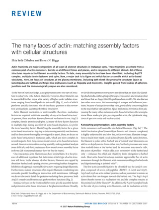
  • 2. REVIEW a Filopodium Lamellipodium Ruffle Lamellum Microvillus Cortical actin Podosome Endosome Phagocytic cup Stress fibre Endocytic pit Cadherin-based Golgi-associated actin adherens junction Nuclear actin b Dorsal ruffle N Peripheral ruffle Lamellipodium Podosome N Substratum Invadopodium Lamellum Figure 1 Actin-based structures in metazoan cells. The helical actin ruffles are distinct structures; podosomes and invadopodia are ventral; filament is used in a myriad of cellular processes. (a) A hypothetical lamellipodia are weakly adherent to the substratum. As far as we know, metazoan cell, migrating upwards and attached to a second cell on all these structures use the same basic helical actin filament. This is by the right. Cellular structures known or strongly suspected to contain no means a complete inventory of metazoan actin-based structures, and actin filaments are indicated. (b) A side view of a cell, emphasizing we strongly suspect filaments to be involved in other, as yet unidentified several points: ruffles are dorsal structures; peripheral ruffles and dorsal processes. N, nucleus. thereby produces a branched filament structure with an angle of 70° the association of profilin with the FH1 domain of formin15. Although between the filaments. Repeated branching leads to a ‘dendritic net- all formins have these basic properties, they vary significantly in their work’ (Fig. 2b). In this manner, the Arp2/3 complex functions not only potency14. In addition, some formins also bundle, sever or depolymer- as a nucleation factor, but also provides structure to the network. Most ize actin filaments16–20. Most organisms have multiple formin isoforms of the Arp2/3 complex subunits are single isoforms in all organisms, (15 in mammals, six each in Drosophila and Caenorhabditis elegans)21. with a few exceptions in mammals10–12 and Drosophila13. The Arp2/3 It is becoming increasingly clear that formins have additional roles in complex requires activation by one of several nucleation promoting fac- microtubule dynamics (see below)22,23. At present, the best understood tors (NPFs), including WASp, N-WASP, Scar/WAVE1, Scar/WAVE2 and mechanism of formin regulation is through direct activation by Rho Scar/WAVE3 in mammals8. family GTPases24. Formins were identified more recently as a second family of actin Spire proteins are a third class of actin nucleators that were initially assembly factors. In contrast with the Arp2/3 complex, formins are identified in Drosophila25. Spire proteins are single polypeptides charac- single polypeptide, multidomain proteins14. All formins studied to date terized by the presence of four Wasp homology 2 (WH2) motifs, which are dimeric, due to dimerization of their formin homology 2 (FH2) are responsible for actin nucleation26. As with formins, spire-nucleated domain. The FH2 domain is responsible for driving actin nucleation filaments are not branched (Fig. 2b). Spire can crosslink microtubules and formin-nucleated filaments are not branched. After nucleation, the and actin filaments27, and also inhibits actin nucleation by the formin FH2 domain remains bound at the barbed end and moves processively cappuccino in Drosophila and its mammalian homologue, formin1 (M. as it elongates, promoting elongation by preventing the access of capping Quinlan, University of California at San Francisco, San Francisco, CA, proteins (Fig. 2b). Formin-mediated elongation is further enhanced by and R. D. Mullins, University of California at Los Angeles, Los Angeles, NATURE CELL BIOLOGY VOLUME 9 | NUMBER 10 | OCTOBER 2007 1111 © 2007 Nature Publishing Group
  • 3. REVIEW CA, unpublished observations). This inhibition is through bind- a ing between KIND domain of spire and the FH2 domain of formin. B P Metazoans have two spire genes, whereas yeast have none. Nucleation factor Assembling filaments for diverse cellular functions A schematic overview of metazoan actin-based structures is shown in Fig. 1. b In this section, we review what is known of the actin-filament architectures underlying some of these structures, and how they are assembled. Sheet-like protrusive structures. Cells extend a variety of sheet-like structures, and we discuss three basic types: lamellipodia/lamella, ruf- fles and phagocytic cups. The adhesive structures known as podosomes and invadopodia are not strictly sheet-like, but require many of the same assembly proteins as lamellipodia. Arp 2/3 Formin Spire Lamellipodium and lamellum. We define the lamellipodia/lamella as surface-attached sheet-like membrane protrusions observed dur- ing crawling cell motility and spreading. The first step in crawling cell motility is the forward protrusion of the cell front, generally as a thin sheet of membrane-enclosed cytoplasm1. In an elegant series of papers, Abercrombie et al. defined the key characteristics of two regions within this protrusive front, now known as the lamellipodium and lamellum28–31 (see Timeline). The lamellipodium is more distal, starting at the leading edge and extending several micrometres back. The lamellum then takes over, extending from the lamellipodium to the cell body. The lamellipodium is thinner (100–160 nm thick versus >200 nm), more densely stained in electron microscopy images31, less Figure 2 Filament polymerization by actin assembly factors. (a) During actin- adherent, seems devoid of organelles and is more dynamic. Although filament assembly from monomers, the dimerization and trimerization steps these properties were revealed by Abercrombie’s landmark studies, are unfavourable, whereas subsequent monomer additions are much more subsequent work better defined additional adhesive differences, with favourable. Nucleation factors overcome these unfavourable dimerization and/or trimerization steps. Actin filaments are polar structures, with the lamellipodia being weakly adherent, and strong adhesion beginning at barbed end (B) being the sole site of elongation in non-muscle cells. Thus, the lamellipodia–lamella boundary32–34. physiologically relevant nucleation factors must allow barbed-end elongation. Two breakthroughs have changed our conception of the lamellipo- (b) The Arp2/3 complex (multi-coloured) nucleates a new filament from the side of an existing filament, causing filament branching at a 70° angle. The dium and lamellum dramatically. The first was the discovery that den- Arp2/3 complex remains at the branch point between the pointed end of dritically branched filaments dominate the actin network at the extreme the new filament and the side of the existing filament. Repeated branching leading edge35,36; the second was the observation that two distinct actin- results in the assembly of a dendritic network. The formin FH2 domain based networks constitute the lamellipodium and lamellum37. The evo- (semi-circles) nucleates a filament, and then moves processively with the barbed end as it elongates. Some formins can also bundle filaments. lution of this field is a beautiful example of two basic facets of modern Spire (circles with black connectors) nucleates a filament by stabilizing biomedical research: the ability of technological advances to change a a longitudinal tetramer. The current model is that spire dissociates from field; and the power of having multiple laboratories investigating over- filaments soon after nucleation. lapping subjects. The first finding, that dendritically branched filaments dominate lamellipodial speckles, is observed at the leading-edge plasma mem- near the leading edge35,36, represented a major change in our view of cell brane and disappears abruptly about 1–3 µm back37, seemingly at the motility. Before these data, the protrusive region of the cell was generally lamellipodial–lamellar boundary where the first stable adhesion to the considered to contain orthogonally crosslinked filaments. The discov- substratum occurs34. A second population appears in a more graded ery of the dendritic network coincided closely with the finding that the fashion, increasing in frequency with distance from the leading edge and Arp2/3 complex could nucleate dendritically branched filaments38. Very persisting through the lamellum. The two populations differ in kinetics, rapidly, the Arp2/3 complex was considered the dominant nucleator with lamellipodial speckles exhibiting rapid retrograde flow and lamel- driving leading-edge protrusion. lar speckles moving more slowly; transitions between the two states are The second finding, that two filament populations exist at the leading extremely rare. Taken together, these data suggest that two sets of actin edge, was a direct result of technological advances and astute observa- filaments exist in the lamellipodia and lamella, which are independently tion37. The technological advance was fluorescent speckle microscopy nucleated and disassembled. (FSM) — the phenomenon of ‘speckle’ movement on cellular structures Similar results have been obtained using Drosophila S2 cells40, and because of low levels of fluorophore incorporation39. Using FSM on actin further support for two independent actin networks comes from ret- filaments in marsupial kidney epithelial cells, two speckle populations rospective examination of fixed cells. In highly motile fish or Xenopus were observed in the protruding region. One population, referred to as keratocytes, the actin network is significantly more dense near the 1112 NATURE CELL BIOLOGY VOLUME 9 | NUMBER 10 | OCTOBER 2007 © 2007 Nature Publishing Group
  • 4. REVIEW TIMELINE – History of the terms “Lamellipodia”, “Lamella” and “Ruffles” LAMELLUM (plural, lamella) 1971 Coined in 1969 to replace In 1971, differences between lamellipodia “ruffled membrane” for the and lamella were defined more fully by protrusive region of a motile EM criteria, in terms of thickness, staining fibroblast133. Also recognized density, presence of organelles, and that only the first 3-5 microns of apparent adhesion31. the lamellum had ruffling activity. 1958 1969 1970 1971 1980 RUFFLE LAMELLIPODIUM (plural, lamellipodia) 1980 Coined in 1958 to describe the Coined in 1970 to differentiate the highly First record of differentiation between protruding leading edge of a motile protrusive, distal portion at the front of lamellipodia and ruffles. In Abercrombie’s cell132. Used interchangeably the motile cell (first several microns) from much-cited review134, lamellipodia defined with “ruffled membrane”. the rest of the lamellum22. No distinction as forward-protruding structures that adhere made between protrusions parallel to the weakly to substratum, with ruffles being of substratum and those that protruded similar morphology, but non-adhered and vertically, although the term “ruffle” was often protruding dorsally. used as well to describe these structures. leading edge than it is farther back, and a large proportion of these dendritic branch points in these cells36, as well as to the leading edge in filaments are labile to extraction35,36,41. Electron microscopy of fish multiple cell types10,45–48. Second, Arp2/3-complex dynamics are confined keratocytes reveals that filaments within 1 µm of the leading edge are largely to lamellipodia in Xenopus49 and Drosophila40. In the latter case, predominantly short and crosslinked in dendritic branches, whereas treatments that vary the width of the lamellipodium proportionally vary most filaments farther back are longer and less dendritically branched35. the width of the Arp2/3 complex-rich region. Third, altering the activity Electron microscopic analysis of the protrusive regions of mouse embryo of the Arp2/3 complex affects lamellipodial structures. Overexpression fibroblasts suggests a similar organization of the two networks42. Where of central acidic (CA) or WH2 central acidic (WCA) constructs that studied, both lamellipodial and lamellar filaments orient with barbed inhibit the Arp2/3 complex also block lamellipodia assembly10,48,50, ends toward the leading edge plasma membrane35 and the movement although efficacy of these constructs is not universally accepted (Box 1). of actin speckles supports this orientation37. Microinjection of antibodies that inhibit dendritic branching by the It has also been suggested that the lamellipodium may lie on top of Arp2/3 complex inhibits lamellipodial protrusion51. Knockdown of an the lamellum43. At the leading edge, a layer of densely packed filaments Arp2/3-complex subunit causes significant lamellipodial loss in mouse seems to lie above a second network of less dense filaments that extends cells52 and strongly inhibits cell spreading in Drosophila cells53. further back. The speculation is that the upper layer represents lamel- Debate continues, however, as to how universal the role of the lipodial filaments, whereas the lower layer is lamellar (Fig. 3b). The Arp2/3 complex at the leading edge is. One RNA interference (RNAi) rear of the lamellipodium is thought to be specified by myosin-II-based study in mouse embryonic fibroblasts argues that the Arp2/3 complex contraction at adhesion sites, which is vital for lamellipodial disassem- is not required in lamellipodia54. Another suggests that the Arp2/3 bly. Although this model of the two networks being vertically separated complex does not localize to the leading edge in neuronal growth needs to be tested further, there may also be support for this idea in the cones, and that overexpression of CA constructs does not alter growth early electron microscopy work by Abercrombie31. cone morphology or slow growth cone motility48. Others have found Based on the above findings, we conclude that two autonomous actin the Arp2/3 complex present in, and necessary for, growth-cone pro- networks are present in the protrusive region, with the lamellipodial trusion (T. Svitkina, University of Pennsylvania, Philadelphia, PA, filaments probably overlaying the lamellar filaments (Fig. 3a). Clearly, unpublished observations). the proportions and characteristics of these networks might vary sub- One issue in these studies may be how efficiently the Arp2/3 complex stantially between cell types, especially between slow-moving (for exam- is inhibited, given that it is highly abundant9. Mathematical models of ple, fibroblasts) and fast-moving cells (for example, keratocytes). In this leading-edge protrusion suggest that the Arp2/3 complex may be in respect, speckle analysis of actin networks in keratocytes would be highly large excess over that needed for ‘normal’ migration on experimental informative, as so much ultrastructural information is available for these surfaces55. It would, therefore, be interesting to evaluate Arp2/3 com- cells. The question also remains concerning the relative importance of plex-inhibited cells by FSM to determine whether the narrow band of these networks in motility. lamellipodial speckles is completely ablated. How are lamellipodia and lamella assembled? Most evidence suggests Less is known about the nucleators required for lamellar actin fila- that lamellipodia are Arp2/3 complex dependent: first, there is a strong ments. The Arp2/3 complex seems largely absent from this region36,40,49, correlation between Arp2/3-complex localization and lamellipodial and inhibiting the Arp2/3 complex or other lamellipodial proteins dynamics. The lamellipodial network contains predominately dendritic does not alter lamellar properties40,50. The formin mDia2 is implicated branches in several instances35,36,44 and the Arp2/3 complex localizes to in lamellar filament assembly, as well as in focal adhesion turnover NATURE CELL BIOLOGY VOLUME 9 | NUMBER 10 | OCTOBER 2007 1113 © 2007 Nature Publishing Group
  • 5. REVIEW a b Lamellipodium Lamellum Substratum Weak adhesion site Stable adhesion site Ruffle c Filopodium Filopodium Figure 3 Schematic representations of models of assembly for lamellipodia, Lamellipodial filaments may lie above lamellar filaments. At the leading lamella, peripheral ruffles and filopodia. (a) Top view of lamellipodia and edge, lamellipodial filaments predominate, but lamellar filaments increase lamella. Lamellipodial actin filaments (blue) are dendritically branched and in abundance further back. A lamellipodium maintaining weak attachment to Arp2/3-complex-dependent. These filaments assemble at the leading edge the substratum is shown in the upper panel. Myosin II activity at the stable plasma membrane (due to assembly factors bound there) and disassemble adhesion site causes retrograde flow of the lamellipodial actin network. abruptly at a band corresponding to the first sites of stable adhesion (green), If the lamellipodial attachment with the substrate is broken, myosin II generally 1–3 µm from the leading edge. Lamellar actin filaments (red) are activity causes this membrane sheet to move rearward as a peripheral ruffle, in a less defined network, and are not Arp2/3 complex-dependent. They can which disassembles at the stable adhesion site (lower panel). (c) Filopodial initiate throughout the lamellipodium and lamellum, but their frequency assembly from the lamellipodium or lamellum. Filopodial assembly from increases as one moves back from the leading edge. Lamellar filaments the lamellipodial network through the convergent-elongation mechanism is persist many micrometres behind the lamellipodium, and disassembly can shown on the left. Possible filopodial assembly from the lamellar network, occur throughout this region. (b) Side views of lamellipodium and lamellum. through an as yet undefined mechanism, is shown on the right. (C.Waterman-Storer, University of Washington, WA, unpublished imaging studies provide convincing evidence that peripheral ruffles observations), suggesting a potential association between lamellar fila- and lamellipodia have common origins29, and recent results support this ments and focal adhesions. However, mDia2 is not expressed in sev- hypothesis43. The Arp2/3 complex seems to be important for assembly eral motile cell types, suggesting it may be functionally redundant with of both types of ruffle56,58. Most recently, the presence of dendritically mDia1, mDia3 or other fomins. branched filaments in peripheral ruffles was observed59. Formins may also function in peripheral-ruffle dynamics: first, active Ruffles. We define ruffles as sheet-like membrane protrusions that RhoA — thought to directly activate the formins mDia1 and mDia2, but do not attach at all to the substratum, as opposed to weakly adherent not the Arp2/3 complex — is enriched at the leading edge of multiple lamellipodia. There are at least two distinct varieties of these transient mammalian cell types, both in lamellipodia and ruffles60–64, and is required structures, both with half-lives of minutes. Peripheral ruffles assemble for ruffle assembly61; second, mDia1 is enriched at ruffle edges60,61,63; and at the leading edge of motile cells, and move rearward29. Circular dorsal third, coexpression of constitutively active Rac1 and dominant-negative ruffles — also called ‘waves’ or ‘actin ribbons’ — assemble on the dorsal RhoA allows spreading, but blocks ruffles61. This result hints at potential surface and constrict into a circular structure before disappearing. These differences between lamellipodia and peripheral ruffles, by suggesting seem to be distinct structures, as rearward-moving peripheral ruffles do that Rac1 activation of the Arp2/3 complex (through Scar or WAVE) is not transition to dorsal ruffles56. Peripheral ruffles are associated with sufficient for lamellipodia, but not ruffle, generation. crawling-cell motility only, whereas dorsal ruffles also affect receptor The proposal that the lamellipodial network lies above the lamel- internalization and possibly macropinocytosis57. A distinction between lar network might also explain the role of mDia1 in peripheral peripheral and dorsal ruffles was made by Abercrombie29. ruffles 43: according to this study, the rear of the lamellipodium It is tempting to speculate that peripheral and dorsal ruffles arise is specified by assembly of a myosin II-based contractile com- through the same mechanisms as lamellipodia. Indeed, early live-cell plex at sites of stable adhesion (Fig. 3a), and its contraction pulls 1114 NATURE CELL BIOLOGY VOLUME 9 | NUMBER 10 | OCTOBER 2007 © 2007 Nature Publishing Group
  • 6. REVIEW BOX 1 TOOLS FOR REGULATING THE ARP2/3 COMPLEX: WCA AND CA CONSTRUCTS WCA constructs refer to the minimum polypeptide sufficient for in vitro activation of the Arp2/3 complex, located at the carboxyl termini of most nucleation-promoting factors9. WCA binds both the Arp2/3 complex and an actin monomer. Overexpression of WCA constructs has been used to inhibit the Arp2/3 complex, on the basis that these constructs would activate the Arp2/3 complex in a mislocalized fashion, preventing localized activation by endogenous nucleation-promoting factors. CA constructs are shorter and only bind to the Arp2/3 complex. The CA construct of WASp competes with Arp2/3-complex activators in vitro, blocking nucleation73. In vivo, CA constructs are predicted to sequester the Arp2/3 complex from endogenous nucleation-promoting factors. Reviews are mixed on the efficacy and specificity of these constructs for Arp2/3-complex inhibition in cells. Many researchers have reported successful cellular inhibition of the Arp2/3 complex with these constructs (see main text). However, one concern with WCA is that high expression might sequester significant amounts of actin monomer, causing additional effects that are not dependent on the Arp2/3 complex. As for CA, some report lack of effect on cellular processes strongly believed to depend on Arp2/3 complex. lamellipodial filaments rearward. If the extreme leading edge Podosomes and invadopodia. Podosomes are integrin-containing struc- remains adhered to the substratum, contraction causes retrograde tures on the basal cell surface, containing an actin-rich core surrounded flow. If, however, the connection is broken, the whole leading edge, by a ring of several actin-associated proteins and signalling proteins including actin filaments and plasma membrane, moves rearward (Fig. 1)57,69,70. They have been studied in detail in a limited number of as a peripheral ruffle (Fig. 3b). Support for this model can be found cells — Src-transformed fibroblasts and monocyte-derived cells (such in the earlier studies of Abercrombie31. The lamellar network makes as macrophages, dendritic cells and osteoclasts). However, the evidence up part of the substrate–adhesion complex34 and so may be essen- is mounting that podosomes are also present in other cell types71,72, and tial for peripheral ruffles. Perhaps mDia formins mediate lamellar- may mediate integrin-based adhesion to extracellular matrix. filament assembly in fibroblasts? Immunofluorescence microscopy Invadopodia have a similar architecture and protein composition to illustrates that mDia1 localizes to the edge of spreading cells in podosomes, but are formed by cells migrating over thick extracellular several cell types (refs 23, 63; K. Eisenmann and A. Alberts, Van matrix, and mediate matrix degradation69,70. Podosomes — which some Andel Institute, Grand Rapids, MI, unpublished observations; have postulated to be ‘invadopodia precursors’ — are smaller (approxi- H.N.H, unpublished observations). mately 0.5 µm diameter and 0.5 µm height), whereas invadopodia are Our current conclusion is that lamellipodia and peripheral ruffles wider (2 µm diameter) and protrude several micrometres into the extra- arise from the same assembly mechanism, and differ in substrate adhe- cellular matrix (Fig. 1). Both structures are ‘hot spots’ for membrane sion. The assembly mechanism for dorsal ruffles is unclear, but probably dynamics, and invadopodia have extensive plasma membrane invagina- involves Arp2/3 complex-generated dendritic networks. tions. Where examined, both podosomes and invadopodia are enriched in a specific matrix metalloprotease57,69,70. Phagocytic cups and pits. Phagocytosis is the cellular uptake of parti- All evidence points to the Arp2/3 complex as the key nucleator for cles larger than 0.5 µm diameter. Although both types of phagocytosis both podosomes and invadopodia. The Arp2/3 complex and several of its conducted by macrophages — Fc receptor-mediated (FcR) and com- regulators are enriched in macrophage podosomes57,69,70. Microinjection plement receptor-mediated (CR) — require actin polymerization, they of CA constructs blocks podosome assembly in macrophages73,74. Human differ substantially in morphology64. In FcR-mediated phagocytosis, Wiskott-Aldrich Syndrome patients, who lack WASp, do not have mac- the macrophage membrane protrudes around the phagocytosed par- rophage and dendritic cell podosomes75,76. N-WASP is enriched at podo- ticle. Following membrane fusion at the distal tip, the particle is then somes of non-haematopoietic cells, and is important for their assembly77. pulled into the macrophage. In CR-mediated phagocytosis, the parti- N-WASP and the Arp2/3 complex are enriched in, and are required for, cle ‘sinks’ into the macrophage and the macrophage does not extend invadopodia assembly78. toward the particle. The ultrastructural details of the actin network are unclear for podo- The morphology of the actin filaments is not known for either proc- somes and invadopodia. Clearly, the filaments are densely packed in ess. Based on the protrusive, sheet-like nature of the phagocytic cups both, but it is uncertain whether they are dendritically branched, straight in FcR-mediated phagocytosis, we speculate that these cups use lamel- or crosslinked into orthogonal networks. In view of the requirement for lipodia-like or lamella-like filaments. One difference, however, is that the Arp2/3 complex in these structures, one would postulate dendritic phagocytic-cup assembly requires extensive vesicle fusion65, which has branching is present, but it will be important to confirm this. not been shown for lamellipodia or lamella. The Arp2/3 complex seems to be required for both types of phago- Finger-like protrusive structures. The range of finger-like protru- cytosis, based on its localization to these structures and on inhibition sions observed in metazoan cells is enormous, and our general view of both processes by WCA constructs66. In addition, WASP–GFP local- is that multiple assembly mechanisms are likely. We define filopodia izes to FcR-mediated phagosomes, and macrophages lacking WASP are and microvilli as thin (less than 200 nm diameter) protrusions, con- compromised in FcR-mediated phagocytosis67. The formins mDia1 and taining parallel bundles of 10–30 actin filaments that run the length mDia2 seem to function in CR-mediated phagocytosis68. of the protrusion and orient their barbed ends towards the membrane. NATURE CELL BIOLOGY VOLUME 9 | NUMBER 10 | OCTOBER 2007 1115 © 2007 Nature Publishing Group
  • 7. REVIEW a Tip complex Ena/Vasp, Formin, myosin X b Electron-dense mass Fascin (and/or formin?) Myosin I ERM protein Villin Fimbrin Epsin Terminal web Figure 4 Schematic representations of models for assembly of filopodia and assembly is thought to occur through re-organization of the Arp2/3 complex- microvilli. (a) A top view of a filopodium — a finger-like, actin-rich protrusion dependent lamellipodial network. (b) A side view of a microvillus, which that is adhered to the substratum. An electron-dense complex at the tip has similar finger-like morphology to filopodia, but is not attached to the seems to be enriched in Ena/Vasp proteins, myosin X and formins. Fascin substratum. The most well characterized microvilli on epithelial cells (brush- seems to be the predominant bundling protein in the most well characterized border microvilli) contain parallel bundles of actin filaments, crosslinked by filopodia systems. One possibility is that formins might contribute to villin, fimbrin and espin. Microvilli contain an electron-dense mass at their bundling in some cases. Most often, the filopodial base is embedded in the tip. At their base, epithelial microvilli seem to be embedded in the terminal lamellipodium and lamellum. The convergent-elongation model of filopodial web, which is composed of actin filaments and myosin II. We define filopodia as finger-like protrusions that are adhered in some Electron microscopy analyses show that filopodia contain bundles manner to a substratum or another cell. We define microvilli as finger- of long parallel filaments36,88–90. These extend deep into the lamellipo- like protrusions that are not adhered. Of course, grey areas exist between dium or lamellum, where they eventually splay at their pointed ends. these definitions, in particular when both adherent and non-adherent The sites and mechanisms for filopodia assembly are currently under finger-like protrusions are experimentally induced79. In addition to these debate. One model is that filopodia assemble by ‘convergent elongation’ simple finger-like protrusions, a wide variety of larger and more complex of lamellipodial filaments44,91, through re-organization of the Arp2/3 protrusions (including Drosophila bristles and hair-cell stereocilia) use complex-assembled dendritic network into bundles. This re-organiza- multiple modules of actin bundles, but these are not discussed here (for tion is initiated when a subset of filament barbed ends associate with a a review, see ref. 80). filopodial-tip protein complex at the leading edge plasma membrane. This complex, containing the Ena/VASP actin-binding proteins and pos- Filopodia. Filopodia protrude from the leading edge of many motile cells, sibly formins, protects barbed ends from capping proteins, thus allowing including fibroblasts and nerve growth cones (Fig. 4)81. In these situations, them to elongate preferentially in the network. Contacting filaments filopodia emanate from a lamellipodial or lamellar sheet, and are believed would then be bundled by the bundling protein fascin to eventually pro- to function as directional sensors82 (perhaps through enrichment of acti- duce a filopodium91. vated integrins in filopodial tips83). Viruses often bind at filopodial tips, The convergent elongation model requires Arp2/3 complex-medi- and are transported back to the cell body before being internalized84,85. A ated nucleation, followed by rearrangement of the dendritic network. similar process occurs for activated epidermal growth factor (EGF) recep- However, recent studies depleting subunits of the Arp2/3 complex tor on adenocarcinoma cells86. Finally, filopodia are abundant on neuronal by RNAi have provided conflicting results in terms of whether filo- dendrites, and seem to be important for dendritic spine development87. podia or just lamellipodia were affected (T. Svitkina, unpublished 1116 NATURE CELL BIOLOGY VOLUME 9 | NUMBER 10 | OCTOBER 2007 © 2007 Nature Publishing Group
  • 8. REVIEW whereas monomer addition rate varies. An interesting possibility is that Formins (mDia1, FRL1)? formins are at the barbed ends of filopodial filaments and their regulation controls elongation rate. The basic structure of filopodia, and how they relate to lamellipodia and lamella, is still open to surprises, and recent results may impact on possible assembly mechanisms. Although most electron microscopy Nucleus APC studies show that filopodia consist of long, parallel filaments, recent cryo-electron microscopy work in Dictyostelium suggests that shorter filaments may also be present that are not necessarily parallel, with very T cell short filaments in a meshwork pattern at the tip99. The suggestion from this work is that these shorter, labile, filaments may have been destroyed Arp2/3 by previous electron microscopy preparations. We reserve judgment on this possibility until similar observations are made for other filopodia, as these Dictyostelium filopodia elongate approximately twentyfold faster Figure 5 Actin assembly factors and immune synapse. The Arp2/3 complex localizes to the actin-rich contact site (red) at the immune synapse in T cells, than most other protrusions (about 1 µmicron s–1) and, thus, may use a and is necessary for full actin accumulation at the immune synapse. mDia1 fundamentally different protrusive mechanism. and FRL1 localize to both the actin-rich site and to regions surrounding The suggestion that the lamellipodium lies on top of the lamellum43 the MTOC (green). Knockdown of either mDia1 or FRL1 inhibits MTOC has implications for filopodia assembly. If filopodia derive from the re-orientation toward the immune synapse. Effects of formins on actin polymerization at the immune synapse are controversial, with one report action of the Arp2/3 complex in the lamellipodium, do they transition indicating that deletion of mDia1 ablates filament accumulation, and at some point to the lower, lamellar layer, as their roots often end up another indicating no effect of either mDia1 or FRL1 knockdown on filament proximal to lamellipodium (Fig. 3c)? Or, do they assemble from the accumulation. FRL1 and mDia1 seem to act independently at the MTOC, dendritic network at the lamellipodium–lamellum interface? Could filo- in view of their differential localization patterns around the MTOC, and the observations that knockdown of either inhibits MTOC re-orientation. APC, podia-like protrusions derive from the lamellum? Clearly, many other antigen-presenting cell. mechanisms are possible. The definition of a filopodium can be quite subjective, if based solely on observations)52. Both of these studies use B16F1 mouse melanoma cells, light microscopy of fixed cells. Care must be taken to ensure that the struc- but with potentially key variations in the substrates on which the cells tures are protrusive, and not ‘retraction fibres’. Some protrusions defined were plated and in the medium added to the cells. A third study reports as filopodia in the literature are less finger-like than others, being wider a slight increase in filopodia number in mouse embryonic fibroblasts overall and splaying to widths of several microns at their intersection with when Arp3 is depleted by RNAi54. Finally, N-WASP has been shown the lamellipodium–lamellum. In addition, fibroblasts can protrude filopo- to trigger filopodia assembly downstream of Cdc42 (ref. 92), but cells dia-like structures from regions containing no obvious lamellipodium or lacking N-WASP still possess filopodia93. lamellum (S. Nicholson-Dykstra, Dartmouth, Hanover, NH, and H.N.H., Regarding formins, mDia2 has been linked to filopodia assembly79,94,95 unpublished observations). We question whether all such structures are (T. Svitkina, unpublished observations). In many of these cases, it is subject to a common assembly mechanism. unclear whether the structures hold to our adhesion-based definitions of filopodia, as both adherent and non-adherent finger-like protrusions Microvilli. Microvilli are most recognizable on the luminal surfaces of are visible. It is possible that overexpression of formins or other proteins intestinal and kidney epithelial cells, where they are densely packed and used in these studies causes assembly of both structures. A formin is of uniform length (around 2 µm; ref. 100). However, shorter (<0.5 µm) also required for filopodia in Dictyostelium96, although the localization and more variable length microvilli are present on many cell types, of a GFP-fusion of this formin to finger-like protrusions irrespective of including circulating lymphocytes and the dorsal surfaces of many substrate-attachment in this study raises questions about our distinction cultured cells101,102. Although the function of epithelial microvilli is to between microvilli and filopodia. increase absorptive surface area, lymphocyte microvilli may segregate Our opinion is that there may be multiple potential mechanisms for cell surface proteins103,104 — a property that may aid extravasation from assembling filopodia. Filopodial assembly requires three basic steps: fila- blood to the periphery. ment nucleation, sustained barbed-end elongation and filament bundling. Both epithelial and lymphocyte microvilli contain long actin fila- A variety of molecules may be capable of accomplishing each task. Moreover, ments arranged in parallel bundles100,102. Although epithelial microvilli bundled structures can be nucleated by the Arp2/3 complex or formins in maintain constant length after initial assembly, their actin filaments are vitro17,19,97,98. We believe that it is perfectly possible that different mechanisms still dynamic, with subunits adding at barbed ends and dissociating at of filament nucleation are used in distinct cellular environments. pointed ends7,105. Shorter microvilli seem to have dynamic lengths, elon- After assembling, leading-edge filopodia elongate at rates between gating and retracting on a timescale of minutes101,102. 1–5 µm min–1 in multiple cell types5,52. However, elongation dynam- The cellular environment from which microvilli grow seems to be fun- ics vary, even during the lifetime of a single filopodium, with periods damentally different from that of most filopodia, and does not involve of elongation and retraction. In growth-cone filopodia, the elongation a clear lamellipodial or lamellar surface. The apical surface of epithelial rate depends on a balance between monomer addition at the tip, and cells contains an actin meshwork called the ‘terminal web’ (Fig. 4) that is retrograde flow combined with depolymerization at the base5. Most rich in myosin II and spectrin, and the actin filaments do not seem to be commonly, retrograde flow and depolymerization seems fairly constant, dendritically branched106–108. The small diameter of blood lymphocytes NATURE CELL BIOLOGY VOLUME 9 | NUMBER 10 | OCTOBER 2007 1117 © 2007 Nature Publishing Group
  • 9. REVIEW (<10 µm) allows about 2 µm of cytoplasm surrounding the nucleus, and WAVE2 in these cells; WAVE2 seems to be the Arp2/3-complex regula- it is unclear how actin filaments here are organized102. It would be useful tor primarily responsible for actin assembly at the synapse112. Another to characterize actin organization at the base of microvilli in more detail, consideration is the time allowed for synapse assembly in these different in light of our new knowledge of the Arp2/3 complex and formins. model systems. We feel that it is unlikely that microvilli require the Arp2/3 complex for The role for these formins in MTOC repositioning is not the first evi- assembly. RNAi-mediated knockdown of Arp2 has no effect on micro- dence of association between formins and microtubule-based structures. villar morphology in a lymphocyte culture line (S. Nicholson-Dykstra, mDia regulates microtubule stabilization in fibroblasts22, and mDia2 can Dartmouth, Hanover, NH, and H.N.H., unpublished observations). interact with microtubule-binding proteins113. In addition, there is evi- However, formins affect microvillar assembly in both Dictyostelium96 and dence that mDia1 effects MTOC positioning in migrating fibroblasts, NIH3T3 cells79. It is unclear how the latter structures relate to Cdc42- although this result is not universally accepted22,114. dependent filopodia in NIH3T3 cells, which also require mDia2 (ref. 94). Are the effects of mDia1 and FRL1 on MTOC re-orientation in T cells In addition, all of these structures deviate from our definition of micro- related to their effects on actin dynamics? Suppression of either formin villi, as they include both attached and unattached protrusions. alone inhibits re-orientation23, and the formins localize in different pat- terns at the MTOC, suggesting distinct roles. One study suggests that the Novel insights from adhesion structures FH2 domain is required for mDia1-mediated effects on microtubules115, Recent studies of adhesion structures have revealed an intricate interplay which suggests a link to actin. However, the FH2 domain is also sufficient between multiple actin-based structures, as well as a role for formins in for the interaction of mDia2 with microtubule-binding proteins113, and controlling microtubule-based structures. the FH2 domain of mDia1 can interact directly with microtubules (E.S.C. and H.N.H., unpublished observations; F. Bartolini and G. Gundersen, Immunological synapse. The term ‘immunological synapse’ refers Columbia University, New York, NY, unpublished observations). It is not to the extensive interaction surface between a T lymphocyte and an currently known whether actin filaments colocalize with mDia1 or FRL1 antigen-presenting cell bound to specific antigen, mediated by both at the MTOC in T cells, although these may be difficult to detect. One integrin-based and T-cell receptor-based interactions109. This structure final point is that the two effects of formins on microtubules (stabiliza- is important in establishing cell–cell adhesion and T-cell polarity, and tion and MTOC repositioning) may involve distinct mechanisms, as they is necessary for T-cell activation. An early and requisite step in syn- presumably occur at different ends of microtubules. apse assembly is massive actin polymerization on the T-cell side, which expands the interaction surface. Following this event, the microtubule Adherens junctions. Adherens junctions are cell–cell adhesions mediated organizing centre (MTOC) (and also the Golgi) re-orients towards the by homophilic interaction of cadherins: their core components are the synapse, and a variety of substances are secreted towards the antigen- transmembrane cadherin and two cytoplasmic proteins, β-catenin and α- presenting cell. catenin116,117. The long-accepted model is that mature adherens junctions The actin-based structure at the synapse is widely considered to be in epithelial cells are associated with a circumferential band of actin and a lamellipodium110, and this is supported by recent RNAi studies23. myosin II that is contractile and runs parallel to the membrane118. This Knockdown of either Arp2 or Arp3 in Jurkat T cells inhibits spreading band of actin filaments is relatively stable, and the actin–adherens junc- or polymerization of a tight F-actin band at the synapse, depending tions interaction is mediated by α-catenin, bound to β-catenin, which is on the experimental setting. However, polarized actin-rich finger-like bound to cadherin. However, new results call this model into question: protrusions persist at the synapse interface in the near absence of the first, α-catenin cannot bind β-catenin and actin filaments simultane- Arp2/3 complex (Fig. 5). Interestingly, although two formins (mDia1 ously in vitro119; second, actin filaments are much more dynamic than and FRL1) colocalize with F-actin at the synapse, suppression of either cadherins or catenins at the adherens junctions119; and third, factors formin alone or in combination affects neither spreading nor the fin- other than core adherens junction components seem to link junctions ger-like protrusions. Instead, mDia1 and FRL1 also localize near the to actin120,121. MTOC and are important for MTOC repositioning towards the synapse. Therefore, our image of the mature adherens junction may well change Another study, using splenic T cells from mDia1 knockout mice, also over the next few years. However, it is clear that extensive actin dynamics results in disrupted MTOC polarization111. However, these cells also have are required to assemble the mature structure. Cells approach each other an almost complete loss of polymerized actin staining at the synapse, in by extension of sheet-like membrane protrusions122. On initial contact, contrast with the Jurkat cell study. the two interacting cells extend actin-rich finger-like protrusions toward Clearly, differences between the experiments could explain these each other, with cadherin concentrated at the tips123–125. These protru- inconsistencies in the role of mDia1 in actin assembly at the immuno- sions interdigitate and then shorten as the junction matures, bringing the logical synapse. One possibility is that the residual mDia1 remaining two cell membranes closer. The initial sheet-like protrusions are likely to after RNAi is sufficient for the actin response, but not for the MTOC be lamellipodia or lamella, although it is less clear whether the finger-like response. Using quantitative immunoblotting, we found that Jurkat protrusions are filopodia-like124 or more like an acto-myosin contractile cells express 170,000 molecules of mDia1 per cell (H.N.H., unpublished structure126. Clearly, myosin II is important for adherens junction assem- observations), so that >90% suppression (estimated from westerns in bly, but its localization has not been determined at high resolution. ref. 23), would result in 10,000–20,000 remaining molecules. Another There is evidence that both the Arp2/3 complex and formins partici- possibility is that mDia1 knockout in T lymphocytes results in second- pate in adherens junctions assembly. The Arp2/3 complex localizes to ary effects on Arp2/3-complex levels or activity. It would be interest- sites of junction assembly127 and its inhibition disrupts this process127,128, ing to probe the levels of Arp2/3-complex subunits, or the WASp and although it is unclear whether this may be due to general inhibition of 1118 NATURE CELL BIOLOGY VOLUME 9 | NUMBER 10 | OCTOBER 2007 © 2007 Nature Publishing Group
  • 10. REVIEW lamellipodial protrusion — recent evidence suggests that the Arp2/3 6. Theriot, J. A. & Mitchison, T. J. Actin microfilament dynamics in locomoting cells. Nature 352, 126–131 (1991). complex accumulates transiently at nascent junctions, but then disperses 7. Rzadzinska, A. K. et al. An actin molecular treadmill and myosins maintain stereocilia rapidly as E-cadherin accumulates129. Formin1 also localizes to assem- functional architecture and self-renewal. J. Cell Biol. 164, 887–897 (2004). 8. Goley, E. D. & Welch, M. D. The ARP2/3 complex: an actin nucleator comes of age. bling adherens junctions, concentrating at the tips of the finger-like Nature Rev. Mol. Cell Biol. 7, 713–726 (2006). protrusions. Formin1 binds α-catenin and overexpression of a formin1 9. Higgs, H. N. & Pollard, T. D. Regulation of actin filament formation through Arp2/3 complex: Activation by a Diverse Array of Proteins. Annu. Rev. Biochem. 70, 649–676 construct containing the α-catenin-binding region disrupts adherens (2001). junction assembly130. Interestingly, α-catenin binding to actin filaments 10. Machesky, L. M. et al. Mammalian actin-related protein 2/3 complex localizes to regions of lamellipodial protrusion and is composed of evolutionarily conserved pro- also inhibits the Arp2/3 complex131. Therefore, the interplay between teins. Biochem. J. 328, 105–112 (1997). α-catenin and the two actin nucleators might be crucial for adheren 11. Jay, P. et al. ARP3β, the gene encoding a new human actin-related protein, is alterna- tively spliced and predominantly expressed in brain neuronal cells. Eur. J. Biochem. junction assembly. An outstanding question is whether α-catenin can 267, 2921–2928 (2000). bind β-catenin–cadherin and formin1 simultaneously. In addition, the 12. Millard, T. H. et al. Identification and characterisation of a novel human isoform of Arp2/3 complex subunit p16-ARC/ARPC5. Cell Motil. Cytoskeleton 54, 81–90 recently identified ability of spire to inhibit formin1 (M. Quinlan & R. D. (2003). Mullins, unpublished observations) suggests that spire may have a role 13. Hudson, A. M. & Cooley, L. A subset of dynamic actin rearrangements in Drosophila requires the Arp2/3 complex. J. Cell Biol. 156, 677–687 (2002). in this process. 14. Higgs, H. N. Formin proteins: a domain-based approach. Trends Biochem. Sci. 30, 342–353 (2005). 15. Kovar, D. R. Molecular details of formin-mediated actin assembly. Curr. Opin. Cell Biol. An intricate web to weave 18, 11–17 (2006). A simplistic conclusion, on reading this review, is that the Arp2/3 com- 16. Harris, E. S., Li, F. & Higgs, H. N. The mouse formin, FRLa, slows actin filament barbed end elongation, competes with capping protein, accelerates polymerization plex mediates dendritically branched structures and that formins control from monomers, and severs filaments. J. Biol. Chem. 279, 20076–20087 (2004). unbranched structures. Although there may be some truth to this, the 17. Harris, E. S., Rouiller, I., Hanein, D. & Higgs, H. N. Mechanistic differences in actin bundling activity of two mammalian formins, FRL1 and mDia2. J. Biol. Chem. 281, number of qualifications to this statement are large. 14383–14392 (2006). One possibility is that formins might function more as elongation 18. Moseley, J. B. & Goode, B. L. Differential activities and regulation of Saccharomyces cerevisiae formin proteins Bni1 and Bnr1 by Bud6. J. Biol. Chem. 280, 28023–28033 factors than nucleation factors in some processes, and so work coordi- (2005). nately with the Arp2/3 complex, spire or other formins. In fact, several 19. Michelot, A. et al. The formin homology 1 domain modulates the actin nucleation and bundling activity of Arabidopsis FORMIN1. Plant Cell 17, 2296–2313 (2005). formins are very poor nucleators in vitro but still bind the barbed ends 20. Chhabra, E. S. & Higgs, H. N. INF2 is a WH2 motif-containing formin that severs actin of filaments strongly, modulating elongation rate and protecting barbed filaments and accelerates both polymerization and depolymerization. J. Biol. Chem. 281, 26754–26767 (2006). ends from capping16 (E. S. Harris, Dartmouth, Hanover, NH, and H.N.H, 21. Higgs, H. N. & Peterson, K. J. Phylogenetic analysis of the formin homology 2 (FH2) unpublished observations). Thus, perhaps the Arp2/3 complex nucleates domain. Mol. Biol. Cell 16, 1–13 (2005). 22. Eng, C. H., Huckaba, T. M. & Gundersen, G. G. The formin mDia regulates GSK3 filaments and formins control their elongation in some circumstances. through novel PKCs to promote microtubule stabilization but not MTOC reorientation Recent results suggest that this situation could occur with mDia2 and in migrating fibroblasts. Mol. Biol. Cell 17, 5004–5016 (2006). 23. Gomez, T. S. et al. Formins regulate the actin-related protein 2/3 complex-independent the Arp2/3 complex in certain filopodia (T. Svitkina, unpublished obser- polarization of the centrosome to the immunological synapse. Immunity 26, 177–190 vations). Similarly, the ability of spire to both nucleate filaments and (2007). 24. Wallar, B. J. & Alberts, A. S. The formins: active scaffolds that remodel the cytoskel- to regulate formin1 suggests an intricate interplay between these two eton. Trends Cell Biol. 13, 435–446 (2003) factors, although spire-dependent actin filaments have not yet been 25. Kerkhoff, E. Cellular functions of the Spir actin-nucleation factors. Trends Cell Biol. 16, 477–483 (2006). identified in cells. 26. Quinlan, M. E., Heuser, J. E., Kerkhoff, E. & Mullins, R. D. Drosophila Spire is an actin A related point is that these simple actin-based structures do not act nucleation factor. Nature 433, 382–388 (2005). 27. Rosales-Nieves, A. E. et al. Coordination of microtubule and microfilament dynam- alone, but interface with other actin-based structures, or microtubule- ics by Drosophila Rho1, Spire and Cappuccino. Nature Cell Biol. 8, 367–376 based structures. The interface between lamellipodia and lamella, and (2006). 28. Abercrombie, M., Heaysman, J. E. & Pegrum, S. M. The locomotion of fibroblasts in between lamellipodia and filopodia, provide clear examples of this. Both culture. I. Movements of the leading edge. Exp. Cell Res. 59, 393–398 (1970). immunological synapses and adherens junctions are even more complex 29. Abercrombie, M., Heaysman, J. E. & Pegrum, S. M. The locomotion of fibroblasts in culture. II. “Ruffling”. Exp. Cell Res. 60, 437–444 (1970). cases. Dissecting these interfaces will be important for understanding the 30. Abercrombie, M., Heaysman, J. E. & Pegrum, S. M. The locomotion of fibroblasts in complexity of cytoskeletal organization and the control of its assembly. culture. 3. Movements of particles on the dorsal surface of the leading lamella. Exp. Cell Res. 62, 389–398 (1970). ACKNOWLEDGEMENTS 31. Abercrombie, M., Heaysman, J. E. & Pegrum, S. M. The locomotion of fibroblasts in We are indebted to many for useful discussions, including A. Alberts, D. Billadeau, culture. IV. Electron microscopy of the leading lamella. Exp. Cell Res. 67, 359–367 (1971). J. Burkhardt, J. Condeelis, F. Flures, G. Gundersen, M. McNiven, D. Mullins, 32. Izzard, C. S. & Lochner, L. R. Cell-to-substrate contacts in living fibroblasts: an inter- S. Nicholson-Dykstra, T. Svitkina and C. Waterman-Storer. We also thank our ference reflexion study with an evaluation of the technique. J. Cell Sci. 21, 129–159 anonymous reviewers, whose comments improved this work immensely. This work (1976). was supported by National Institutes of Health grant GM069818 and by a Pew 33. Bailly, M. et al. Regulation of protrusion shape and adhesion to the substratum during Biomedical Scholars Award. chemotactic responses of mammalian carcinoma cells. Exp. Cell Res. 241, 285–299 (1998). 1. Pollard, T. D., Blanchoin, L. & Mullins, R. D. Molecular mechanisms controlling actin 34. Gupton, S. L. & Waterman-Storer, C. M. Spatiotemporal feedback between actomyosin filament dynamics in nonmuscle cells. Annu. Rev. Biophys. Biomol. Struct. 29, 545– and focal-adhesion systems optimizes rapid cell migration. Cell 125, 1361–1374 576 (2000). (2006). 2. Moseley, J. B. & Goode, B. L. The yeast actin cytoskeleton: from cellular function to 35. Svitkina, T., Verkhovsky, A. B., McQuade, K. M. & Borisy, G. G. Analysis of the actin- biochemical mechanism. Microbiol. Mol. Biol. Rev. 70, 605–645 (2006). myosin II system in fish epidermal keratocytes: mechanism of cell body translocation. 3. Pollard, T. D. & Cooper, J. A. Actin and actin-binding proteins. A critical evaluation of J. Cell Biol. 139, 397–415 (1997). mechanisms and functions. Annu. Rev. Biochem. 55, 987–1035 (1986). 36. Svitkina, T. M. & Borisy, G. G. Arp2/3 complex and actin depolymerizing factor/cofilin 4. Wang, Y. L. Exchange of actin subunits at the leading edge of living fibroblasts: possible in dendritic organization and treadmilling of actin filament array in lamellipodia. J. role of treadmilling. J. Cell Biol. 101, 597–602 (1985). Cell Biol. 145, 1009–1026 (1999). 5. Mallavarapu, A. & Mitchison, T. Regulated actin cytoskeleton assembly at filopodium 37. Ponti, A. et al. Two distinct actin networks drive the protrusion of migrating cells. tips controls their extension and retraction. J. Cell Biol. 146, 1097–1106 (1999). Science 305, 1782–1786 (2004). NATURE CELL BIOLOGY VOLUME 9 | NUMBER 10 | OCTOBER 2007 1119 © 2007 Nature Publishing Group
  • 11. REVIEW 38. Mullins, R. D., Heuser, J. A. & Pollard, T. D. The interaction of Arp2/3 complex with 72. Moreau, V. et al. Cdc42-driven podosome formation in endothelial cells. Eur. J. Cell actin: nucleation, high affinity pointed end capping, and formation of branching net- Biol. 85, 319–325 (2006). works of filaments. Proc. Natl Acad. Sci. USA 95, 6181–6186 (1998). 73. Hufner, K. et al. The VC region of Wiskott-Aldrich syndrome protein induces Arp2/3 39. Waterman-Storer, C. M., Desai, A., Bulinski, J. C. & Salmon, E. D. Fluorescent speckle complex-dependent actin nucleation. J. Biol. Chem. 276, 35761–35767 (2001). microscopy, a method to visualize the dynamics of protein assemblies in living cells. 74. Linder, S. et al. The polarization defect of Wiskott-Aldrich syndrome macrophages is Curr. Biol. 8, 1227–1230 (1998). linked to dislocalization of the Arp2/3 complex. J. Immunol. 165, 221–225 (2000). 40. Iwasa, J. H. & Mullins, R. D. Spatial and temporal relationships between actin-filament 75. Burns, S. et al. Configuration of human dendritic cell cytoskeleton by Rho GTPases, nucleation, capping, and disassembly. Curr. Biol. 17, 395–406 (2007). the WAS protein, and differentiation. Blood 98, 1142–1149 (2001). 41. Small, J. V., Herzog, M. & Anderson, K. Actin filament organization in the fish keratocyte 76. Linder, S., Nelson, D., Weiss, M. & Aepfelbacher, M. Wiskott-Aldrich syndrome protein lamellipodium. J. Cell Biol. 129, 1275–1286 (1995). regulates podosomes in primary human macrophages. Proc. Natl Acad. Sci. USA 96, 42. Svitkina, T. M., Shevelev, A. A., Bershadsky, A. D. & Gelfand, V. I. Cytoskeleton of 9648–9653 (1999). mouse embryo fibroblasts. Electron microscopy of platinum replicas. Eur. J. Cell Biol. 77. Mizutani, K. et al. Essential role of neural Wiskott-Aldrich syndrome protein in podo- 34, 64–74 (1984). some formation and degradation of extracellular matrix in src-transformed fibroblasts. 43. Giannone, G. et al. Lamellipodial actin mechanically links myosin activity with adhe- Cancer Res. 62, 669–674 (2002). sion-site formation. Cell 128, 561–575 (2007). 78. Yamaguchi, H. et al. Molecular mechanisms of invadopodium formation: the role of the 44. Svitkina, T. M. et al. Mechanism of filopodia initiation by reorganization of a dendritic N-WASP-Arp2/3 complex pathway and cofilin. J. Cell Biol. 168, 441–452 (2005). network. J. Cell Biol. 160, 409–421 (2003). 79. Pellegrin, S. & Mellor, H. The Rho family GTPase Rif induces filopodia through mDia2. 45. Welch, M. D. et al. The human Arp2/3 complex is composed of evolutionarily conserved Curr. Biol. 15, 129–133 (2005). subunits and is localized to cellular regions of dynamic actin filament assembly. J. Cell 80. DeRosier, D. J. & Tilney, L. G. F-actin bundles are derivatives of microvilli: What does Biol. 138, 375–384 (1997). this tell us about how bundles might form? J. Cell Biol. 148, 1–6 (2000). 46. Bailly, M. et al. Relationship between Arp2/3 complex and the barbed ends of actin 81. Faix, J. & Rottner, K. The making of filopodia. Curr. Opin. Cell Biol. 18, 18–25 filaments at the leading edge of carcinoma cells after epidermal growth factor stimula- (2006). tion. J. Cell Biol. 145, 331–345 (1999). 82. Zheng, J. Q., Wan, J. J. & Poo, M. M. Essential role of filopodia in chemotropic turning 47. Falet, H. et al. Importance of free actin filament barbed ends for Arp2/3 complex of nerve growth cone induced by a glutamate gradient. J. Neurosci. 16, 1140–1149 function in platelets and fibroblasts. Proc. Natl Acad. Sci. USA 99, 16782–16787 (1996). (2002). 83. Galbraith, C. G., Yamada, K. M. & Galbraith, J. A. Polymerizing actin fibers position 48. Strasser, G. A. et al. Arp2/3 is a negative regulator of growth cone translocation. Neuron integrins primed to probe for adhesion sites. Science 315, 992–995 (2007). 43, 81–94 (2004). 84. Lehmann, M. J. et al. Actin- and myosin-driven movement of viruses along filopodia 49. Miyoshi, T. et al. Actin turnover-dependent fast dissociation of capping protein in the precedes their entry into cells. J. Cell Biol. 170, 317–325 (2005). dendritic nucleation actin network: evidence of frequent filament severing. J. Cell Biol. 85. Sherer, N. M. et al. Retroviruses can establish filopodial bridges for efficient cell-to-cell 175, 947–955 (2006). transmission. Nature Cell Biol. 9, 310–315 (2007). 50. Gupton, S. L. et al. Cell migration without a lamellipodium: translation of actin 86. Lidke, D. S. et al. Reaching out for signals: filopodia sense EGF and respond by directed dynamics into cell movement mediated by tropomyosin. J. Cell Biol. 168, 619–631 retrograde transport of activated receptors. J. Cell Biol. 170, 619–626 (2005). (2005). 87. Jontes, J. D. & Smith, S. J. Filopodia, spines, and the generation of synaptic diversity. 51. Bailly, M. et al. The F-actin side binding activity of the Arp2/3 complex is essential for Neuron 27, 11–14 (2000). actin nucleation and lamellipod extension. Curr. Biol. 11, 620–625 (2001). 88. Lindberg, U., Hoglund, A. S. & Karlsson, R. On the ultrastructural organization of the 52. Steffen, A. et al. Filopodia formation in the absence of functional WAVE- and Arp2/3- microfilament system and the possible role of profilactin. Biochimie 63, 307–323 complexes. Mol. Biol. Cell. 17, 2581–2591 (2006). (1981). 53. Rogers, S. L., Wiedemann, U., Stuurman, N. & Vale, R. D. Molecular requirements for 89. Small, J. V., Rinnerthaler, G. & Hinssen, H. Organization of actin meshworks in cultured actin-based lamella formation in Drosophila S2 cells. J. Cell Biol. 162, 1079–1088 cells: the leading edge. Cold Spring Harb. Symp. Quant. Biol. 46, 599–611 (1982). (2003). 90. Lewis, A. K. & Bridgman, P. C. Nerve growth cone lamellipodia contain two populations 54. Di Nardo, A. et al. Arp2/3 complex-deficient mouse fibroblasts are viable and have of actin filaments that differ in organization and polarity. J. Cell Biol. 119, 1219–1243 normal leading-edge actin structure and function. Proc. Natl Acad. Sci. USA 102, (1992). 16263–16268 (2005). 91. Vignjevic, D. et al. Role of fascin in filopodial protrusion. J. Cell Biol. 174, 863–875 55. Mogilner, A. & Edelstein-Keshet, L. Regulation of actin dynamics in rapidly moving (2006). cells: a quantitative analysis. Biophys. J. 83, 1237–1258 (2002). 92. Miki, H., Sasaki, T., Takai, Y. & Takenawa, T. Induction of filopodium formation by a 56. Suetsugu, S., Yamazaki, D., Kurisu, S. & Takenawa, T. Differential roles of WAVE1 and WASP-related actin-depolymerizing protein N-WASP. Nature 391, 93–96 (1998). WAVE2 in dorsal and peripheral ruffle formation for fibroblast cell migration. Dev. Cell 93. Snapper, S. B. et al. N-WASP deficiency reveals distinct pathways for cell surface pro- 5, 595–609 (2003). jections and microbial actin-based motility. Nature Cell Biol. 3, 897–904 (2001). 57. Buccione, R., Orth, J. D. & McNiven, M. A. Foot and mouth: podosomes, invadopodia 94. Peng, J. et al. Disruption of the Diaphanous-related formin Drf1 gene encoding mDia1 and circular dorsal ruffles. Nature Rev. Mol. Cell Biol. 5, 647–657 (2004). reveals a role for Drf3 as an effector for Cdc42. Curr. Biol. 13, 534–545 (2003). 58. Legg, J. A. et al. N-WASP involvement in dorsal ruffle formation in mouse embryonic 95. Wallar, B. J. et al. The basic region of the diaphanous-autoregulatory domain (DAD) is fibroblasts. Mol. Biol Cell. 18, 678–687 (2007). required for autoregulatory interactions with the diaphanous-related formin inhibitory 59. Svitkina, T. Electron microscopic analysis of the leading edge in migrating cells. domain. J. Biol. Chem. 281, 4300–4307 (2006). Methods Cell Biol. 79, 295–319 (2007). 96. Schirenbeck, A. et al. The Diaphanous-related formin dDia2 is required for the forma- 60. Goulimari, P. et al. Gα12/13 is essential for directed cell migration and localized Rho- tion and maintenance of filopodia. Nature Cell Biol. 7, 619–625 (2005). Dia1 function. J. Biol. Chem. 280, 42242–42251 (2005). 97. Brieher, W. M., Coughlin, M. & Mitchison, T. J. Fascin-mediated propulsion of Listeria 61. Kurokawa, K. & Matsuda, M. Localized RhoA activation as a requirement for the induc- monocytogenes independent of frequent nucleation by the Arp2/3 complex. J. Cell Biol. tion of membrane ruffling. Mol. Biol. Cell 16, 4294–4303 (2005). 165, 233–242 (2004). 62. Pertz, O., Hodgson, L., Klemke, R. L. & Hahn, K. M. Spatiotemporal dynamics of RhoA 98. Vignjevic, D. et al. Formation of filopodia-like bundles in vitro from a dendritic network. activity in migrating cells. Nature 440, 1069–1072 (2006). J. Cell Biol. 160, 951–962 (2003). 63. Watanabe, N. et al. p140mDia, a mammalian homolog of Drosophila diaphanous, 99. Medalia, O. et al. Organization of actin networks in intact filopodia. Curr. Biol. 17, is a target protein for Rho small GTPase and is a ligand for profilin. EMBO J. 16, 79–84 (2007). 3044–3056 (1997). 100.Mooseker, M. S. & Tilney, L. G. Organization of an actin filament-membrane complex. 64. Aderem, A. & Underhill, D. M. Mechanisms of phagocytosis in macrophages. Annu. Filament polarity and membrane attachment in the microvilli of intestinal epithelial Rev. Immunol. 17, 593–623 (1999). cells. J. Cell Biol. 67, 725–743 (1975). 65. Niedergang, F. & Chavrier, P. Signaling and membrane dynamics during phagocytosis: 101.Gorelik, J. et al. Dynamic assembly of surface structures in living cells. Proc. Natl many roads lead to the phagos(R)ome. Curr. Opin. Cell Biol. 16, 422–428 (2004). Acad. Sci. USA 100, 5819–5822 (2003). 66. May, R. C., Caron, E., Hall, A. & Machesky, L. M. Involvement of the Arp2/3 complex 102.Majstoravich, S. et al. Lymphocyte microvilli are dynamic, actin-dependent structures in phagocytosis mediated by FcγR or CR3. Nature Cell Biol. 2, 246–248 (2000). that do not require Wiskott-Aldrich syndrome protein (WASp) for their morphology. 67. Lorenzi, R. et al. Wiskott-Aldrich syndrome protein is necessary for efficient IgG-medi- Blood 104, 1396–1403 (2004). ated phagocytosis. Blood 95, 2943–2946 (2000). 103.von Andrian, U. H. et al. A central role for microvillous receptor presentation in leu- 68. Colucci-Guyon, E. et al. A role for mammalian diaphanous-related formins in comple- kocyte adhesion under flow. Cell 82, 989–999 (1995). ment receptor (CR3)-mediated phagocytosis in macrophages. Curr. Biol. 15, 2007– 104.Singer, II et al. CCR5, CXCR4, and CD4 are clustered and closely apposed on microvilli 2012 (2005). of human macrophages and T cells. J. Virol. 75, 3779–3790 (2001). 69. Linder, S. The matrix corroded: podosomes and invadopodia in extracellular matrix 105.Tyska, M. J. & Mooseker, M. S. MYO1A (brush border myosin I) dynamics in the brush degradation. Trends Cell Biol. 17, 107–117 (2007). border of LLC–PK1–CL4 cells. Biophys. J. 82, 1869–1883 (2002). 70. Yamaguchi, H., Pixley, F. & Condeelis, J. Invadopodia and podosomes in tumor invasion. 106.Hirokawa, N., Tilney, L. G., Fujiwara, K. & Heuser, J. E. Organization of actin, myosin, Eur. J. Cell Biol. 85, 213–218 (2006). and intermediate filaments in the brush border of intestinal epithelial cells. J. Cell 71. Goicoechea, S. et al. Palladin binds to Eps8 and enhances the formation of dorsal Biol. 94, 425–443 (1982). ruffles and podosomes in vascular smooth muscle cells. J. Cell Sci. 119, 3316–3324 107.Heintzelman, M. B. & Mooseker, M. S. Assembly of the intestinal brush border (2006). cytoskeleton. Curr. Top. Dev. Biol. 26, 93–122 (1992). 1120 NATURE CELL BIOLOGY VOLUME 9 | NUMBER 10 | OCTOBER 2007 © 2007 Nature Publishing Group
  • 12. REVIEW 108.Bretscher, A. Microfilament structure and function in the cortical cytoskeleton. Annu. 122.Adams, C. L., Nelson, W. J. & Smith, S. J. Quantitative analysis of cadherin–cat- Rev. Cell Biol. 7, 337–374 (1991). enin–actin reorganization during development of cell–cell adhesion. J. Cell Biol. 135, 109.Dustin, M. L. A dynamic view of the immunological synapse. Semin. Immunol. 17, 1899–1911 (1996). 400–410 (2005). 123.Adams, C. L., Chen, Y. T., Smith, S. J. & Nelson, W. J. Mechanisms of epithelial 110.Billadeau, D. D. & Burkhardt, J. K. Regulation of cytoskeletal dynamics at the immune cell–cell adhesion and cell compaction revealed by high-resolution tracking of E-cad- synapse: new stars join the actin troupe. Traffic 7, 1451–1460 (2006). herin-green fluorescent protein. J. Cell Biol. 142, 1105–1119 (1998). 111.Eisenmann, K. M. et al. T cell responses in mammalian Diaphanous-related formin 124.Vasioukhin, V., Bauer, C., Yin, M. & Fuchs, E. Directed actin polymerization is the mDia1 knock-out mice. J. Biol. Chem. 282, 25152–25158 (2007). driving force for epithelial cell–cell adhesion. Cell 100, 209–219 (2000). 112.Nolz, J. C. et al. The WAVE2 complex regulates actin cytoskeletal reorganization 125.Yonemura, S., Itoh, M., Nagafuchi, A. & Tsukita, S. Cell-to-cell adherens junc- and CRAC-mediated calcium entry during T cell activation. Curr. Biol. 16, 24–34 tion formation and actin filament organization: similarities and differences between (2006). non-polarized fibroblasts and polarized epithelial cells. J. Cell Sci. 108, 127–142 113.Wen, Y. et al. EB1 and APC bind to mDia to stabilize microtubules downstream of Rho (1995). and promote cell migration. Nature Cell Biol. 6, 820–830 (2004). 126.Vaezi, A., Bauer, C., Vasioukhin, V. & Fuchs, E. Actin cable dynamics and Rho/Rock 114.Yamana, N. et al. The Rho–mDia1 pathway regulates cell polarity and focal adhesion orchestrate a polarized cytoskeletal architecture in the early steps of assembling a turnover in migrating cells through mobilizing Apc and c-Src. Mol. Cell. Biol. 26, stratified epithelium. Dev. Cell 3, 367–381 (2002). 6844–6858 (2006). 127.Ivanov, A. I. et al. Differential roles for actin polymerization and a myosin II motor in 115.Ishizaki, T. et al. Coordination of microtubules and the actin cytoskeleton by the Rho assembly of the epithelial apical junctional complex. Mol. Biol Cell. 16, 2636–2650 effector mDia1. Nature Cell Biol. 3, 8–14 (2001). (2005). 116.Aberle, H. et al. Assembly of the cadherin-catenin complex in vitro with recombinant 128.Verma, S. et al. Arp2/3 activity is necessary for efficient formation of E-cadherin proteins. J. Cell Sci. 107, 3655–3663 (1994). adhesive contacts. J. Biol. Chem. 279, 34062–34070 (2004). 117.Nathke, I. S. et al. Defining interactions and distributions of cadherin and catenin 129.Yamada, S. & Nelson, W. J. Localized zones of Rho and Rac activities drive initiation complexes in polarized epithelial cells. J. Cell Biol. 125, 1341–1352 (1994). and expansion of epithelial cell cell adhesion. J. Cell Biol. 178, 517–527 (2007). 118.Gates, J. & Peifer, M. Can 1000 reviews be wrong? Actin, α-catenin, and adherens 130.Kobielak, A., Pasolli, H. A. & Fuchs, E. Mammalian formin-1 participates in adhe- junctions. Cell 123, 769–772 (2005). rens junctions and polymerization of linear actin cables. Nature Cell Biol. 6, 21–30 119.Yamada, S. et al. Deconstructing the cadherin-catenin-actin complex. Cell 123, (2004). 889–901 (2005). 131.Drees, F. et al. α-catenin is a molecular switch that binds E-cadherin–β-catenin and 120.Pilot, F., Philippe, J. M., Lemmers, C. & Lecuit, T. Spatial control of actin organization regulates actin-filament assembly. Cell 123, 903–915 (2005). at adherens junctions by a synaptotagmin-like protein Btsz. Nature 442, 580–584 132.Abercrombie, M. & Ambrose, E. J. Interference microscope studies of cell contacts in (2006). tissue culture. Exp Cell Res. 15, 332–345 (1958). 121.Tamada, M., Perez, T. D., Nelson, W. J. & Sheetz, M. P. Two distinct modes of myosin 133.Ingram, V. M. A side view of moving fibroblasts. Nature 222, 641–644 (1969). assembly and dynamics during epithelial wound closure. J. Cell Biol. 176, 27–33 134.Abercrombie, M. The crawling movement of metazoan cells. Proc. R. Soc. Lond. 207, (2007). 129–147 (1980). NATURE CELL BIOLOGY VOLUME 9 | NUMBER 10 | OCTOBER 2007 1121 © 2007 Nature Publishing Group