
Recomendados
Más contenido relacionado
Similar a Pnas 2009-caceres-9099-104
Similar a Pnas 2009-caceres-9099-104 (20)
Último
Último (20)
Pnas 2009-caceres-9099-104
- 1. A sensory neuronal ion channel essential for airway inflammation and hyperreactivity in asthma Ana I. Caceresa, Marian Brackmanna, Maxwell D. Eliaa, Bret F. Bessaca, Donato del Caminob, Marc D’Amoursb, JoAnn S. Witekb, Chistopher M. Fangerb, Jayhong A. Chongb, Neil J. Haywardb, Robert J. Homerc, Lauren Cohnd, Xiaozhu Huange, Magdalene M. Moranb,1, and Sven-Eric Jordta,1 aDepartment of Pharmacology, Yale University School of Medicine, 333 Cedar St., New Haven, CT 06520; bHydra Biosciences, Inc., 790 Memorial Drive, Cambridge, MA 02139; cDepartment of Pathology, Yale University School of Medicine, 333 Cedar St., New Haven, CT 06520; dSection of Pulmonary and Critical Care Medicine, Yale University School of Medicine, 333 Cedar St., New Haven, CT 06520; and eLung Biology Center, University of California, Box 2922, San Francisco, CA 94143-2922 Edited by Lily Y. Jan, University of California, San Francisco, CA, and approved April 22, 2009 (received for review January 21, 2009) Asthma is an inflammatory disorder caused by airway exposures to play a key role in the response of sensory neurons to inflam- allergens and chemical irritants. Studies focusing on immune, matory mediators (10–12). The 2 major pro-inflammatory TRP smooth muscle, and airway epithelial function revealed many ion channels in sensory neurons are TRPV1, the capsaicin aspects of the disease mechanism of asthma. However, the limited receptor, and TRPA1, activated by mustard oil (13–16). efficacies of immune-directed therapies suggest the involvement Agonists of TRPV1 and TRPA1, such as capsaicin, acrolein, of additional mechanisms in asthmatic airway inflammation. or chlorine, are potent tussive agents and have been associated TRPA1 is an irritant-sensing ion channel expressed in airway with allergic and occupational asthma and reactive airway chemosensory nerves. TRPA1-activating stimuli such as cigarette dysfunction syndrome (RADS) (12, 17–23). Potential endoge- smoke, chlorine, aldehydes, and scents are among the most prev- nous TRPA1 agonists include reactive oxygen species, hypochlo- alent triggers of asthma. Endogenous TRPA1 agonists, including rite, and lipid peroxidation products (18, 24–26). Similar to reactive oxygen species and lipid peroxidation products, are potent TRPV1, TRPA1 is activated or sensitized downstream of in- drivers of allergen-induced airway inflammation in asthma. Here, flammatory PLC-coupled receptor pathways and mediates in- we examined the role of TRPA1 in allergic asthma in the murine flammatory pain sensitization (12–14, 27). In animal models, ovalbumin model. Strikingly, genetic ablation of TRPA1 inhibited TRPA1 antagonists block chemically induced inflammatory ther- allergen-induced leukocyte infiltration in the airways, reduced mal and mechanical hyperalgesia, neuropathic pain, and diminish cytokine and mucus production, and almost completely abolished acute airway responses to chemical exposures (17, 19, 28). airway hyperreactivity to contractile stimuli. This phenotype is The roles of TRPV1 and TRPA1 in asthmatic airway inflam- recapitulated by treatment of wild-type mice with HC-030031, a mation remain unknown. Using a murine model of acute asthma, TRPA1 antagonist. HC-030031, when administered during airway we identify a critical role of TRPA1 in this disease. We show that allergen challenge, inhibited eosinophil infiltration and prevented genetic deletion of TRPA1 or pharmacological channel inhibi- the development of airway hyperreactivity. Trpa1؊/؊ mice dis- tion diminishes allergen-induced inflammatory leukocyte infil- played deficiencies in chemically and allergen-induced neuropep- tration, mucus production, cytokine and chemokine levels, and tide release in the airways, providing a potential explanation for airway hyperreactivity. Trpa1Ϫ/Ϫ mice also show impaired acute the impaired inflammatory response. Our data suggest that TRPA1 and inflammatory neuropeptide release in the airways. In con- is a key integrator of interactions between the immune and trast, all aspects of asthmatic airway inflammation were normal nervous systems in the airways, driving asthmatic airway inflam- in Trpv1Ϫ/Ϫ mice. These results suggest that TRPA1 is a major mation following inhaled allergen challenge. TRPA1 may represent neuronal mediator of allergic airway inflammation and may a promising pharmacological target for the treatment of asthma represent a promising target for suppression of inflammation and other allergic inflammatory conditions. and airway hyperreactivity in asthma. airway hyperreactivity ͉ TRP channel ͉ TRPA1 Results Diminished Leukocyte Airway Infiltration and Airway Hyperreactivity in OVA-Challenged Trpa1؊/؊ Mice. We used the ovalbumin (OVA) T he dramatic increase in the number of asthma cases over the last decades is of great concern for public health in the United States and world-wide (1, 2). The inflammatory response mouse model of asthma to induce a Th2-directed allergic re- sponse, comparing leukocyte levels in the bronchoalveolar la- in asthma is orchestrated by CD4 Th2 cells inducing eosinophil vage fluid (BALF) of OVA-challenged wild-type, Trpa1Ϫ/Ϫ, and Trpv1Ϫ/Ϫ mice (Fig. 1A and B). Leukocyte numbers were greatly infiltration and mast cell activation, followed by tissue remod- elevated in BALF of OVA-challenged wild-type C57BL/6 mice eling, mucus hypersecretion, and airway hyperresponsiveness (3). While it is clear that immune mechanisms play a significant role in the development and maintenance of asthma, the limited Author contributions: N.J.H., L.C., X.H., M.M.M., and S.-E.J. designed research; A.I.C., M.B., efficacy of immune therapies suggests the involvement of addi- M.D.E., B.F.B., M.D., J.S.W., J.A.C., R.J.H., and X.H. performed research; D.d.C. and L.C. tional mechanisms and physiological systems in the disease contributed new reagents/analytic tools; A.I.C., M.B., M.D.E., B.F.B., J.A.C., R.J.H., X.H., and S.-E.J. analyzed data; and A.I.C., C.M.F., L.C., M.M.M., and S.-E.J. wrote the paper. process (4). The airways are densely innervated by peripheral sensory Conflict of interest statement: S.-E.J. is serving on the scientific advisory board of Hydra Biosciences, Cambridge, MA. Hydra Biosciences developed the TRPA1-antagonist, HC- neurons expressing specific receptors activated by noxious chem- 030031, used in the present study. D.d.C., M.D., J.S.W., C.M.F., J.A.C., N.J.H., and M.M.M. are icals contained in the inhaled air (5). Over the last decades, employees of Hydra Biosciences, and receive options. PHARMACOLOGY evidence has mounted for bi-directional feedback between im- This article is a PNAS Direct Submission. munogenic and neurogenic mechanisms in airway inflammation Freely available online through the PNAS open access option. (6, 7). Neuronal activation causes pain and irritation, neurogenic 1To whom correspondence may be addressed. E-mail: mmoran@hydrabiosciences.com or inflammation, mucus secretion, and reflex responses, such as sven.jordt@yale.edu. cough, sneezing, and bronchoconstriction (8, 9). Members of the This article contains supporting information online at www.pnas.org/cgi/content/full/ transient receptor potential (TRP) superfamily of ion channels 0900591106/DCSupplemental. www.pnas.org͞cgi͞doi͞10.1073͞pnas.0900591106 PNAS ͉ June 2, 2009 ͉ vol. 106 ͉ no. 22 ͉ 9099 –9104
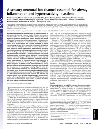
- 2. A *** B Trpa1 +/+ veh Trpv1 +/+ veh 250 250 Trpa1 -/- veh Trpv1 -/- veh *** 4 4 Trpa1 +/+ OVA Trpv1 +/+ OVA B AL cells x 10 B AL cells x 10 200 200 Trpa1 -/- OVA Trpv1 -/- OVA 150 150 100 100 50 50 0 0 tal ils es tes phils tal ils es tes phils T o inoph phag ocy tro T o inoph phag ocy tro E os acro ymph Neu E os acro ymph Neu M L M L C Trpa1+/+ OVA *** D E ** Density of Inflammation 14 Trpa1-/- OVA 35 4 O/ml/s) OVA IgE (s erum) 12 Trpa1+/+ veh 30 (cells/area unit) 10 Trpa1-/- veh ** 25 3 8 R (cm H 2 20 6 15 2 4 ** 10 2 1 5 0 0 0 eh eh VA VA e Trpa1+/+ Trpa1-/- 03 1 3 0 0 /+ v 1 -/- v +/+ O -/- O lin 0. 0. 1. 3. 1+ 0. se ACh (mg/ml) pa Trpa rpa1 rpa1 Ba Tr T T Fig. 1. Decreased inflammatory response to inhaled OVA in TRPA1-deficient mice. (A) Reduced leukocyte infiltration in airways of OVA-challenged Trpa1Ϫ/Ϫ mice. Cell differentials are shown for total cells, eosinophils, macrophages, lymphocytes, and neutrophils in BALF collected from vehicle (veh, PBS)- and OVA-challenged Trpa1ϩ/ϩ and Trpa1Ϫ/Ϫ mice. Animal groups: Trpa1ϩ/ϩ OVA: n ϭ 8, Trpa1Ϫ/Ϫ OVA: n ϭ 8, Trpa1ϩ/ϩ veh: n ϭ 7, Trpa1Ϫ/Ϫ veh: n ϭ 6. *, P Ͻ 0.05; **, P Ͻ 0.01; ***, P Ͻ 0.001 . (B) Normal inflammatory leukocyte infiltration in OVA-challenged Trpv1Ϫ/Ϫ mice. BALF leukocyte cell differentials are shown for vehicle (veh, PBS)- and OVA-challenged Trpv1ϩ/ϩ and Trpv1Ϫ/Ϫ mice. Animal groups: Trpv1ϩ/ϩ OVA: n ϭ 6, Trpv1Ϫ/Ϫ OVA: n ϭ 4, Trpv1ϩ/ϩ veh: n ϭ 6, Trpv1Ϫ/Ϫ veh: n ϭ 4. (C) Comparison of airway resistance (R) in OVA-challenged Trpa1ϩ/ϩ (blue) and Trpa1Ϫ/Ϫ mice (red), as well as vehicle (veh)-treated Trpa1ϩ/ϩ (green) and Trpa1Ϫ/Ϫ (purple) mice, measured by forced oscillation in response to increasing dosages of acetylcholine. Animal groups: Trpa1ϩ/ϩ OVA: n ϭ 4, Trpa1Ϫ/Ϫ OVA: n ϭ 4, Trpa1ϩ/ϩ veh: n ϭ 6, Trpa1Ϫ/Ϫ veh: n ϭ 6. (*, ␣ ϭ 0.05; **, ␣ ϭ 0.01; ***, ␣ ϭ 0.001). (D) Induction of OVA-reactive Ig E in OVA-challenged wild-type and TRPA1-deficient mice, as determined by ELISA. Animal groups as in Fig. 1 A. (E) Density of inflammation in H&E-stained airway sections from OVA-challenged Trpa1ϩ/ϩ and Trpa1Ϫ/Ϫ mice, scored by counting of inflammatory cells near bronchial bundles (n ϭ 4 mice per group). (Fig. 1 A). Eosinophils represented the majority of leukocytes Reduced Mucus Production and Th2 Cytokine Levels in Airways of (Fig. 1 A). In OVA-challenged TRPA1-deficient mice, we ob- OVA-Challenged Trpa1؊/؊ Mice. Using quantitative Taqman PCR, served a remarkable reduction in BALF leukocyte numbers we compared the transcriptional levels of the muc5ac mucin (Ͼ50%), with eosinophilia reduced by Ͼ80% (Fig. 1 A). In genes in whole lung cDNA (Fig. 2A). Mucins are mucus proteins contrast, OVA-challenged Trpv1Ϫ/Ϫ mice showed robust leuko- highly expressed in asthmatic airways. OVA-challenged wild- cyte infiltration, with BALF cell counts indistinguishable from type mice displayed robust induction of muc5ac transcription (Fig. those of wild-type mice (Fig. 1B). 2 A). In contrast, muc5ac levels were reduced by 50% in lungs of Airway hyperreactivity (AHR) is another important hallmark OVA-challenged Trpa1Ϫ/Ϫ mice (Fig. 2 A). Mucin5ac induction was of asthma. Airway resistance was measured by forced oscillation normal in OVA-challenged Trpv1Ϫ/Ϫ mice (Fig. S1B). in response to i.v. administration of increasing concentrations of Th2 leukocytes orchestrate the allergic inflammatory response acetylcholine (Fig. 1C). OVA-challenged wild-type C57BL/6 in the airways through the release of cytokines, such as inter- mice developed robust AHR (Fig. 1C). In OVA-challenged leukin 5 (IL-5). We examined transcriptional activity of the IL-5 Trpa1Ϫ/Ϫ mice, AHR was very mild, only differing from control gene by Taqman PCR of whole lung cDNA from wild-type, animals at the highest doses of acetylcholine. Basal reactivity of Trpa1Ϫ/Ϫ, and Trpv1Ϫ/Ϫ mice as a measure for Th2 leukocyte vehicle-treated wild-type and Trpa1Ϫ/Ϫ mice was identical. We infiltration and activity (Fig. 2B). Strikingly, while OVA- conclude that TRPA1 plays an essential role in asthma-related challenged wild-type mice showed a robust increase in IL-5 airway hyperreactivity. transcriptional activity, IL-5 levels in OVA-challenged Trpa1Ϫ/Ϫ Airway eosinophilia and hyperreactivity are consequences of mice were indistinguishable from those in vehicle-treated mice an allergen-induced Th2-lymphocyte response leading to the (Fig. 2B). Trpv1Ϫ/Ϫ mice showed normal induction of IL-5 production of allergen-specific IgE antibodies. We measured expression (Fig. S1C). OVA-reactive IgE in serum by EIA to verify whether Trpa1Ϫ/Ϫ A systematic comparison of peptide concentrations of cyto- mice produce a normal Th2-response following immunization kines and chemokines was performed using Luminex multiplex and airway challenge with OVA (Fig. 1D). OVA-reactive IgE protein analysis of BAL fluid (Fig. 2C). As predicted from our serum levels in Trpa1Ϫ/Ϫ mice were indistinguishable from those qPCR analysis, IL-5 protein levels in BALF of OVA-challenged in wild-type C57BL/6 mice, suggesting a normal Th2-dependent Trpa1Ϫ/Ϫ mice were much lower (Ͻ20%) than in wild-type mice systemic immune response to OVA (Fig. 1D). These data (Fig. 2C, Inset). Trpa1Ϫ/Ϫ mice also showed significantly dimin- indicate that TRPA1 has a crucial role in later events leading to ished levels of IL-13, IL-17, eotaxin, MCP-1, RANTES, and airway inflammation following allergen challenge. TNF␣, suggesting a profound defect in the Th2-directed local Quantitative comparison of inflammatory cell densities near inflammatory response in the airways (Fig. 2C). Levels of IFN airways in lung sections of OVA-challenged mice confirmed ␥, an indicator for a Th1 leukocyte activity, were below the diminished eosinophilia in Trpa1Ϫ/Ϫ mice and reduced hyper- detection limit in all mouse groups, showing that the observed plasia of mucus-producing goblet cells (Fig. 1E and supporting reduction in airway eosinophilia was not due to a shift toward a information (SI) Fig. S1 A). Th1-directed immune response in Trpa1Ϫ/Ϫ mice. Cytokine 9100 ͉ www.pnas.org͞cgi͞doi͞10.1073͞pnas.0900591106 Caceres et al.
- 3. showed much lower densities of inflammatory cells (Figs. 3E and A Mucin 5AC (RQ) * B *** S1E). Similar to Trpa1Ϫ/Ϫ mice, treatment with HC-030031 did 100 12 not affect serum levels of OVA-specific IgE in OVA-challenged 10 wild-type BALB/C mice, indicating a normal Th2-directed sys- 80 Il-5 (R Q) 8 temic inflammatory response (Fig. S1F). These data suggest that 60 6 TRPA1 plays a crucial role in the development of asthma during 40 4 airway allergen challenge, enabling inflammatory leukocyte 20 2 infiltration, airway hyperreactivity, and mucus production. 0 0 h ve /- veh OVA OVA h h ve /- ve OVA OVA Trpa1 is Essential for Chemically Induced and Inflammatory Neuropep- +/+ - /+ /- +/+ - /+ /- a1 rpa1 pa1+ pa1- a1 rpa1 a1+ pa1 - T rp T Tr Tr T rp T T rp T r tide Release in the Airways. It is unclear whether the pro- inflammatory action of TRPA1 in asthma can be explained through purely neurogenic effects. TRPA1 may play an as yet C 100 Trpa1+/+ 300 250 undetected role in cells of the immune system or in airway tissue. Trpa1-/- pg/ml 200 150 To assess this possibility, we used Taqman quantitative PCR to 80 100 compare TRPA1 transcript levels in cDNA derived from spleen 50 *** harboring a large variety of leukocyte precursors, Th2 lympho- pg/ml 60 0 * IL-5 cytes, whole mouse lung and BALF leukocytes of OVA- 40 challenged mice, and DRG. Relative transcript quantities in * spleen, Th2 cells, whole lung, and leukocytes were minimal, with 20 * DRG expression several 100-fold higher (Fig. S1G). Additional ** ** qPCR experiments using cDNA prepared from primary leuko- 0 * cytes and leukocyte cell lines failed to detect the presence of -2 -4 -6 0 3 7 in 1 S Fα IL IL IL IL -1 IL -1 IL -1 t ax P- TE TN TRPA1 cDNA. These results point to a key role for sensory C Eo M AN neuronal TRPA1 channels in allergic airway inflammation. R TRPA1 may be a critical trigger for neuropeptide release Fig. 2. Impaired induction of mucin, cytokines, and chemokines in OVA- crucial for leukocyte infiltration and inflammatory progression challenged airways of Trpa1-deficient mice. (A) Relative quantities (RQ) of in asthmatic airways. To investigate this possibility, we compared mucin5ac gene transcript, determined by Taqman qPCR of whole mouse lung neuropeptide release in airways of wild-type and and Trpa1 Ϫ/Ϫ cDNA. Mucin5ac induction is diminished in Trpa1Ϫ/Ϫ OVA mice. GAPDH tran- mice in response to 2-chloroacetophenone (CN), a potent in- script levels were used for normalization as endogenous control. Animal flammatory TRPA1 agonist (20). We performed a 30-s BAL in groups: Trpa1ϩ/ϩ veh: n ϭ 4, Trpa1Ϫ/Ϫ veh: n ϭ 4, Trpa1ϩ/ϩ OVA: n ϭ 6, Trpa1Ϫ/Ϫ mice with CN (4 mM) contained in the BAL buffer (PBS) and OVA: n ϭ 7. *, P Ͻ 0.05. (B) Relative quantities (RQ) of interleukin 5 (IL-5) gene transcript, as determined by Taqman real-time quantitative PCR of whole measured the resultant release of CGRP, substance P (SP) and mouse lung cDNA. OVA-challenged Trpa1Ϫ/Ϫ mice show no significant neurokinin A (NKA) using EIA (Fig. 4 A–C). CN induced strong changes in IL-5 transcription compared with vehicle-treated mice. GAPDH increases in the levels of all 3 neuropeptides in BALF of transcript levels were used for normalization as endogenous control. Animal wild-type C57BL/6 mice (Fig. 4 A–C). In Trpa1Ϫ/Ϫ mice CN- groups: Trpa1ϩ/ϩ veh: n ϭ 4, Trpa1Ϫ/Ϫ veh: n ϭ 4, Trpa1ϩ/ϩ OVA: n ϭ 6, Trpa1Ϫ/Ϫ induced peptide release was clearly diminished (Ͻ50% of wild- OVA: n ϭ 6. ***, P Ͻ 0.001. (C) Comparison of cytokine and chemokine levels type levels), supporting a specific and essential role for TRPA1 in BALF of OVA-challenged Trpa1ϩ/ϩ (white) and Trpa1Ϫ/Ϫ (black) mice, as in chemically stimulated neurogenic peptide release in the measured by Luminex peptide analysis. Groups: Trpa1ϩ/ϩ n ϭ 8 –10, Trpa1Ϫ/Ϫ airways (Fig. 4 A–C). Acute CN-induced neuropeptide release n ϭ 8 –10 for each analyte. **, P Ͻ 0.01; ***, P Ͻ 0.001 was suppressed by prior treatment with HC-030031 in Balb/C mice (Fig. 4D). levels were normally elevated in OVA-challenged Trpv1Ϫ/Ϫ mice Exogenous TRPA1 agonists such as CN are likely to mimic the (Fig. S1D). actions of endogenous reactive products and inflammatory signaling pathways activating TRPA1 (16). Since Trpa1Ϫ/Ϫ mice showed clear deficiencies in acute neurogenic peptide release in A TRPA1 Antagonist Reduces Airway Inflammation and Hyperreactiv- the airways, we asked whether neuropeptide levels would also be ity when Administered During OVA Airway Challenge. TRPA1- reduced during airway allergen challenge. We compared neu- antagonists showed efficacy in animal models of acute and ropeptide levels in BALF of OVA-challenged wild-type and inflammatory pain and diminished the noxious effects of TRPA1 Trpa1Ϫ/Ϫ-deficient C57/BL6 mice, and in antagonist treated agonists known to cause asthma-related conditions (17, 19, 20, Balb/C mice, focusing on NK-A, the most abundant neuropep- 28, 29). We asked whether a TRPA1 antagonist would prevent tide in airway lining fluid (30) (Fig. 4 E and F). Indeed, we or diminish airway inflammation when administered to OVA- observed that the OVA-induced increase in BALF NK-A levels sensitized Balb/C mice during the airway challenge phase of the was clearly diminished in Trpa1Ϫ/Ϫ mice and in antagonist- OVA protocol. HC-030031, the most thoroughly studied TRPA1 treated Balb/C mice (Fig. 4 E and F). antagonist, was injected i.p. into OVA-sensitized animals on the day before (200 mg/kg) and twice daily (100 mg/kg) during the Discussion 4 days of OVA airway challenge. While vehicle (methyl cellulose, Our results reveal a crucial role for the sensory neuronal ion MC)-treated mice showed robust OVA-induced increases in channel TRPA1 in experimental asthma. TRPA1-deficient mice BALF leukocyte numbers, cell numbers were diminished in showed profound deficits in airway infiltration by inflammatory OVA-challenged HC-030031-treated mice (Fig. 3A). Moreover, leukocytes in the OVA model of allergic airway inflammation, treatment with HC-030031 led to a nearly complete suppression accompanied by a reduction in inflammatory Th2 cytokines, PHARMACOLOGY of airway hyperreactivity in OVA-challenged Balb/C mice (Fig. such as IL-5 and IL-13, and pro-inflammatory chemokines, such 3B). Mucin5ac transcription was strongly suppressed by HC- as TNF␣ and the eosinophil attractant, eotaxin. As a conse- 030031 treatment, indicating diminished production of airway quence, OVA-induced mucus production is impaired in mucus (Fig. 3C). Antagonist-injected mice also showed dimin- Trpa1Ϫ/Ϫ mice. Airway hyperreactivity, another important hall- ished levels of Th2 cytokines IL-5 and IL-13 in BALF (Fig. 3D). mark of asthma, was strongly reduced. Pharmacological inhibi- Histological sections of airways from antagonist-treated mice tion of TRPA1 during the airway challenge phase of the OVA Caceres et al. PNAS ͉ June 2, 2009 ͉ vol. 106 ͉ no. 22 ͉ 9101
- 4. A 250 * B MC veh 35 MC OVA *** HC-030031 veh B AL cells x 10 4 200 HC-030031 OVA R (cm H2 O/ml/s) MC OVA 30 MC veh HC-030031 OVA 25 HC-030031 veh *** 150 20 100 * 15 10 * *** 50 * 5 ** *** 0 0 tal es tes e ils ils 03 1 3 0 0 lin T o inoph phag hocy troph 0. 0. 1. 3. 0. se s ro p u ACh (mg/ml) Ba E o Mac Lym Ne C * D ** E MC ** Mucin 5AC (RQ) Density of Inflammation 50 2.5 HC-030031 60 40 (cells/area unit) 2.0 30 pg/ml 40 1.5 20 1.0 20 * 10 0.5 0 0 0.0 h h A IL-4 IL-5 IL-13 Eotaxin MC HC-030031 ve 1 ve OVA OV MC 3003 MC 0031 -0 -03 HC HC Fig. 3. Decreased inflammatory response in Balb/C mice treated with the TRPA1 antagonist HC-030031 during the OVA airway challenge phase. (A) Cell differentials for total leukocytes, eosinophils, macrophages, lymphocytes, and neutrophils in BALF collected from vehicle (veh, PBS)- or OVA-challenged mice injected i.p. with HC-030031 or with methyl cellulose (MC) during the airway challenge phase. Animal groups: MC veh: n ϭ 8, HC-030031 veh: n ϭ 9, MC OVA, n ϭ 10, HC-030031 OVA: n ϭ 8. *, P Ͻ 0.05. (B) Comparison of airway hyperresponsiveness to i.v. acetylcholine in vehicle (veh, PBS) - or OVA-challenged mice injected i.p. with HC-030031 or with just methyl cellulose (MC) during OVA airway challenge. Animal groups: MC veh: n ϭ 7, HC-030031 veh: n ϭ 7, MC OVA, n ϭ 7, HC-030031 OVA: n ϭ 6 (*, ␣ ϭ 0.05; **, ␣ ϭ 0.01; ***, ␣ ϭ 0.001). (C) Decreased lung mucin5ac transcription in OVA-challenged mice treated with TRPA1 antagonist HC-030031, determined by Taqman q PCR. RQ of mucin5ac transcript are shown for vehicle (veh.) or OVA-challenged mice injected i.p. with HC-030031 or with just methyl cellulose (MC) during OVA airway challenge. GAPDH transcript levels were used for normalization as endogenous control. Animal groups: MC veh: n ϭ 4, HC-030031 veh: n ϭ 4, MC OVA, n ϭ 8, HC-030031 OVA: n ϭ 8. *, P Ͻ 0.05. (D) Cytokine and eotaxin levels in bronchoalveolar lavage fluid (BALF) of OVA-challenged Balb/C mice treated with TRPA1 antagonist HC-030031 (black) or carrier methyl cellulose (white). (n ϭ 4 mice/group) *, P Ͻ 0.05; **, P Ͻ 0.01. (E) Density of inflammation in H&E-stained airway sections from OVA-challenged HC-030031-treated and Ϫuntreated (MC) Balb/C mice, scored by counting of inflammatory cells near bronchial bundles (n ϭ 4 mice per group). protocol confirmed the essential function of this receptor, local inflammatory responses to allergen challenge is also sup- blocking leukocyte infiltration, cytokine and neuropeptide re- ported by the observation that genetic deletion of TRPA1, or lease, mucus production, and abolishing airway hyperreactivity. treatment with the TRPA1 antagonist HC-030031, did not seem Trpa1Ϫ/Ϫ mice are deficient in the neuronal detection of to affect the systemic Th2-mediated response to allergen immu- multiple pro-inflammatory exogenous and endogenous agents. nization, as evidenced by normal serum levels of OVA-reactive These include asthma-inducing agents such as chlorine, unsat- IgE. Future experiments are needed to address the detailed urated aldehydes in smoke and smog, chloramines, tear gas mechanistic role of TRPA1 in neurogenic responses affecting agents, and industrial isocyanates, as well as endogenous reactive Th2 lymphocyte migration into the airways, cytokine production, oxidative species and lipid mediators (12, 17–20, 24, 25). Some leukocyte recruitment, as well as in airway hyperreactivity. of these endogenous mediators are produced by infiltrating Our experiments clearly show that TRPV1, the capsaicin leukocytes or inflamed airway tissue and can reach concentra- receptor, is not required for allergic airway inflammation in the tions high enough to chronically activate TRPA1 in airway nerve OVA mouse model of asthma. A TRPA1-specific stimulus, endings (31). Chemosensory deficiency in Trpa1Ϫ/Ϫ mice may cause possibly a reactive TRPA1-specific mediator, appears to be a lack of neuronal excitation and Ca2ϩ influx activated by these required to drive the pro-inflammatory activity of airway C- reactive compounds during inflammatory progression in asthma, fibers in asthma. Nevertheless, TRPV1 is clearly involved in the resulting in reduced reflex hyperreactivity and neuropeptide re- symptomatic consequences of airway inflammation, as evi- lease. Indeed, we find that sensory neuropeptide release, a prereq- denced by a recent report showing anti-tussive activity of a uisite for inflammatory leukocyte infiltration in mice, is impaired in TRPV1 antagonist in a model of chronic cough (32). the airways of Trpa1Ϫ/Ϫ mice. This defect applies to both acute The data in our present study support the idea that TRPA1 neuropeptide release, induced by a TRPA1 agonist, and inflam- may function as an integrator of chemical and immunological matory peptide release following OVA challenge in the airways. stimuli modulating inflammation in the airways. This integrative The ability of a TRPA1 antagonist to recapitulate the knock- activity can explain the pro-inflammatory effects of chemical out phenotype suggests that TRPA1 fulfills an acute role in exposures in asthma patients (16). By activating TRPA1, chem- promoting local inflammation in the airways, rather than causing ical irritants may trigger the release of neuropeptides and a developmental defect in immune system function. Adminis- chemokines in the airways, thereby exacerbating the cellular and tration of HC-030031 during the OVA airway challenge phase tissue inflammatory response observed in our present study. Our was sufficient to potently suppress airway leukocyte infiltration, study opens an avenue for asthma pharmacology, revealing TRPA1 mucus production and hyperreactivity. The role of TRPA1 in as a potential target for anti-asthmatic drugs. Future studies will 9102 ͉ www.pnas.org͞cgi͞doi͞10.1073͞pnas.0900591106 Caceres et al.
- 5. Francisco, and Hydra Biosciences. Mice were housed at facilities accredited A CGRP release B SP release * by the Association for Assessment and Accreditation of Laboratory Animal * Substance P (ng/ml) 70 0.20 Care in standard environmental conditions (12-h light-dark cycle and CGR P (pg/ml) 60 50 0.15 23 °C). Food and water were provided ad libitum. Trpa1Ϫ/Ϫ mice were a gift 40 30 0.10 from David Julius (University of California, San Francisco). The Trpa1- 20 0.05 knockout allele was backcrossed into the C57BL/6 background (Ͼ99.5%) by 10 0 0.00 marker assisted accelerated backcrossing (Charles River Laboratories). S N N S N N Trpv1Ϫ/Ϫ mice were purchased from Jackson Laboratories and C57/Bl6 and P B +/+ C 1-/- C P B +/+ C 1-/- C A1 A A1 P A BALB/c mice from Charles River Laboratories. For experiments on C57BL/6, RP RP P R T T TR T Trpv1Ϫ/Ϫ, and Trpa1Ϫ/Ϫ mice, animals were matched for age (12–22 weeks) and gender. Six- to 8-week-old BALB/c mice were used for OVA sensitiza- C NK-A release * D SP release / HC tion and antagonist studies. * Substance P (pg/ml) 0.12 14 NK-A (ng/ml) 0.10 12 Sensitization and Airway Challenge Procedure. Mice were sensitized on days 0, 0.08 10 0.06 8 7, and 14 by i.p. injection of 50 g ovalbumin (OVA) (Sigma-Aldrich) adsorbed in 6 0.04 4 2 mg alum gel (Sigma-Aldrich) in a total volume of 200 L PBS. Control animals 0.02 2 0.00 0 received alum only. Subsequently, lightly anesthetized mice (isoflurane) were S N N S -C N -C N intranasally challenged with OVA [100 g in 40 L PBS (PBS)] or with PBS alone on P B +/+ C 1-/- C PB 1 A MC HC days 21, 22, and 23. For therapeutic intervention with TRP channel antagonist PA R P TR T HC-030031, mice were given the compound (200 mg/kg mouse body weight) i.p. once on day 20 and 100 mg/kg twice a day on days 21, 22, and 23. On the day of E OVA NK-A release * F OVA NK-A release / HC lung mechanics measurement, all mice received 100 mg/kg compound once in the 0.30 * morning and subject to the end point of the experiment. All measurements and 0.12 NK-A (ng/ml) NK-A (ng/ml) 0.25 0.10 sample collection were performed 24 h after the final intranasal challenge. 0.20 0.08 Following lung mechanics measurements plasma levels of HC-030031 along with 0.15 0.10 0.06 samples of dose solutions were analyzed by LC/MS/MS using a standard protein 0.05 0.04 precipitation extraction with the addition of an internal standard. 0.00 eh eh OVA OVA h h A A /+ v /- v - Ve Ve OV OV 1+ 1 - 1+/+ 1 -/ M C HC MC HC Measurement of Airway Reactivity. Twenty-four hours following the last OVA Trpa Trpa Trpa Trpa challenge, mice were anesthetized with pentobarbital (60 mg/kg of body weight) and urethane (1 g/kg). A tracheostomy was performed, and the Fig. 4. Role of TRPA1 in chemically induced and inflammatory neuropeptide trachea was cannulated. Mice were attached to a Flexivent pulmonary me- release in the airways, measured by EIA. (A) Diminished chemically induced release of CGRP in Trpa1Ϫ/Ϫ mice following lung exposure to CN (2- chanics analyzer (SCIREQ) and ventilated at a tidal volume of 9 mL/kg, at a chloroacetophenone) during BAL. Averaged CGRP levels in BAL fluid of frequency of 150 bpm. Positive end-expiratory pressure was set at 2 cm H2O. TRPA1ϩ/ϩ mice, treated with PBS (n ϭ 4) or 4 mM CN (n ϭ 6), and TRPA1Ϫ/Ϫ mice Mice were paralyzed with pancuronium (0.1 mg/kg i.p.). A 27-gauge needle treated with 4 mM CN (n ϭ 5) are shown. *, P Ͻ 0.05. (B) Diminished chemically was used to administer acetylcholine (0.03, 0.1, 0.3, 1.0, and 3.0 mg/mL) induced release of Substance P (SP) in Trpa1Ϫ/Ϫ mice following lung exposure through the subclavial/tail vein to generate a concentration-response curve. to CN (2-chloroacetophenone) during BAL. Treatments and mouse groups as Measurements of airway mechanics were made continuously applying the in Fig. 4A. (C) Diminished chemically induced release of neurokinin A (NK-A) single-compartment model. in Trpa1Ϫ/Ϫ mice following lung exposure to CN (2-chloroacetophenone) during BAL. Treatments and mouse groups as in Fig. 4A. (D) Diminished Quantitative Analysis of Cytokines, Chemokines, and Neuropetides in BAL Fluid. CN-induced release of SP in mice injected i.p. with TRPA1 antagonist HC- Cytokines and chemokines in BAL fluid (50 L) were measured using a Milliplex 030031. Averaged SP concentrations are shown in BAL fluid of Balb/C mice MAP Mouse Cytokine/Chemokine Kit (Millipore) on Luminex 200 analyzer treated with PBS (n ϭ 4), 200 M CN and methylcellulose vehicle (MC CN, n ϭ (Luminex), following the manufacturer’s recommendations. Neuropeptide 4), or with 200 M CN and HC-030031 (HC CN, n ϭ 4) are shown. *, P Ͻ 0.05. levels in BAL were measured by EIA (CGRP: Cayman Chemical; Substance P: (E) Reduced level of neurokinin A in BAL fluid of OVA-challenged Trpa1Ϫ/Ϫ Phoenix Pharmaceuticals; Neurokinin A: Bachem). To measure acute peptide mice. NK-A levels were compared by EIA in BAL fluid of vehicle-treated wild-type mice (Trpa1ϩ/ϩ veh, n ϭ 11), vehicle-treated Trpa1Ϫ/Ϫ mice (n ϭ 7), release, 4 mM CN (2-chloroacetophenone) was added to the BAL buffer. Lungs OVA-challenged wild-type mice (Trpa1ϩ/ϩ OVA, n ϭ 12), and OVA-challenged were inflated with CN-BAL buffer for 30 sec. HC-030031 was injected i.p. in Trpa1Ϫ/Ϫ mice (n ϭ 12). *, ␣Ͻ0.05. (F) Reduction of NK-A in BAL fluid of OVA- Balb/C mice at 200 mg/kg 24 h, 100 mg/kg 6 h, and 100 mg/kg 30 min before challenged Balb/C mice due to injection of TRPA1-antagonist HC-030031. NK-A CN challenge (200 M). levels were compared by EIA in BAL fluid of methyl cellulose and vehicle-treated mice (MC veh, n ϭ 4), HC-030031- and vehicle-treated (HC veh, n ϭ 4), methyl ACKNOWLEDGMENTS. This work was supported by grants from the American cellulose treated and OVA-challenged (MC OVA, n ϭ 7), and HC-030031-treated Asthma Foundation (Grant 07– 0212 to S.J.), the National Institute of Environ- and OVA-challenged mice (HC OVA, n ϭ 8). *, ␣Ͻ0.05 mental Health Sciences (ES015056 and ES017218 to S.J.), a Spanish Ministry of Science and Innovation (MICINN)/Fulbright fellowship to A.I.C. (2007– 0480), a fellowship from the German Research Foundation to M. B. (BR 3773/1–1.) and address the action of TRPA1 antagonists in additional animal National Institutes of Health (NIH) training grant 5T32GM07205 to M. D. E. Its models of asthma and in other allergic inflammatory conditions. contents are solely the responsibility of the authors and do not necessarily represent the official views of the NIH or federal government. S. J. serves on Materials and Methods the Scientific Advisory Board of Hydra Biosciences. All members of Hydra Animals. Experimental procedures were approved by the Institutional Animal Biosciences receive options. We are grateful to Aiwei Sui and Lesley Devine for Care and Use Committees of Yale University, the University of California, San technical support. 1. Eder W, Ege MJ, von Mutius E (2006) The asthma epidemic. N Engl J Med 355:2226 –2235. 6. Dakhama A, et al. (2002) Regulation of airway hyperresponsiveness by calcitonin 2. Maddox L, Schwartz DA (2002) The pathophysiology of asthma. Annu Rev Med gene-related peptide in allergen sensitized and challenged mice. Am J Respir Crit Care 53:477– 498. Med 165:1137–1144. PHARMACOLOGY 3. Cohn L, Elias JA, Chupp GL (2004) Asthma: Mechanisms of disease persistence and 7. White FA, Wilson NM (2008) Chemokines as pain mediators and modulators. Curr Opin progression. Annu Rev Immunol 22:789 – 815. Anaesthesiol 21:580 –585. 4. Flood-Page P, et al. (2007) A study to evaluate safety and efficacy of mepolizumab in 8. Davis B, Roberts AM, Coleridge HM, Coleridge JC (1982) Reflex tracheal gland secretion patients with moderate persistent asthma. Am J Respir Crit Care Med 176:1062–1071. evoked by stimulation of bronchial C-fibers in dogs. J Appl Physiol 53:985–991. 5. Undem BJ, Carr MJ (2002) The role of nerves in asthma. Curr Allergy Asthma Rep 9. Groneberg DA, Quarcoo D, Frossard N, Fischer A (2004) Neurogenic mechanisms in 2:159 –165. bronchial inflammatory diseases. Allergy 59):1139 –1152. Caceres et al. PNAS ͉ June 2, 2009 ͉ vol. 106 ͉ no. 22 ͉ 9103
- 6. 10. Caterina MJ, et al. (2000) Impaired nociception and pain sensation in mice lacking the 22. Shakeri MS, Dick FD, Ayres JG (2008) Which agents cause reactive airways dysfunction capsaicin receptor. Science 288:306 –313. syndrome (RADS)? A systematic review. Occup Med (Lond) 58:205–211. 11. Basbaum AI, Julius D (2006) Toward better pain control. Sci Am 294:60 – 67. 23. Redlich CA, Karol MH (2002) Diisocyanate asthma: Clinical aspects and immunopatho- 12. Bautista DM, et al. (2006) TRPA1 mediates the inflammatory actions of environmental genesis. Int Immunopharmacol 2:213–224. irritants and proalgesic agents. Cell 124:1269 –1282. 24. Trevisani M, et al. (2007) 4-Hydroxynonenal, an endogenous aldehyde, causes pain and 13. Jordt SE, et al. (2004) Mustard oils and cannabinoids excite sensory nerve fibres through neurogenic inflammation through activation of the irritant receptor TRPA1. Proc Natl the TRP channel ANKTM1. Nature 427:260 –265. Acad Sci USA 104:13519 –13524. 14. Bandell M, et al. (2004) Noxious cold ion channel TRPA1 is activated by pungent 25. Andersson DA, Gentry C, Moss S, Bevan S (2008) Transient receptor potential A1 is compounds and bradykinin. Neuron 41:849 – 857. a sensory receptor for multiple products of oxidative stress. J Neurosci 28:2485– 15. Jia Y, Lee LY (2007) Role of TRPV receptors in respiratory diseases. Biochim Biophys Acta 2494. 1772:915–927. 26. Cruz-Orengo L, et al. (2008) Cutaneous nociception evoked by 15-delta PGJ2 via 16. Bessac BF, Jordt SE (2008) Breathtaking TRP Channels: TRPA1 and TRPV1 in Airway activation of ion channel TRPA1. Mol Pain 4:30. Chemosensation and Reflex Control. Physiology (Bethesda) 23:360 –370. 27. Dai Y, et al. (2007) Sensitization of TRPA1 by PAR2 contributes to the sensation of 17. McNamara CR, et al. (2007) TRPA1 mediates formalin-induced pain. Proc Natl Acad Sci inflammatory pain. J Clin Invest 117:1979 –1987. USA 104:13525–13530. 28. Eid SR, et al. (2008) HC-030031, a TRPA1 selective antagonist, attenuates inflammatory- 18. Bessac BF, et al. (2008) TRPA1 is a major oxidant sensor in murine airway sensory neurons. J Clin Invest 118:1899 –1910. and neuropathy-induced mechanical hypersensitivity. Mol Pain 4:48. 19. Andre E, et al. (2008) Cigarette smoke-induced neurogenic inflammation is mediated 29. Petrus M, et al. (2007) A role of TRPA1 in mechanical hyperalgesia is revealed by by alpha,beta-unsaturated aldehydes and the TRPA1 receptor in rodents. J Clin Invest pharmacological inhibition. Mol Pain 3:40. 118:2574 –2582. 30. Heaney LG, et al. (1998) Neurokinin A is the predominant tachykinin in 20. Bessac BF, et al. (2009) Transient receptor potential ankyrin 1 antagonists block the human bronchoalveolar lavage fluid in normal and asthmatic subjects. Thorax noxious effects of toxic industrial isocyanates and tear gases. FASEB J 23:1102–1114. 53:357–362. 21. Taylor-Clark TE, Kiros F, Carr MJ, McAlexander MA (2008) Transient receptor potential 31. Weiss SJ (1989) Tissue destruction by neutrophils. N Engl J Med 320:365–376. ankyrin 1 mediates toluene diisocyanate-evoked respiratory irritation. Am J Respir Cell 32. McLeod RL, et al. (2006) TRPV1 antagonists attenuate antigen-provoked cough in Mol Biol. 10.1165/rcmb. 2008 – 0292OC (epub ahead of print). ovalbumin sensitized guinea pigs. Cough 2:10. 9104 ͉ www.pnas.org͞cgi͞doi͞10.1073͞pnas.0900591106 Caceres et al.