
Recomendados
Recomendados
Más contenido relacionado
La actualidad más candente
La actualidad más candente (20)
Destacado
Destacado (20)
Similar a maturity index
Similar a maturity index (20)
maturity index
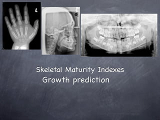
- 1. Skeletal Maturity Indexes Growth prediction
- 2. Outline
- 3. Outline Growth prediction in Orthodontics Skeletal Maturity Index Hand-wrist radiographs Greulich and Pyle (1959) Fishman LH (1982) Björk (1972) and Grave and Brown (1976) Hagg and Tanranger method (1982) Reviews of related studies Cervical vertebra maturation Hassel and Farman (1995) Baccetti et al.(2005) Reviews of related studies Dental age Reviews of related studies
- 4. Growth Prediction in orthodontics In orthodontic treatment planning, knowledge of facial growth velocity and percentage of facial growth remaining is very important for effective growth modification interventions. Assessment of skeletal maturation is essential in planning individual orthodontic treatment because of marked individual variations in timing, duration, and intensity of pubertal growth. Use of cervical vertebral maturation to determine skeletal age Ricky W. K. Wong,a Hessa A. Alkhal,b and A. Bakr M. Rabiec (Am J Orthod Dentofacial Orthop 2009;136:484.e1-484.e6)
- 5. Growth Prediction in orthodontics Clinical decisions regarding functional appliances or orthognathic surgeries are modulated by the patient’s degree of physiological maturity.
- 6. Growth Prediction in orthodontics Many studies have shown an association between peak velocity of facial growth and peak velocity of statural growth during puberty (Bergerson EO. 1972; Singh J1 et al.1967; Fishman LS.1979) Thompson G et al. (1974) demonstrated that the pattern of mandibular growth coincides with body height growth in adolescents
- 7. Growth Prediction in orthodontics General skeletal maturity usually is used as an indicator to predict timing of mandibular growth velocity peak
- 8. Growth Prediction in orthodontics chronological age alone is considered a poor indicator of the level of skeletal maturity due to significant individual growth variation (Roche AF. 1978.;Fishman LS 1979.)
- 9. Growth Prediction in orthodontics Growth prediction can be assessed using physiological parameters such as Peak growth velocity in standing height Pubertal markers ( voice change in male,menarche in female etc. ) Dental development Radiological analysis of skeletal maturation. Hand-wrist radiographs Lateral cephalometric radiographs assessment cervical vertebral maturation
- 10. Growth Prediction in orthodontics Secondary sexual characteristics and physical body measurements , cannot be used to predict the timing of maximum growth due to their retrospective nature (Bjork A., Helm S. 1967) Dental development indicators are not reliable predictors of an individual’s stage of skeletal development. (Demirjian A et al.,1985;Chertkow S., 1980)
- 11. Skeletal Maturity Index Hand-wrist radiographs cervical vertebral maturation
- 13. Hand-wrist radiographs The hand-wrist radiograph is considered to be the most standardized method of skeletal maturation assessment ( Hassel B,Farman AG,1995.;Gandini P et al.2006) Assessment based upon time and the sequence of appearance of the carpal bones and certain ossification events
- 14. Hand-wrist radiographs The ossification sequence and timing of the skeletal maturity within the hand-wrist area show polymorphism and sexual dimorphism. (Bowden BD 1976;Houston WJ 1980;Smith RJ 1980;Garn SM et al 1966) Hand-wrist radiograph of male patient provide more valuable information for orthodontic treatment (Smith 1980)
- 15. Hand-wrist radiographs The usual means by which to assess the hand-wrist radiograph are The comparison atlas of Greulich and Pyle (1959) The processes that use specific indicators that relate skeletal maturation to the pubertal growth curve, such as the methods described by Bjork (1972), Fishman (1982), Hagg and Taranger (1982)
- 16. Greulich and Pyle (1959)
- 17. Greulich and Pyle (1959)
- 18. Greulich and Pyle (1959)
- 19. Greulich and Pyle (1959)
- 20. Greulich and Pyle (1959)
- 21. Greulich and Pyle (1959)
- 22. Radiographic evaluation of Skeletal maturity A clinically orient method based on Hand-wrist films Leonard S. Fishman, Angle orthodontist Vol. 52,No.2,April 1982
- 23. Björk’s method of assessment Bjork utilized certain anatomical sites located on the phalanges, abductor sesamoid, carpal and radius bone, which have predictable and consistent time of onset of ossification.
- 24. Method of Björk (1972) Skeletal maturation of each hand-wrist radiograph 9 stages Stage 1 (PP2): Epiphysis of proximal phalanx of index finger (PP2) is same width as diaphysis Stage 2 (MP3): Epiphysis of middle phalanx of middle finger (MP3) is same width as diaphysis Stage 3 (Pisi-H1-R): Pisi, visible ossification of pisiform; H1: ossification of the hamular process of the hamatum; R, same width of epiphysis and diaphysis of radius Stage 4 (S-H2): S, first mineralization of ulnar sesamoid bone of metacarpophalangeal joint of hamatum; H2, progressive ossification of hamular process of hamatum Chronologic age and skeletal maturation of the cervical vertebrae and hand-wrist: Is there a relationship? Tancan Uysal,a Sabri Ilhan Ramoglu,b Faruk Ayhan Basciftci,c and Zafer Saric, Kayseri and Konya, Turkey (Am J Orthod Dentofacial Orthop 2006;130:622-8)
- 25. Method of Björk (1972) Skeletal maturation of each hand-wrist radiograph 9 stages (method of Björk and Grave and Brown) Stage 5 (MP3cap-PP1cap-Rcap): Diaphysis is covered by cap-shaped epiphysis; in MP3cap, process begins at middle phalanx of third finger; in PP1cap, at proximal phalanx of thumb; in Rcap, at radius Stage 6 (DP3u): Visible union of epiphysis and diaphysis at distal phalanx of middle finger (DP3) Stage 7 (PP3u): Visible union of epiphysis and diaphysis at proximal phalanx of little finger (PP3) Stage 8 (MP3u): Union of epiphysis and diaphysis at middle phalanx of middle finger is clearly visible (MP3) Stage 9 (Ru): Complete union of epiphysis and diaphysis of radius Chronologic age and skeletal maturation of the cervical vertebrae and hand-wrist: Is there a relationship? Tancan Uysal,a Sabri Ilhan Ramoglu,b Faruk Ayhan Basciftci,c and Zafer Saric, Kayseri and Konya, Turkey (Am J Orthod Dentofacial Orthop 2006;130:622-8)
- 26. Method of Björk (1972) Bjork 9 stages of skeletal maturation were reduced to five intervals and ranked 1 to 5 according to growth completion. 1.Growth preceeding acceleration = Bjork stage 1-3 2.Stage of growth acceleration = Bjork stage 4 3.Stage of peak growth = Bjork stage 5 4.Stage of growth deceleration = Bjork stage 6-7 5.Stage of growth completion = Bjork stage 8-9
- 27. Skeletal maturation determine by cervical vertebrae development. Paloma San Roman et al. European Journal of Orthodontic 24 (2002) 303-311
- 28. Skeletal Maturation Assessment (SMA) L.Fishman (1982) Based on the observation of ossification events localized in the area of the finger phalanges, carpal bone and radius. There is a close correlation between sequence of hand- wrist ossification and mandibular growth status Radiographic evaluation of Skeletal maturity A clinically orient method based on Hand-wrist films Leonard S. Fishman, Angle orthodontist Vol. 52,No.2,April 1982
- 29. Skeletal Maturation Assessment (SMA) L.Fishman (1982) Radiographic evaluation of Skeletal maturity A clinically orient method based on Hand-wrist films Leonard S. Fishman, Angle orthodontist Vol. 52,No.2,April 1982
- 30. Skeletal Maturation Assessment (SMA) L.Fishman (1982) SMI 1: The third finger proximal phalanx shows equal width of epiphysis and diaphysis(PP3width) SMI 2: Width of epiphysis equal to that of diaphysis in the middle phalanx of third finger(MP3Width) SMI 3: Width of epiphysis equal to that of diaphysis in the middle phalanx of fifth finger (MP5Width) SMI 4: Appearance of adductor sesamoid of the thumb (S) Radiographic evaluation of Skeletal maturity A clinically orient method based on Hand-wrist films Leonard S. Fishman, Angle orthodontist Vol. 52,No.2,April 1982
- 31. Skeletal Maturation Assessment (SMA) L.Fishman (1982) SMI 5: Capping of epiphysis seen in distal phalanx of third finger(DP3cap) SMI 6: Capping of epiphysis seen in middle phalanx of third finger(MP3cap) SMI 7: Capping of epiphysis seen in middle phalanx of fifth finger(MP5cap) Radiographic evaluation of Skeletal maturity A clinically orient method based on Hand-wrist films Leonard S. Fishman, Angle orthodontist Vol. 52,No.2,April 1982
- 32. Skeletal Maturation Assessment (SMA) L.Fishman (1982) SMI 8: Fusion of epiphysis and diaphysis in the distal phalanx of third finger(DP3fuse) SMI 9: Fusion of epiphysis and diaphysis in proximal phalanx of third finger (PP3fuse) SMI 10: Fusion of epiphysis and diaphysis in middle phalanx of third finger (MP3fuse) SMI 11: Fusion of epiphysis and diaphysis seen in the radius (Rfuse) Radiographic evaluation of Skeletal maturity A clinically orient method based on Hand-wrist films Leonard S. Fishman, Angle orthodontist Vol. 52,No.2,April 1982
- 33. Skeletal Maturation Assessment (SMA) L.Fishman (1982) Accelerating growth velocity period = SMI 1-4 High growth velocity period = SMI 4-7 Decelerating growth velocity period = SMI 7-11
- 34. Skeletal Maturation Assessment (SMA) L.Fishman (1982) PP3width appears approximately 3 years before the peak of pubertal growth spurt(SMI 1) Ossification of adductor sesamoid of thumb (S) occurs shortly before or at the beginning of the pubertal growth spurt. (SMI 4) The MP3cap stage marks the peak of the pubertal growth.(SMI 6) Mandibular Pubertal Growth Spurt Prediction. Part One: Method Based on the Hand-Wrist Radiographs Antana S et al. Stomatologija, Baltic Dental and Maxillofacial Journal, 7:16-20, 2005
- 35. Skeletal Maturation Assessment (SMA) L.Fishman (1982) PP3width appears approximately 3 years before the peak of pubertal growth spurt(SMI 1) Ossification of adductor sesamoid of thumb (S) occurs shortly before or at the beginning of the pubertal growth spurt. (SMI 4) The MP3cap stage marks the peak of the pubertal growth.(SMI 6) Mandibular Pubertal Growth Spurt Prediction. Part One: Method Based on the Hand-Wrist Radiographs Antana S et al. Stomatologija, Baltic Dental and Maxillofacial Journal, 7:16-20, 2005
- 36. Skeletal Maturation Assessment (SMA) L.Fishman (1982) PP3width appears approximately 3 years before the peak of pubertal growth spurt(SMI 1) Ossification of adductor sesamoid of thumb (S) occurs shortly before or at the beginning of the pubertal growth spurt. (SMI 4) The MP3cap stage marks the peak of the pubertal growth.(SMI 6) Mandibular Pubertal Growth Spurt Prediction. Part One: Method Based on the Hand-Wrist Radiographs Antana S et al. Stomatologija, Baltic Dental and Maxillofacial Journal, 7:16-20, 2005
- 37. Skeletal Maturation Assessment (SMA) L.Fishman (1982) Union of epiphysis and diaphysis at the distal phalanx of the middle finger (DP3fusion) signify the end of pubertal growth. (SMI 8) Complete union of epiphysis and diaphysis of the radius (R fusion) indicate that skeletal growth is finished. (SMI 11) Mandibular Pubertal Growth Spurt Prediction. Part One: Method Based on the Hand-Wrist Radiographs Antana S et al. Stomatologija, Baltic Dental and Maxillofacial Journal, 7:16-20, 2005
- 38. Skeletal Maturation Assessment (SMA) L.Fishman (1982) Union of epiphysis and diaphysis at the distal phalanx of the middle finger (DP3fusion) signify the end of pubertal growth. (SMI 8) Complete union of epiphysis and diaphysis of the radius (R fusion) indicate that skeletal growth is finished. (SMI 11) Mandibular Pubertal Growth Spurt Prediction. Part One: Method Based on the Hand-Wrist Radiographs Antana S et al. Stomatologija, Baltic Dental and Maxillofacial Journal, 7:16-20, 2005
- 39. Hagg and Taranger Method(1982) MP3 Ossification Stages: Hagg and Taranger Method 1. MP3-E stage: Epiphysis is 3/4 wide as diaphysis. 2. MP3-F stage: Start of the curve of pubertal growth spurt Epiphysis is as wide as metaphysis. Radiolucent gap between epiphysis and metaphysis is wide. 3. MP3-FG stage: Acceleration of the curve of pubertal growth spurt.Distinct medial and/or lateral border of epiphysis forms line of demarcation at right angle to distal border. 4. MP3-G stage: Maximum point of pubertal growth spurt Sides of epiphysis have thickened and cap its metaphysis, forming sharp distal edge on one or both sides. Marked undulations in metaphysis give it “Cupid’s bow” appearance. 5. MP3-H stage: Deceleration of the curve of pubertal growth spurt.Fusion of epiphysis and metaphysis begins. Radiolucent gap between epiphysis and metaphysic is narrower. 6. MP3-I stage: End of pubertal growth spurt Fusion of epiphysis and metaphysis complete
- 40. Skeletal maturation evaluation using cervical vertebrae Brent Hassel, BA, DDS, MS, and Allan G. Farman, BDS, PhD(odont), Dip ABOMR, EdS, MBA (AMJ ORTHOD DENTOFAC ORTHOP 1995;107:58-66.)
- 41. Conclusion MP3cap MP3width S Ru
- 42. Conclusion MP3cap MP3width S Ru
- 43. Conclusion MP3cap MP3width S Ru
- 44. Conclusion MP3cap MP3width S Ru
- 45. Use of Skeletal Maturation Based on Hand-Wrist Radiographic Analysis as a Predictor of Facial Growth: A Systematic Review Carlos Flores-Mir, DDS, MSc, Cert Orth, PhDa; Brian Nebbe, BDS, Mdent, FFD(SA)Ortho, PhDb; Paul W. Major, DDS, MSc, MRCD(c) The purpose of this systematic review was to evaluate the predictive value of hand- wrist radiographic assessment of skeletal maturity in estimating facial growth timing and velocity. A search of PubMed, Medline, Cochrane Database of Systematic Reviews, Embase, Web of Sciences, and Lilacs 16 articles that met the following inclusion criteria: use of hand-wrist radiographs for skeletal maturation determination facial growth evaluated through cephalometric radiographs and cross-sectional or longitudinal studies. Five articles were rejected because of major methodological issues. Most of the remaining articles had small sample size, and there was no report of randomization or method error. Angle Orthod 2004;74:118–124.
- 46. USE OF SKELETAL MATURATION BASED ON HAND-WRIST RADIOGRAPHIC ANALYSIS AS A PREDICTOR OF FACIAL GROWTH: A SYSTEMATIC REVIEW CARLOS FLORES-MIR, DDS, MSC, CERT ORTH, PHDA; BRIAN NEBBE, BDS, MDENT, FFD(SA)ORTHO, PHDB; PAUL W. MAJOR, DDS, MSC, MRCD(C) Angle Orthod 2004;74:118–124.
- 47. USE OF SKELETAL MATURATION BASED ON HAND-WRIST RADIOGRAPHIC ANALYSIS AS A PREDICTOR OF FACIAL GROWTH: A SYSTEMATIC REVIEW CARLOS FLORES-MIR, DDS, MSC, CERT ORTH, PHDA; BRIAN NEBBE, BDS, MDENT, FFD(SA)ORTHO, PHDB; PAUL W. MAJOR, DDS, MSC, MRCD(C) Skeletal maturity determined by hand-wrist radiographic analysis was well related to overall facial growth velocity. Maxillary and mandibular growth velocities were related to skeletal maturity, but their relationship was less robust than that for overall facial growth. Angle Orthod 2004;74:118–124.
- 48. Reliability of growth prediction with hand–wrist radiographs Damian Verma, Timo Peltomäki and Andreas Jäger The aim of this study was to investigate the validity of hand–wrist radiographic analysis in estimating the amount of remaining craniofacial growth. The material and method cephalograms of 22 males and 27 females with a Class I malocclusion. The median age of the females at the beginning (T1) was 11 years 10 months and at the end (T2) of treatment 14 years 7 months The median age of the males at the beginning (T1) was 12 years 6 months and at the end (T2) of treatment 15 years 3 months, respectively. European Journal of Orthodontics 31 (2009) 438–442
- 49. Reliability of growth prediction with hand–wrist radiographs Damian Verma, Timo Peltomäki and Andreas Jäger The results showed a highly significant correlation between statural growth increases and growth prediction assessed from the hand–wrist radiographs (females: r = 0.68; males: r = 0.7). Concerning craniofacial structures, the increase in mandibular corpus (Go-Gn) showed the highest correlation with growth prediction (females: r = 0.21; males: r = 0.52), but this association would not allow a reliable growth prediction. There was no significant correlation between growth increases of the cranial base, the maxilla, the ramus, and the effective length of the mandible (Co-Gn) and growth prediction assessed with the help of hand–wrist radiographs. European Journal of Orthodontics 31 (2009) 438–442
- 50. Reliability of growth prediction with hand–wrist radiographs Damian Verma, Timo Peltomäki and Andreas Jäger As each patient has an individual growth pattern and different craniofacial structures show individual growth potential, it is questionable if quantitative craniofacial growth prediction with the help of hand–wrist radiographs is reliable. However, in an individual case for the assessment of the timing of the growth process, a hand–wrist radiograph can contribute to treatment planning. European Journal of Orthodontics 31 (2009) 438–442
- 51. Cervical Vertebral Maturation method(CVM) The cervical vertebral maturation method has proved effective in assessing the adolescent growth peak both in body height and mandibular size. Hellsing (1991) found significant correlation between height and length of the cervical vertebral bodies and statural height. Mitani and Sato (1992) reported changes in the cervical vertebrae correlated significantly with increases in mandibular size. Mappes et al (1992) indicated that predominant ethnic origin, climate, nutrition, socioeconomic level, and urbanization are causative factors of these racial variations.
- 52. Cervical Vertebral Maturation method(CVM) The cervical vertebral maturation method has proved effective in assessing the adolescent growth peak both in body height and mandibular size. Hassel and Farman (1995) and Garcia-Fernandez et al (1998) found high correlations between cervical vertebral maturation and the skeletal maturation of the hand-wrist. Franchi and coworkers (2000) confirmed the validity of six CVM stages as a biologic indicator for both mandibular and somatic skeletal maturity in 24 growing untreated subjects
- 53. Cervical Vertebral Maturation method (CVM) Lamparski (1972) created method analyzed size and shape changes in the bodies of five cervical vertebrae (from the second one through the sixth). Thereafter, several cervical vertebral maturation (CVM) methods were proposed, and some modifications were made and improvements achieved.
- 54. Cervical Vertebral Maturation method(CVM) Hassel and Farman (1995) compiled a new index of CVM, evaluated the second, third, and fourth cervical vertebrae. Baccetti et al (2002) devised new CVM method,comprised of five maturationa stage (CVMS I through CVMS V instead of Cvs 1 through Cvs 6 in the former CVM method) Baccetti et al (2005) present a further modified and refined version of the CVM method
- 55. Hassel and Farman(1995) Six stages in evaluation of cervical vertebrae maturation according to method of Hassel and Farman(1995) I.Initiation stage (CVMI 1) Very significant amount of adolescent growth expected C2, C3, and C4 inferior vertebral body borders are flat Superior vertebral borders are tapered posterior to anterior II. Acceleration stage (CVMI 2) Significant amount of adolescent growth expected Concavities developing in lower borders of C2 and C3 Lower border of C4 vertebral body is flat C3 and C4 are more rectangular in shape III.! Transition stage (CVMI 3) Moderate amount of adolescent growth expected Distinct concavities in lower borders of C2 and C3 C4 developing concavity in lower border of body C3 and C4 are rectangular in shape IV. Deceleration stage (CVMI 4) Small amount of adolescent growth expected Distinct concavities in lower borders of C2, C3, and C4 C3 and C4 are nearly square in shape V. Maturation stage (CVMI 5) Insignificant amount of adolescent growth expected Accentuated concavities of inferior verte- bral body borders of C2, C3, and C4 C3 and C4 are square in shape VI. Completion stage (CVMI 6) Adolescent growth is completed Deep concavities are present for inferior vertebral body borders of C2, C3, and C4 C3 and C4 heights are greater than widths
- 56. Hassel and Farman(1995) I.Initiation stage (CVMI 1) Very significant amount of adolescent growth expected C2, C3, and C4 inferior vertebral body borders are flat Superior vertebral borders are tapered posterior to anterior II. Acceleration stage (CVMI 2) Significant amount of adolescent growth expected Concavities developing in lower borders of C2 and C3 Lower border of C4 vertebral body is flat C3 and C4 are more rectangular in shape
- 57. Hassel and Farman(1995) III.! Transition stage (CVMI 3) Moderate amount of adolescent growth expected Distinct concavities in lower borders of C2 and C3 C4 developing concavity in lower border of body C3 and C4 are rectangular in shape IV. Deceleration stage (CVMI 4) Small amount of adolescent growth expected Distinct concavities in lower borders of C2, C3, and C4 C3 and C4 are nearly square in shape
- 58. Hassel and Farman(1995) V. Maturation stage (CVMI 5) Insignificant amount of adolescent growth expected Accentuated concavities of inferior vertebral body borders of C2, C3, and C4 C3 and C4 are square in shape VI. Completion stage (CVMI 6) Adolescent growth is completed Deep concavities are present for inferior vertebral body borders of C2, C3, and C4 C3 and C4 heights are greater than widths
- 60. Baccetti et al 2002 An Improved Version of the Cervical Vertebral Maturation (CVM) Method for the Assessment of Mandibular Growth (Angle Orthod 2002;72:316–323.)
- 61. The Cervical Vertebral Maturation (CVM) Method for the Assessment of Optimal Treatment Timing in Dentofacial Orthopedics Tiziano Baccetti,Lorenzo Franchi,and James A. McNamara, Jr. The aim of the present article is to present a further modified and refined version of the CVM method and its validity for the appraisal of mandibular skeletal maturity in the individual patient in light of the findings of recent studies in which the CVM method has been used to assess optimal timing for the treatment of malocclusions in the transverse, sagittal, and vertical planes of space. Semin Orthod 11:119–129 © 2005
- 62. The Cervical Vertebral Maturation (CVM) Method for the Assessment of Optimal Treatment Timing in Dentofacial Orthopedics Tiziano Baccetti,Lorenzo Franchi,and James A. McNamara, Jr. Subjects and Methods The total 30 sujects (18 males, 12 females) that comprises the cephalometric files of the University of Michigan Elementary and Secondary School Growth Study was evaluated. Total mandibular length (Co-Gn) was measured on the longitudinal sets of lateral cephalograms The maximum increase in Co-Gn between two consecutive annual cephalograms was used to define the peak in mandibular growth at puberty in the individual subjects. Two consecutive cephalograms comprising the interval of maximum mandibular growth, together with two earlier consecutive cephalograms and two later consecutive cephalograms, The morphology of the bodies of the second (C2 odontoid process), third (C3), and fourth (C4) cervical vertebrae were analyzed in the six consecutive annual observations (T1 through T6). The analysis consisted of both visual and cephalometric appraisals of morphological characteristics of the cervical vertebrae. Semin Orthod 11:119–129 © 2005
- 63. The Cervical Vertebral Maturation (CVM) Method for the Assessment of Optimal Treatment Timing in Dentofacial Orthopedics Tiziano Baccetti,Lorenzo Franchi,and James A. McNamara, Jr. Semin Orthod 11:119–129 © 2005
- 64. Baccetti et al 2005 Cervical stage 1 The lower borders of all the three vertebrae (C2-C4) are flat. The bodies of both C3 and C4 are trapezoid in shape (the superior border of the vertebral body is tapered from posterior to anterior). The peak in mandibular growth will occur on average 2 years after this stage.
- 65. Baccetti et al 2005 Cervical stage 2 A concavity is present at the lower border of C2 (in four of five cases, with the remaining subjects still showing a cervical stage 1). The bodies of both C3 and C4 are still trapezoid in shape. The peak in mandibular growth will occur on average 1 year after this stage.
- 66. Baccetti et al 2005 Cervical stage 3 Concavities at the lower borders of both C2 and C3 are present. The bodies of C3 and C4 may be either trapezoid or rectangular horizontal in shape. The peak in mandibular growth will occur during the year after this stage.
- 67. Baccetti et al 2005 Cervical stage 4 Concavities at the lower borders of C2, C3, and C4 now are present. The bodies of both C3 and C4 are rectangular horizontal in shape. The peak in mandibular growth has occurred within 1 or 2 years before this stage.
- 68. Baccetti et al 2005 Cervical stage 5 The concavities at the lower borders of C2, C3, and C4 still are present. At least one of the bodies of C3 and C4 is squared in shape. If not squared, the body of the other cervical vertebra still is rectangular horizontal. The peak in mandibular growth has ended at least 1 year before this stage.
- 69. Baccetti et al 2005 Cervical stage 6 The concavities at the lower borders of C2, C3, and C4 still are evident. At least one of the bodies of C3 and C4 is rectangular vertical in shape. If not rectangular vertical, the body of the other cervical vertebra is squared. The peak in mandibular growth has ended at least 2 years before this stage.
- 70. Baccetti et al 2005 The Cervical Vertebral Maturation (CVM) Method for the Assessment of Optimal Treatment Timing in Dentofacial Orthopedics Tiziano Baccetti,Lorenzo Franchi,and James A. McNamara, Jr. Semin Orthod 11:119–129 © 2005
- 71. The Cervical Vertebral Maturation (CVM) Method for the Assessment of Optimal Treatment Timing in Dentofacial Orthopedics Tiziano Baccetti,Lorenzo Franchi,and James A. McNamara, Jr. Semin Orthod 11:119–129 © 2005
- 72. The Cervical Vertebral Maturation (CVM) Method for the Assessment of Optimal Treatment Timing in Dentofacial Orthopedics Tiziano Baccetti,Lorenzo Franchi,and James A. McNamara, Jr. Application to Dentofacial Orthopedics 1. Class II treatment is most effective when it includes the peak in mandibular growth (CS3) 2. Class III treatment with maxillary expansion and protraction is effective when performed before the peak (CS1 or CS2), whereas it is effective in the mandible during both prepubertal and pubertal stages; 3. skeletal effects of rapid maxillary expansion for the correction of transverse maxillary deficiency are greater at prepubertal stages(CS1-CS3), 4. deficiency of mandibular ramus height : the peak in mandibular growth (CS3). Semin Orthod 11:119–129 © 2005
- 73. Cervical vertebral maturation as a biologic indicator of skeletal maturity A systematic review Rodrigo Ce ́sar Santiagoa; Luiz Felipe de Miranda Costaa; Robert Willer Farinazzo Vitralb; Marcelo Reis Fragac; Ana Maria Bolognesed; Lucianne Cople Maiae (Angle Orthod. 0000;00:000–000.)
- 74. Cervical vertebral maturation as a biologic indicator of skeletal maturity A systematic review Rodrigo Ce ́sar Santiagoa; Luiz Felipe de Miranda Costaa; Robert Willer Farinazzo Vitralb; Marcelo Reis Fragac; Ana Maria Bolognesed; Lucianne Cople Maiae There was a moderate to high reproducibility of the CVM method Although some studies indicate that the CVM method shows good correlation with the HWM method, with considerable levels of reproducibility These parameters are not good enough for determining the validity of the CVM method. Furthermore, these conclusions were based on six articles, which points to a low level of evidence, and The biggest question remains unanswered: How reliable is CVM staging for predicting the pubertal spurt? (Angle Orthod. 0000;00:000–000.)
- 75. Cervical vertebrae maturation method morphologic criteria: Poor reproducibility Trenton S. Nestman,Steven D. Marshall, Fang Qian, Nathan Holton,Robert G. Franciscus, and Thomas E. Southardf Brighton, Colo, and Iowa City, Iowa The weakness of the CVM method Difficulty in classifying the vertebral bodies of C3 and C4 as trapezoidal, rectangular horizontal, square, or rectangular vertical. This led to the overall poor reproducibility of the CVM method and our inability to support its use as a strict clinical guideline for the timing of orthodontic treatment. (Am J Orthod Dentofacial Orthop 2011;140:182-8)
- 76. Validity and reliability of a method for assessment of cervical vertebral maturation Xiao-Guang Zhaoa; Jiuxiang Linb; Jiu-Hui Jiangc; Qingzhu Wanga; Sut Hong NGa The reliability of the CVM method was strong interobserver agreement and substantial intraobserver agreement. The percent of Interobserver agreement was below 50% at both times. Percent intraobserver agreement varied widely among observers (range, 40.7%–79.1%). The validity of the CVM method was moderate agreement between the gold standard and observer staging at the initial time. Percent agreement, especially at cervical stage 3, left much to be desired. The CVM method should be used with other growth indicators in the evaluation of skeletal maturation. (Angle Orthod. 2012;82:229–234.)
- 77. Validity and reliability of a method for assessment of cervical vertebral maturation Xiao-Guang Zhaoa; Jiuxiang Linb; Jiu-Hui Jiangc; Qingzhu Wanga; Sut Hong NGa The accuracy and reproducibility of the CVM method leave much to be desired. Factors that affect them are listed as follows. 1) The ‘‘measurements’’ are purely subjective. It is difficult to define and identify the gradual appearance of the concavity in the inferior border of the vertebral bodies. The difference between horizontally rectangular, square, and vertically rectangular shapes depends on the researcher’s arbitrary decision (Angle Orthod. 2012;82:229–234.)
- 78. Validity and reliability of a method for assessment of cervical vertebral maturation Xiao-Guang Zhaoa; Jiuxiang Linb; Jiu-Hui Jiangc; Qingzhu Wanga; Sut Hong NGa The accuracy and reproducibility of the CVM method leave much to be desired. Factors that affect them are listed as follows. 2) The shapes of cervical vertebrae show marked variation from subject to subject. Sometimes the shape and inferior borders of C2–C4 cannot fulfill the definition of a cervical stage at the same time. Furthermore, The shapes of C3 and C4 bodies were still horizontally rectangular in some adult patients (Angle Orthod. 2012;82:229–234.)
- 79. Validity and reliability of a method for assessment of cervical vertebral maturation Xiao-Guang Zhaoa; Jiuxiang Linb; Jiu-Hui Jiangc; Qingzhu Wanga; Sut Hong NGa The accuracy and reproducibility of the CVM method leave much to be desired. Factors that affect them are listed as follows. 3) Could the mandibular growth rate be taken as a reference to establish the CVM method ? Controversy about the relationship between facial (mandibular) growth and general bodily growth continues (Angle Orthod. 2012;82:229–234.)
- 80. Dental maturation Tooth emergence, dental maturity detected through radiographic methods has been investigated as a marker of skeletal maturity Tooth emergence has been investigated as a marker of skeletal maturity; however, this has been shown to be poorly correlated with individual skeletal maturity (Bjork and Helm, 1967; Hagg and Taranger, 1982; Franchi et al., 2008).
- 81. Dental maturity VS Skeletal maturity Dental maturity detected through radiographic methods appears to be highly related to skeletal maturity (Sierra, 1987; Coutinho et al., 1993; Krailassiri et al., 2002; Uysal et al., 2004; Basaran et al., 2007). The maturity of several mandibular teeth, excluding the third molar, has been reported to be correlated with the skeletal maturation phases, with correlation coefficients of 0.63–0.81 (Sierra, 1987), 0.56–0.69 (Krailassiri et al., 2002), 060–0.91 (Basaran et al., 2007), and 0.63–0.84 (Uysal et al., 2004).
- 82. Dental maturity VS Skeletal maturity In contrast, one study failed to show significant correlations between dental maturation and other indices of skeletal maturation (Demirjian et al., 1985). However, this last study recorded dental maturity as 90 per cent of development of the whole dentition rather than using individual tooth maturity.
- 83. Dental Calcification Stages Using Demirjian Index (DI) Stage A Calcification of single occlusal points A without fusion of different calcifications Stage B Fusion of mineralization points; the B contour of the occlusal surface is recognizable Stage C Enamel formation has been completed C at the occlusal surface, and dentin formation has commenced The pulp chamber is curved,and no pulp horns are visible D Stage D Crown formation has been completed to the level of the cementoenamel junction Root formation has commenced. The pulp horns are beginning to differentiate, but the walls of the
- 84. Dental Calcification Stages Using Demirjian Index (DI) Stage E The root length remains shorter than the crown height. The walls of the pulp chamber are straight, and the E pulp horns have become more differentiated than in the previous stage. In molars, the radicular bifurcation has commenced to calcify. F Stage F The walls of the pulp chamber now form an isosceles triangle, and the root length is equal to or greater than the crown height. In molars, the bifurcation has developed sufficiently to give the roots a distinct form G Stage G The walls of the root canal are now parallel, but the apical end is partially open. In molars, only the distal root is rated. H Stage H The root apex is completely closed (distal root in molars). The periodontal membrane surrounding the root and apex is uniform in width throughout..
- 85. Eruption of the maxillary canines in relation to skeletal maturity This study was to assess the relationship between the eruption of the permanent maxillary canines and skeletal maturity in subjects with different skeletal relationships in the sagittal and vertical planes. Methods: A sample of 152 subjects (63 boys, 89 girls) with erupting permanent maxillary canines was analyzed. On the lateral cephalograms, the stage of cervical vertebral maturation was assessed. The subjects were divided into prepeak (before the pubertal growth spurt, cervical stage [CS]1 and CS2) peak (during the pubertal growth spurt, CS3 and CS4) postpeak (after the pubertal growth spurt, CS5 and CS6) groups. Skeletal relationships in the sagittal and vertical planes were evaluated, and relationships to timing of canine eruption were tested statistically Tiziano Baccetti, Lorenzo Franchi, Simona De Lisa, and Veronica Giuntini (Am J Orthod Dentofacial Orthop 2008;133:748-51)
- 86. Eruption of the maxillary canines in relation to skeletal maturity Eruption of the permanent maxillary canine can occur at any stage in skeletal maturation before the end the pubertal growth spurt (CS1-CS4). A post- pubertal stage (CS5 or CS6) without an erupted maxillary canine indicates delayed canine eruption and suggests canine impaction. Eruption of the permanent maxillary canine occurs more frequently in the prepubertal stages of skeletal development in subjects with increased vertical skeletal relationships. Tiziano Baccetti, Lorenzo Franchi, Simona De Lisa, and Veronica Giuntini (Am J Orthod Dentofacial Orthop 2008;133:748-51)
- 87. Relationships Between Dental Calcification Stages and Skeletal Maturity Indicators in Thai Individuals the canine stage F may represent the MP3width stage evaluating the onset of the accelerating growth period. The second molar stage E for female subjects, coinciding with the S stage the second molar stage G for male subjects, coinciding with the MP3cap stage, were indicative of a very high rate of growth acceleration. Suleekorn Krailassiri, DDS; Niwat Anuwongnukroh, DDS, MSD; Surachai Dechkunakorn, DDS, D Ortho (Angle Orthod 2002;72:155–166.)
- 88. Relationships Between Dental Calcification Stages and Skeletal Maturity Indicators in Thai Individuals This suggests that tooth calcification stages from panoramic radiographs might be clinically useful as a maturity indicator of the pubertal growth period However, further study is recommended in a larger sample size, and future studies should address development of the canines and second molars Suleekorn Krailassiri, DDS; Niwat Anuwongnukroh, DDS, MSD; Surachai Dechkunakorn, DDS, D Ortho (Angle Orthod 2002;72:155–166.)
- 89. Conclusion No single method of assessing maturity level has been shown to be effective and accurate in all patients. The hand-wrist radiograph is considered to be the most standardized method of skeletal maturation assessment Additonal radiation exposure to the patient should be concern In an individual case, for the assessment of the timing of growth process, hand-wrist radiograph can contribute for treatment plan
- 90. Conclusion The accuracy of predicting growth may be improved if other parameters (morphologic, biological, or genetic indicators), in addition to hand-wrist radiographic evaluation are used. Sato K, Mito T, Mitani H. An accurate method of predicting man- dibular growth potential based on bone maturity. Am J Orthod Dentofacial Orthop. 2001;120:286–293.
- 91. Conclusion Validity and reliability of CVM method : still controversy The CVM method should be used with other growth indicator in the evaluation of skeletal maturation So do dental maturity index
- 93. If you look for the peak of growth
- 94. If you look for the peak of growth MP3cap
- 95. If you look for the peak of growth MP3cap
- 96. If you look for the peak of growth MP3cap
- 97. If you look for the peak of growth
- 98. If you look for the peak of growth CS 3
- 99. If you look for the peak of growth
- 100. If you look for the peak of growth
- 101. If you look for the peak of growth
- 102. If you look for the peak of growth 2nd molar stage G (Male)
- 103. If you look for the peak of growth 2nd molar stage G (Male)
- 104. If you look for the peak of growth 2nd molar stage G (Male)
- 105. If you look for the peak of growth
- 106. If you look for the peak of growth 2nd molar stage E(Female) : S stage
- 107. If you look for the peak of growth 2nd molar stage E(Female) : S stage
- 109. Thank you
Notas del editor
- \n
- ความสำคัญของ Growth prediction ในทางทันตกรรมจัดฟัน\nตัวชี้วัดอายุกระดูก ได้แก่\n Handwrist radiograph ประเมินอายุกระดูกจากการเจริญเติบโตของกระดูกมือ โดยวิธีต่างๆ ในที่นี้จะพูดถึงวิธีของ Fishman LH (1982)- Björk (1972) and Grave and Brown (1976) -Hagg and Tanranger method (1982) และกล่าวถึง studies ที่เกี่ยวข้อง\nการใช้การเจริญเติบโต ของกระดูก สัน หลัง ส่วนคอในการประเมิน อายุกระดูก CVM โดยวิธีของ Hassel and Farman (1995) และ Baccetti et al. (2005) \n การ ประเมินอายุกระดูกจากอายุของฟัน โดยจะพูดถึงการดูอายุของฟันและการศึกษาที่เกี่ยวข้องกับการใช้อายุของฟันในการประเมินอายุกระดูก\n\n\n
- ในทางทันตกรรมจัดฟันความรู้ที่เกี่ยวกับการเจริญเติบโตของกระโหลกศรีษะและใบหน้าเป็นสิ่งที่สำคัญในการวางแผนการรักษาที่เกี่ยวข้องกับการควบคุม เปลี่ยนแปลง เจริญเติบโตของผู้ป่วย เนื่องจากผู้ป่วยแต่ละ คนมีรูปแบบการเจริญเติบโตที่แตกต่าง การประเมินระดับการเจริญเติบโตจึงมีความสำคัญในการวางแผน การรักษา \n\n
- Bjork meaning\nการประเมินการเจริญเติบโต ของขากรรไกรล่างมีความสำคัญในการรักษาที่เกี่ยวข้องกับการใช้ functional appliance therapy และการทำ orthognathic surgery เนื่องจากการเจริญเติบโตจะมีผลต่อผลลัพธ์ของการรักษาเหล่านี้ อายุตามปฏิทินไม่สามารถใช้เป็นตัวชี้วัดระดับของการเจริญเติบโตที่น่าเชื่อถือได้ เนื่องจากผู้ป่วยแต่ละคนมีการ รูปแบบเจริญเติบโตที่แตกต่างกัน (Roche AF.1978: Fishman LS.1979.) \n\n
- มีหลายการศึกษาที่บ่งชี้ว่าการเจริญบริเวณกระโหลกศรีษะและใบหน้ามีความสัมพันธ์กับการเจริญ เติบโตของร่างกาย Thompson G et al. (1974) พบว่ารูปแบบการเจริญเติบโตของขากรรไกรล่าง มีความสอดคล้องกับการเจริญของร่างกายด้านส่วนสูงในวัยรุ่น การประเมินการเจริญเติบโตของขากรรไกรล่าง (mandibular growth) จึงมักอ้างอิงจากการเจริญเติบโตของร่างกาย \n
- มีหลายการศึกษาที่บ่งชี้ว่าการเจริญบริเวณกระโหลกศรีษะและใบหน้ามีความสัมพันธ์กับการเจริญ เติบโตของร่างกาย Thompson G et al. (1974) พบว่ารูปแบบการเจริญเติบโตของขากรรไกรล่าง มีความสอดคล้องกับการเจริญของร่างกายด้านส่วนสูงในวัยรุ่น การประเมินการเจริญเติบโตของขากรรไกรล่าง (mandibular growth) จึงมักอ้างอิงจากการเจริญเติบโตของร่างกาย \n
- Bjork meaning\nการประเมินการเจริญเติบโต ของขากรรไกรล่างมีความสำคัญในการรักษาที่เกี่ยวข้องกับการใช้ functional appliance therapy และการทำ orthognathic surgery เนื่องจากการเจริญเติบโตจะมีผลต่อผลลัพธ์ของการรักษาเหล่านี้ อายุตามปฏิทินไม่สามารถใช้เป็นตัวชี้วัดระดับของการเจริญเติบโตที่น่าเชื่อถือได้ เนื่องจากผู้ป่วยแต่ละคนมีการ รูปแบบเจริญเติบโตที่แตกต่างกัน (Roche AF.1978: Fishman LS.1979.) \n\n
- การคาดการณ์การเจริญเติบโตของผู้ป่วยโดยใช้ physiologic parameter มีวิธีต่างๆดังนี้\n1. การใช้อัตราการเปลี่ยนแปลงความสูง PVH\n2. การใช้อายุตามลักษณะทางเพศ\n3. การใช้อายุฟัน\n4. การใช้อายุกระดูก โดยการวิเคราะห์จากภาพรังสี ได้แก่ ประเมินอายุกระดูกจากการเจริญเติบโตของกระดูกมือและ การใช้การเจริญเติบโต ของกระดูก สัน หลัง ส่วนคอในการประเมิน\n \n\n
- Bjork A., และHelm S (1967) การเปลี่ยนแปลงของลักษณะทางเพศและรูปร่าง ได้แก่ ความสูง เป็นตัวชี้วัดการเจริญเติบโตที่ดีแต่ด้วยลักษณะของตัวชี้วัดแล้วไม่สามารถนำมาใช้ในการคาดการณ์การ อัตราเจริญเติบโตของผู้ป่วยได้ \n Dental development indicators are not reliable predictors of an individual’s stage of skeletal development. (Demirjian A et al.,1985;Chertkow S.,1980)\nUse of Skeletal Maturation Based on Hand-Wrist Radiographic Analysis as a Predictor of Facial Growth: A Systematic Review\nCarlos Flores-Mir et al.(Angle Orthod 2004;74:118–124.)\nCompare cvm and wrist\n
- \n
- \n
- วิธีดูการเจริญเติบโตของกระดูกมือจากภาพรังสีเป็นวิธีมาตรฐานในการประเมินอายุกระดูก ( Hassel B,Farman AG,1995 ;Gandini P et al.2006) โดยประเมินจากลำดับและกระบวนการสร้างกระดูกชิ้นต่างๆ \n
- แต่ลำดับกระบวน การสร้างกระดูกมีหลากหลายรูปแบบและมีความแตกต่างในแต่ละบุคคลและเพศ จึงทำให้ยังมีข้อจำกัดในการ ใช้งาน (Bowden BD. 1976;HoustonWJ. 1980;Smith RJ 1980;Garn SM. et al 1966)\n Smith ในปี 1980 กล่าวว่า การประเมินอายุกระดูกจากภาพรังสีกระดูกข้อมือในเพศชายนั้น มีคุณค่าต่อ งานทางทันตกรรมจัดฟันมากกว่า เนื่องจากการศึกษาพบว่าในเพศชายมีความสัมพันธ์ของการเจริญเติบโตของ กระดูกมือจากภาพรังสีกับการเจริญเติบโตของขากรรไกรล่างมากกว่าในเพศหญิง\n\n Systemic CVmThe ossification sequence and timing of the skeletal maturity within the hand-wrist area show polymorphism and sexual dimorphism, which can limit their clinical predictive use. (Bowden BD 1976;Houston WJ 1980;Smith RJ 1980;Garn SM et al 1966) แต่ลำดับกระบวนการสร้างกระดูกมีหลากหลายรูปแบบ และมีความแตกต่างในแต่ละเพศ จึงทำให้มีข้อจำกัดในการใช้งาน \n Reliability HW : โดย smith ในปี 1980 กล่าวว่า การประเมินอายุกระดูกภาพรังสีกระดูกข้อมือในเพศชายนั้นมีคุณค่าต่องานทางทันตกรรมจัดฟันมากกว่าเนื่องจากมีความสัมพันธ์กับการเจริญเติบโตของ ขากรรไกรล่างมากกว่าในเพศหญิง\n
- โดยทั่วไปแล้ววิธีที่ใช้ในการประเมินอายุกระดูกจากภาพรังสีกระดูกมือ มี 2 วิธีหลักๆ ได้แก่ (2)\n 1.การประเมินอายุกระดูกจากภาพรังสีกระดูกมือโดยการเปรียบเทียบกับหนังสือภาพ Atlas ของ Greulich and Pyle (1959)(3) ที่ประกอบด้วยรูปภาพของกระดูกมือที่เป็นต้นแบบ ในแต่ละช่วงอายุ เป็นระยะทุกๆ 6 เดือนตามอายุปฏิทิน\n2.การประเมินอายุกระดูกจากภาพรังสีกระดูกมือโดยการใช้ specific indicators ที่มีความ สัมพันธ์ระยะการเจริญเติบโตในช่วงวัยรุ่น เช่น วิธีการของ Bjork (1972), Fishman (1982), Hagg and Taranger (1982) เป็นต้น\n Systemic CVM : โดยทั่วไปแล้ววิธีที่ใช้ในการประเมินอายุกระดูกจากภาพรังสีกระดูกมือ มี 2 วิธีหลักๆ ได้แก่ 1.การประเมินอายุกระดูกจากภาพรังสีกระดูกมือโดยการเปรียบเทียบกับหนังสือภาพ Atlas ของ Greulich and Pyle (1959) and Tanner et al.(1980) ที่ประกอบด้วยรูปภาพต้นแบบของกระดูกมือที่ถ่ายเป็นระยะทุกๆ 6 เดือนตามอายุปฏิทิน และนำภาพรังสีกระดูกข้อมือที่มีมาเปรียบเทียบกับอายุการเจริญของกระดูกใน Atlas ซึ่งเป็นอายุตามค่ามาตรฐาน และ 2. การประเมินอายุกระดูกจากภาพรังสีกระดูกมือโดยการใช้ specific indicator ที่มีความสัมพันธ์กับกราฟการเจริญในช่วงวัยรุ่น เช่น วิธีการของ Fishman (1982)\n
- การประเมินอายุกระดูกจากภาพรังสีกระดูกมือโดยการเปรียบเทียบกับหนังสือภาพ Atlas ของ Greulich and Pyle (1959)(3) ที่ประกอบด้วยรูปภาพของกระดูกมือที่เป็นต้นแบบ ในแต่ละช่วงอายุ เป็นระยะทุกๆ 6 เดือนตามอายุปฏิทิน\n\n
- การประเมินอายุกระดูกจากภาพรังสีกระดูกมือโดยการเปรียบเทียบกับหนังสือภาพ Atlas ของ Greulich and Pyle (1959)(3) ที่ประกอบด้วยรูปภาพของกระดูกมือที่เป็นต้นแบบ ในแต่ละช่วงอายุ เป็นระยะทุกๆ 6 เดือนตามอายุปฏิทิน\n\n
- การประเมินอายุกระดูกจากภาพรังสีกระดูกมือโดยการเปรียบเทียบกับหนังสือภาพ Atlas ของ Greulich and Pyle (1959)(3) ที่ประกอบด้วยรูปภาพของกระดูกมือที่เป็นต้นแบบ ในแต่ละช่วงอายุ เป็นระยะทุกๆ 6 เดือนตามอายุปฏิทิน\n\n
- การประเมินอายุกระดูกจากภาพรังสีกระดูกมือโดยการเปรียบเทียบกับหนังสือภาพ Atlas ของ Greulich and Pyle (1959)(3) ที่ประกอบด้วยรูปภาพของกระดูกมือที่เป็นต้นแบบ ในแต่ละช่วงอายุ เป็นระยะทุกๆ 6 เดือนตามอายุปฏิทิน\n\n
- การประเมินอายุกระดูกจากภาพรังสีกระดูกมือโดยการเปรียบเทียบกับหนังสือภาพ Atlas ของ Greulich and Pyle (1959)(3) ที่ประกอบด้วยรูปภาพของกระดูกมือที่เป็นต้นแบบ ในแต่ละช่วงอายุ เป็นระยะทุกๆ 6 เดือนตามอายุปฏิทิน\n\n
- กระดูกนิ้ว พัฒนามาจาก cartilage ในระยะ Embryo ต่อมาเกิดแทนที่ด้วยกระดูกโดยกระบวนการ endochondral ossification \n กระดูกนิ้วทุกชิ้นประกอบด้วย primary ossification center : Diaphysis และ Secondary ossification center : Epiphysis\n การเจริญของ epiphysis ผ่านกระบวนการต่างๆดังนี้ Finger phalanx epiphysis passes three bone formation stages (widening, capping, and fusion).\n Epiphyseal widening การเจริญในแนวกว้างของ Epiphysis เพิ่มขึ้นเรื่อยๆจากจุดเล็กๆใกล้ๆ diaphysis \n Capping of epiphysis เป็นระยะเปลี่ยนผ่านระหว่างการขยายออกในแนวกว้างของ epiphysis กับการเชื่อมกันของ epiphysis dyo Diaphysis โดยในระยะนี้ขอบด้านนอกของ epiphysis จะเปลี่ยนแปลงจาก ขอบที่กลมมนเป็น ขอบที่แบนและทำมุมเข้า \n Fusion between the epiphysis and diaphysis คือระยะที่เกิดขึ้นหลังจาก capping โดยจะพบการเชื่อมกันเริ่มจากตรงกลางแล้วขยายออกมาด้านข้างทั้งสอง\n
- วิธีของ Bjork อาศัยลำดับของการสร้างกระดูกนิ้ว , การปรากฎของ abductor sesamoid ,กระดูกข้อมือและกระดูกแขน ที่มีความแน่นอนมาให้ในการดูอายุกระดูก\n
- ลำดับการเจริญของกระดูกมือของ Bjork แบ่งออกเป็น 9 ระยะ ดังนี้\n \n\n\nand Grave and Brown (1976):Chronologic age and skeletal maturation of the cervical vertebrae and hand-wrist: Is there a relationship?\nTancan Uysal,a Sabri Ilhan Ramoglu,b Faruk Ayhan Basciftci,c and Zafer Saric, Kayseri and Konya, Turkey\n(Am J Orthod Dentofacial Orthop 2006;130:622-8)\n\n
- Chronologic age and skeletal maturation of the cervical vertebrae and hand-wrist: Is there a relationship?\nTancan Uysal,a Sabri Ilhan Ramoglu,b Faruk Ayhan Basciftci,c and Zafer Saric, Kayseri and Konya, Turkey\n(Am J Orthod Dentofacial Orthop 2006;130:622-8)\n\n
- A Comparison of Hand-wrist Bone and Cervical Vertebral Analyses in Measuring Skeletal Maturation.Paola Gandinia; Marta Mancinib; Federico Andreanic Angle Orthodontist, Vol 76, No 6, 2006\n
- Bjork 2002\n
- Skeletal maturation determine by cervical vertebrae development.\nPaloma San Roman et al.\nEuropean Journal of Orthodontic 24 (2002) 303-311\n
- Skeletal maturation determine by cervical vertebrae development.\nPaloma San Roman et al.\nEuropean Journal of Orthodontic 24 (2002) 303-311\n
- Skeletal maturation determine by cervical vertebrae development.\nPaloma San Roman et al.\nEuropean Journal of Orthodontic 24 (2002) 303-311\n
- Skeletal maturation determine by cervical vertebrae development.\nPaloma San Roman et al.\nEuropean Journal of Orthodontic 24 (2002) 303-311\n
- Skeletal maturation determine by cervical vertebrae development.\nPaloma San Roman et al.\nEuropean Journal of Orthodontic 24 (2002) 303-311\n
- Skeletal maturation determine by cervical vertebrae development.\nPaloma San Roman et al.\nEuropean Journal of Orthodontic 24 (2002) 303-311\n
- \n
- \n
- \n
- \n
- \n
- \n
- \n
- \n
- \n
- \n
- \n
- \n
- การประเมินอายุกระดูกโดยวิธีของ L.Fishman \nเป็นวิธีที่ดูลำดับกระบวนการสร้างกระดูกในบริเวณ กระดูกนิ้วมือ กระดูกข้อมือ กระดูกแขน Radius\nซึ่งลำดับกระบวนการสร้างกระดูกมีความสัมพันธ์กับการเจริญของกระดูกขากรรไกรล่าง\n
- \n
- SMA โดยวิธีของ Fishman ประกอบด้วยระยะต่างๆ 11 ระยะ ได้แก่\n1. Width of epiphysis as wide as diaphysis: (PP3width) (MP3Width) (MP5Width) และระยะ S\n ซึ่ง 1-4 ระยะ แรกจัดอยู่ในช่วง เริ่มเข้าสู่ Spurt\n\n
- 2.ระยะ Capping of epiphysis: จัดอยู่ในช่วงที่พบการเจริญสูงสุด หรือ ระยะ Spurt\n
- Fusion of epiphysis and diaphysis คือ ระยะหลัง spurt คือการเจริญเริ่มลดลง\n
- \n
- สรุป คือ ระยะ PP3width จะพบก่อนเข้าสู่ช่วง peak of pubertal growth ประมาณ 3 ปี\n การปรากฎของกระดูก adductor sesamoid บริเวณ นิ้วโป้งจะเกิดขึ้นก่อนช่วงที่มีการเจริญสูงสุด เพียงเล็กน้อย\n ระยะ MP3cap คือระยะที่มีการเจริญสูงสุด\n \n
- สรุป คือ ระยะ PP3width จะพบก่อนเข้าสู่ช่วง peak of pubertal growth ประมาณ 3 ปี\n การปรากฎของกระดูก adductor sesamoid บริเวณ นิ้วโป้งจะเกิดขึ้นก่อนช่วงที่มีการเจริญสูงสุด เพียงเล็กน้อย\n ระยะ MP3cap คือระยะที่มีการเจริญสูงสุด\n \n
- สรุป คือ ระยะ PP3width จะพบก่อนเข้าสู่ช่วง peak of pubertal growth ประมาณ 3 ปี\n การปรากฎของกระดูก adductor sesamoid บริเวณ นิ้วโป้งจะเกิดขึ้นก่อนช่วงที่มีการเจริญสูงสุด เพียงเล็กน้อย\n ระยะ MP3cap คือระยะที่มีการเจริญสูงสุด\n \n
- (DP3fusion) เป็นระยะที่บ่งชี้ถึงช่วงสุดท้ายของการเจริญเติบโตที่กำลังจะหมดลง\n ระยะ R fusion เป็นระยะที่บ่งชี้ว่า การเจริญเติบโตสิ้นสุดลงแล้ว \n According to Bjork the pubertal growth spurt ends even earlier, with complete fusion of the third distal phalanx (DP3 fusion).\n
- (DP3fusion) เป็นระยะที่บ่งชี้ถึงช่วงสุดท้ายของการเจริญเติบโตที่กำลังจะหมดลง\n ระยะ R fusion เป็นระยะที่บ่งชี้ว่า การเจริญเติบโตสิ้นสุดลงแล้ว \n According to Bjork the pubertal growth spurt ends even earlier, with complete fusion of the third distal phalanx (DP3 fusion).\n
- \n
- \n
- \n
- วิธีการดูการเจริญของกระดูกมือของ Hagg and Taranger (1982) อาศัยการลำดับการเจริญของกระดูกนิ้วกลางบริเวณข้อต่อตรงกลาง โดยแบ่งออกได้ 6 ระยะ ดังนี้\nSkeletal maturation evaluation using cervical vertebrae\nBrent Hassel, BA, DDS, MS, and Allan G. Farman, BDS, PhD(odont), Dip ABOMR, EdS, MBA \n(AMJ ORTHOD DENTOFAC ORTHOP 1995;107:58-66.)\n
- MP3 Ossification Stages: Hagg and Taranger Method\n MP3-F stage: Start of the curve of pubertal growth spurt Epiphysis is as wide as metaphysis. Radiolucent gap between epiphysis and metaphysis is wide.MP3-FG stage: Acceleration of the curve of pubertal growth spurt.Distinct medial and/or lateral border of epiphysis forms line of demarcation at right angle to distal border\n MP3-G stage: Maximum point of pubertal growth spurt Sides of epiphysis have thickened and cap its metaphysis, forming sharp distal edge on one or both sides. Marked undulations in metaphysis give it “Cupid’s bow” appearance.\n MP3-H stage: Deceleration of the curve of pubertal growth spurt.Fusion of epiphysis and metaphysis begins. Radiolucent gap between epiphysis and metaphysic is narrower.MP3-I stage: End of pubertal growth spurt Fusion of epiphysis and metaphysis complete\n
- \n
- \n
- \n
- \n
- \n
- \n
- \n
- การศึกษา systemic review ของ Carlos Flores-Mir et al.(2004) Canadaเกี่ยวกับการใช้ภาพรังสี กระดูกมือในการประเมินการเจริญบริเวณใบหน้าส่วนต่างๆ พบว่าความสัมพันธ์ระหว่างอัตราการ เจริญของใบหน้ากับการวิเคราะห์การเจริญของกระดูกจากภาพรังสีกระดูกมือ\nอัตราการเจริญของขากรรไกรบนและขากรรไกรล่างมีความสัมพันธ์กับการวิเคราะห์ การ เจริญของกระดูกจากภาพรังสีกระดูกมือ แต่ความสัมพันธ์น้อยลงเมื่อเทียบกับทุกส่วนของใบหน้า\nแต่บทความที่ใช้ในการศึกษาครั้งนี้ยังไม่เพียงพอที่จะหาความสัมพันธ์ของอัตราการเจริญของ Cranial base กับการเจริญของกระดูกมือได้\n \n
- \n
- \n
- \n
- การศึกษา Reliability ของ การทำนายการเจริญเติบโตโดยใช้ภาพรังสีของกระดูกมือ ของ Damian Verma et al. พบว่า\nพบความสัมพันธ์ของอัตราการเจริญของร่างกาย (ส่วนสูง) กับการประเมินการเจริญของกระดูกภาพรังสีของกระดูกมือ\n\nการเพิ่มขึ้นของความยาวของขากรรไกรล่าง (Go-Gn) มีความสัมพันธ์กับการประเมินการเจริญของกระดูกภาพรังสีของกระดูกมือมากที่สุด แต่มีค่ามากพอที่จะเชื่อถือได้\nและพบว่าอัตราการเจริญของ Cranial base,Maxilla,ramus และ ความยาวของขากรรไกร (Co-Gn) ไม่มีความสัมพันธ์อย่างมีนัยสำคัญกับการประเมินการเจริญของกระดูกภาพรังสีของกระดูกมือ\n
- \n
- \n
- \n
- การเจริญเติบโตของกระดูกสันหลังส่วนคอเป็นอีกวิธีในการประเมินอายุกระดูกและขนาดของขากรรไกรล่าง Hellsing (1991) ศึกษาพบความสัมพันธ์ระหว่างการเปลี่ยนแปลงของส่วนสูงและรูปร่างของกระดูกสันหลังส่วนคอกับส่วนสูง Mitani and Sato (1992) รายงานถึงความสัมพันธ์ของการเปลี่ยนแปลงรูปร่างของกระดูกสันหลังส่วนคอกับการเจริญของขากรรไกรล่าง Mappes et al (1992) ระบุว่าความแตกต่างของรูปแบบการเจริญของ CVM แปรผันไปตาม เชื้อชาติ สภาพภูมิอากาศ การได้รับสารอาหาร เศรษฐสถานะ ลักษณะเมือง ชนบท\n \n \n \n \n \n Chronological\n \n\n
- การเจริญเติบโตของกระดูกสันหลังส่วนคอเป็นอีกวิธีในการประเมินอายุกระดูกและขนาดของขากรรไกรล่าง Hassel and Farman (1995) and Garcia-Fernandez et al (1998) รายงานพบความสัมพันธ์ในระดับสูงระหว่าง การเจริญของกระดูกสันหลังส่วนคอกับการเจริญของกระดูกที่ประเมินจากภาพรังสีกระดูกข้อมือ Franchi and coworker (2000) ยืนยันความเที่ยงตรงของการใช้ระยะต่างๆของการเจริญของกระดูกสันหลังส่วนคอเป็นตัวชี้วัดการเจริญเติบโตของขากรรไกรล่างและการเจริญของร่างกายในกลุ่มทดลอง 24 คน\n \n \n \n \n Chronological\n \n
- ใในปี 1972 Lamparski ได้เสนอวิธีในการดูลักษณะของกระดูกสันหลังส่วนคอเปรียบเทียบกับอายุ ตามปฏิทินและอายุกระดูกที่ประเมินจาก Hand-wrist radiograph โดยเป็นการวิเคราะห์ขนาดและ รูปร่างของกระดูกสันหลังส่วนคอ 5 ชิ้น (C2-C6) \n\n( c2-c6) The use of a lead collar to protect the thyroid may hinder full vision of the cervical spine.\n\n
- \nในปี 1995 Hassel and Farman ได้เสนอวิธีชี้วัด CVM แบบใหม่โดยการประเมินจาก กระดูกสันหลังส่วนคอ 3ชิ้น( c2-c4) ซึ่งแบ่งได้ 6 ระยะเช่นเดียวกับ Lamparski และยังมีความสัมพันธ์ใกล้ชิดกับอายุกระดูกที่ประเมินโดยวิธีของ Fishman\nจนกระทั่งในปี 2002 Baccetti และคณะ(ซึ่งได้แก่ McNamara และ Franchi) ได้นำเสนอวิธีในการแบ่งระยะของ กระดูกสันหลังส่วนคอเพื่อใช้ประเมินการเจริญเติบโตของ ขากรรไกรล่าง(Mandibular lendht (Co-Gn)) โดยแบ่งเป็น 5 ระยะจากเมื่อก่อนที่เคยแบ่งเป็น 6 ระยะ คือ รวมเอาระยะ Cvs 1 และ Cvs 2 ในวิธีก่อนๆ มารวมกันเนื่องจากการศึกษาพบว่าไม่พบความแตกต่างของลักษณะของ C2 convavity ระหว่าง 2 ระยะนี้\nและล่าสุด ในปี 2005 Baccetti และคณะ(ซึ่งได้แก่ McNamara และ Franchi) ได้ทำการศึกษาเกี่ยวกับ CVM stage และการเจริญของขากรรไกรล่าง(total mandibular length (Co-Gn)) ซึ่งเป็นวิธีที่ได้รับความนิยมใช้อย่างแพร่หลายในการใช้ CVM method ในการประเมินการเจริญของขากรรไกนล่างเพื่อิจารณาช่วงเวลาที่เหมาะสมในการรักษาความผิดปกติบริเวณ dentofacial\n
- Hassel and Farman (1995) ได้นำเสนอวิธีในการดูระยะการเจริญของกระดูกสันหลังส่วนคอมาใช้ในการประเมินของร่างกาย ซึ่งประกอบด้วยระยะต่างๆ 6 ระยะ ดังนี้\n\n\nChronage Hassel Bjork\n
- CVM 1 ก่อน spurt ในระยะแรก พบ C2, C3, and C4 inferior vertebral body borders are flat, superior tapered posterior to anterior\nCVM 2 ระยะ spurt พบความเว้า Concavities developing in lower borders of C2,and C3, แต่ C4 flat c3 c4 มีลักษณะเป็นสี่เหลี่ยมผืนผ้ามากขึ้น \nChronage Hassel Bjork\n
- \nChronage Hassel Bjork\n
- Chronage Hassel Bjork\n
- วิธีของ Hassel and Farman ที่ดูเฉพาะ C3 โดยระยะ CVM 2 เป็นระยะที่มีการเจริญอย่างมีนัยสำคัญจะพบว่ารูปร่างของ C3 จะมีลักษณะเป็น สี่เหลี่ยมผืนผ้ามากขึ้น ในขณะที่ขอบล่าง เว้า\nChronage Hassel Bjork\n
- \n ในปี 2002 Baccetti และคณะ ได้นำเสนอวิธีในการแบ่งระยะของกระดูกสันหลังส่วนคอเพื่อ ใช้ประเมินการเจริญเติบโตของขากรรไกรล่าง (Mandibular lendht : Co-Gn) โดยแบ่งเป็น 5 ระยะซึ่งต่างจากวิธีก่อนๆ ที่แบ่งเป็น 6 ระยะ โดยวิธีของ Baccetti รวมเอาระยะ Cvs 1 และ Cvs 2 ในวิธีก่อนๆ ไว้ในระยะเดียวกัน เนื่องจากการศึกษาพบว่าไม่พบความแตกต่างของลักษณะของ C2 convavity ระหว่าง 2 ระยะนี้\n CVMS I: the lower borders of all the three vertebrae are flat, with the possible exception of a concavity at the lower border of C2 in almost half of the cases. The bodies of both C3 and C4 are trapezoid in shape (the superior border of the vertebral body is tapered from posterior to anterior). The peak in mandibular growth will occur not earlier than one year after this stage.\n CVMS II: Concavities at the lower borders of both C2 and C3 are present. The bodies of C3 and C4 may be either trapezoid or rectangular horizontal in shape. The peak in mandibular growth will occur within one year after this stage.\n CVMS III: Concavities at the lower borders of C2, C3, and C4 now are present. The bodies of both C3 and C4 are rectangular horizontal in shape. The peak in mandibular growth has occurred within one or two years before this stage.\n CVMS IV: The concavities at the lower borders of C2, C3, and C4 still are present. At least one of the bodies of C3 and C4 is squared in shape. If not squared, the body of the other cervical vertebra still is rectangular horizontal. The peak in mandibular growth has occurred not later than one year before this stage.\n
- \n
- \n
- \n
- Subjects and Methods\n The total 30 sujects (18 males, 12 females) that comprises the cephalometric files of the University of Michigan Elementary and Secondary School Growth Study was evaluated.\n Total mandibular length (Co-Gn) was measured on the longitudinal sets of lateral cephalograms \n The maximum increase in Co-Gn between two consecutive annual cephalograms was used to define the peak in mandibular growth at puberty in the individual subjects. Two consecutive cephalograms comprising the interval of maximum mandibular growth, together with two earlier consecutive cephalograms and two later consecutive cephalograms, \n The morphology of the bodies of the second (C2 odontoid process), third (C3), and fourth (C4) cervical vertebrae were analyzed in the six consecutive annual observations (T1 through T6). The analysis consisted of both visual and cephalometric appraisals of morphological characteristics of the cervical vertebrae.\n
- \n
- \n
- \n
- \n
- \n
- \nCervical stage 1 \nThe lower borders of all the three vertebrae (C2-C4) are flat. \n\nThe bodies of both C3 and C4 are trapezoid in shape (the superior border of the vertebral body is tapered from posterior to anterior). \n\nThe peak in mandibular growth will occur on average 2 years after this stage.\n\n
- \n \n Cervical stage 2 \n A concavity is present at the lower border of C2 (in four of five cases, with the remaining subjects still showing a cervical stage 1). \n \n The bodies of both C3 and C4 are still trapezoid in shape. \n \n The peak in mandibular growth will occur on average 1 year after this stage.\n
- \nCervical stage 3 \nConcavities at the lower borders of both C2 and C3 are present. \n\nThe bodies of C3 and C4 may be either trapezoid or rectangular horizontal in shape.\n\nThe peak in mandibular growth will occur during the year after this stage.\n\n
- Cervical stage 4 \n Concavities at the lower borders of C2, C3, and C4 now are present.\n \n The bodies of both C3 and C4 are rectangular horizontal in shape. \n \n The peak in mandibular growth has occurred within 1 or 2 years before this stage.\n\n
- Cervical stage 5 \n The concavities at the lower borders of C2, C3, and C4 still are present. \n \n At least one of the bodies of C3 and C4 is squared in shape. If not squared, the body of the other cervical vertebra still is rectangular horizontal. \n \n The peak in mandibular growth has ended at least 1 year before this stage.\n\n
- Cervical stage 6 \n The concavities at the lower borders of C2, C3, and C4 still are evident.\n At least one of the bodies of C3 and C4 is rectangular vertical in shape. If not rectangular vertical, the body of the other cervical vertebra is squared. \n The peak in mandibular growth has ended at least 2 years before this stage.\n
- \nCervical stage 3 \nConcavities at the lower borders of both C2 and C3 are present. \n\nThe bodies of C3 and C4 may be either trapezoid or rectangular horizontal in shape.\n\nThe peak in mandibular growth will occur during the year after this stage.\n Cervical stage 4 \n Concavities at the lower borders of C2, C3, and C4 now are present.\n \n The bodies of both C3 and C4 are rectangular horizontal in shape. \n \n The peak in mandibular growth has occurred within 1 or 2 years before this stage.\n\n\n
- \n
- การศึกษาของ Baccetti และคณะในปี 2005 เกี่ยวกับการใช้ CVM ในการประเมินช่วงเวลาในการรักษาแก้ไขการเจริญเติบโตที่ผิดปกติของใบหน้าและขากรรไกร พบว่า\nในผู้ป่วยที่มี การเจริญของขากรรไกรล่างที่น้อยกว่าปกติ และพบว่าในระยะ CS1,CS2 ควรจะรอประมาณ 1 ปีก่อนเริ่มรักษาด้วยเครื่องมือฟังชั่นนอล จนถึงระยะ CS3 ซึ่งเป็นระยะที่มีการเจริญสูงสุด เป็นระยะที่เหมาะสมในการเริ่มรักษาที่สุด Concavity at the lower border of C2 : the growth spurt is approaching, the year of the peak will start approximately 1 year after this stage. \n\n\n
- \n
- \n
- Application to Dentofacial Orthopedics สรุปเกี่ยวกับช่วงเวลาที่เหมาะสมในการเริ่มรักษาผู้ป่วยด้วยวิธี dentofacial orthopedics\n 1. Class II treatment is most effective when it includes the peak in mandibular growth (CS3 represents the ideal stage to begin functional jaw orthopedics, as the peak in mandibular growth) ในผู้ป่วย Skeletal Class II ช่วงเวลาที่เหมาะสมในการรักษาคือช่วงที่มีการเจริญเติบโตสูงสุด คือเริ่มรักษาในระยะ CS3 \n 2. Class III treatment with maxillary expansion and protraction is effective in the maxilla only when it is performed before the peak (CS1 or CS2), whereas it is effective in the mandible during both prepubertal and pubertal stages; ในผู้ป่วยที่มีความผิดปกติแบบ Skeletal Class III,การทำ maxilla expansion และ protraction อย่างมีประสิทธิภาพควรเริ่มรักษาก่อนช่วงที่จะมีการเจริญสูงสุด คือ ระยะ CS1 ,CS2 (ช่วงก่อน peak) ขณะที่ในขากรรไกรล่างจะได้ผลทั้งในช่วง ก่อนและช่วงที่มีการเจริญสูงสุดของขากรรไกร\n 3. skeletal effects of rapid maxillary expansion for the correction of transverse maxillary deficiency are greater at prepubertal stages(CS1-CS3), while pubertal or post pubertal use of the rapid maxillary expander entails more dentoalveolar effects ผลในการรักษาโดยการขยายขากรรไกรบนในแนวความกว้างจะได้ผลมากกว่าเมื่อรักษาในช่วงก่อนที่จะมีการเจริญสูงสุด (CS!-CS3)\n 4. deficiency of mandibular ramus height can be enhanced significantly in subjects with increased vertical facial dimension when orthopedic treatment is performed at the peak in mandibular growth (CS3). การแก้ไข deficiency of mandibular ramus ได้ผลดีในช่วง ที่มีการเจริญสูงสุด\n
- \n
- การศึกษาของ Rodrigo Ce ́sar Santiagoa (Brazil) et al. systemic review CVM method reproducibility คือความสามารถในการทำซำ้ได้ของ CVM อยู่ในระดับ ปานกลางถึงสูง ถึงแม้ว่ามีการศึกษาที่ระบุว่า CVM method มีความสัมพันธ์กับ HW method และมีความสามารถของการทำซำ้ในระดับที่สูง Reproducibility แต่ก็ยังไม่เพียงพอที่จะสามารถชี้วัดระดับความเที่ยงตรงได้ เพราะว่าการศึกษานี้เป็นการรีวิวจาก article เพียง 6 ฉบับเท่านั้น\n \n \n Conclusions: This systematic review has shown that the studies on CVM method for radiographic assessment of skeletal maturation stages suffer from serious methodological failures. Better- designed studies with adequate accuracy, reproducibility, and correlation analysis, including studies with appropriate sensitivity-specificity analysis, should be performed. (Angle Orthod. 0000;00:000–000.)\n
- การศึกษาของ Rodrigo Ce ́sar Santiagoa (Brazil) et al. systemic review CVM method reproducibility คือความสามารถในการทำซำ้ได้ของ CVM อยู่ในระดับ ปานกลางถึงสูง ถึงแม้ว่ามีการศึกษาที่ระบุว่า CVM method มีความสัมพันธ์กับ HW method และมีความสามารถของการทำซำ้ในระดับที่สูง Reproducibility แต่ก็ยังไม่เพียงพอที่จะสามารถชี้วัดระดับความเที่ยงตรงได้ เพราะว่าการศึกษานี้เป็นการรีวิวจาก article เพียง 6 ฉบับเท่านั้น\n \n \n Conclusions: This systematic review has shown that the studies on CVM method for radiographic assessment of skeletal maturation stages suffer from serious methodological failures. Better- designed studies with adequate accuracy, reproducibility, and correlation analysis, including studies with appropriate sensitivity-specificity analysis, should be performed. (Angle Orthod. 0000;00:000–000.)\n
- \n
- การศึกษา ของ TRenton S. Nestman ศึกษาเกี่ยวกับ CVM method ในแง่ของการดูรูปร่าง เพื่อประเมิน reproducibility พบว่า\nจุดอ่อนของวิธี CVM ได้แก่\nการระบุรูปร่างของ C3 และ C4 ว่าเป็นสี่เหลี่ยมคางหมู สี่เหลี่ยมผืนผ้าในแนวนอน สี่เหลี่ยมจตุรัส จนถึงสี่เหลี่ยมผืนผ้าในแนวตั้ง นั้นค่อนข้างทำได้ยาก ทำให้ความสามารถในการทำซำ้ได้ลดลง และผลจากการศึกษานี้จึงสรุปได้ว่า CVM method ไม่สามรถใช้เป็นแนวทางในการประเมินช่วงเวลาของการรักษาทางทันตกรรมจัดฟันได้\n\n\n\n
- \n
- \n
- การศึกษาของ Xiao Guang Zhaoa et al.เกี่ยวกับ ความเที่ยงตรง (validity) และความน่าเชื่อถือ (reliability) ของ CVM method พบว่า\n Reliability ของ CVM method พบว่า มี interobserver agreement ในระดับสูง และ intraobserver agreement ในระดับปานกลาง แต่อย่างไรก็ตามพบว่า interobserver agreement อยู่ในระดับที่ตำ่กว่า 50% และ intraobserver agreement มีช่วงที่กว้างมาก คือ ตั้งแต่ 40.7%-79.1%\nValidity มีค่าทางสถิติในระดับปานกลางเมื่อเทียบระหว่าง gold standard กับจากการประเมินในครั้งแรกของ observer อย่างไรก็ตามในระยะ CS3 ยังมีระดับของ agreement ที่ไม่น่าพอใจนัก\nสรุป คือ CVM method ควรนำมาให้ร่วมกับวิธีอื่นๆในการประเมินการเจริญเติบโตของผู้ปวย\n
- The accuracy and reproducibility of the CVM method leave much to be desired. Factors that affect them are listed as follows.\n ปัจจัยที่มีผลต่อความเที่ยงตรงและความสามารถในการทำซำ้ได้ของ CVM method ได้แก่\n การประเมินเป็นความคิดเห็นส่วนตัวของแต่ละคน ในการแยกแยะระหว่าง ความเว้าของฐาน C2 และรูปร่าง C3,C4 ที่จะเป็น ขึ้นอยู่กับการตัดสินใจของผู้ศึกษาแต่ละคน \n 1) The ‘‘measurements’’ are purely subjective. \nThe transition in the shape of cervical vertebral bodies is a consecutive and gradual process. It is difficult to exactly define and identify the gradual appearance of the concavity in the inferior border of the vertebral bodies. \n By definition, a square shape has equal length and height of the vertebral body. An arbitrarily set border of what is considered square must be applied. So, the difference between horizontally rectangular, square, and vertically rectangular shapes depends on the researcher’s arbitrary decision\n\n
- The accuracy and reproducibility of the CVM method leave much to be desired. Factors that affect them are listed as follows.\n รูปร่างของกระดูกสันหลังส่วนคอที่มีความแปรผันและแตกต่างกันในผู้ป่วยแต่ละราย ในบางครั้งรูปร่างของ C2-C4 มีความไม่สอดคล้องกับระยะของการเจริญของ CVM ที่กำหนด ยกตัวอย่างในการศึกษาครั้งนี้ ในผู้ป่วยกลุ่มหนึ่ง พบว่ารูปร่างของ C3 มีลักษณะเป็น สี่เหลี่ยมผืนผ้าในแนวนอน หรือเป็นสี่เหลี่ยมจตุรัสแล้วแต่ยังไม่พบว่ามีลักษณะเว้าที่ฐาน\n และยังพบลักษณะของ สี่เหลี่ยมผืนผ้าแนวนอนในผู้ใหญ่บางคน \n2) The shapes of cervical vertebrae show marked variation from subject to subject. Sometimes the shape and inferior borders of C2–C4 cannot fulfill the definition of a cervical stage at the same time. \n For example, in some cases (eg, subject #17 in this study), the shape of the C3 body has changed to horizontally rectangular or even square, but a distinct concavity of the inferior border of C3 does not appear. Furthermore, we noted in the clinic that the shapes of C3 and C4 bodies were still horizontally rectangular in some adult patients \n\n
- The accuracy and reproducibility of the CVM method leave much to be desired. Factors that affect them are listed as follows.\n ความสามารถในการใช้อัตราการเจริญของขากรรไกรล่างอ้างอิง CVM method\n ยังมีการโต้เถียงเกี่ยวกับความสัมพันธ์ระหว่างอัตราการเจริญของขากรรไกรล่างกันการเจริญเติบโตของร่างกาย ทำให้ยังคงไม่ได้ตอบที่แน่ชัดในเรื่องของการใช้อัตราการเจริญของขากรรไกรล่างอ้างอิง CVM method ซึ่งถ้าไม่สามารถใช้ได้ CVM method ของ Baccetti ก็ไม่สามารถเชื่อภือได้อีกต่อไป\n \nทำให้\n\n3) Could the mandibular growth rate be taken as a reference to establish the CVM method? When most CVM methods were developed, except those proposed by Baccetti et al. hand-wrist skeletal ages (stages) were used as the gold standard to measure the general growth rate. \n Controversy about the relationship between facial (mandibular) growth and general bodily growth continues\n Even though mandibular growth plays an important role in adolescent orthodontic treatment, the response of other parts of the face to adolescent growth should be taken into consideration. \n Therefore, we believe there is no positive answer yet to the question of whether the mandibular growth rate can be used as a reference to establish the CVM method. If it cannot be used, the validity of this method (Baccetti et al) will be unreliable.\n
- \n
- การประเมินอายุกระดูกโดยดูจาก dental maturation การเจริญของฟัน หรือ อายุฟัน \nโดยทั่วไปแล้วการดูอายุฟันสามารถดูได้จาก การขึ้นของฟันและ ลำดับการสร้างฟันจากภาพรังสี ซึ่ง ทั้งสองวิธีนี้ถูกใช้ในการประเมินการเจริญเติบโตของกระดูก\nมีการศึกษาพบว่า ความสัมพันธ์ของอายุกระดูกและการขึ้นของฟันอยู่ในระดับที่ตำ่ ดังนั้นการขึ้นของฟันจึงเป็นตัวชี้วัดที่ไม่น่าเชื่อถือในการประเมินอายุกระดูก (Bjork and Helm, 1967; Hagg and Taranger, 1982; Franchi et al., 2008). \n
- \n
- ในขณะที่ ีมีหลายการศึกษาที่พบว่าการดูอายุฟันจากภาพรังสีความสัมพันธ์สูงกับอายุของกระดูก\n อายุของฟันในขากรรไกรล่าง ยกเว้นฟัน third molar มีความสัมพันธ์กับระยะการเจริญของกระดูกจากค่า Correlation coefficients จากการศึกษาต่างๆ \n The correlation coefficients between the mandibular canine and the skeletal maturity were reported to be from 0.53 to 0.85 (Coutinho et al., 1993). \n
- In contrast, one study failed to show significant correlations between dental maturation and other indices of skeletal maturation (Demirjian et al., 1985). However, this last study recorded dental maturity as 90 per cent of development of the whole dentition rather than using individual tooth maturity.\n\n
- การดูอายุของฟันจากระยะการสร้างฟันตามวิธีของ Demirjian et al. (1973) 8 ระยะ ดังนี้\nStage A เริ่มปรากฎ calcified บริเวณ cusp tip และยังไม่มีการ fused\nStage B มีการ calcified เพิ่มขึ้นจนเห็นเป็น outline ของ occlusal surface\nStage C มีการสะสมของเคลือบฟันบริเวณ occlusal surface เสร็จสมบูรณ์ พบ dentinal deposite บริเวณ outline ของ pulp chamber\nStage D การสร้างตัวฟันเสร็จสมบูรณ์ ที่ระดับ cementoenamel junction,pulp chamber ของฟันที่มีรากเดียวมีลักษณะโค้ง และเว้าเข้าหา cervical region ในฟัน molar pulp chamber มีลักษณะเป็นรูปสี่เหลี่ยมคางหมู pulpal horn และ root เริ่มปรากฎ\nStage E ผนังของ pulp chamber มีลักษณะเป็นเส้นตรง ความยาวของรากฟันน้อยกว่าความสูงตัวฟัน ในฟันกรามเริ่มปรากฎลักษณะของ bifurcation\nStage F ผนังของ pulp chamber มีลักษณะคล้ายสามเหลี่ยมด้านเท่า ความยาวของรากเท่ากับความสูงตัวฟันหรือมากกว่า bifurcation ของฟันกรามพัฒนาขึ้นจนปรากฎลักษณะของแนวรากฟันที่คอดลงไป\nStage G ผนังของ root canal ขนานกันลงไป ปลายรากฟันยังปิดไม่สมบูรณ์\nStage H ปลายรากฟันปิดสนิท ความกว้างของ periodontal membrane เท่ากันตลอดแนวรากฟัน \n\n
- การดูอายุของฟันจากระยะการสร้างฟันตามวิธีของ Demirjian et al. (1973) 8 ระยะ ดังนี้\nStage A เริ่มปรากฎ calcified บริเวณ cusp tip และยังไม่มีการ fused\nStage B มีการ calcified เพิ่มขึ้นจนเห็นเป็น outline ของ occlusal surface\nStage C มีการสะสมของเคลือบฟันบริเวณ occlusal surface เสร็จสมบูรณ์ พบ dentinal deposite บริเวณ outline ของ pulp chamber\nStage D การสร้างตัวฟันเสร็จสมบูรณ์ ที่ระดับ cementoenamel junction,pulp chamber ของฟันที่มีรากเดียวมีลักษณะโค้ง และเว้าเข้าหา cervical region ในฟัน molar pulp chamber มีลักษณะเป็นรูปสี่เหลี่ยมคางหมู pulpal horn และ root เริ่มปรากฎ\nStage E ผนังของ pulp chamber มีลักษณะเป็นเส้นตรง ความยาวของรากฟันน้อยกว่าความสูงตัวฟัน ในฟันกรามเริ่มปรากฎลักษณะของ bifurcation\nStage F ผนังของ pulp chamber มีลักษณะคล้ายสามเหลี่ยมด้านเท่า ความยาวของรากเท่ากับความสูงตัวฟันหรือมากกว่า bifurcation ของฟันกรามพัฒนาขึ้นจนปรากฎลักษณะของแนวรากฟันที่คอดลงไป\nStage G ผนังของ root canal ขนานกันลงไป ปลายรากฟันยังปิดไม่สมบูรณ์\nStage H ปลายรากฟันปิดสนิท ความกว้างของ periodontal membrane เท่ากันตลอดแนวรากฟัน \n\n
- การดูอายุของฟันจากระยะการสร้างฟันตามวิธีของ Demirjian et al. (1973) 8 ระยะ ดังนี้\nStage A เริ่มปรากฎ calcified บริเวณ cusp tip และยังไม่มีการ fused\nStage B มีการ calcified เพิ่มขึ้นจนเห็นเป็น outline ของ occlusal surface\nStage C มีการสะสมของเคลือบฟันบริเวณ occlusal surface เสร็จสมบูรณ์ พบ dentinal deposite บริเวณ outline ของ pulp chamber\nStage D การสร้างตัวฟันเสร็จสมบูรณ์ ที่ระดับ cementoenamel junction,pulp chamber ของฟันที่มีรากเดียวมีลักษณะโค้ง และเว้าเข้าหา cervical region ในฟัน molar pulp chamber มีลักษณะเป็นรูปสี่เหลี่ยมคางหมู pulpal horn และ root เริ่มปรากฎ\nStage E ผนังของ pulp chamber มีลักษณะเป็นเส้นตรง ความยาวของรากฟันน้อยกว่าความสูงตัวฟัน ในฟันกรามเริ่มปรากฎลักษณะของ bifurcation\nStage F ผนังของ pulp chamber มีลักษณะคล้ายสามเหลี่ยมด้านเท่า ความยาวของรากเท่ากับความสูงตัวฟันหรือมากกว่า bifurcation ของฟันกรามพัฒนาขึ้นจนปรากฎลักษณะของแนวรากฟันที่คอดลงไป\nStage G ผนังของ root canal ขนานกันลงไป ปลายรากฟันยังปิดไม่สมบูรณ์\nStage H ปลายรากฟันปิดสนิท ความกว้างของ periodontal membrane เท่ากันตลอดแนวรากฟัน \n\n
- \n
- การศึกษาความสัมพันธ์ระหว่างการขึ้นของฟัน Maxillary canine กับการเจริญของกระดูกร่างกายของ Baccetti et al.(2008) พบว่า ฟัน Maxillary canine จะเริ่มปรากฎในช่องปากและขึ้นจนถึงระดับ 1mm. จาก occlusal plane ในช่วงระยะตั้งแต่เริ่มจนจบช่วงที่มีการเจริญสูงสุดของร่างกาย (CS1-CS4)\nหากว่าไม่มีการขึ้นของ ฟัน Maxillary canine เมื่อผ่านช่วง post pubertal stage ไปแล้ว แสดงถึงการขึ้นของฟันที่ช้าผิดปกติ หรือการเกิดฟันคุด หรือฟันฝัง\nในผู้ที่มีลักษณะ increase vertical skeletal relationship การขึ้นของฟัน Maxillary canine ในช่วง prepubetal stage จะพบได้บ่อย\n
- \n
- มีการศึกษาเกี่ยวกับ dental calcification stage กับ skeletal maturity ในคนไทย\nพบว่า ฟัน 2nd premolar มีความสัมพันธ์กับการเจริญของกระดูกในระยะต่างๆมากที่สุด (r = 0.66 in male subjects, r = 0.69 female subjects). \nในขณะที่ 3rd molar ทีความสัมพันธ์น้อยที่สุด\n พบว่า canine stage F มีความสัมพันธ์กับระยะ MP3 width (ในระยะ MP3 cap พบการสร้างฟัน canine stage F มากที่สุดในทั้งสองเพศ)\n The second molar stage E สัมพันธ์กับระยะ S ในเพศหญิง\n The second molar stage G สัมพันธ์กับระยะ MP3 cap ในเพศ ชาย\n \n\n \n
- มีการศึกษาเกี่ยวกับ dental calcification stage กับ skeletal maturity ในคนไทย\nพบว่า ฟัน 2nd premolar มีความสัมพันธ์กับการเจริญของกระดูกในระยะต่างๆมากที่สุด (r = 0.66 in male subjects, r = 0.69 female subjects). \nในขณะที่ 3rd molar ทีความสัมพันธ์น้อยที่สุด\n พบว่า canine stage F มีความสัมพันธ์กับระยะ MP3 width (ในระยะ MP3 cap พบการสร้างฟัน canine stage F มากที่สุดในทั้งสองเพศ)\n The second molar stage E สัมพันธ์กับระยะ S ในเพศหญิง\n มีการศึกษาเกี่ยวกับ dental calcification stage กับ skeletal maturity ในคนไทย\nพบว่า ฟัน 2nd premolar มีความสัมพันธ์กับการเจริญของกระดูกในระยะต่างๆมากที่สุด (r = 0.66 in male subjects, r = 0.69 female subjects). \nในขณะที่ 3rd molar ทีความสัมพันธ์น้อยที่สุด\n พบว่า canine stage F มีความสัมพันธ์กับระยะ MP3 width (ในระยะ MP3 cap พบการสร้างฟัน canine stage F มากที่สุดในทั้งสองเพศ)\n The second molar stage E สัมพันธ์กับระยะ S ในเพศหญิง\n The second molar stage G สัมพันธ์กับระยะ MP3 cap ในเพศ ชาย\n \n\n \n\n \n
- สรุปว่าการดูอายุฟันจากภาพรังสีน่าจะเป็นวิธีที่สามารถใช้ดู ช่วงการเจริญของกระดูกได่\nอย่างไรก็ตามควรจะมีการศึกษาต่อเนื่องไปและมีขนาดกลุ่มทดลองที่มากขึ้น โดยมุ่งเน้นดูความสัมพันธ์ของฟัน canine และ second molar\n
- การประเมินอายุกระดูกจากภาพรังสีกระดูกมือเป็นวิธีที่เป็นมาตรฐาน ข้อเสียของวิธีนี้คือผู้ป่วยจะต้องได้รับรังสีที่มากขึ้นจากจากการถ่ายภาพรังสี ดังนั้นในการส่งผู้ป่วยถ่ายภาพรังสีกระดูกมือจึงต้องพิจารณาเป็นรายๆไป ในรายที่ต้องมีการประเมินเจริญเติบโตภาพรังสีกระดูกมือจะมีส่วนสำคัญในการรักษา\n
- ความเที่ยงตรงในการทำนายการเจริญเติบโตของการใช้ภาพรังสีกระดูกมือจะมากขึ้นหากใช้ร่วมกับตัวชี้วัดอื่นๆที่ใช้ทำนายการเจริญเติบโต เช่น การเปลี่ยนแปลงรูปร่างและส่วนสูง ลักษณะทางพันธุกรรม\n\n
- ความเที่ยงตรงและความน่าเชื่อถือของวิธีการประเมินอายุกระดูกจากระดูกคอยังเป็นที่ถกเถียงอยู่ การใช้วิธีการประเมินอายุกระดูกจากระดูกคอจึงควรทำร่วมกับวิธีอื่นๆ เช่นเดียวกับประเมินอายุกระดูกจากการดูอายุุฟัน\n
- \n
- \n
- \n
- \n
- \n
- \n
- \n
- \n
- \n
- \n
- \n
- \n
- \n
- \n
- \n
- \n
- \n
- \n
- \n
- \n
- \n
- \n
- \n