SlideShare una empresa de Scribd logo
1 de 91
Descargar para leer sin conexión
04p179.qxd     29/07/2004          17:27          Page 179


       http://dentalbooks-drbassam.blogspot.com/                                                                       PRACTICE
             IN BRIEF
           ●   An overview of the new 12-part BDJ series on Endodontics.


       Endodontics — a series overview
       P. Carrotte1


                                                      Stock and Nehammer's BDJ textbook Endodontics in Practice was first published in
          ENDODONTICS                                 1985, and almost immediately became a standard text for both undergraduate students
          1. The modern concept of                    and general practitioners. In the first sentence of the first chapter the authors observed
              root canal treatment                    ‘during the last three decades research in the field of endodontics has modified the
          2. Diagnosis and treatment                  approach to treatment’, and that observation was retained in the extensively revised
              planning                                second edition, published in 1990.
          3. Treatment of endodontic                        With an inordinate amount of research of an increasingly high standard taking
              emergencies                             place, the changes in the field of endodontics during the last decade have been even
          4. Morphology of the root                   greater, and a third edition was required to keep practitioners up to date with current
              canal system                            thinking and practice. Sadly, because of their research and other commitments, Chris
                                                      Stock and Carl Nehammer did not have the time to devote to such a task, and I am
          5. Basic instruments and
                                                      therefore honoured and delighted to have edited this edition of their text and converted
              materials for root canal
                                                      it to the new BDJ Clinical Guide format.
              treatment
                                                            In some aspects of the subject there has been little change, whilst the developments
          6. Rubber dam and access
                                                      in others have been immense. I may be criticised for retaining some historical material
              activities
                                                      which could seem outdated to the modern practitioner using the latest canal
          7. Preparing the root canal                 preparation techniques. However, few dental schools have the resources necessary to
          8. Filling the root canal                   introduce many of the recent developments, and undergraduate students still learn
              system                                  conventional techniques. It is important that they understand how these have
          9. Calcium hydroxide, root                  developed, and it is essential, as with most things in life, that they develop basic skills
              resorption, endo-perio                  before advanced ones!
              lesions                                       The subject is covered in 12 parts. The first part emphasises the modern concept of
          10. Endodontic treatment for                endodontics, surprisingly founded upon research published almost forty years ago.
              children                                Root canal treatment must be seen as essentially the treatment of a disease process.
          11. Surgical endodontics                    The procedures must both remove all infection from the root canal system, and
          12. Endodontic problems                     prevent contamination by other pathogenic organisms. Failure to achieve either of
                                                      these aspects may compromise success and lead to eventual failure. Parts 2 and 3
                                                      consider the importance of diagnosis in treatment planning, and how emergency
                                                      events may be quickly diagnosed and treated. Part 4 shows how research into root
                                                      canal morphology continues, knowledge of which is essential for effective shaping
                                                      and cleaning.
                                                            Parts 5 to 9 cover the technical procedures, but the wise reader will realise that
                                                      there is no ‘best way' to clean, shape and obturate a tooth. Various manufacturers make
                                                      claims that their own product is the latest and best. The emphasis in the series is the
                                                      understanding of the objectives of treatment. The actual technical procedures must be
                                                      secondary to this. Dentists are clinicians, not technicians, and should use whichever
                                                      procedure works best in their own hands to resolve the diagnosed disease process.
                                                      ‘Step-back and apical stops’ may be old-fashioned, one brand of rotary instruments
                                                      may have been around longer than another, but if the technique works for you then
       1*Clinical Lecturer, Department of Adult
                                                      why change it?
       Dental Care, Glasgow Dental Hospital and             As in Stock and Nehammer's original text, the final three parts of the series
       School, 378 Sauchiehall Street, Glasgow        consider some wider aspects of endodontic treatment. These subjects are not considered
       G2 3JZ                                         in great detail, but hopefully direct readers to deeper study of the subjects concerned in
       *Correspondence to: Peter Carrotte
       Email: p.carrotte@dental.gla.ac.uk
                                                      texts dedicated to that specific aspect.
                                                            However, it is hoped that readers will consider this is a practical series written for
       Refereed Paper                                 the practice of endodontics. The research which underpins this practice is discussed
       doi:10.1038/sj.bdj.4811582                     where necessary, but the prime aim of the series is to guide practitioners through their
       © British Dental Journal 2004; 197:
       179                                            everyday treatment of teeth with endodontic problems.

       BRITISH DENTAL JOURNAL VOLUME 197 NO. 4 AUGUST 28 2004                                                                                        179
04p181-183.qxd       26/07/2004           15:09    Page 181


                                                                                                                                            PRACTICE


                                                                                                                                       1
       http://dentalbooks-drbassam.blogspot.com/
          IN BRIEF
         ●   Root canal treatment is normally prescribed to treat an infection, and as with all surgical
             procedures an aseptic technique is essential throughout.
         ●   As research has shown that success is only achieved when all microorganisms are removed
             from the entire root canal system, the anatomy of this system must be understood for each
             tooth.
         ●   Modern endodontic practice is concerned not with the old cliché of cleaning, shaping and
             filling, but with shaping first, to open the canals wide, so that cleaning can be effectively
             carried out prior to three-dimensional filling.
                                                                                                                                     VERIFIABLE
                                                                                                                                     CPD PAPER

       Endodontics: Part 1
       The modern concept of root canal treatment
       P. Carrotte1                                                                                                                 NOW AVAILABLE
                                                                                                                                    AS A BDJ BOOK

       Root canal treatment has changed considerably since the hollow tube theory was first postulated in 1930. Research continues
       into the elaborate anatomy of root canal systems, and also into the microbial causes of endodontically related diseases. Only by
       understanding these aspects in detail can the practitioner quickly and effectively shape the main root canals to facilitate
       thorough cleaning of the entire system, and easy and effective filling.


             ENDODONTICS
             1. The modern concept of              In 1965 Kakehashi, Stanley and Fitzgerald1                reduce the level of microbial contamination as
                 root canal treatment              showed conclusively that pulpal and endodontic            far as is practical, and to entomb any remaining
                                                   problems are primarily related to microbial con-          microorganisms with an effective three-dimen-
             2. Diagnosis and treatment
                                                   tamination of the root canal system. Since that           sional seal.
                 planning
                                                   time endodontology has increasingly focussed                 The prime aim when preparing the root canal
             3. Treatment of endodontic
                                                   on the ways and means of eliminating micro-               has long been stated as cleaning and shaping.
                 emergencies
                                                   organisms from the entire root canal system.              One of the prime aims of this text will be to
             4. Morphology of the root                The majority of patients who require root              encourage the practitioner to see this in
                 canal system                      canal treatment will have been diagnosed as suf-          reverse, ie shaping and cleaning. Modern
             5. Basic instruments and              fering from the disease of periradicular peri-            instruments and techniques will be described
                 materials for root canal          odontitis. The treatment of this disease must             which rapidly open and shape the main root
                 treatment                         address the microbial contamination of the
             6. Rubber dam and access              entire root canal system. It must also be carried         Fig. 1 The root
                 cavities                          out under aseptic conditions in order to prevent          canal system of
             7. Preparing the root canal           further microbial ingress, in particular from saliva.     this lower molar
                                                                                                             has been stained
             8. Filling the root canal             The use of a rubber dam very much reflects the            and the tooth
                 system                            use of a surgical drape in other invasive medical         totally decalcified,
             9. Calcium hydroxide, root            procedures. Such a biological approach will be            showing the
                                                   emphasized throughout this text. The temptation           complex nature of
                 resorption, endo-perio                                                                      the root canal
                 lesions                           to regard root canal treatment as a purely                system. (Courtesy
             10. Endodontic treatment for          mechanical procedure, producing excellent                 of Professor R T
                 children                          post-operative radiographs but with little regard         Walker.)
                                                   to diagnosis and prognosis, must be resisted in
             11. Surgical endodontics
                                                   today’s practice.
             12. Endodontic problems
                                                      Research into the morphology of the pulp has
                                                   shown the wide variety of shapes, and the occur-
        1*Clinical Lecturer, Department of Adult   rence of two or even three canals in a single
        Dental Care, Glasgow Dental Hospital and   root.2 There is a high incidence of fins which run
        School, 378 Sauchiehall Street, Glasgow    longitudinally within the wall of the canal and a
        G2 3JZ
        *Correspondence to: Peter Carrotte
                                                   network of communications between canals
        Email: p.carrotte@dental.gla.ac.uk         lying within the same root (Fig. 1). The many
                                                   nooks and crannies within the root canal system
        Refereed Paper                             make it impossible for any known technique,
        doi:10.1038/sj.bdj.4811565
        © British Dental Journal 2004; 197:        either chemical or mechanical, to render it total-
        181–183                                    ly sterile. The objective of treatment must be to

       BRITISH DENTAL JOURNAL VOLUME 197 NO. 4 AUGUST 28 2004                                                                                            181
04p181-183.qxd   26/07/2004        15:11      Page 182


       PRACTICE


             a                                                                          b


         Fig. 2 (a) The pre-operative radiograph of tooth LR6 (46) shows a large       the patient finally returned to continue treatment shows evidence of bony
         radiolucent area associated with the root apex and the furcation area. Root   repair with a return to a normal periodontal ligament space around the
         canal treatment was commenced. (b) A radiograph 6 months later when           apex and in the furcation.


                                               canals, thus permitting the effective access of             of root canals, but the experimental group were
                                               antimicrobial irrigants to the entire root canal            not obturated. They ensured that an effective,
                                               system, including lateral canals, fins, anasta-             well-sealed, coronal restoration was placed.
                                               moses and other canal aberrations. It is imper-             They found that healing occurred in every case.
                                               ative that these instruments are not seen as                Figure 2 shows a lower molar with a large peri-
                                               providing a route to quick and speedy root                  radicular lesion. The root canal system was
                                               canal treatment. To achieve success the time                shaped and cleaned, and an intervisit dressing of
                                               saved by the rapid opening of the canal system              calcium hydroxide placed. The patient did not
                                               must be spent in thorough and effective                     return for further treatment for 6 months, when a
                                               antimicrobial irrigation.                                   radiograph revealed that complete healing had
                                                  Research has also shown that when an infect-             taken place.
                                               ed root canal is accessed, the number of different             Of course, this does not mean that obturation is
                                               species of microorganisms is small, rarely above            unimportant. It is essential for the reasons
                                               single figures.3 Treatment will become far more             described earlier. It does prove, however, the old
                                               difficult and extended, and success may well be             cliché that it is what is removed from the canal
                                               compromised, if this flora is altered by the                that is important, not what is put in. Similarly,
                                               ingress of saliva. Isolation of the tooth under             Ray and Trope6 found that root-treated teeth with
                                               treatment is essential not only for medicolegal             a poor obturation on radiograph but a good coro-
                                               reasons to protect the airway, but, far more                nal restoration had a better prognosis than teeth
                                               importantly, to prevent further contamination of            with a good obturation but a poor restoration.
                                               the root canal system and to permit the use of                 The majority of root canal sealers are soluble
                                               strong intracanal medicaments.                              and their only function is to fill the minute
                                                  Other areas of research have had the signifi-            spaces between the wall of the root canal and the
                                               cant effect of changing the approach to                     root filling material. Their importance, judged
                                               endodontic treatment. The hollow tube theory                by the number of products advertised in the den-
                                               put forward by Rickert and Dixon in 19314                   tal press, has been over-emphasized. Despite
                                               postulated that tissue fluids entering the root             much research, gutta-percha remains the root
                                               canal stagnated and formed toxic breakdown                  filling of choice, although it is recognized that a
                                               products which then passed out into the peri-               biologically inert, insoluble and injectable paste
                                               apical tissues. This theory, that dead spaces               may be better suited for obturation of the root
                                               within the body must be obturated, originally               canal. Most of the new root canal filling tech-
                                               formed the basis for filling root canals. How-              niques are concerned with methods of heating
                                               ever, a variety of different studies have                   gutta-percha, making it softer and easier to
                                               demonstrated that, on the contrary, hollow                  adapt to the irregular shape of the canal wall. It
                                               tubes are tolerated by the body. As a result                must be emphasized, however, that, whatever
                                               there are currently two indications for filling a           the obturation system used, if the root canal sys-
                                               root canal, once the canal system has been                  tem has not been adequately cleaned healing
                                               shaped and cleaned. Firstly, to prevent the                 may not occur (Fig. 3).
                                               entry of microorganisms to the root canal sys-                 Finally, lesions of endodontic origin which
                                               tem from either the oral cavity, should the                 appear radiographically as areas of radiolucency
                                               coronal restoration leak or fail, or via the                around the apices or lateral aspects of the roots
                                               bloodstream (anachoresis). Secondly, to pre-                of teeth are, in the majority of cases, sterile.7,8
                                               vent the ingress of tissue fluid which would                The lesions are the result of toxins produced by
                                               provide a culture medium for any bacteria                   microorganisms lying within the root canal sys-
                                               remaining within the tooth following treatment.             tem. This finding suggests that the removal of
                                                  A report by Klevant and Eggink5 is particular-           microorganisms from the root canal followed by
                                               ly relevant. They shaped and cleaned a number               root filling is the first treatment of choice, and

       182                                                                                                 BRITISH DENTAL JOURNAL VOLUME 197 NO. 4 AUGUST 28 2004
04p181-183.qxd    26/07/2004        17:50     Page 183


                                                                                                                                        PRACTICE

                                                                                       Fig. 3 A radiograph of tooth LL7
                                                                                       (37) showing a root canal
                                                                                       treatment carried out 12 months
                                                                                       previously, with what appears to be
                                                                                       an effective obturation yet no
                                                                                       evidence of healing of the
                                                                                       periradicular lesion.


       that periradicular surgery, including an apicec-         1.   Kakehashi S, Stanley H R, Fitzgerald R. The effects of
                                                                     surgical exposures of dental pulps in germfree and
       tomy with a retrograde filling, can only be sec-              conventional laboratory rats. J South California Dent
       ond best.9 Apicectomy with a retrograde filling               Assoc 1966; 334: 449–451.
       at the apex is carried out in the hope of merely         2.   Burns R C, Herbranson E J. Tooth Morphology and
       incarcerating microorganisms within the tooth,                Cavity Preparation, Chapter 7 in Cohen S and Burns
                                                                     R C, Pathways of the Pulp, St Louis 2002: Mosby.
       but does not take into account the fact that             3.   Molven S, Olsen I, Kerekes K. Scanning electron
       approximately 50% of teeth have at least one                  microscopy of bacteria in endodontically treated teeth.
       lateral canal. The long-term success rate of                  III In vivo study. J Endod 1991; 7: 226–229.
       apicectomy must inevitably be lower than                 4.   Rickert U G, Dixon C M. The controlling of root
                                                                     surgery. In Transactions of the Eighth International
       orthograde root treatment.                                    Dental Congress. Section 111a p15. Paris, 1931.
          In summary, the principles of treatment of the        5.   Klevant F J, Eggink C O. The effect of canal preparation
       disease of periapical periodontitis are as follows.           on periapical disease. Int Endod J 1983; 16: 68–75.
       Shape: Produce a gradual smooth taper in the             6.   Ray H A, Trope M. Periapical status of endodontically
                                                                     treated teeth in relation to the technical quality of the
                root canal with its widest part coronally            root filling and the coronal restoration. Int Endod J
                and the narrowest part at the apical                 1995; 28: 12–18.
                constriction, which, as discussed in            7.   Grossman L I. Bacteriologic status of periapical tissue
                Part 4, is normally about 1 mm short of              in 150 cases of infected pulpless teeth. J Endod (Special
                                                                     Issue) 1982; 8: 513–515.
                the apex.                                       8.   Siqueira J F, Lopes H P. Bacteria on the apical root
       Clean: Use antimicrobial agents to remove                     surfaces of untreated teeth with periradicular lesions:
                microorganisms and pulpal debris from                a scanning electron microscopy study. Int Endod J
                                                                     2001; 34: 216–220.
                the entire root canal system.
                                                                9.   Pitt Ford T R. Surgical treatment of apical
       Fill:    Obturate the canal system with an inert,             periodontitis. Chapter12 in Ørstavik D and Pitt Ford
                insoluble filling material.                          T R, Essential Endodontology. Oxford 1998: Blackwell.


                                                One Hundred Years Ago
                                                     British Dental Journal, July 1904
                                                             Letter to the Editor
                                                              Fluorine in Food
                 Sir,
                     No doubt after our Annual General Meeting you will be dealing with fluorine. It is my conviction that fluorine
                 should be given to children in the natural foodstuffs such as unrefined cereals, sea foods and cod liver oil, etc.
                 Yesterday I saw an 11-month baby chewing and enjoying raw carrot. A short time ago I saw a girl of 16 brought
                 up on Hovis toast from the age of 12 months with excellent teeth.
                     The fluorine should be ingested at weaning time and onwards. Adding fluorine to drinking water may do a lit-
                 tle good but it is very doubtful if it is of permanent value. I preach the building up of the atomic structure.

                                                                                                                                   C.N. Peacock
                                                                                                                                 Br Dent J 1904


       BRITISH DENTAL JOURNAL VOLUME 197 NO. 4 AUGUST 28 2004                                                                                     183
05p231-238.qxd       12/08/2004          11:51    Page 231


                                                                                                                                                PRACTICE


                                                                                                                                          2
        http://dentalbooks-drbassam.blogspot.com/
          IN BRIEF
         ●   An accurate diagnosis of the patient’s condition is essential before an appropriate treatment
             plan can be formulated for that individual.
         ●   A logical approach to clinical examination should be adopted.
         ●   A high quality long-cone parallel radiograph is mandatory before commencing root canal
             treatment, and should be carefully examined to obtain all possible information.
         ●   Root canal treatment may not be the most appropriate therapy, and treatment plans should
             take into account not only the expected prognosis but also the patient’s dental condition,
             expectations and wishes.
                                                                                                                                      VERIFIABLE
                                                                                                                                      CPD PAPER

       Endodontics: Part 2
       Diagnosis and treatment planning
       P. Carrotte1                                                                                                              NOW AVAILABLE
                                                                                                                                 AS A BDJ BOOK

       As with all dental treatment, a detailed treatment plan can only be drawn up when a correct and accurate diagnosis has been
       made. It is essential that a full medical, dental and demographic history be obtained, together with a thorough extra-oral and
       intra-oral examination. This part considers the classification of diseases of the dental pulp, together with various diagnostic
       aids to help in determining which condition is present, and the appropriate therapy.


          ENDODONTICS                             The importance of correct diagnosis and treat-        practitioner should be consulted before any
          1. The modern concept of                ment planning must not be underestimated.             endodontic treatment is commenced. This also
              root canal treatment                There are many causes of facial pain and the dif-     applies if the patient is on medication, such as
          2. Diagnosis and treatment              ferential diagnosis can be both difficult and         corticosteroids or an anticoagulant. An example
              planning                            demanding. All the relevant information must be       of the particulars required on a patient’s folder is
          3. Treatment of endodontic              collected; this includes a case history and the       illustrated in Table 1.
              emergencies                         results of both a clinical examination and diag-         Antibiotic cover has been recommended for
                                                  nostic tests. The practitioner should be fully        certain medical conditions, depending upon the
          4. Morphology of the root
              canal system
                                                  conversant with the prognosis for different           complexity of the procedure and the degree of
                                                  endodontic clinical situations, discussed in          bacteraemia expected, but the type of antibiotic
          5. Basic instruments and
                                                  Part 12. Only at this stage can the cause of the      and the dosage are under continual review and
              materials for root canal
                                                  problem be determined, a diagnosis made, the          dental practitioners should be aware of current
              treatment
                                                  appropriate treatments discussed with the patient     opinion. The latest available edition of the Dental
          6. Rubber dam and access                and informed or valid consent obtained.               Practitioners’ Formulary1 should be consulted for
              cavities                                                                                  the current recommended antibiotic regime.
          7. Preparing the root canal             CASE HISTORY                                          However, when treating patients who may be
          8. Filling the root canal               The purpose of a case history is to discover          considered predisposed to endocarditis, it may be
              system                              whether the patient has any general or local con-     advisable to liaise with the patient’s cardiac
          9. Calcium hydroxide, root              dition that might alter the normal course of
              resorption, endo-perio              treatment. As with all courses of treatment, a         Table 1 A simple check list for a medical history
              lesions                             comprehensive demographic, medical and previ-          (Scully and Cawson2)
          10. Endodontic treatment for            ous dental history should be recorded. In addi-        Anaemia
              children                            tion, a description of the patient’s symptoms in
                                                                                                         Bleeding disorders
          11. Surgical endodontics                his or her own words and a history of relevant
                                                                                                         Cardiorespiratory disorders
          12. Endodontic problems                 dental treatment should be noted.
                                                                                                         Drug treatment and allergies
       1*Clinical Lecturer, Department of Adult   Medical history                                        Endocrine disease
       Dental Care, Glasgow Dental Hospital and   There are no medical conditions which specifi-         Fits and faints
       School, 378 Sauchiehall Street, Glasgow                                                           Gastrointestinal disorders
       G2 3JZ                                     cally contra-indicate endodontic treatment, but
       *Correspondence to: Peter Carrotte         there are several which require special care. The      Hospital admissions and attendances
       Email: p.carrotte@dental.gla.ac.uk         most relevant conditions are allergies, bleeding       Infections
       Refereed Paper
                                                  tendencies, cardiac disease, immune defects or         Jaundice or liver disease
       doi:10.1038/sj.bdj.4811612                 patients taking drugs acting on the endocrine or       Kidney disease
       © British Dental Journal 2004; 197:        CNS system. If there is any doubt about the state      Likelihood of pregnancy or pregnancy itself
       231–238                                    of health of a patient, his/her general medical

       BRITISH DENTAL JOURNAL VOLUME 197 NO. 5 SEPTEMBER 11 2004                                                                                             231
05p231-238.qxd     12/08/2004   11:54   Page 232


       PRACTICE


       Fig. 1 A facial sinus                                                                       It is usually possible to decide, as a result of
       associated with a                                                                       questioning the patient, whether the pain is of
       severe periapical
       abscess on the upper                                                                    pulpal, periapical or periodontal origin, or if it is
       canine.                                                                                 non-dental in origin. As it is not possible to
                                                                                               diagnose the histological state of the pulp from
                                                                                               the clinical signs and symptoms, the terms acute
                                                                                               and chronic pulpitis are not appropriate. In cases
                                                                                               of pulpitis, the decision the operator must make
                                                                                               is whether the pulpal inflammation is reversible,
                                                                                               in which case it may be treated conservatively,
                                                                                               or irreversible, in which case either the pulp or
                                                                                               the tooth must be removed, depending upon the
                                                                                               patient’s wishes.
                                                                                                  If symptoms arise spontaneously, without
                                                                                               stimulus, or continue for more than a few sec-
                                                                                               onds after a stimulus is withdrawn, the pulp may
                                                                                               be deemed to be irreversibly damaged. Applica-
                                                                                               tions of sedative dressings may relieve the pain,
                                        specialist or general medical practitioner. Not all    but the pulp will continue to die until root canal
                                        patients with cardiac lesions require antibiotic       treatment becomes necessary. This may then
                                        prophylaxis, and such regimes are not generally        prove more difficult if either the root canals
                                        supported by the literature.2 However, if it is        have become infected or if sclerosis of the root
                                        agreed that the patient is at risk, they would nor-    canal system has occurred. The correct diagno-
                                        mally be prescribed the appropriate prophylactic       sis, once made, must be adhered to with the
                                        antibiotic regime, and should be advised to            appropriate treatment.
                                        report even a minor febrile illness which occurs          In early pulpitis the patient often cannot
                                        up to 2 months following root canal treatment.         localise the pain to a particular tooth or jaw
                                        Prior to endodontic surgery, it is useful to pre-      because the pulp does not contain any proprio-
                                        scribe aqueous chlorhexidine (2%) mouthwash.           ceptive nerve endings. As the disease advances
                                                                                               and the periapical region becomes involved, the
                                        Patient’s complaints                                   tooth will become tender and the proprioceptive
                                        Listening carefully to the patient’s description       nerve endings in the periodontal ligament are
                                        of his/her symptoms can provide invaluable             stimulated.
                                        information. It is quicker and more efficient to
                                        ask patients specific, but not leading, ques-          CLINICAL EXAMINATION
                                        tions about their pain. Examples of the type of        A clinical examination of the patient is carried
                                        questions which may be asked are given                 out after the case history has been completed.
                                        below.                                                 The temptation to start treatment on a tooth
                                        1. How long have you had the pain?                     without examining the remaining dentition must
                                        2. Do you know which tooth it is?                      be resisted. Problems must not be dealt with in
                                        3. What initiates the pain?                            isolation and any treatment plan should take the
                                        4. How would you describe the pain?                    entire mouth and the patient’s general medical
                                            Sharp or dull                                      condition and attitude into consideration.
                                            Throbbing
                                            Mild or severe                                     Extra-oral examination
                                            Localised or radiating                             The patient’s face and neck are examined and
                                        5. How long does the pain last?                        any swelling, tender areas, lymphadenopathy, or
                                        6. Does it hurt most during the day or night?          extra-oral sinuses noted, as shown in Figure 1.
                                        7. Does anything relieve the pain?
                                                                                               Intra-oral examination
                                                                                               An assessment of the patient’s general dental
       Fig. 2 An assessment                                                                    state is made, noting in particular the following
       should be made of the
       patient’s general
                                                                                               aspects (Fig. 2).
       dental condition.                                                                       • Standard of oral hygiene.
                                                                                               • Amount and quality of restorative work.
                                                                                               • Prevalence of caries.
                                                                                               • Missing and unopposed teeth.
                                                                                               • General periodontal condition.
                                                                                               • Presence of soft or hard swellings.
                                                                                               • Presence of any sinus tracts.
                                                                                               • Discoloured teeth.
                                                                                               • Tooth wear and facets.

                                                                                               Diagnostic tests
                                                                                               Most of the diagnostic tests used to assess the
                                                                                               state of the pulp and periapical tissues are

       232                                                                                    BRITISH DENTAL JOURNAL VOLUME 197 NO. 5 SEPTEMBER 11 2004
05p231-238.qxd    12/08/2004        12:17     Page 7


                                                                                                                                            PRACTICE


                                                                                                                           Fig. 3 The anatomical detail
         a                                                          b                                                      obtained from a radiograph taken by
                                                                                                                           the long-cone paralleling technique
                                                                                                                           (a) is far clearer and more accurate
                                                                                                                           than when the bisecting angle
                                                                                                                           technique (b) is used.


       relatively crude and unreliable. No single test,            Radiography
       however positive the result, is sufficient to make a        In all endodontic cases, a good intra-oral paral-
       firm diagnosis of reversible or irreversible pulpi-         lel radiograph of the root and periapical region
       tis. There is a general rule that before drilling into      is mandatory. Radiography is the most reliable
       a pulp chamber there should be two independent              of all the diagnostic tests and provides the most
       positive diagnostic tests. An example would be a            valuable information. However, it must be
       tooth not responding to the electric pulp tester            emphasised that a poor quality radiograph not
       and tender to percussion.                                   only fails to yield diagnostic information, but
                                                                   also, and more seriously, causes unnecessary
       Palpation                                                   radiation of the patient. The use of film holders,
       The tissues overlying the apices of any suspect             recommended by the National Radiographic
       teeth are palpated to locate tender areas. The site         Guidelines3 and illustrated in Part 4, has two dis-
       and size of any soft or hard swellings are noted            tinct advantages. Firstly a true image of the
       and examined for fluctuation and crepitus.                  tooth, its length and anatomical features, is
                                                                   obtained (Fig. 3), and, secondly, subsequent
       Percussion                                                  films taken with the same holder can be more
       Gentle tapping with a finger both laterally and             accurately compared, particularly at subsequent
       vertically on a tooth is sufficient to elicit any ten-      review when assessing the degree of healing of a
       derness. It is not necessary to strike the tooth with       periradicular lesion.
       a mirror handle, as this invites a false-                      A radiograph may be the first indication of the
       positive reaction from the patient.                         presence of pathology (Fig. 4). A disadvantage of
                                                                   the use of radiography in diagnosis, however,
       Mobility                                                    can be that the early stages of pulpitis are not
       The mobility of a tooth is tested by placing a fin-         normally evident on the radiograph.
       ger on either side of the crown and pushing with               If a sinus is present and patent, a small-sized
       one finger while assessing any movement with                (about #40) gutta-percha point should be
       the other. Mobility may be graded as:                       inserted and threaded, by rolling gently
       1 — slight (normal)                                         between the fingers, as far along the sinus tract
       2 — moderate                                                as possible. If a radiograph is taken with the
       3 — extensive movement in a lateral or                      gutta-percha point in place, it will lead to an
           mesiodistal direction combined with a                   area of bone loss showing the cause of the
           vertical displacement in the alveolus.                  problem (Fig. 5).
                                                                                     Fig. 4 A radiograph taken as part
                                                                                     of a periodontal assessment also
                                                                                     reveals a previously undiagnosed
                                                                                     and asymptomatic periradicular
                                                                                     lesion on the palatal root of tooth
                                                                                     UL6 (26).


       BRITISH DENTAL JOURNAL VOLUME 197 NO. 5 SEPTEMBER 11 2004                                                                                            233
05p231-238.qxd     12/08/2004        11:57    Page 234


       PRACTICE


                                                                                                        between them. Most pulp testers manufactured
                                                            Fig. 5 A gutta-percha point has been
             a
                                                                                                        today are monopolar (Fig. 6).
                                                            threaded into a sinus tract adjacent to
                                                            a recently root-treated canine (a). The        As well as the concerns expressed earlier
                                                            radiograph (b) reveals the source of the    about pulp testing, electric pulp testers may give
                                                            infection to be the first premolar.         a false-positive reading due to stimulation of
                                                                                                        nerve fibres in the periodontium. Again, posteri-
                                                                                                        or teeth may give misleading readings since a
                                                                                                        combination of vital and non-vital root canal
                                                                                                        pulps may be present. The use of gloves in the
                                                                                                        treatment of all dental patients has produced
                                                                                                        problems with electric pulp testing. A lip elec-
                                                                                                        trode attachment is available which may be
                                                                                                        used, but a far simpler method is to ask the
                                                                                                        patient to hold on to the metal handle of the pulp
                                                                                                        tester. The patient is asked to let go of the handle
                                                                                                        if they feel a sensation in the tooth being tested.
                                                                                                           The teeth to be tested are dried and isolated
             b                                                                                          with cotton wool rolls. A conducting medium
                                                                                                        should be used; the one most readily available is
                                                                                                        toothpaste. Pulp testers should not be used on
                                                                                                        patients with pacemakers because of the possi-
                                                                                                        bility of electrical interference.
                                                                                                           Teeth with full crowns present problems with
                                                                                                        pulp testing. A pulp tester is available with a
                                                                                                        special point fitting which may be placed
                                                                                                        between the crown and the gingival margin.
                                                                                                        There is little to commend the technique of cut-
                                                                                                        ting a window in the crown in order to pulp test.

                                                                                                        Thermal pulp testing
                                                                                                        This involves applying either heat or cold to a
                                                                                                        tooth, but neither test is particularly reliable and
                                                                                                        may produce either false-positive or false-
                                                                                                        negative results.

                                                                                                        Heat
                                              Pulp testing                                              There are several different methods of applying
                                              Pulp testing is often referred to as ‘vitality’ test-     heat to a tooth. The tip of a gutta-percha stick
                                              ing. In fact, a moribund pulp may still give a            may be heated in a flame and applied to a tooth.
                                              positive reaction to one of the following tests           Take great note that hot gutta-percha may stick
                                              as the nervous tissue may still function in               fast to enamel, and it is essential to coat the
                                              extreme states of disease. It is also, of course,         tooth with vaseline to prevent the gutta-percha
                                              possible in a multirooted tooth for one root              sticking and causing unnecessary pain to the
                                              canal to be diseased, but another still capable           patient. Another method is to ask the patient to
                                              of giving a vital response. Pulp testers should           hold warm water in the mouth, which will act on
                                              only be used to assess vital or non-vital pulps;          all the teeth in the arch, or to isolate individual
                                              they do not quantify disease, nor do they meas-           teeth with rubber dam and apply warm water
                                              ure health and should not be used to judge the            directly to the suspected tooth. This is explored
                                              degree of pulpal disease. Pulp testing gives no           further under local anaesthesia.
                                              indication of the state of the vascular supply
                                              which would more accurately indicate the                  Cold
                                              degree of pulp vitality. The only way pulpal              Three different methods may be used to apply a
                                              blood-flow may be measured is by using a                  cold stimulus to a tooth. The most effective is the
                                              Laser-Doppler Flow Meter, not usually avail-              use of a –50°C spray, which may be applied using
                                              able in general practice!                                 a cotton pledget (Fig. 7). Alternatively, though
                                                Doubt has been cast on the efficacy of pulp             less effectively, an ethyl chloride spray may be
                                              testing the corresponding tooth on the other side         used. Finally, ice sticks may be made by filling
                                              of the mid-line for comparison, and it is sug-            the plastic covers from a hypodermic needle with
                                              gested that only the suspect teeth are tested.            water and placing in the freezing compartment
                                                                                                        of a refrigerator. When required for use one cover
                                              Electronic                                                is warmed and removed to provide the ice stick.
                                              The electric pulp tester is an instrument which           However, false readings may be obtained if the
                                              uses gradations of electric current to excite a           ice melts and flows onto the adjacent tissues.
       Fig. 6 A modern electric pulp tester   response from the nervous tissue within the pulp.
       combined with an endodontic apex       Both alternating and direct current pulp testers          Local anaesthetic
       locator.                               are available, although there is little difference        In cases where the patient cannot locate the pain

       234                                                                                             BRITISH DENTAL JOURNAL VOLUME 197 NO. 5 SEPTEMBER 11 2004
05p231-238.qxd     12/08/2004       11:58     Page 235


                                                                                                                                               PRACTICE


       and routine thermal tests have been negative, a             of the perforation, followed by the provision of
       reaction may be obtained by asking the patient to           new posts and cores, and crowns.
       sip hot water from a cup. The patient is instructed            However, success in this case may depend
       to hold the water first against the mandibular              upon the correct planning of treatment. For
       teeth on one side and then by tilting the head, to          example, what provisional restorations will be
       include the maxillary teeth. If a reaction occurs,          used during the root canal treatment, and during
       an intraligamental injection may be given to                the following re-evaluation period. Temporary
       anaesthetise the suspect tooth and hot water is             post-crowns have been shown to be very poor at
       then again applied to the area; if there is no reac-        resisting microleakage.4 The provision of a tem-
       tion, the pulpitic tooth has been identified. It            porary over denture, enabling the total sealing
       should be borne in mind that a better term for              of the access cavities, would seem an appropri-
       intraligamental is intra-osseous, as the local              ate alternative, but if this has not been properly
       anaesthetic will pass into the medullary spaces             planned for, problems may arise and successful
       round the tooth and may possibly also affect the            treatment may be compromised.
       proximal teeth.
                                                                   INDICATIONS FOR ROOT CANAL TREATMENT
       Wooden stick                                                All teeth with pulpal or periapical pathology are        Fig. 7 A more effective source of
       If a patient complains of pain on chewing and               candidates for root canal treatment. There are also      cold stimulus for sensibility testing.
       there is no evidence of periapical inflammation,            situations where elective root canal treatment is
       an incomplete fracture of the tooth may be sus-             the treatment of choice.
       pected. Biting on a wood stick in these cases can
       elicit pain, usually on release of biting pressure.         Post space
                                                                   A vital tooth may have insufficient tooth sub-
       Fibre-optic light                                           stance to retain a jacket crown so the tooth may
       A powerful light can be used for transilluminat-            have to be root-treated and restored with a post-
       ing teeth to show interproximal caries, fracture,           retained crown (Fig. 9).
       opacity or discoloration. To carry out the test,
       the dental light should be turned off and the               Overdenture
       fibre-optic light placed against the tooth at the           Decoronated teeth retained in the arch to pre-
       gingival margin with the beam directed through              serve alveolar bone and provide support or
       the tooth. If the crown of the tooth is fractured,          removable prostheses must be root-treated.
       the light will pass through the tooth until it
       strikes the stain lying in the fracture line; the           Teeth with doubtful pulps
       tooth beyond the fracture will appear darker.               Root treatment should be considered for any
                                                                   tooth with doubtful vitality if it requires an exten-
       Cutting a test cavity                                       sive restoration, particularly if it is to be a bridge
       When other tests have given an indeterminate                abutment. Such elective root canal treatment has
       result, a test cavity may be cut in a tooth which           a good prognosis as the root canals are easy to
       is believed to be pulpless. In the author’s opin-           access and are not infected. If the indications are
       ion, this test can be unreliable as the patient may
       give a positive response although the pulp is
       necrotic. This is because nerve tissues can con-
       tinue to conduct impulses for some time in the
       absence of a blood supply.

       TREATMENT PLANNING
       Having taken the case history and carried out the
       relevant diagnostic tests, the patient’s treatment is
       then planned. The type of endodontic treatment
       chosen must take into account the patient’s med-
       ical condition and general dental state. The indi-
       cations and contra-indications for root canal
       treatment are given below and the problems of re-
       root treatment discussed. The treatment of frac-
       tured instruments, perforations and perio-endo
       lesions are discussed in subsequent chapters.
          It should be emphasised here that there is a
       considerable difference between a treatment
       plan and planning treatment. Figure 8 shows a
       radiograph of a patient with a severe endodontic
       problem. A diagnosis of failed root canal treat-
       ments, periapical periodontitis (both apically
       and also associated with a perforation of one
       root), and failed post crowns could be made. A              Fig. 8 This complicated case exhibits a number of
       treatment plan for this patient may be                      different endodontic problems, and requires careful
       orthograde re-root canal treatment, with repair             treatment planning if success is to be achieved.


       BRITISH DENTAL JOURNAL VOLUME 197 NO. 5 SEPTEMBER 11 2004                                                                                               235
05p231-238.qxd     12/08/2004        11:59    Page 236


       PRACTICE


       Fig. 9 Tooth UL1 (21) requires a                                                               sure. In some cases these teeth should be elec-
       crown, but there is insufficient                                                               tively root-treated.
       coronal tissue remaining. One
       possible treatment plan would be
       elective endodontic treatment                                                                  Periodontal disease
       followed by the provision of a post-                                                           In multirooted teeth there may be deep pocket-
       retained core build-up and crown.                                                              ing associated with one root or the furcation.
                                                                                                      The possibility of elective devitalisation follow-
                                                                                                      ing the resection of a root should be considered
                                                                                                      (see Part 9).

                                                                                                      Pulpal sclerosis following trauma
                                                                                                      Review periapical radiographs should be taken
                                                                                                      of teeth which have been subject to trauma.
                                                                                                      If progressive narrowing of the pulp space is
                                                                                                      seen due to secondary dentine, elective root
                                                                                                      canal treatment may be considered while the
                                                                                                      coronal portion of the root canal is still patent.
                                                                                                      This may occasionally apply after a pulpotomy
                                                                                                      has been carried out. However, Andreasen6
                                                                                                      refers to a range of studies that show a maxi-
                                                                                                      mum of 16% of sclerosed teeth subsequently
                                                                                                      cause problems, and the decision over root
                                                                                                      canal treatment must be arrived at after full
                                              ignored and the treatment deferred until the pulp       consultation with the patient. If the sclerosing
                                              becomes painful or even necrotic, access through        tooth is showing the classic associated discol-
                                              the crown or bridge will be more restricted, and        oration the patient may elect for treatment, but
                                              treatment will be significantly more difficult, with    otherwise the tooth may better be left alone
                                              a lower prognosis.5                                     (Fig. 10).

                                              Risk of exposure                                        CONTRA-INDICATIONS TO ROOT CANAL
                                              Preparing teeth for crowning in order to align          TREATMENT
                                              them in the dental arch can risk traumatic expo-        The medical conditions which require special
                                                                                                      precautions prior to root canal treatment have
                                                                                                      already been listed. There are, however, other
             a                                                                                        conditions both general and local, which may
                                                                                                      contra-indicate root canal treatment.

                                                                                                      General
                                                                                                      Inadequate access
                                                                                                      A patient with restricted opening or a small
                                                                                                      mouth may not allow sufficient access for root
                                                                                                      canal treatment. A rough guide is that it must
                                                                                                      be possible to place two fingers between the
                                                                                                      mandibular and maxillary incisor teeth so that
                                                                                                      there is good visual access to the areas to be
                                                                                                      treated. An assessment for posterior endodon-
                                                                                                      tic surgery may be made by retracting the
                                                         Fig. 10 A 23-year-old female patient         cheek with a finger. If the operation site can
             b                                           suffered trauma to tooth UL1 (21) when       be seen directly with ease, then the access is
                                                         aged 16, and is complaining about the
                                                         yellow discoloration of the tooth (a). A     sufficient.
                                                         radiograph (b) reveals that the pulp
                                                         space has sclerosed.                         Poor oral hygiene
                                                                                                      As a general rule root canal treatment should
                                                                                                      not be carried out unless the patient is able to
                                                                                                      maintain his/her mouth in a healthy state, or
                                                                                                      can be taught and motivated to do so. Excep-
                                                                                                      tions may be patients who are medically or
                                                                                                      physically compromised, but any treatment
                                                                                                      afforded should always be in the best long-term
                                                                                                      interests of the patient.

                                                                                                      Patient’s general medical condition
                                                                                                      The patient’s physical or mental condition due to,
                                                                                                      for example, a chronic debilitating disease or old
                                                                                                      age, may preclude endodontic treatment. Similar-
                                                                                                      ly, the patient at high risk to infective endocardi-

       236                                                                                           BRITISH DENTAL JOURNAL VOLUME 197 NO. 5 SEPTEMBER 11 2004
05p231-238.qxd     12/08/2004       12:00     Page 237


                                                                                                                                                 PRACTICE


       Fig. 11 Tooth UL1 (21) was so extensively decayed           Fig. 12 The vertical root fracture can be clearly seen in
       subgingivally that restoration would have proved            this extracted tooth which had been fitted with a post
       impossible even if endodontic treatment had been carried    crown.
       out.
       tis, for example one who has had a previous                 pathological fracture of the tooth. Internal resorp-
       attack, may not be considered suitable for com-             tion ceases immediately the pulp is removed and,
       plex endodontic therapy.                                    provided the tooth is sufficiently strong, it may be
                                                                   retained. Most forms of external resorption will
       Patient’s attitude                                          continue (see Part 9) unless the defect can be
       Unless the patient is sufficiently well motivated,          repaired and made supragingival, or arrested with
       a simpler form of treatment is advised.                     calcium hydroxide therapy.

       Local                                                       Bizarre anatomy
       Tooth not restorable                                        Exceptionally curved roots (Fig. 13), dilacer-
       It must be possible, following root canal treat-            ated teeth, and congenital palatal grooves
       ment, to restore the tooth to health and function           may all present considerable difficulties if
       (Fig. 11). The finishing line of the restoration must       root canal treatment is attempted. In addition,
       be supracrestal and preferably supragingival.               any unusual anatomical features related to the
          An assessment of possible restorative problems           roots of the teeth should be noted as these
       should always be made before root canal treat-              may affect prognosis.
       ment is prescribed.
                                                                   Re-root treatment
       Insufficient periodontal support                            One problem which confronts the general
       Provided the tooth is functional and the                    dental practitioner is to decide whether an
       attachment apparatus healthy, or can be made                inadequate root treatment requires replace-
       so, root canal treatment may be carried out.                ment (Fig. 14). The questions the operator
                                                                   should consider are given below.
       Non-strategic tooth
                                                                                                                               Fig. 13 The tooth UR4 (14) has such a
       Extraction should be considered rather than                                                                             bizarre root canal anatomy that
       root canal treatment for unopposed and non-                                                                             endodontic treatment would probably
       functional teeth.                                                                                                       be impossible.

       Root fractures
       Incomplete fractures of the root have a poor prog-
       nosis if the fracture line communicates with the
       oral cavity as it becomes infected. For this reason,
       vertical fractures will often require extraction of
       the tooth while horizontal root fractures have a
       more favourable prognosis (Fig. 12).

       Internal or external resorption
       Both types of resorption may eventually lead to

       BRITISH DENTAL JOURNAL VOLUME 197 NO. 5 SEPTEMBER 11 2004                                                                                                237
05p231-238.qxd     12/08/2004       12:01     Page 238


       PRACTICE


                                                                                                                           Fig. 14 Tooth UL4 (24) has previously
                                                                                                                           been root treated (and obturated with
                                                                                                                           silver points) but is symptomless.
                                                                                                                           However, the tooth now requires a full
                                                                                                                           crown restoration. A decision must be
                                                                                                                           made as to whether the tooth should
                                                                                                                           be re-treated before fitting the
                                                                                                                           advanced restoration.


                                                1    Is there any evidence that the old root filling   root canal filling of a tooth with a periradicu-
                                                     has failed?                                       lar lesion falls to about 65%.
                                                     • Symptoms from the tooth.                           The final decision by the operator on the treat-
                                                     • Radiolucent area is still present or has        ment plan for a patient should be governed by the
                                                          increased in size.                           level of his/her own skill and knowledge. General
                                                     • Presence of sinus tract.                        dental practitioners cannot become experts in all
                                                2    Does the crown of the tooth need restoring?       fields of dentistry and should learn to be aware of
                                                                                                       their own limitations. The treatment plan pro-
                                                3    Is there any obvious fault with the present
                                                                                                       posed should be one which the operator is confi-
                                                     root filling which could lead to failure?
                                                                                                       dent he/she can carry out to a high standard.
                                                  Practitioners should be particularly aware
                                                of the prognosis of root canal re-treatments.          1.   Dental Practitioners’ Formulary 2000/2002. British
                                                                                                            Dental Association. BMA Books, London
                                                As a rule of thumb, taking the average of the          2.   Scully C, Cawson R A. Medical problems in dentistry.
                                                surveys reported in the endodontic literature               Oxford: Butterworth-Heinemann, p74, 1998.
                                                (see Part 12) suggests a prognosis of 90–95%           3.   National Radiographic Protection Board. Guidance
                                                for an initial root canal treatment of a tooth              Notes for Dental Practitioners on the safe use of
                                                                                                            x-ray equipment. 2001. Department of Health,
                                                with no radiographic evidence of a periradicu-              London, UK.
                                                lar lesion. When such a lesion is present prog-        4.   Fox K, Gutteridge D L. An in vitro study of coronal
                                                nosis will fall to around 80–85%, and the                   microleakage in root-canal-treated teeth restored by
                                                                                                            the post and core technique. Int Endod J 1997; 30:
                                                longer the lesion has been present the more                 361–368.
                                                established will be the infection, treatment (ie       5.   Ørstavik D. Time-course and risk analysis of the
                                                removal of that infection from the entire root              development and healing of chronic apical
                                                canal system) will be more difficult and the                periodontitis in man. Int Endod J 1996; 29: 150–155.
                                                                                                       6.   Andreasen J O, Andreasen F M. Chapter 9 in Textbook
                                                prognosis significantly lower. The average                  and colour atlas of traumatic injuries to the teeth. 3rd
                                                reported prognosis for re-treatment of a failed             Ed, Denmark, Munksgard 1994.


                                           Fifty years ago today
                                                    British Dental Journal, January 1954
                                                                   Subscriptions 1954
                         Members are reminded that the Annual Subscription for the Association was due on January 1,
                         1954. If not already paid this should be sent at the earliest possible moment. The rates of subscrip-
                         tion for 1954 are as follows:

                                                                                                 £          s.           d.
                            Ordinary members                                                     6          6            0
                            Service members                                                      4          14           6
                            Retired members                                                      2          12           6
                            Overseas members                                                     3          13           6
                            Members within three years from qualifying                           3          13           6
                            Affiliated members                                                   2          12           6


       BRITISH DENTAL JOURNAL VOLUME 197 NO. 5 SEPTEMBER 11 2004                                                                                                  238
06p299-305.qxd       24/08/2004           11:24   Page 299


        http://dentalbooks-drbassam.blogspot.com/                                                                    PRACTICE

                                                                                                                                      3
          IN BRIEF
         ●   Before any dental treatment is provided it is essential that the patient’s symptoms have been
             correctly diagnosed.
         ●   Conditions causing dental pain on first presentation may include pulpitis (reversible or
             irreversible), periapical periodontitis, dental abscess, as well as cracked tooth syndrome and
             other oro-facial pain disorders.
         ●   Conditions arising during treatment may include high restorations, (probably the most
             common), root or crown fractures, problems with root canal instrumentation and infection.
         ●   Following treatment pain may be due to any of the above, or failure of the root canal treatment.
             However, patients should always be cautioned to expect a certain amount of post-treatment
             discomfort.
                                                                                                                                   VERIFIABLE
                                                                                                                                   CPD PAPER

       Endodontics: Part 3
       Treatment of endodontic emergencies
                                                                                                                               NOW AVAILABLE
       P. Carrotte1                                                                                                            AS A BDJ BOOK

       The swift and correct diagnosis of emergency problems is essential when providing treatment, especially in a busy dental
       practice. A diagnosis must be made and appropriate treatment provided in usually just a few minutes. The sequence considered
       here encompasses problems presenting before, during and after dental treatment. Various diagnostic aids are considered, and
       some unusual presenting conditions discussed.


          ENDODONTICS
                                                  The aim of emergency endodontic treatment is            considered together with the medical history.
          1. The modern concept of                to relieve pain and control any inflammation or         The following points are particularly relevant
              root canal treatment                infection that may be present. Although insuffi-        and are covered more fully in Part 2.
          2. Diagnosis and treatment              cient time may prevent ideal treatment from             1. Where is the pain?
              planning                            being carried out, the procedures followed              2. When was the pain first noticed?
          3. Treatment of endodontic              should not prejudice any final treatment plan. It       3. Description of the pain.
              emergencies                         has been reported that nearly 90% of patients           4. Under what circumstances does the pain
          4. Morphology of the root               seeking emergency dental treatment have symp-              occur?
              canal system                        toms of pulpal or periapical disease.1,2                5. Does anything relieve it?
          5. Basic instruments and                  Patients who present as endodontic emergen-           6. Any associated tenderness or swelling.
              materials for root canal            cies can be divided into three main groups.             7. Previous dental history:
              treatment                             Before treatment:                                        a) recent treatment;
          6. Rubber dam and access                1. Pulpal pain                                             b) periodontal treatment;
              cavities                                a) Reversible pulpitis                                 c) any history of trauma to the teeth.
          7. Preparing the root canal                 b) Irreversible pulpitis
          8. Filling the root canal               2. Acute periapical abscess                                Particular note should be made of any disor-
              system                              3. Cracked tooth syndrome                               ders which may affect the differential diagnosis
                                                                                                          of dental pain, such as myofascial pain dysfunc-
          9. Calcium hydroxide, root
                                                    Patients under treatment:                             tion syndrome (MPD), neurological disorders
              resorption, endo-perio
                                                  1. Recent restorative treatment                         such as trigeminal neuralgia, vascular pain
              lesions
                                                  2. Periodontal treatment                                syndromes and maxillary sinus disorders.
          10. Endodontic treatment for
              children
                                                  3. Exposure of the pulp
                                                  4. Fracture of the root or crown                        Diagnostic aids
          11. Surgical endodontics
                                                  5. Pain as a result of instrumentation                  • Periapical radiographs taken with a parallel-
          12. Endodontic problems                    a) acute apical periodontitis                           ing technique.
                                                     b) Phoenix abscess                                   • Electric pulp tester for testing pulpal
       1*Clinical Lecturer, Department of Adult                                                              responses.
       Dental Care, Glasgow Dental Hospital and        Post-endodontic treatment:                         • Ice sticks, hot gutta-percha, cold spray and
       School, 378 Sauchiehall Street, Glasgow    1.    High restoration                                     hot water for testing thermal responses.3
       G2 3JZ
       *Correspondence to: Peter Carrotte         2.    Overfilling                                       • Periodontal probe.
       Email: p.carrotte@dental.gla.ac.uk         3.    Root filling
                                                  4.    Root fracture                                     Pulpal pain
       Refereed Paper                                                                                     The histological state of the pulp cannot be
       doi:10.1038/sj.bdj.4811641
       © British Dental Journal 2004; 197:        BEFORE TREATMENT                                        assessed clinically.4,5 Nevertheless, the signs and
       299–305                                    Details of the patient’s complaint should be            symptoms associated with progressive pulpal

       BRITISH DENTAL JOURNAL VOLUME 197 NO. 6 SEPTEMBER 25 2004                                                                                         299
An overview of the new 12-part BDJ series on Endodontics

Más contenido relacionado

Similar a An overview of the new 12-part BDJ series on Endodontics

A clinical guide to endodontics
A clinical guide to endodonticsA clinical guide to endodontics
A clinical guide to endodonticsNay Aung
 
Why the Future of Endodontics is Bioactive Endodontics - ICPA Health Products...
Why the Future of Endodontics is Bioactive Endodontics - ICPA Health Products...Why the Future of Endodontics is Bioactive Endodontics - ICPA Health Products...
Why the Future of Endodontics is Bioactive Endodontics - ICPA Health Products...ICPA Care
 
Endodontic Course in Delhi | Short Term Endo Course | Delhi
 Endodontic Course in Delhi | Short Term Endo Course | Delhi Endodontic Course in Delhi | Short Term Endo Course | Delhi
Endodontic Course in Delhi | Short Term Endo Course | DelhiDr. Rajat Sachdeva
 
Alternative Procedure to Improve the Stability of Mandibular Complete Denture...
Alternative Procedure to Improve the Stability of Mandibular Complete Denture...Alternative Procedure to Improve the Stability of Mandibular Complete Denture...
Alternative Procedure to Improve the Stability of Mandibular Complete Denture...Miriam E. Catalina Rojas Tapia
 
Grossman 13 preview endodontics
Grossman 13 preview endodonticsGrossman 13 preview endodontics
Grossman 13 preview endodonticsemailym
 
Orthodontic assessment
Orthodontic assessmentOrthodontic assessment
Orthodontic assessmentammar905
 
cleaning-and-shaping-2006-final.pdf
cleaning-and-shaping-2006-final.pdfcleaning-and-shaping-2006-final.pdf
cleaning-and-shaping-2006-final.pdfDrSurajYeshwantRane
 
Determination of root canal working length /certified fixed orthodontic cours...
Determination of root canal working length /certified fixed orthodontic cours...Determination of root canal working length /certified fixed orthodontic cours...
Determination of root canal working length /certified fixed orthodontic cours...Indian dental academy
 
Non surgical retreatment in endodontics / endodontics courses
Non surgical retreatment in endodontics / endodontics coursesNon surgical retreatment in endodontics / endodontics courses
Non surgical retreatment in endodontics / endodontics coursesIndian dental academy
 
Prosthodontics ( inhibition of denture plaque)
Prosthodontics ( inhibition of denture plaque)Prosthodontics ( inhibition of denture plaque)
Prosthodontics ( inhibition of denture plaque)DHANANJAYSHETH1
 
From plaster monkey to dental professional - 22-07-08_v2
From plaster monkey to dental professional - 22-07-08_v2From plaster monkey to dental professional - 22-07-08_v2
From plaster monkey to dental professional - 22-07-08_v2Dirk Annaars
 
Root canal obturation timing materials and techniques
Root canal obturation timing materials and techniquesRoot canal obturation timing materials and techniques
Root canal obturation timing materials and techniquesSilas Toka
 
Lasers in pediatric dentistry
Lasers in pediatric dentistryLasers in pediatric dentistry
Lasers in pediatric dentistryBhumi Patel
 
Lasers in dentistry, invited editorial
Lasers in dentistry, invited editorialLasers in dentistry, invited editorial
Lasers in dentistry, invited editorialJan Tunér
 

Similar a An overview of the new 12-part BDJ series on Endodontics (20)

A clinical guide to endodontics
A clinical guide to endodonticsA clinical guide to endodontics
A clinical guide to endodontics
 
Why the Future of Endodontics is Bioactive Endodontics - ICPA Health Products...
Why the Future of Endodontics is Bioactive Endodontics - ICPA Health Products...Why the Future of Endodontics is Bioactive Endodontics - ICPA Health Products...
Why the Future of Endodontics is Bioactive Endodontics - ICPA Health Products...
 
Endodontic Course in Delhi | Short Term Endo Course | Delhi
 Endodontic Course in Delhi | Short Term Endo Course | Delhi Endodontic Course in Delhi | Short Term Endo Course | Delhi
Endodontic Course in Delhi | Short Term Endo Course | Delhi
 
166th publication jamdsr- 7th name
166th publication  jamdsr- 7th name166th publication  jamdsr- 7th name
166th publication jamdsr- 7th name
 
Alternative Procedure to Improve the Stability of Mandibular Complete Denture...
Alternative Procedure to Improve the Stability of Mandibular Complete Denture...Alternative Procedure to Improve the Stability of Mandibular Complete Denture...
Alternative Procedure to Improve the Stability of Mandibular Complete Denture...
 
Grossman 13 preview endodontics
Grossman 13 preview endodonticsGrossman 13 preview endodontics
Grossman 13 preview endodontics
 
Orthodontic assessment
Orthodontic assessmentOrthodontic assessment
Orthodontic assessment
 
cleaning-and-shaping-2006-final.pdf
cleaning-and-shaping-2006-final.pdfcleaning-and-shaping-2006-final.pdf
cleaning-and-shaping-2006-final.pdf
 
Determination of root canal working length /certified fixed orthodontic cours...
Determination of root canal working length /certified fixed orthodontic cours...Determination of root canal working length /certified fixed orthodontic cours...
Determination of root canal working length /certified fixed orthodontic cours...
 
Non surgical retreatment in endodontics / endodontics courses
Non surgical retreatment in endodontics / endodontics coursesNon surgical retreatment in endodontics / endodontics courses
Non surgical retreatment in endodontics / endodontics courses
 
Prosthodontics ( inhibition of denture plaque)
Prosthodontics ( inhibition of denture plaque)Prosthodontics ( inhibition of denture plaque)
Prosthodontics ( inhibition of denture plaque)
 
Copy removable orthodontic appliances
Copy removable orthodontic appliancesCopy removable orthodontic appliances
Copy removable orthodontic appliances
 
Implants dio
Implants dioImplants dio
Implants dio
 
Furcation - session II
Furcation - session IIFurcation - session II
Furcation - session II
 
From plaster monkey to dental professional - 22-07-08_v2
From plaster monkey to dental professional - 22-07-08_v2From plaster monkey to dental professional - 22-07-08_v2
From plaster monkey to dental professional - 22-07-08_v2
 
ART.ppt
ART.pptART.ppt
ART.ppt
 
Pedodontic endodontics-and4951
Pedodontic endodontics-and4951Pedodontic endodontics-and4951
Pedodontic endodontics-and4951
 
Root canal obturation timing materials and techniques
Root canal obturation timing materials and techniquesRoot canal obturation timing materials and techniques
Root canal obturation timing materials and techniques
 
Lasers in pediatric dentistry
Lasers in pediatric dentistryLasers in pediatric dentistry
Lasers in pediatric dentistry
 
Lasers in dentistry, invited editorial
Lasers in dentistry, invited editorialLasers in dentistry, invited editorial
Lasers in dentistry, invited editorial
 

An overview of the new 12-part BDJ series on Endodontics

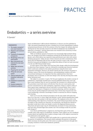
  • 1. 04p179.qxd 29/07/2004 17:27 Page 179 http://dentalbooks-drbassam.blogspot.com/ PRACTICE IN BRIEF ● An overview of the new 12-part BDJ series on Endodontics. Endodontics — a series overview P. Carrotte1 Stock and Nehammer's BDJ textbook Endodontics in Practice was first published in ENDODONTICS 1985, and almost immediately became a standard text for both undergraduate students 1. The modern concept of and general practitioners. In the first sentence of the first chapter the authors observed root canal treatment ‘during the last three decades research in the field of endodontics has modified the 2. Diagnosis and treatment approach to treatment’, and that observation was retained in the extensively revised planning second edition, published in 1990. 3. Treatment of endodontic With an inordinate amount of research of an increasingly high standard taking emergencies place, the changes in the field of endodontics during the last decade have been even 4. Morphology of the root greater, and a third edition was required to keep practitioners up to date with current canal system thinking and practice. Sadly, because of their research and other commitments, Chris Stock and Carl Nehammer did not have the time to devote to such a task, and I am 5. Basic instruments and therefore honoured and delighted to have edited this edition of their text and converted materials for root canal it to the new BDJ Clinical Guide format. treatment In some aspects of the subject there has been little change, whilst the developments 6. Rubber dam and access in others have been immense. I may be criticised for retaining some historical material activities which could seem outdated to the modern practitioner using the latest canal 7. Preparing the root canal preparation techniques. However, few dental schools have the resources necessary to 8. Filling the root canal introduce many of the recent developments, and undergraduate students still learn system conventional techniques. It is important that they understand how these have 9. Calcium hydroxide, root developed, and it is essential, as with most things in life, that they develop basic skills resorption, endo-perio before advanced ones! lesions The subject is covered in 12 parts. The first part emphasises the modern concept of 10. Endodontic treatment for endodontics, surprisingly founded upon research published almost forty years ago. children Root canal treatment must be seen as essentially the treatment of a disease process. 11. Surgical endodontics The procedures must both remove all infection from the root canal system, and 12. Endodontic problems prevent contamination by other pathogenic organisms. Failure to achieve either of these aspects may compromise success and lead to eventual failure. Parts 2 and 3 consider the importance of diagnosis in treatment planning, and how emergency events may be quickly diagnosed and treated. Part 4 shows how research into root canal morphology continues, knowledge of which is essential for effective shaping and cleaning. Parts 5 to 9 cover the technical procedures, but the wise reader will realise that there is no ‘best way' to clean, shape and obturate a tooth. Various manufacturers make claims that their own product is the latest and best. The emphasis in the series is the understanding of the objectives of treatment. The actual technical procedures must be secondary to this. Dentists are clinicians, not technicians, and should use whichever procedure works best in their own hands to resolve the diagnosed disease process. ‘Step-back and apical stops’ may be old-fashioned, one brand of rotary instruments may have been around longer than another, but if the technique works for you then 1*Clinical Lecturer, Department of Adult why change it? Dental Care, Glasgow Dental Hospital and As in Stock and Nehammer's original text, the final three parts of the series School, 378 Sauchiehall Street, Glasgow consider some wider aspects of endodontic treatment. These subjects are not considered G2 3JZ in great detail, but hopefully direct readers to deeper study of the subjects concerned in *Correspondence to: Peter Carrotte Email: p.carrotte@dental.gla.ac.uk texts dedicated to that specific aspect. However, it is hoped that readers will consider this is a practical series written for Refereed Paper the practice of endodontics. The research which underpins this practice is discussed doi:10.1038/sj.bdj.4811582 where necessary, but the prime aim of the series is to guide practitioners through their © British Dental Journal 2004; 197: 179 everyday treatment of teeth with endodontic problems. BRITISH DENTAL JOURNAL VOLUME 197 NO. 4 AUGUST 28 2004 179
  • 2. 04p181-183.qxd 26/07/2004 15:09 Page 181 PRACTICE 1 http://dentalbooks-drbassam.blogspot.com/ IN BRIEF ● Root canal treatment is normally prescribed to treat an infection, and as with all surgical procedures an aseptic technique is essential throughout. ● As research has shown that success is only achieved when all microorganisms are removed from the entire root canal system, the anatomy of this system must be understood for each tooth. ● Modern endodontic practice is concerned not with the old cliché of cleaning, shaping and filling, but with shaping first, to open the canals wide, so that cleaning can be effectively carried out prior to three-dimensional filling. VERIFIABLE CPD PAPER Endodontics: Part 1 The modern concept of root canal treatment P. Carrotte1 NOW AVAILABLE AS A BDJ BOOK Root canal treatment has changed considerably since the hollow tube theory was first postulated in 1930. Research continues into the elaborate anatomy of root canal systems, and also into the microbial causes of endodontically related diseases. Only by understanding these aspects in detail can the practitioner quickly and effectively shape the main root canals to facilitate thorough cleaning of the entire system, and easy and effective filling. ENDODONTICS 1. The modern concept of In 1965 Kakehashi, Stanley and Fitzgerald1 reduce the level of microbial contamination as root canal treatment showed conclusively that pulpal and endodontic far as is practical, and to entomb any remaining problems are primarily related to microbial con- microorganisms with an effective three-dimen- 2. Diagnosis and treatment tamination of the root canal system. Since that sional seal. planning time endodontology has increasingly focussed The prime aim when preparing the root canal 3. Treatment of endodontic on the ways and means of eliminating micro- has long been stated as cleaning and shaping. emergencies organisms from the entire root canal system. One of the prime aims of this text will be to 4. Morphology of the root The majority of patients who require root encourage the practitioner to see this in canal system canal treatment will have been diagnosed as suf- reverse, ie shaping and cleaning. Modern 5. Basic instruments and fering from the disease of periradicular peri- instruments and techniques will be described materials for root canal odontitis. The treatment of this disease must which rapidly open and shape the main root treatment address the microbial contamination of the 6. Rubber dam and access entire root canal system. It must also be carried Fig. 1 The root cavities out under aseptic conditions in order to prevent canal system of 7. Preparing the root canal further microbial ingress, in particular from saliva. this lower molar has been stained 8. Filling the root canal The use of a rubber dam very much reflects the and the tooth system use of a surgical drape in other invasive medical totally decalcified, 9. Calcium hydroxide, root procedures. Such a biological approach will be showing the emphasized throughout this text. The temptation complex nature of resorption, endo-perio the root canal lesions to regard root canal treatment as a purely system. (Courtesy 10. Endodontic treatment for mechanical procedure, producing excellent of Professor R T children post-operative radiographs but with little regard Walker.) to diagnosis and prognosis, must be resisted in 11. Surgical endodontics today’s practice. 12. Endodontic problems Research into the morphology of the pulp has shown the wide variety of shapes, and the occur- 1*Clinical Lecturer, Department of Adult rence of two or even three canals in a single Dental Care, Glasgow Dental Hospital and root.2 There is a high incidence of fins which run School, 378 Sauchiehall Street, Glasgow longitudinally within the wall of the canal and a G2 3JZ *Correspondence to: Peter Carrotte network of communications between canals Email: p.carrotte@dental.gla.ac.uk lying within the same root (Fig. 1). The many nooks and crannies within the root canal system Refereed Paper make it impossible for any known technique, doi:10.1038/sj.bdj.4811565 © British Dental Journal 2004; 197: either chemical or mechanical, to render it total- 181–183 ly sterile. The objective of treatment must be to BRITISH DENTAL JOURNAL VOLUME 197 NO. 4 AUGUST 28 2004 181
  • 3. 04p181-183.qxd 26/07/2004 15:11 Page 182 PRACTICE a b Fig. 2 (a) The pre-operative radiograph of tooth LR6 (46) shows a large the patient finally returned to continue treatment shows evidence of bony radiolucent area associated with the root apex and the furcation area. Root repair with a return to a normal periodontal ligament space around the canal treatment was commenced. (b) A radiograph 6 months later when apex and in the furcation. canals, thus permitting the effective access of of root canals, but the experimental group were antimicrobial irrigants to the entire root canal not obturated. They ensured that an effective, system, including lateral canals, fins, anasta- well-sealed, coronal restoration was placed. moses and other canal aberrations. It is imper- They found that healing occurred in every case. ative that these instruments are not seen as Figure 2 shows a lower molar with a large peri- providing a route to quick and speedy root radicular lesion. The root canal system was canal treatment. To achieve success the time shaped and cleaned, and an intervisit dressing of saved by the rapid opening of the canal system calcium hydroxide placed. The patient did not must be spent in thorough and effective return for further treatment for 6 months, when a antimicrobial irrigation. radiograph revealed that complete healing had Research has also shown that when an infect- taken place. ed root canal is accessed, the number of different Of course, this does not mean that obturation is species of microorganisms is small, rarely above unimportant. It is essential for the reasons single figures.3 Treatment will become far more described earlier. It does prove, however, the old difficult and extended, and success may well be cliché that it is what is removed from the canal compromised, if this flora is altered by the that is important, not what is put in. Similarly, ingress of saliva. Isolation of the tooth under Ray and Trope6 found that root-treated teeth with treatment is essential not only for medicolegal a poor obturation on radiograph but a good coro- reasons to protect the airway, but, far more nal restoration had a better prognosis than teeth importantly, to prevent further contamination of with a good obturation but a poor restoration. the root canal system and to permit the use of The majority of root canal sealers are soluble strong intracanal medicaments. and their only function is to fill the minute Other areas of research have had the signifi- spaces between the wall of the root canal and the cant effect of changing the approach to root filling material. Their importance, judged endodontic treatment. The hollow tube theory by the number of products advertised in the den- put forward by Rickert and Dixon in 19314 tal press, has been over-emphasized. Despite postulated that tissue fluids entering the root much research, gutta-percha remains the root canal stagnated and formed toxic breakdown filling of choice, although it is recognized that a products which then passed out into the peri- biologically inert, insoluble and injectable paste apical tissues. This theory, that dead spaces may be better suited for obturation of the root within the body must be obturated, originally canal. Most of the new root canal filling tech- formed the basis for filling root canals. How- niques are concerned with methods of heating ever, a variety of different studies have gutta-percha, making it softer and easier to demonstrated that, on the contrary, hollow adapt to the irregular shape of the canal wall. It tubes are tolerated by the body. As a result must be emphasized, however, that, whatever there are currently two indications for filling a the obturation system used, if the root canal sys- root canal, once the canal system has been tem has not been adequately cleaned healing shaped and cleaned. Firstly, to prevent the may not occur (Fig. 3). entry of microorganisms to the root canal sys- Finally, lesions of endodontic origin which tem from either the oral cavity, should the appear radiographically as areas of radiolucency coronal restoration leak or fail, or via the around the apices or lateral aspects of the roots bloodstream (anachoresis). Secondly, to pre- of teeth are, in the majority of cases, sterile.7,8 vent the ingress of tissue fluid which would The lesions are the result of toxins produced by provide a culture medium for any bacteria microorganisms lying within the root canal sys- remaining within the tooth following treatment. tem. This finding suggests that the removal of A report by Klevant and Eggink5 is particular- microorganisms from the root canal followed by ly relevant. They shaped and cleaned a number root filling is the first treatment of choice, and 182 BRITISH DENTAL JOURNAL VOLUME 197 NO. 4 AUGUST 28 2004
  • 4. 04p181-183.qxd 26/07/2004 17:50 Page 183 PRACTICE Fig. 3 A radiograph of tooth LL7 (37) showing a root canal treatment carried out 12 months previously, with what appears to be an effective obturation yet no evidence of healing of the periradicular lesion. that periradicular surgery, including an apicec- 1. Kakehashi S, Stanley H R, Fitzgerald R. The effects of surgical exposures of dental pulps in germfree and tomy with a retrograde filling, can only be sec- conventional laboratory rats. J South California Dent ond best.9 Apicectomy with a retrograde filling Assoc 1966; 334: 449–451. at the apex is carried out in the hope of merely 2. Burns R C, Herbranson E J. Tooth Morphology and incarcerating microorganisms within the tooth, Cavity Preparation, Chapter 7 in Cohen S and Burns R C, Pathways of the Pulp, St Louis 2002: Mosby. but does not take into account the fact that 3. Molven S, Olsen I, Kerekes K. Scanning electron approximately 50% of teeth have at least one microscopy of bacteria in endodontically treated teeth. lateral canal. The long-term success rate of III In vivo study. J Endod 1991; 7: 226–229. apicectomy must inevitably be lower than 4. Rickert U G, Dixon C M. The controlling of root surgery. In Transactions of the Eighth International orthograde root treatment. Dental Congress. Section 111a p15. Paris, 1931. In summary, the principles of treatment of the 5. Klevant F J, Eggink C O. The effect of canal preparation disease of periapical periodontitis are as follows. on periapical disease. Int Endod J 1983; 16: 68–75. Shape: Produce a gradual smooth taper in the 6. Ray H A, Trope M. Periapical status of endodontically treated teeth in relation to the technical quality of the root canal with its widest part coronally root filling and the coronal restoration. Int Endod J and the narrowest part at the apical 1995; 28: 12–18. constriction, which, as discussed in 7. Grossman L I. Bacteriologic status of periapical tissue Part 4, is normally about 1 mm short of in 150 cases of infected pulpless teeth. J Endod (Special Issue) 1982; 8: 513–515. the apex. 8. Siqueira J F, Lopes H P. Bacteria on the apical root Clean: Use antimicrobial agents to remove surfaces of untreated teeth with periradicular lesions: microorganisms and pulpal debris from a scanning electron microscopy study. Int Endod J 2001; 34: 216–220. the entire root canal system. 9. Pitt Ford T R. Surgical treatment of apical Fill: Obturate the canal system with an inert, periodontitis. Chapter12 in Ørstavik D and Pitt Ford insoluble filling material. T R, Essential Endodontology. Oxford 1998: Blackwell. One Hundred Years Ago British Dental Journal, July 1904 Letter to the Editor Fluorine in Food Sir, No doubt after our Annual General Meeting you will be dealing with fluorine. It is my conviction that fluorine should be given to children in the natural foodstuffs such as unrefined cereals, sea foods and cod liver oil, etc. Yesterday I saw an 11-month baby chewing and enjoying raw carrot. A short time ago I saw a girl of 16 brought up on Hovis toast from the age of 12 months with excellent teeth. The fluorine should be ingested at weaning time and onwards. Adding fluorine to drinking water may do a lit- tle good but it is very doubtful if it is of permanent value. I preach the building up of the atomic structure. C.N. Peacock Br Dent J 1904 BRITISH DENTAL JOURNAL VOLUME 197 NO. 4 AUGUST 28 2004 183
  • 5. 05p231-238.qxd 12/08/2004 11:51 Page 231 PRACTICE 2 http://dentalbooks-drbassam.blogspot.com/ IN BRIEF ● An accurate diagnosis of the patient’s condition is essential before an appropriate treatment plan can be formulated for that individual. ● A logical approach to clinical examination should be adopted. ● A high quality long-cone parallel radiograph is mandatory before commencing root canal treatment, and should be carefully examined to obtain all possible information. ● Root canal treatment may not be the most appropriate therapy, and treatment plans should take into account not only the expected prognosis but also the patient’s dental condition, expectations and wishes. VERIFIABLE CPD PAPER Endodontics: Part 2 Diagnosis and treatment planning P. Carrotte1 NOW AVAILABLE AS A BDJ BOOK As with all dental treatment, a detailed treatment plan can only be drawn up when a correct and accurate diagnosis has been made. It is essential that a full medical, dental and demographic history be obtained, together with a thorough extra-oral and intra-oral examination. This part considers the classification of diseases of the dental pulp, together with various diagnostic aids to help in determining which condition is present, and the appropriate therapy. ENDODONTICS The importance of correct diagnosis and treat- practitioner should be consulted before any 1. The modern concept of ment planning must not be underestimated. endodontic treatment is commenced. This also root canal treatment There are many causes of facial pain and the dif- applies if the patient is on medication, such as 2. Diagnosis and treatment ferential diagnosis can be both difficult and corticosteroids or an anticoagulant. An example planning demanding. All the relevant information must be of the particulars required on a patient’s folder is 3. Treatment of endodontic collected; this includes a case history and the illustrated in Table 1. emergencies results of both a clinical examination and diag- Antibiotic cover has been recommended for nostic tests. The practitioner should be fully certain medical conditions, depending upon the 4. Morphology of the root canal system conversant with the prognosis for different complexity of the procedure and the degree of endodontic clinical situations, discussed in bacteraemia expected, but the type of antibiotic 5. Basic instruments and Part 12. Only at this stage can the cause of the and the dosage are under continual review and materials for root canal problem be determined, a diagnosis made, the dental practitioners should be aware of current treatment appropriate treatments discussed with the patient opinion. The latest available edition of the Dental 6. Rubber dam and access and informed or valid consent obtained. Practitioners’ Formulary1 should be consulted for cavities the current recommended antibiotic regime. 7. Preparing the root canal CASE HISTORY However, when treating patients who may be 8. Filling the root canal The purpose of a case history is to discover considered predisposed to endocarditis, it may be system whether the patient has any general or local con- advisable to liaise with the patient’s cardiac 9. Calcium hydroxide, root dition that might alter the normal course of resorption, endo-perio treatment. As with all courses of treatment, a Table 1 A simple check list for a medical history lesions comprehensive demographic, medical and previ- (Scully and Cawson2) 10. Endodontic treatment for ous dental history should be recorded. In addi- Anaemia children tion, a description of the patient’s symptoms in Bleeding disorders 11. Surgical endodontics his or her own words and a history of relevant Cardiorespiratory disorders 12. Endodontic problems dental treatment should be noted. Drug treatment and allergies 1*Clinical Lecturer, Department of Adult Medical history Endocrine disease Dental Care, Glasgow Dental Hospital and There are no medical conditions which specifi- Fits and faints School, 378 Sauchiehall Street, Glasgow Gastrointestinal disorders G2 3JZ cally contra-indicate endodontic treatment, but *Correspondence to: Peter Carrotte there are several which require special care. The Hospital admissions and attendances Email: p.carrotte@dental.gla.ac.uk most relevant conditions are allergies, bleeding Infections Refereed Paper tendencies, cardiac disease, immune defects or Jaundice or liver disease doi:10.1038/sj.bdj.4811612 patients taking drugs acting on the endocrine or Kidney disease © British Dental Journal 2004; 197: CNS system. If there is any doubt about the state Likelihood of pregnancy or pregnancy itself 231–238 of health of a patient, his/her general medical BRITISH DENTAL JOURNAL VOLUME 197 NO. 5 SEPTEMBER 11 2004 231
  • 6. 05p231-238.qxd 12/08/2004 11:54 Page 232 PRACTICE Fig. 1 A facial sinus It is usually possible to decide, as a result of associated with a questioning the patient, whether the pain is of severe periapical abscess on the upper pulpal, periapical or periodontal origin, or if it is canine. non-dental in origin. As it is not possible to diagnose the histological state of the pulp from the clinical signs and symptoms, the terms acute and chronic pulpitis are not appropriate. In cases of pulpitis, the decision the operator must make is whether the pulpal inflammation is reversible, in which case it may be treated conservatively, or irreversible, in which case either the pulp or the tooth must be removed, depending upon the patient’s wishes. If symptoms arise spontaneously, without stimulus, or continue for more than a few sec- onds after a stimulus is withdrawn, the pulp may be deemed to be irreversibly damaged. Applica- tions of sedative dressings may relieve the pain, specialist or general medical practitioner. Not all but the pulp will continue to die until root canal patients with cardiac lesions require antibiotic treatment becomes necessary. This may then prophylaxis, and such regimes are not generally prove more difficult if either the root canals supported by the literature.2 However, if it is have become infected or if sclerosis of the root agreed that the patient is at risk, they would nor- canal system has occurred. The correct diagno- mally be prescribed the appropriate prophylactic sis, once made, must be adhered to with the antibiotic regime, and should be advised to appropriate treatment. report even a minor febrile illness which occurs In early pulpitis the patient often cannot up to 2 months following root canal treatment. localise the pain to a particular tooth or jaw Prior to endodontic surgery, it is useful to pre- because the pulp does not contain any proprio- scribe aqueous chlorhexidine (2%) mouthwash. ceptive nerve endings. As the disease advances and the periapical region becomes involved, the Patient’s complaints tooth will become tender and the proprioceptive Listening carefully to the patient’s description nerve endings in the periodontal ligament are of his/her symptoms can provide invaluable stimulated. information. It is quicker and more efficient to ask patients specific, but not leading, ques- CLINICAL EXAMINATION tions about their pain. Examples of the type of A clinical examination of the patient is carried questions which may be asked are given out after the case history has been completed. below. The temptation to start treatment on a tooth 1. How long have you had the pain? without examining the remaining dentition must 2. Do you know which tooth it is? be resisted. Problems must not be dealt with in 3. What initiates the pain? isolation and any treatment plan should take the 4. How would you describe the pain? entire mouth and the patient’s general medical Sharp or dull condition and attitude into consideration. Throbbing Mild or severe Extra-oral examination Localised or radiating The patient’s face and neck are examined and 5. How long does the pain last? any swelling, tender areas, lymphadenopathy, or 6. Does it hurt most during the day or night? extra-oral sinuses noted, as shown in Figure 1. 7. Does anything relieve the pain? Intra-oral examination An assessment of the patient’s general dental Fig. 2 An assessment state is made, noting in particular the following should be made of the patient’s general aspects (Fig. 2). dental condition. • Standard of oral hygiene. • Amount and quality of restorative work. • Prevalence of caries. • Missing and unopposed teeth. • General periodontal condition. • Presence of soft or hard swellings. • Presence of any sinus tracts. • Discoloured teeth. • Tooth wear and facets. Diagnostic tests Most of the diagnostic tests used to assess the state of the pulp and periapical tissues are 232 BRITISH DENTAL JOURNAL VOLUME 197 NO. 5 SEPTEMBER 11 2004
  • 7. 05p231-238.qxd 12/08/2004 12:17 Page 7 PRACTICE Fig. 3 The anatomical detail a b obtained from a radiograph taken by the long-cone paralleling technique (a) is far clearer and more accurate than when the bisecting angle technique (b) is used. relatively crude and unreliable. No single test, Radiography however positive the result, is sufficient to make a In all endodontic cases, a good intra-oral paral- firm diagnosis of reversible or irreversible pulpi- lel radiograph of the root and periapical region tis. There is a general rule that before drilling into is mandatory. Radiography is the most reliable a pulp chamber there should be two independent of all the diagnostic tests and provides the most positive diagnostic tests. An example would be a valuable information. However, it must be tooth not responding to the electric pulp tester emphasised that a poor quality radiograph not and tender to percussion. only fails to yield diagnostic information, but also, and more seriously, causes unnecessary Palpation radiation of the patient. The use of film holders, The tissues overlying the apices of any suspect recommended by the National Radiographic teeth are palpated to locate tender areas. The site Guidelines3 and illustrated in Part 4, has two dis- and size of any soft or hard swellings are noted tinct advantages. Firstly a true image of the and examined for fluctuation and crepitus. tooth, its length and anatomical features, is obtained (Fig. 3), and, secondly, subsequent Percussion films taken with the same holder can be more Gentle tapping with a finger both laterally and accurately compared, particularly at subsequent vertically on a tooth is sufficient to elicit any ten- review when assessing the degree of healing of a derness. It is not necessary to strike the tooth with periradicular lesion. a mirror handle, as this invites a false- A radiograph may be the first indication of the positive reaction from the patient. presence of pathology (Fig. 4). A disadvantage of the use of radiography in diagnosis, however, Mobility can be that the early stages of pulpitis are not The mobility of a tooth is tested by placing a fin- normally evident on the radiograph. ger on either side of the crown and pushing with If a sinus is present and patent, a small-sized one finger while assessing any movement with (about #40) gutta-percha point should be the other. Mobility may be graded as: inserted and threaded, by rolling gently 1 — slight (normal) between the fingers, as far along the sinus tract 2 — moderate as possible. If a radiograph is taken with the 3 — extensive movement in a lateral or gutta-percha point in place, it will lead to an mesiodistal direction combined with a area of bone loss showing the cause of the vertical displacement in the alveolus. problem (Fig. 5). Fig. 4 A radiograph taken as part of a periodontal assessment also reveals a previously undiagnosed and asymptomatic periradicular lesion on the palatal root of tooth UL6 (26). BRITISH DENTAL JOURNAL VOLUME 197 NO. 5 SEPTEMBER 11 2004 233
  • 8. 05p231-238.qxd 12/08/2004 11:57 Page 234 PRACTICE between them. Most pulp testers manufactured Fig. 5 A gutta-percha point has been a today are monopolar (Fig. 6). threaded into a sinus tract adjacent to a recently root-treated canine (a). The As well as the concerns expressed earlier radiograph (b) reveals the source of the about pulp testing, electric pulp testers may give infection to be the first premolar. a false-positive reading due to stimulation of nerve fibres in the periodontium. Again, posteri- or teeth may give misleading readings since a combination of vital and non-vital root canal pulps may be present. The use of gloves in the treatment of all dental patients has produced problems with electric pulp testing. A lip elec- trode attachment is available which may be used, but a far simpler method is to ask the patient to hold on to the metal handle of the pulp tester. The patient is asked to let go of the handle if they feel a sensation in the tooth being tested. The teeth to be tested are dried and isolated b with cotton wool rolls. A conducting medium should be used; the one most readily available is toothpaste. Pulp testers should not be used on patients with pacemakers because of the possi- bility of electrical interference. Teeth with full crowns present problems with pulp testing. A pulp tester is available with a special point fitting which may be placed between the crown and the gingival margin. There is little to commend the technique of cut- ting a window in the crown in order to pulp test. Thermal pulp testing This involves applying either heat or cold to a tooth, but neither test is particularly reliable and may produce either false-positive or false- negative results. Heat Pulp testing There are several different methods of applying Pulp testing is often referred to as ‘vitality’ test- heat to a tooth. The tip of a gutta-percha stick ing. In fact, a moribund pulp may still give a may be heated in a flame and applied to a tooth. positive reaction to one of the following tests Take great note that hot gutta-percha may stick as the nervous tissue may still function in fast to enamel, and it is essential to coat the extreme states of disease. It is also, of course, tooth with vaseline to prevent the gutta-percha possible in a multirooted tooth for one root sticking and causing unnecessary pain to the canal to be diseased, but another still capable patient. Another method is to ask the patient to of giving a vital response. Pulp testers should hold warm water in the mouth, which will act on only be used to assess vital or non-vital pulps; all the teeth in the arch, or to isolate individual they do not quantify disease, nor do they meas- teeth with rubber dam and apply warm water ure health and should not be used to judge the directly to the suspected tooth. This is explored degree of pulpal disease. Pulp testing gives no further under local anaesthesia. indication of the state of the vascular supply which would more accurately indicate the Cold degree of pulp vitality. The only way pulpal Three different methods may be used to apply a blood-flow may be measured is by using a cold stimulus to a tooth. The most effective is the Laser-Doppler Flow Meter, not usually avail- use of a –50°C spray, which may be applied using able in general practice! a cotton pledget (Fig. 7). Alternatively, though Doubt has been cast on the efficacy of pulp less effectively, an ethyl chloride spray may be testing the corresponding tooth on the other side used. Finally, ice sticks may be made by filling of the mid-line for comparison, and it is sug- the plastic covers from a hypodermic needle with gested that only the suspect teeth are tested. water and placing in the freezing compartment of a refrigerator. When required for use one cover Electronic is warmed and removed to provide the ice stick. The electric pulp tester is an instrument which However, false readings may be obtained if the uses gradations of electric current to excite a ice melts and flows onto the adjacent tissues. Fig. 6 A modern electric pulp tester response from the nervous tissue within the pulp. combined with an endodontic apex Both alternating and direct current pulp testers Local anaesthetic locator. are available, although there is little difference In cases where the patient cannot locate the pain 234 BRITISH DENTAL JOURNAL VOLUME 197 NO. 5 SEPTEMBER 11 2004
  • 9. 05p231-238.qxd 12/08/2004 11:58 Page 235 PRACTICE and routine thermal tests have been negative, a of the perforation, followed by the provision of reaction may be obtained by asking the patient to new posts and cores, and crowns. sip hot water from a cup. The patient is instructed However, success in this case may depend to hold the water first against the mandibular upon the correct planning of treatment. For teeth on one side and then by tilting the head, to example, what provisional restorations will be include the maxillary teeth. If a reaction occurs, used during the root canal treatment, and during an intraligamental injection may be given to the following re-evaluation period. Temporary anaesthetise the suspect tooth and hot water is post-crowns have been shown to be very poor at then again applied to the area; if there is no reac- resisting microleakage.4 The provision of a tem- tion, the pulpitic tooth has been identified. It porary over denture, enabling the total sealing should be borne in mind that a better term for of the access cavities, would seem an appropri- intraligamental is intra-osseous, as the local ate alternative, but if this has not been properly anaesthetic will pass into the medullary spaces planned for, problems may arise and successful round the tooth and may possibly also affect the treatment may be compromised. proximal teeth. INDICATIONS FOR ROOT CANAL TREATMENT Wooden stick All teeth with pulpal or periapical pathology are Fig. 7 A more effective source of If a patient complains of pain on chewing and candidates for root canal treatment. There are also cold stimulus for sensibility testing. there is no evidence of periapical inflammation, situations where elective root canal treatment is an incomplete fracture of the tooth may be sus- the treatment of choice. pected. Biting on a wood stick in these cases can elicit pain, usually on release of biting pressure. Post space A vital tooth may have insufficient tooth sub- Fibre-optic light stance to retain a jacket crown so the tooth may A powerful light can be used for transilluminat- have to be root-treated and restored with a post- ing teeth to show interproximal caries, fracture, retained crown (Fig. 9). opacity or discoloration. To carry out the test, the dental light should be turned off and the Overdenture fibre-optic light placed against the tooth at the Decoronated teeth retained in the arch to pre- gingival margin with the beam directed through serve alveolar bone and provide support or the tooth. If the crown of the tooth is fractured, removable prostheses must be root-treated. the light will pass through the tooth until it strikes the stain lying in the fracture line; the Teeth with doubtful pulps tooth beyond the fracture will appear darker. Root treatment should be considered for any tooth with doubtful vitality if it requires an exten- Cutting a test cavity sive restoration, particularly if it is to be a bridge When other tests have given an indeterminate abutment. Such elective root canal treatment has result, a test cavity may be cut in a tooth which a good prognosis as the root canals are easy to is believed to be pulpless. In the author’s opin- access and are not infected. If the indications are ion, this test can be unreliable as the patient may give a positive response although the pulp is necrotic. This is because nerve tissues can con- tinue to conduct impulses for some time in the absence of a blood supply. TREATMENT PLANNING Having taken the case history and carried out the relevant diagnostic tests, the patient’s treatment is then planned. The type of endodontic treatment chosen must take into account the patient’s med- ical condition and general dental state. The indi- cations and contra-indications for root canal treatment are given below and the problems of re- root treatment discussed. The treatment of frac- tured instruments, perforations and perio-endo lesions are discussed in subsequent chapters. It should be emphasised here that there is a considerable difference between a treatment plan and planning treatment. Figure 8 shows a radiograph of a patient with a severe endodontic problem. A diagnosis of failed root canal treat- ments, periapical periodontitis (both apically and also associated with a perforation of one root), and failed post crowns could be made. A Fig. 8 This complicated case exhibits a number of treatment plan for this patient may be different endodontic problems, and requires careful orthograde re-root canal treatment, with repair treatment planning if success is to be achieved. BRITISH DENTAL JOURNAL VOLUME 197 NO. 5 SEPTEMBER 11 2004 235
  • 10. 05p231-238.qxd 12/08/2004 11:59 Page 236 PRACTICE Fig. 9 Tooth UL1 (21) requires a sure. In some cases these teeth should be elec- crown, but there is insufficient tively root-treated. coronal tissue remaining. One possible treatment plan would be elective endodontic treatment Periodontal disease followed by the provision of a post- In multirooted teeth there may be deep pocket- retained core build-up and crown. ing associated with one root or the furcation. The possibility of elective devitalisation follow- ing the resection of a root should be considered (see Part 9). Pulpal sclerosis following trauma Review periapical radiographs should be taken of teeth which have been subject to trauma. If progressive narrowing of the pulp space is seen due to secondary dentine, elective root canal treatment may be considered while the coronal portion of the root canal is still patent. This may occasionally apply after a pulpotomy has been carried out. However, Andreasen6 refers to a range of studies that show a maxi- mum of 16% of sclerosed teeth subsequently cause problems, and the decision over root canal treatment must be arrived at after full ignored and the treatment deferred until the pulp consultation with the patient. If the sclerosing becomes painful or even necrotic, access through tooth is showing the classic associated discol- the crown or bridge will be more restricted, and oration the patient may elect for treatment, but treatment will be significantly more difficult, with otherwise the tooth may better be left alone a lower prognosis.5 (Fig. 10). Risk of exposure CONTRA-INDICATIONS TO ROOT CANAL Preparing teeth for crowning in order to align TREATMENT them in the dental arch can risk traumatic expo- The medical conditions which require special precautions prior to root canal treatment have already been listed. There are, however, other a conditions both general and local, which may contra-indicate root canal treatment. General Inadequate access A patient with restricted opening or a small mouth may not allow sufficient access for root canal treatment. A rough guide is that it must be possible to place two fingers between the mandibular and maxillary incisor teeth so that there is good visual access to the areas to be treated. An assessment for posterior endodon- tic surgery may be made by retracting the Fig. 10 A 23-year-old female patient cheek with a finger. If the operation site can b suffered trauma to tooth UL1 (21) when be seen directly with ease, then the access is aged 16, and is complaining about the yellow discoloration of the tooth (a). A sufficient. radiograph (b) reveals that the pulp space has sclerosed. Poor oral hygiene As a general rule root canal treatment should not be carried out unless the patient is able to maintain his/her mouth in a healthy state, or can be taught and motivated to do so. Excep- tions may be patients who are medically or physically compromised, but any treatment afforded should always be in the best long-term interests of the patient. Patient’s general medical condition The patient’s physical or mental condition due to, for example, a chronic debilitating disease or old age, may preclude endodontic treatment. Similar- ly, the patient at high risk to infective endocardi- 236 BRITISH DENTAL JOURNAL VOLUME 197 NO. 5 SEPTEMBER 11 2004
  • 11. 05p231-238.qxd 12/08/2004 12:00 Page 237 PRACTICE Fig. 11 Tooth UL1 (21) was so extensively decayed Fig. 12 The vertical root fracture can be clearly seen in subgingivally that restoration would have proved this extracted tooth which had been fitted with a post impossible even if endodontic treatment had been carried crown. out. tis, for example one who has had a previous pathological fracture of the tooth. Internal resorp- attack, may not be considered suitable for com- tion ceases immediately the pulp is removed and, plex endodontic therapy. provided the tooth is sufficiently strong, it may be retained. Most forms of external resorption will Patient’s attitude continue (see Part 9) unless the defect can be Unless the patient is sufficiently well motivated, repaired and made supragingival, or arrested with a simpler form of treatment is advised. calcium hydroxide therapy. Local Bizarre anatomy Tooth not restorable Exceptionally curved roots (Fig. 13), dilacer- It must be possible, following root canal treat- ated teeth, and congenital palatal grooves ment, to restore the tooth to health and function may all present considerable difficulties if (Fig. 11). The finishing line of the restoration must root canal treatment is attempted. In addition, be supracrestal and preferably supragingival. any unusual anatomical features related to the An assessment of possible restorative problems roots of the teeth should be noted as these should always be made before root canal treat- may affect prognosis. ment is prescribed. Re-root treatment Insufficient periodontal support One problem which confronts the general Provided the tooth is functional and the dental practitioner is to decide whether an attachment apparatus healthy, or can be made inadequate root treatment requires replace- so, root canal treatment may be carried out. ment (Fig. 14). The questions the operator should consider are given below. Non-strategic tooth Fig. 13 The tooth UR4 (14) has such a Extraction should be considered rather than bizarre root canal anatomy that root canal treatment for unopposed and non- endodontic treatment would probably functional teeth. be impossible. Root fractures Incomplete fractures of the root have a poor prog- nosis if the fracture line communicates with the oral cavity as it becomes infected. For this reason, vertical fractures will often require extraction of the tooth while horizontal root fractures have a more favourable prognosis (Fig. 12). Internal or external resorption Both types of resorption may eventually lead to BRITISH DENTAL JOURNAL VOLUME 197 NO. 5 SEPTEMBER 11 2004 237
  • 12. 05p231-238.qxd 12/08/2004 12:01 Page 238 PRACTICE Fig. 14 Tooth UL4 (24) has previously been root treated (and obturated with silver points) but is symptomless. However, the tooth now requires a full crown restoration. A decision must be made as to whether the tooth should be re-treated before fitting the advanced restoration. 1 Is there any evidence that the old root filling root canal filling of a tooth with a periradicu- has failed? lar lesion falls to about 65%. • Symptoms from the tooth. The final decision by the operator on the treat- • Radiolucent area is still present or has ment plan for a patient should be governed by the increased in size. level of his/her own skill and knowledge. General • Presence of sinus tract. dental practitioners cannot become experts in all 2 Does the crown of the tooth need restoring? fields of dentistry and should learn to be aware of their own limitations. The treatment plan pro- 3 Is there any obvious fault with the present posed should be one which the operator is confi- root filling which could lead to failure? dent he/she can carry out to a high standard. Practitioners should be particularly aware of the prognosis of root canal re-treatments. 1. Dental Practitioners’ Formulary 2000/2002. British Dental Association. BMA Books, London As a rule of thumb, taking the average of the 2. Scully C, Cawson R A. Medical problems in dentistry. surveys reported in the endodontic literature Oxford: Butterworth-Heinemann, p74, 1998. (see Part 12) suggests a prognosis of 90–95% 3. National Radiographic Protection Board. Guidance for an initial root canal treatment of a tooth Notes for Dental Practitioners on the safe use of x-ray equipment. 2001. Department of Health, with no radiographic evidence of a periradicu- London, UK. lar lesion. When such a lesion is present prog- 4. Fox K, Gutteridge D L. An in vitro study of coronal nosis will fall to around 80–85%, and the microleakage in root-canal-treated teeth restored by the post and core technique. Int Endod J 1997; 30: longer the lesion has been present the more 361–368. established will be the infection, treatment (ie 5. Ørstavik D. Time-course and risk analysis of the removal of that infection from the entire root development and healing of chronic apical canal system) will be more difficult and the periodontitis in man. Int Endod J 1996; 29: 150–155. 6. Andreasen J O, Andreasen F M. Chapter 9 in Textbook prognosis significantly lower. The average and colour atlas of traumatic injuries to the teeth. 3rd reported prognosis for re-treatment of a failed Ed, Denmark, Munksgard 1994. Fifty years ago today British Dental Journal, January 1954 Subscriptions 1954 Members are reminded that the Annual Subscription for the Association was due on January 1, 1954. If not already paid this should be sent at the earliest possible moment. The rates of subscrip- tion for 1954 are as follows: £ s. d. Ordinary members 6 6 0 Service members 4 14 6 Retired members 2 12 6 Overseas members 3 13 6 Members within three years from qualifying 3 13 6 Affiliated members 2 12 6 BRITISH DENTAL JOURNAL VOLUME 197 NO. 5 SEPTEMBER 11 2004 238
  • 13. 06p299-305.qxd 24/08/2004 11:24 Page 299 http://dentalbooks-drbassam.blogspot.com/ PRACTICE 3 IN BRIEF ● Before any dental treatment is provided it is essential that the patient’s symptoms have been correctly diagnosed. ● Conditions causing dental pain on first presentation may include pulpitis (reversible or irreversible), periapical periodontitis, dental abscess, as well as cracked tooth syndrome and other oro-facial pain disorders. ● Conditions arising during treatment may include high restorations, (probably the most common), root or crown fractures, problems with root canal instrumentation and infection. ● Following treatment pain may be due to any of the above, or failure of the root canal treatment. However, patients should always be cautioned to expect a certain amount of post-treatment discomfort. VERIFIABLE CPD PAPER Endodontics: Part 3 Treatment of endodontic emergencies NOW AVAILABLE P. Carrotte1 AS A BDJ BOOK The swift and correct diagnosis of emergency problems is essential when providing treatment, especially in a busy dental practice. A diagnosis must be made and appropriate treatment provided in usually just a few minutes. The sequence considered here encompasses problems presenting before, during and after dental treatment. Various diagnostic aids are considered, and some unusual presenting conditions discussed. ENDODONTICS The aim of emergency endodontic treatment is considered together with the medical history. 1. The modern concept of to relieve pain and control any inflammation or The following points are particularly relevant root canal treatment infection that may be present. Although insuffi- and are covered more fully in Part 2. 2. Diagnosis and treatment cient time may prevent ideal treatment from 1. Where is the pain? planning being carried out, the procedures followed 2. When was the pain first noticed? 3. Treatment of endodontic should not prejudice any final treatment plan. It 3. Description of the pain. emergencies has been reported that nearly 90% of patients 4. Under what circumstances does the pain 4. Morphology of the root seeking emergency dental treatment have symp- occur? canal system toms of pulpal or periapical disease.1,2 5. Does anything relieve it? 5. Basic instruments and Patients who present as endodontic emergen- 6. Any associated tenderness or swelling. materials for root canal cies can be divided into three main groups. 7. Previous dental history: treatment Before treatment: a) recent treatment; 6. Rubber dam and access 1. Pulpal pain b) periodontal treatment; cavities a) Reversible pulpitis c) any history of trauma to the teeth. 7. Preparing the root canal b) Irreversible pulpitis 8. Filling the root canal 2. Acute periapical abscess Particular note should be made of any disor- system 3. Cracked tooth syndrome ders which may affect the differential diagnosis of dental pain, such as myofascial pain dysfunc- 9. Calcium hydroxide, root Patients under treatment: tion syndrome (MPD), neurological disorders resorption, endo-perio 1. Recent restorative treatment such as trigeminal neuralgia, vascular pain lesions 2. Periodontal treatment syndromes and maxillary sinus disorders. 10. Endodontic treatment for children 3. Exposure of the pulp 4. Fracture of the root or crown Diagnostic aids 11. Surgical endodontics 5. Pain as a result of instrumentation • Periapical radiographs taken with a parallel- 12. Endodontic problems a) acute apical periodontitis ing technique. b) Phoenix abscess • Electric pulp tester for testing pulpal 1*Clinical Lecturer, Department of Adult responses. Dental Care, Glasgow Dental Hospital and Post-endodontic treatment: • Ice sticks, hot gutta-percha, cold spray and School, 378 Sauchiehall Street, Glasgow 1. High restoration hot water for testing thermal responses.3 G2 3JZ *Correspondence to: Peter Carrotte 2. Overfilling • Periodontal probe. Email: p.carrotte@dental.gla.ac.uk 3. Root filling 4. Root fracture Pulpal pain Refereed Paper The histological state of the pulp cannot be doi:10.1038/sj.bdj.4811641 © British Dental Journal 2004; 197: BEFORE TREATMENT assessed clinically.4,5 Nevertheless, the signs and 299–305 Details of the patient’s complaint should be symptoms associated with progressive pulpal BRITISH DENTAL JOURNAL VOLUME 197 NO. 6 SEPTEMBER 25 2004 299