Anatomical consideration for local anesthesia - sensory innervation of the face
•
233 recomendaciones•39,894 vistas

This document discusses the anatomical considerations for local anesthesia, focusing on the trigeminal nerve, its branches (maxillary, mandibular), and associated structures like the sphenopalatine ganglion. It provides detailed descriptions and diagrams of the nerves that innervate the face and oral cavity, including their branches and sensory/motor functions. Key points covered include the branches and distributions of the maxillary and mandibular nerves, as well as the innervation of the maxillary teeth from structures like the posterior superior alveolar nerve.

Recomendados
Recomendados
Más contenido relacionado
La actualidad más candente
La actualidad más candente (20)
Similar a Anatomical consideration for local anesthesia - sensory innervation of the face
Similar a Anatomical consideration for local anesthesia - sensory innervation of the face (20)
Más de Umm Al-Qura University Faculty of Dentistry
Más de Umm Al-Qura University Faculty of Dentistry (20)
Último
Último (20)
Anatomical consideration for local anesthesia - sensory innervation of the face
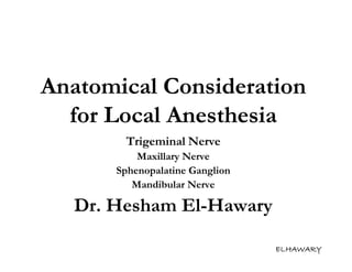
- 1. Anatomical Consideration for Local Anesthesia Trigeminal Nerve Maxillary Nerve Sphenopalatine Ganglion Mandibular Nerve Dr. Hesham El-Hawary ELHAWARY
- 2. Trigeminal nerve Largest of the cranial nerves It is a mixed nerve, which is made up of Large sensory part Smaller motor part ELHAWARY
- 3. Trigeminal nerve Cont. The sensory root arises from sensory cells of the trigeminal ganglion which fills the trigeminal impression in the floor of the middle cranial fossa The motor root arises from motor cells in the medulla oblongata Its name derives from the fact that it has three major branches: Ophthalmic nerve (V1) Maxillary nerve (V2) Mandibular nerve (V3) ELHAWARY
- 4. Trigeminal nerve Cont. The ophthalmic and maxillary nerves are purely sensory The mandibular nerve has both sensory and motor functions ELHAWARY
- 5. ELHAWARY
- 6. The maxillary nerve ELHAWARY
- 7. The Maxillary Nerve It is the 2nd division of the trigeminal nerve It arises from the middle of the gasserian ganglion It is intermediate in size between the ophthalmic and Mandibular divisions It is purely sensory in function ELHAWARY
- 8. The Maxillary Nerve Cont. It leaves the middle cranial fossa through foramen rotandum Before it leaves the middle cranial fossa it gives off the middle meningeal nerve to supply the dura mater ELHAWARY
- 9. Branches of Maxillary nerve 1. Middle meningeal nerve 2. Twiges to the sphenopalatine ganglion 3. Posterior superior alveolar nerve 4. Zygomatic nerve 5. Infra-Orbital Nerve 1. Middle superior alveolar nerve 2. Anterior superior alveolar nerve 3. Terminal branches 1. Inferior palpebral nerve 2. External nasal nerve 3. Superior labial nerve ELHAWARY
- 10. The Posterior Superior Alveolar Neve ELHAWARY
- 11. Posterior superior alveolar nerve It arises from the maxillary nerve just before it enter the infraorbital groove Generally , posterior superior alveolar nerve is two in number One branch descends on the post. Surface of the maxilla & gives off several twigs to m.m. of the cheek Other branch enters small foramen on the post. Surface of the maxilla to run in the post. Sup. Alveolar canal located in the posterior & lateral walls of the max. sinus ELHAWARY
- 12. Posterior superior alveolar nerve Cont. It communicate freely with the middle & anterior superior alveolar nerves to form a superior dental plexus from which small twigs enter the apical foramina of the maxillary teeth & other twigs supply the overlying buccal mucoperiosteum ELHAWARY
- 13. Posterior superior alveolar nerve Cont. Specifically posterior superior alveolar nerve supplies The Pulp The Investing structures The buccal mucoperiosteum of Upper Molars except MB root of 1st molar(6) ELHAWARY
- 14. The Zygomatic Nerve ELHAWARY
- 15. The Zygomatic Nerve Arises from the maxillary nerve as it enters the orbital cavity Divides into two branches: Zygomatico-Tempopral Zygomatico-Facial ELHAWARY
- 16. Infra-Orbital Nerve ELHAWARY
- 17. The Infra-Orbital Nerve Infra- The maxillary nerve enters the inferior orbital fissure to pass into the orbital cavity It turns slightly laterally in the infra-orbital groove and continues forward in the orbital canal In the middle of the course in the canal it gives off the middle superior alveolar nerve Just before the exit of the maxillary nerve from the infra-orbital foramen and still in the infra-orbital canal it gives off the anterior superior alveolar nerve As it leaves the infra-orbital foramen it gives off the terminal branches ELHAWARY
- 18. Middle superior alveolar nerve Approximately in the middle of the infraorbital canal it gives off the middle superior alveolar nerve It runs downward and forwards in a canal in the lateral wall of the maxillary sinus to form part of the superior dental plexus It Supplies Pulp Investing structures buccal mucoperiosteum of Premolars & MB root of 1st molar(6) ELHAWARY
- 19. Anterior superior alveolar nerve Just before the exit of the maxillary nerve from the infraorbital foramen, and while still in infraorbital canal It gives off the anterior superior alveolar nerve which runs in a canal in the anterior wall of the maxillary sinus to form part of the superior dental plexus It supplies Pulp Investing structures labial mucoperiosteum of anterior teeth(1,2,3). ELHAWARY
- 20. Terminal branches of the maxillary nerve As the maxillary nerve leaves the infraorbital foramen, it gives off its three terminal branches, which are: Inferior palpebral nerve External nasal nerve Superior labial nerve ELHAWARY
- 21. The Sphenopalatine Ganglion ELHAWARY
- 22. Sphenopalatine ganglion Nature: parasympathetic ganglion Site: pterygopalatine fossa hanges from the maxillary nerve Roots: Sensory root: twigs from the maxillary nerve to the sphenopalatine ganglion Sympathetic root: deep petrosal nerve Parasympathetic root: great superficial petrosal nerve of the 7th cranial nerve (Vidian nerve of facial nerve) ELHAWARY
- 23. Sphenopalatine ganglion Cont. Branches: Orbital nerve Nasal nerve Palatine nerve Pharyngeal nerve ELHAWARY
- 24. Palatine Branches Palatine Branches Divides into: Greater (anterior) palatine nerve Lesser (posterior) palatine nerve ELHAWARY
- 25. Palatine Branches Cont. The greater (anterior) palatine nerve Emerges from greater (anterior) palatine foramen in the horizontal part of the palatine bone medial to the upper third molar Supplies palatal mucoperiosteum of maxillary molars & premolars ELHAWARY
- 26. Palatine Branches Cont. The lesser ( posterior ) palatine nerve Emerges from Lesser (Posterior) palatine foramen in the horizontal part of the palatine bone medial to the upper third molar supplies uvula, soft palate & posterior part of the hard palate ELHAWARY
- 27. Nasal Branches Nasal Branches Divides into: Long sphenopalatine nerve (nasopalatine nerve) Short sphenopalatine nerve ELHAWARY
- 28. Nasal Branches Cont. The Long sphenopalatine nerve (nasopalatine nerve) Emerges from the incisive foramen in the anterior part of the median plane of the palate supplies palatal mucoperiosteum of maxillary anterior teeth ELHAWARY
- 29. Nasal Branches Cont. At the palatal side of the canine region, there is anastomosis between the nasopalatine nerve & greater palatine nerve The origin of these two nerves (nasopalatine nerve& greater palatine nerve ) comes from the sensory root of the sphenopalatine gangalion (twigs from the maxillary nerve) ELHAWARY
- 30. Nerve supply to the maxillary teeth From the buccal aspect: Posterior superior alveolar nerve It supplies the pulp, investing structures (periodontium, periosteum and the alveolar bone) and the buccal mucoperiosteum of the maxillary third, second and the first molars except the mesiobuccal root of the first molar Middle superior alveolar nerve It supplies the pulp, investing structure and the buccal mucoperiosteum of the mesiobuccal root of the first molar, and the maxillary premolars Anterior superior alveolar nerve It supplies the pulp, investing structures and the labial mucoperiosteum of the anterior teeth ELHAWARY
- 31. Nerve supply to the maxillary teeth Cont. From the palatal aspect: The greater (anterior) palatine nerve It supplies the palatal mucoperiosteum opposite to the maxillary molars, premolars and canine The nasopalatine nerve It supplies the palatal mucoperiosteum opposite to the anterior region (including the canine) ELHAWARY
- 32. Nerve supply of Maxillary teeth Pulp , Investing structures & Labial (buccal) mucoperiosteum Anterior teeth Anterior superior alveolar nerve (1,2,3) Premolars (4,5) & MB root of 1st Middle superior alveolar nerve molar(6) Molars except MB root of 1st Posterior superior alveolar nerve molar(6) ELHAWARY
- 33. Nerve supply of Maxillary teeth Cont. Palatal mucoperiosteum Anterior Nasopalatine nerve teeth (1,2,3) Premolars (4,5) Greater ( Anterior) palatine nerve & Molars (678) ELHAWARY
- 34. The accessory innervation of the teeth The upper anterior teeth cross innervation may take place i.e. branch from the anterior superior nerve of the other side for the pulp, investing structures and labial mucoperiostieum The palatal mucoperiostieum opposite to the uppar canine take innervation mainly from the nasopalatine nerve but sometimes it is supplied by interlacing fibers between the nasopalatine and the greater palatine nerve. ELHAWARY
- 35. The Mandibular Nerve ELHAWARY
- 36. It is the 3rd division of the trigeminal nerve It is the largest branch of the trigeminal ganglion It has undivided trunk & then divides into anterior & posterior division The anterior division is smaller than the posterior division & mainly motor while the posterior division is mainly sensory ( mixed nerve) ELHAWARY
- 37. It leaves the middle cranial fossa through foramen Ovale After it leaves the middle cranial fossa it gives off the nervous spinosum nerve that enter the middle cranial fossa again through foramen spinosum to supply the dura mater Also it gives the nerve to medial pterygoid muscle which passes through the otic ganglion to supply the tensor tempani & palatini muscles Both divisions pass medial to the lateral pterygoid muscle and then branch ELHAWARY
- 38. Branches of Mandibular Nerve 1. Main Trunk 2. Nervous Spimosum 3. Nerve to medial pterygoid 4. Anterior Division 1. N. to Temporalis msc. 2. N. to Massetter msc. 3. N. to Lateral Pterygoid msc. 4. Long Buccal N. 5. Posterior Division 1. Auriculo-temporal N. 2. Lingual Nerve 3. Inferior Alveolar N. ELHAWARY
- 39. Sensory braches of Mandibular nerve 1. Nervous spinosum nerve (trunk) 2. Long buccal nerve ( Ant. division) 3. Auriculotemporal nerve 4. Lingual nerve ( post. division) 5. Inferior alveolar nerve Incisive nerve Mental nerve ELHAWARY
- 40. Motor braches of Mandibular nerve 1. Nerve to medial pterygoid muscle 2. Nerve to tensor palatini muscle 3. Nerve to tensor tympani muscle 4. Nerve to lateral pterygoid muscle 5. Nerve to masseter muscle 6. Nerve to temporalis muscle 7. Nerve to mylohyoid muscle 8. Nerve to anterior belly of digastric muscle ELHAWARY
- 41. The Auriculo-Temporal nerve ELHAWARY
- 42. It arises by a medial & a lateral root that unite below the foramen spinosum The united nerve passes deep to the lateral pterygoid muscle It gives off : Parotid branch Articular branch Auricular branch Temporal branch Terminal branch ELHAWARY
- 43. The Long Buccal Nerve ELHAWARY
- 44. Long Buccal Nerve Purely sensory nerve Passes between two heads of the lateral pterygoid muscles It emerges beneath the anterior border of the masseter muscle It crosses the anterior border of the ramus at the level of the occlusal plane of the lower 7 & 8 ELHAWARY
- 45. Long Buccal Nerve Cont. It sends fibers to: M.M. of the cheek Except the posterosuperior area which receive sensory fibers from posterior superior alveolar nerve Skin of the cheek M.M. of the lower buccal vestibule Buccal mucoperiosteum of the lower molars (6,7,&8) ,&8 ELHAWARY
- 46. The Lingual Nerve ELHAWARY
- 47. Purely sensory nerve Passes between the medial pterygoid muscle and the ramus of the mandible in the pterygomandibular space In the pterygomandibular space, it lies parallel to the inferior alveolar nerve, but anterior and medial (lingual) to it ELHAWARY
- 48. Then , it passes below the mandibular attachment of the superior constrictor muscle of the pharynx to reach the side of the base of the tongue a short distance below & behind the lower third molar (8) ELHAWARY
- 49. It supplies: M.M. of the ant. 2/3 of the tongue M.M. of the floor of the mouth M.M. of the lateral lingual vestibule Lingual mucoperiosteum of the all lower teeth (1, 2, 3, 4, 5, 6, 7, & 8) ELHAWARY
- 50. The Inferior Alveolar Nerve ELHAWARY
- 51. The Posterior Division 1. N. To Mylohyoid msc. 2. Inferior alveolar nerve 3. Terminal Branches 1. Mental Nerve 2. Incisive Nerve ELHAWARY
- 52. The Inferior Alveolar nerve Mixed nerve (Sensory & Motor) Passes in the pterygomandibular space Before it enters the mandibular foramen, it gives off the mylohyoid nerve, which contains sensory & motor fibers ELHAWARY
- 53. The Inferior Alveolar Nerve Cont. Mylohyoid nerve Motor Fibers supply the Mylohyoid muscle Anterior belly of digastric muscles Sensory fibers supply the Skin of the anterior & inferior surfaces of the mental protuberance ELHAWARY
- 54. The Inferior Alveolar Nerve Cont. Within the mandible , the inferior alveolar nerve descends in the inferior alveolar canal In the region of the mental foramen, it divides into two terminal branches The mental nerve The Incisive nerve ELHAWARY
- 55. The Inferior Alveolar Nerve Cont. Mental nerve leaves the mandible through the mental foramen It supply: Skin of the chin Skin of the lower lip M.M. of the lower lip ELHAWARY
- 56. The Inferior Alveolar Nerve Cont. Incisive nerve Supplies pulp & investing structures of the lower anterior teeth (1, 2,& 3) Main trunk of the inferior alveolar nerve which lies in the mandibular canal supplies Pulp & investing structures of the lower premolar (4, & 5) & molars teeth (6, 7,& 8) ELHAWARY
- 57. The Inferior Alveolar Nerve Cont. Inferior alveolar nerve gives branches to form the inferior dental plexus above the main trunk of the nerve, It supplies The investing structures of the corresponding teeth In the premolar , canine & anterior area it supply labial or buccal mucoperiosteum of the corresponding teeth ELHAWARY
- 58. Nerve supply to the mandibular teeth Inferior alveolar nerve It is a branch from the posterior division of the mandibular nerve It supplies the pulp and the investing structures of the Mandibular molars Premolars Anterior teeth (through its terminal branch; the incisive nerve) The lingual nerve It is a branch from the posterior division of the Mandibular nerve It supplies the lingual mucoperiosteum of all the lower teeth, anterior two thirds of the tongue and the floor of the mouth. The long buccal nerve It is a branch from the anterior division of the Mandibular nerve. It supplies the buccal mucoperiosteum opposite to the lower molars. ELHAWARY
- 59. Nerve supply to the mandibular teeth Pulp , Investing structures Anterior teeth Incisive nerve (1,2,3) Premolars (4,5) & Inferior alveolar nerve Molars (678) ELHAWARY
- 60. Nerve supply to the mandibular teeth Cont. Labial (buccal) mucoperiosteum Inferior Dental Plexus/mental nerve Anterior teeth Cutaneous coli nerve (1,2,3) ( branch of cervical plexus C2 & c3 ) & gives additional sensory supply for Premolars (45) premolars ( 4 , 5 ) in about 20 % of the pt. Molars (678) Long buccal nerve ELHAWARY
- 61. Nerve supply to the mandibular teeth Cont. Lingual mucoperiosteum Anterior teeth (1,2,3) Premolars Lingual nerve (4,5) & Molars (678) ELHAWARY
- 62. The accessory innervation of teeth The lower anterior teeth cross innervations may take place i.e. branch from the incisive nerve of the other side for the pulp, investing structures and the labial mucoperiostieum The lower premolar may receive additional nerve supply from the cutaneous coli nerve (branch from the cervical plexus of nerves) for their labial mucoperiostieum The lower molars may receive additional nerve supply from the nerve to mylohyoid for the pulp and investing structures ELHAWARY
- 63. THANK YOU www.elhawarydentalclinic.com ELHAWARY