
Recomendados
Más contenido relacionado
La actualidad más candente
La actualidad más candente (20)
Similar a Proteomics course 1
Similar a Proteomics course 1 (20)
Proteomics course 1
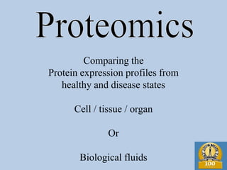
- 1. Comparing the Protein expression profiles from healthy and disease states Cell / tissue / organ Or Biological fluids
- 2. Today 1. Understanding Proteomics 2. How is it different from protein analytical chemistry? 3. Why not genomics? 4. Approaches in Proteomics 5. Tools in Proteomics 6. Applications
- 3. General scheme of proteomic analysis
- 4. Approaches in Proteomics 1. Mining 2. Expression proteomics 3. Network mapping 4. Structural proteomics
- 5. Genomics and Proteomics Term coined by Marc Wilkins and co-workers 1990 to complement genome and genomics DNA - RNA - Protein Allows study of actual functional units of the cell Limitations of genomics Levels of RNA do not correlate with protein Differing stabilities of mRNA and different translational efficiencies No information about the regulatory state of the proteins
- 6. Proteomics Can be defined as the total protein complement of a system (be that an organelle/cell/tissue/organism). This includes:- all proteins all states of proteins The genome is for the most part static (within a generation) whereas the proteome is more dynamic cell cycle, development, response to environment, etc
- 7. • Defined as “the analysis of the entire protein complement in a given cell, tissue, or organism.” • Proteomics “also assesses activities, modifications, localization, and interactions of proteins in complexes.” • Proteomes of organisms share intrinsic differences across species and growth conditions.
- 8. The virtue of proteomics • Proteome= Protein complement of the genome • Proteomics= Study of the proteome • Proteome world= Study of less abundant protein • Transcriptomic= often insufficient study to study most functional aspect of genomics
- 9. Challenges in proteomics The number of different proteins is much larger than the number of genes or mRNAs: – Alternative splicing – Post-translational modifications – Proteolytic cleavage • Changes in protein “states” (e.g. phosphorylation) occur typically much faster than changes in transcriptional regulation • DNA/RNA has four different building blocks and a (mainly) uniform hydrophilic negatively charged structure – proteins have 20 building blocks, highly variable structure, hydrophobicity, and charge • Proteins need to be kept in a functional correctly folded state for most proteomics applications
- 10. Number of proteins in one cell 1. High expression: 105-106 2. Moderate expression: 103-104 3. Low expression: 101-102
- 11. Challenges facing Proteomic Technologies • Limited/variable sample material • Sample degradation (occurs rapidly, even during sample preparation) • Vast dynamic range required • Post-translational modifications (often skew results) • Specificity among tissue, developmental and temporal stages • Perturbations by environmental (disease/drugs) conditions • Researchers have deemed sequencing the genome “easy,” as PCR was able to assist in overcoming many of these issues in genomics.
- 12. Different from Protein chemistry? Protein Chemistry Proteomics Individual protein Complex mixtrure Complete sequence Partial sequence Structure and Function Identification Structural Biology Systems Biology
- 13. Types of proteomics • Protein Expression – Quantitative study of protein expression between samples that differ by some variable • Structural Proteomics – Goal is to map out the 3-D structure of proteins and protein complexes • Functional Proteomics - Protein Networks
- 14. Applications of Proteomics • Characterization of Protein Complexes • Protein Expression Profiling • Yeast Genomics and Proteomics • Proteome Mining • Protein Arrays
- 15. Tools in Proteomics 1. Databases 2. Mass spectrometry 3. Softwares 4. Protein resolution methods
- 17. Approaches in Proteomics 1. Mining 2. Expression proteomics 3. Network mapping 4. Structural proteomics
- 18. Studying the proteome Cell culture extraction method separation (i.e. lipid solubilisation) methods (2D gels) protein protein extraction from comparison methods fragmentation and gels (spot cutting) to identify changed mass spec. proteins in gels matching spectrum to virtual spectrum for protein identification
- 19. Two dimensional gel electrophoresis Comparison of complete protein profiles or proteomes under different conditions. e.g. healthy and disease samples Mass spectrometry Identification of specific proteins. -biomarkers for diagnosis, drug targets for therapeutics
- 20. Two dimensional gel electrophoresis 1) Sample preparation Source: Biological source (tissue, cell, body fluid) Removal of the interfering components (Extra- cellular matrix, salts, oils) Isolation of the fraction of interest (whole cells or sub cellular fraction) Lysis or homogenization of the isolated fraction in homogenization buffer (With protease inhibitor or any other specific inhibitors) Precipitation of proteins from the lysate using plus one 2D cleanup kit. 2) Rehydration (overnight) of the IPG Dry strips (of desired pH range) with the 2 D lysis buffer containing proteins. Solubilization of precipitated proteins in 2D lysis buffer. 3) IEF of the rehydrated gel strips using Ettan IPGphore. 4) Equilibration of the strips in SDS equilibration buffer and second dimension on the SDS PAGE. 5) Staining of the SDS PAGE gels using coomassie Brilliant blue. 6) Scanning of the stained gels using Umax scanner. Image analysis and gels comparison using Ettan progenesis software.
- 21. Proteomics Overview Normal cell Diseased cell Cell Lysates Two dimentional gel-electrophoresis 2-D Results analysed and compared using software. Warped image The spot of interest is picked by spot-picker. In-gel digestion of the desired spot is performed Peptide-mapping by mass spectrometry.
- 22. Detection technologies in proteome analysis General detection methods. Differential display proteomics. Specific detection methods for post-translational modifications.
- 23. Different strategies for proteome purification and protein separation for identification by MS A. Separation of individual proteins by 2-DE. B. Separation of protein complexes by non-denaturing 2-DE (BN-PAGE) C. Purification of protein complexes by immuno- affinity chromatography and SDS-PAGE. D. Multidimensional chromatography. E. Organic solvent fractionation for separation of complex protein mixtures of hydrobhobic membrane proteins.
- 24. Differential display proteomics Detection techniques: Difference gel electrophoresis (DIGE). Multiplexed proteomics (MP) Isotope-coded affinity tagging (ICAT) Differential gel exposure.
- 25. Summary of protein expression profile analysis
- 26. Difference gel electrophoresis (DIGE) (Unlu, 1997, electrophoresis 18, 2071)
- 27. Differential gel exposure (Monribot-Espagne, 2002, Proteomics 2, 229-240) Coelectrophoresis on 2DE of two protein samples. In vivo labelling, using 14C and 3H -isotopes. 2DE separation. Transfer on a PVDF membrane. 3H /14C ratio by exposure to two types of imaging plates. Investigate changes in the rate of synthesis of individual proteins.
- 28. Multiplexed proteomics (MP) technology platform
- 29. • Proteomics is undoubtedly a critical component of systems biology, however: – The lack of hypothesis-driven experiments isn’t necessarily “good” for science. Discovery-based science should be guided by hypotheses, IMO. – Along these lines, as with the HGP, when it comes to literature, what do you do, just publish the whole thing? • This is another stumbling block of what to do with all of this information. – Proteomics needs its “own PCR,” or “miracle” tool, to increase the throughput. • A new technology, or instrument that combines other approaches, would be useful, esp. in structural proteomics, quantification, and sample reproduction.