
Recomendados
Más contenido relacionado
La actualidad más candente
La actualidad más candente (20)
Similar a Guide to Proximal Tibial Fractures
Similar a Guide to Proximal Tibial Fractures (20)
Más de visheshrohatgi
Último
Último (20)
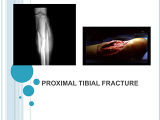
Guide to Proximal Tibial Fractures
- 2. OVERVIEW TIBIAL FRACTURE and TYPES MECHANISM OF INJURY EVALUATION MANAGEMENT TIBIAL SPINE INJURIES TIBIA TUBERCLE FRACTURE TIBIA SHAFT FRACTURE PROXIMAL TIBIAL EPIPHYSIS FRACTURE COMPLICATIONS OF TIBIAL FRACTURE
- 3. TIBIAL FRACTURE AND TYPES
- 4. TIBIAL FRACTURES Most common long bone fracture 492,000 fractures yearly Average 7.4 day hospital stay 100,000 nonunions per year Orthopaedia Trauma Association
- 5. DEMOGRAPHICS • 1% of all fractures • 8% of all fractures in the elderly • Lateral plateau involved 55-70% • Medial plateau involved 10-20% • Both involved 10-30% Sports medicine consult
- 6. TYPES :- 1. Articular (Hohl and Moore`s classification ) Plateau fracture Fracture dislocation 2. Nonarticular
- 7. PLATEAU FRACTURE Minimally displaced Local compression Split compression Total condylar depression Bicondylar fracture
- 8. FRACTURE DISLOCATIONS 1. Split fracture 2. Entire condyle fracture 3. Rim avulsion fracture ( Involves lateral condyle, associated with capsular tears and vascular injuries ) 4. Rim compression type (Unstable associated with avulsion of cruciates) 5. Four part fracture (Unstable with Collateral avulsed And neurovascular injuries)
- 10. MECHANISM OF INJURY Can have both shear and compressive forces Valgus (or varus) force split Axial load depression Combined force split/depression (80%) Higher velocity (bumper fracture) leads to an increase Comminution Displacement Soft tissue injury
- 11. EVALUATION
- 12. EVALUATION Trauma Evaluation ABCs Associated Injuries Evaluation of Limb Gentle exam for knee stability Observation of soft tissues Neurovascular evaluation Evaluate for compartmental syndrome Imaging Evaluation Browner and Jupiter, Skeletal Trauma, 3rd Ed
- 13. PHYSICAL EXAMINATION Neurologic Exam Peroneal nerve especially with valgus force R/O compartment syndrome with severe injuries Vascular Exam Palpable pulses do not exclude injury popliteal artery and medial plateau injuries beware the of the knee dislocation posing as a fracture beware of posteriorly displaced fracture fragments ABI <0.9 urgent arterial study
- 14. PHYSICAL EXAMINATION Soft Tissue Assessment Tscherne & Goetzen (Closed injury) Gustillo and Anderson (Open injury)
- 15. TSCHERNE & GOETZEN CLOSED FRACTURE CLASSIFICATION Grade 0 Minimal Soft Tissue Injury Simple Fracture Pattern Grade 1 Superficial Abrasion Mild to Moderate Fracture Pattern Grade 2 Deep Abrasion Impending Compartment Syndrome Severe Fracture Pattern Grade 3 Extensive Crush Severe Fracture Pattern +/- Vascular Injury
- 16. GUSTILLO-ANDERSON OPEN FRACTURE CLASSIFICATION Grade I - Wound < 1cm Grade II - Wound 1-10 cm Grade III - Wound > 10 cm IIIA = Adequate Soft Tissue Coverage IIIB = Requires Soft Tissue Coverage IIIC = Arterial Injury Requiring Repair
- 17. CLINICAL FEATURES Pain Swelling Deformity Haemarthrosis Decrease movement of knee Instability in valgus or varus
- 18. PHYSICAL EXAMINATION Hemarthrosis, aspirate for: Pain relief Fat evaluation Assessment of stability after local anesthetic Varus/valgus in full extension Compartment syndrome Pain with passive stretch Pain out of proportion
- 19. RADIOGRAPHIC EVALUATION AP, Lateral on Large Cassettes Obliques Internal rotation view Shows postero-lateral fragment Traction Films Defines fragments Bridging Ex-fix can provide traction CT scan with reconstruction Obtain after ex fix if using Coronal Sagittal Arteriography when necessary ? MRI – unsuspected fxs or soft tissue injury
- 20. RADIOGRAPHS Plain films Interobserver agreement = 62% Unchanged by CT scan Addition of CT scan increased agreement for surgery plan from 58 to 71% Treatment plan changed in > 25% FROM CT CT shows Comminution Depression FX lines Chan, JOT,1997
- 21. CLASSIFICATION Type I - Split Lateral Tibial Plateau Fx Type II - Split/Depression Lateral Plateau Fx Type III - Pure Depression Lateral Plateau Fx Type IV - Medial Tibial Plateau Fx Type V - Bicondylar Split Fx Type VI - Tibial Plateau Fx with Metaphyseal - Diaphyseal Separation Schatzker, Clin Orthop, 1979
- 23. SCHATZKER I: Definition:. Lateral split Etiology: Often due to valgus stress. Occurs in younger patients with stronger bones, which are resistant to depression. Often due to a bumper injury. Common associated injuries: Lateral meniscal tear. The lateral meniscus may also become entrapped in the fracture and require arthroscopy. Treatment: Typically, lateral fixation.
- 24. SCHATZKER II Most common tibial plateau fracture. Definition: Lateral split with depression. Etiology: Often due to valgus or axial stress. Occurs in older patients with osteoporosis with bones that do not resist depression. Common associated injuries: Lateral meniscus, medial meniscus, and medial collateral ligament. Treatment: Typically, lateral fixation. The depressed fragments are elevated and supported with bone graft.
- 25. SCHATZKER III: Definition: Pure lateral depression; no splitting Etiology: Older patients with osteoporosis. Often just due to a fall. Common associated injuries: If the depressed fragments are lateral and posterior, it is associated with joint instability. Treatment: If there is instability, the fractured fragments are elevated and supported with bone graft and lateral internal fixation.
- 26. SCHATZKER IV: Definition: Medial tibial plateau fracture that may be a split or split depression type fracture. Etiology: Varus stress. Often severe trauma. Common associated injuries: Associated with avulsion of the intercondylar eminence, which may indicate anterior cruciate ligament injury. Lateral collateral ligament injury. Peroneal nerve injury. Popliteal artery injury. Treatment: Medial plate and screws.
- 27. SCHATZKER V: Definition: Split medial and lateral tibial plateau (Bicondylar). Metaphysis is still in continuity with the diaphysis. Etiology: Often pure axial stress with severe trauma. Common associated injuries: Neurovascular, ACL, and meniscal injuries. Treatment: Typically, medial and lateral internal fixation.
- 28. SCHATZKER VI: Definition: Metaphyseal fracture that separates the articular surface from the diaphysis. Etiology: High-energy trauma. Common associated injuries: Neurovascular injury and compartment syndrome. Also meniscal, ACL, and collateral ligament injuries. Treatment: Typically medial and lateral internal fixation
- 29. AO/OTA CLASSIFICATION Type A - Extraarticular Type B - Partial Articular Type C - Intra-articular and Metaphyseal Orthopaedia Trauma Association
- 31. AO/OTA CLASSIFICATION Type II Type III Type I Type IV
- 33. MANAGEMENT
- 34. TIMING OF SURGERY Stable, resuscitated patient Define fracture Soft tissue envelope Swelling Ecchymosis “Damage Control Orthopaedics” Positioning of patient Other injuries
- 35. “DAMAGE CONTROL ORTHOPAEDICS” Temporary Stabilization Soft Tissue Rest Bony Stabilization Bridging ExFix Across the Knee Pins Out of Zone of ORIF in Tibia Types V & VI Primarily ORIF
- 36. ARTICULAR FRACTURES A decrease in joint surface area leads to increase stresses with subsequent deformity and post – traumatic osteoarthritis Alignment and stable fixation of the articular surface results in the formation of hyaline cartilage NOT fibrocartlilage Mitchell and Shepard, JBJS, 1980
- 37. SURGICAL INDICATIONS Open Fracture – I&D, spanning ex-fix Extensive soft tissue contusion – spanning ex-fix Closed fracture Varus/valgus instability of the knee Varus or valgus tilt of the proximal tibia Meniscal injury/previous meniscectomy Articular displacement or gapping Rasmussen P, JBJS 55, 1973 Lansinger O, JBJS 68, 1986
- 38. SURGICAL APPROACHES Straight Midline Lateral Parapatellar Medial Parapatellar Posteromedial Two approaches for bicondylar fractures AVOID Mercedes incision or midline with stripping of soft tissues, especially for bicondylars www2.aofoundation.org
- 39. Entry site is critical Reference is Lateral Tibial Spine
- 40. JUST RIGHT
- 41. TOO LOW! TOO MEDIAL! Procurvatum Valgus
- 42. APPROACH Lateral parapatellar approach Leg extended Incision starts over distal head of vastus lateralis Continues just lateral to patella and patellar tendon
- 43. APPROACH Complex Fractures Long, straight anterior midline incision Two Incisions – Preferred Bridge knee with distractor or ex-fix Apply moderate distraction to stabilize soft tissues
- 44. POSTEROMEDIAL APPROACH Useful for bicondylar Medial injury often Posteromedial Coronal
- 45. SURGICAL TECHNIQUES Ligamentotaxis Helps with Condyle Architecture Does not reduce Joint Depression Femoral Distraction Indirect Reduction Preserve Meniscus Bone Graft
- 47. TIBIAL SPINE INJURIES aka intercondyle eminence Same mechanism as ACL rupture (hyperextension, rotation, ab/adduction) In young patients ACL stronger than tibial spine – thus tibial spine injury Suspect with ACL-like presentation (positive Lachman, etc.) AND inability to weight bear
- 48. TIBIAL SPINE INJURIES Type I Incomplete avulsion with no displacement Type II incomplete avulsion with displacement Type III Completely avulsed fragment http://remergs.com/WEBPAGE%20Notes/Trauma/50%20--%20Knee%20and%20Leg.pdf
- 49. TIBIAL SPINE INJURY Type II tibial spine avulsion fracture
- 50. TIBIAL SPINE INJURIES TREATMENT Consider arthroscopy for definitive classification Type I: conservative; cast immobilization in full extension X 6 weeks Type II: arthroscopic or open reduction; internal fixation with screws and wires Arthroscopic or ORIF: significant displacement, associated ligamentous injury, Type III fractures, failure of closed reduction Displaced or rotated fractures require ORIF to restore normal ACL function http://remergs.com/WEBPAGE%20Notes/Trauma/50%20--%20Knee%20and%20Leg.pdf
- 52. 52 FRACTURE OF THE TIBIA TUBERCLE Tibia tubercle: patella tendon insertion, most anterior and distal portion of the proximal tibia epiphysis Result from jumping or a rapid quadriceps contraction against a flexed knee
- 53. 53 OSGOOD-SCHLATTER DISEASE Active children over the age of 8 Pain in tibia tubercle Superficial microfracture of the cartilage at the insertion of the tendon
- 54. 54 EVALUATION Pain, swelling, tenderness Limited displacement: may not be an effusion, capable of limited active extension Displaced frx: active knee extension is impossible, effusion,
- 56. 56 TREATMENT Non-displaced type I: long leg cast in extension for 4~6 wks Others: ORIF with screws and washers, post-op immobilization for 4~6 wks Complication: Genu recurvatum
- 58. TIBIAL SHAFT FRACTURE Most commonly fractured long bone Commonly open (1/3 of surface area just subcutaneous) Precarious blood supply Hinge joints at knee and ankle are unforgiving of post-reduction deformity emedicine.medscape.com/article/1249984-overview
- 59. TIBIAL SHAFT FRACTURES CLASSIFICATION No universally accepted classification scheme. Describe the following Location (prox, middle, distal third) Configuration (transverse, spiral, comminuted) Displacement Angulation Length rotation
- 60. TIBIAL SHAFT FRACTURE Closed distal third comminuted fracture of left tibia Nondisplaced as <5% angulation, no rotation
- 61. TIBIAL SHAFT FRACTURE ED TREATMENT Manage neurovascular status Carefully inspect any soft tissue defect for open fracture Splint in long-leg, padded, posterior splint Beware of compartment syndrome
- 62. TIBIAL SHAFT FRACTURE DEFINITIVE MANAGEMENT No consensus exists re: definitive treatment Multifactorial decision Possible management ORIF Intramedullary rod Cast immobilization Early progressive weight bearing after two weeks
- 64. 64 FRACTURE OF THE PROXIMAL TIBIAL EPIPHYSIS Uncommon, 3% of epiphyseal injuries of lower extremity Proximal tibial epiphysis: 55% of the length of the tibia 25% of the entire length of the limb 0.6 cm per year Popliteal artery: lies close to the epiphysis in the popliteal fossa, at risk of injury with displaced fracture www.ncbi.nlm.nih.gov/pubmed/1405569
- 65. 65 EVALUATION Pain, swelling, decreased ROM of knee Neurovascular assessment Vascular injury is suspected arteriogram Classification: Salter-Harris classification Associated injury: Popliteal artery and peroneal nerve injury
- 66. 66 TREATMENT Salter-Harris type I, II: close reduction + immobilization 4~6 weeks Salter-Harris type III, IV: CRIF with percutaneous pins or cannulated screws Displaced frx with vascular injury: urgent reduction and vascular status reassess Irreducible or vascular injury present: open reduction After reduction, splint in 10 ~20 degree flexion Cast when risk of compartment has decreased
- 68. COMPLICATIONS Knee stiffness Infection Compartment syndrome Malunion or nonunion Posttraumatic osteoarthritis Peroneal nerve injury Popliteal artery laceration. Avascular necrosis
- 69. 69 COMPLICATION – COMPARTMENT SYNDROME Potential devastating complication Poorly controlled pain is the earliest sign Discomfort during passive stretch of the muscle Partial fibulectomy: lead to valgus deformity in child, should not be performed
- 70. 70 COMPLICATION – DELAY UNION OR NONUNION Fail to heal within 6 months Unusual Mean healing time 10 weeks in close frx 5 months in open frx Iliac crest bone grafting: usually successful in healing the nonunion
- 71. 71 COMPLICATION – ANGULAR DEFORMITY Result from poor alignment or over-growth Valgus deformity from frx of proximal tibia metaphyseal Frequently correct spontaneously over several years Observation is recommended
- 72. 72 COMPLICATION – ROTATIONAL DEFORMITY Result from inadequate reduction Does not spontaneously correct >20 degree rotational osteotomy
- 73. COMPLICATION – PROXIMAL TIBIAL PHYSEAL CLOSURE Rare complication Cause a genu recurvatum deformity Corrected with an opening wedge osteotomy
- 74. THANK YOU