
Recomendados
Recomendados
Más contenido relacionado
La actualidad más candente
Similar a Flexor Tendon Repair with a Barbed Suture
Similar a Flexor Tendon Repair with a Barbed Suture (20)
Más de W. Thomas McClellan, MD FACS
Más de W. Thomas McClellan, MD FACS (20)
Último
Último (20)
Flexor Tendon Repair with a Barbed Suture
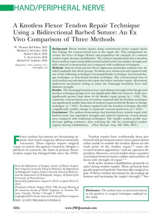
- 1. HAND/PERIPHERAL NERVE A Knotless Flexor Tendon Repair Technique Using a Bidirectional Barbed Suture: An Ex Vivo Comparison of Three Methods W. Thomas McClellan, M.D. Background: Flexor tendon repairs using conventional suture require knots Matthew J. Schessler, M.D. that enlarge the cross-sectional area at the repair site. This enlargement in- David S. Ruch, M.D. creases the force of finger flexion and jeopardizes the integrity of a nascent L. Scott Levin, M.D. tendon repair during rehabilitation. The authors hypothesized that a knotless Richard D. Goldner, M.D. flexor tendon repair using bidirectional barbed suture has similar strength and Morgantown, W.Va.; Durham, N.C.; with reduced cross-sectional area compared with traditional techniques. Philadelphia, Pa. Methods: Sixty-six fresh porcine flexor digitorum profundus tendons were di- vided randomly into three groups. Tendons were transected and repaired with one of the following techniques: two-strand Kessler technique, four-strand Sav- age technique, or four-strand knotless technique. The cross-sectional area of each tendon was calculated at the repair site before and after repair. All tendons underwent mechanical testing to assess the 2-mm-gap formation force and ultimate strength. Results: The 2-mm-gap formation force and ultimate strength of the Savage and knotless technique groups were not significantly different; however, both were significantly greater than those of the Kessler repair group (p 0.05). The repair-site cross-sectional area of tendons repaired with the knotless technique was significantly smaller than that of tendons repaired with the Kessler or Savage technique (p 0.01). Tendons repaired with the knotless technique also had a significantly smaller change in repair-site cross-sectional area (p 0.01). Conclusions: The authors demonstrate that knotless flexor tendon repair with barbed suture has equivalent strength and reduced repair-site cross-sectional area compared with traditional techniques. The smaller tendon profile may decrease gliding resistance, thus reducing the risk for postsurgical tendon rupture during rehabilitation. (Plast. Reconstr. Surg. 128: 322e, 2011.) F lexor tendon lacerations are devastating in- Tendon repairs have traditionally been per- juries that hand surgeons all-too-commonly formed with permanent suture that requires knots encounter. These injuries require surgical either inside or outside the tendon. Knots are the repair to restore the patient’s function. Despite a weak point of the tendon repair,1,2 cause de- plethora of research, the basis of primary flexor creased tendon apposition,3 and are operator de- tendon repair has changed little since the 1970s. pendent. However, several studies show a positive correlation between suture caliber and number of knots with strength of repair.4 – 6 Early active motion rehabilitation protocols re- From the Department of Surgery, Section of Plastic Surgery, quire strong tendon repairs. These protocols have West Virginia University School of Medicine, the Department greatly improved patients’ function after surgical re- of Orthopaedic Surgery, Duke University School of Medicine, pair of flexor tendon lacerations by decreasing ad- and the Department of Orthopaedic Surgery, Hospital of the hesions and increasing the repair’s strength.5,7 Sev- University of Pennsylvania. Received for publication August 6, 2010; accepted March 8, 2011. Presented at Plastic Surgery 2010: 79th Annual Meeting of the American Society of Plastic Surgeons, in Toronto, On- Disclosure: The authors have no financial interest tario, Canada, October 1 through 5, 2010. in the products or surgical techniques employed in Copyright ©2011 by the American Society of Plastic Surgeons this study. DOI: 10.1097/PRS.0b013e3182268c1f 322e www.PRSJournal.com
- 2. Volume 128, Number 4 • Knotless Flexor Tendon Repair eral studies indicate that friction created by Table 1. Brief Description of Tendon Repair Methods tendons gliding against pulleys during active finger Group flexion increases load.5,8,9 In addition, increased su- ture caliber and number of knots increases tendon Group A B C cross-sectional area, causing increased gliding No. of resistance.4,10 This increased load endangers the nas- tendons 22 22 22 Repair Modified Modified cent repair during active rehabilitation. Clearly, one technique Kessler Savage Knotless must balance repair strength with the increased ten- No. of don cross-sectional area within the tendon sheath. strands 2 4 4 Suture 3-0 Ethibond 3-0 Ethibond 0 barbed We hypothesized that a four-strand knotless tendon repair using a bidirectional barbed suture has com- parable strength and reduced repair-site cross-sec- tional area when compared with traditional flexor nonabsorbable, 3-0 braided polyester suture (Ethi- tendon repairs. bond Excel; Ethicon, Inc., Somerville, N.J.), whereas the knotless repair was performed using barbed, nonabsorbable, 0-diameter monofilament polypro- MATERIALS AND METHODS pylene suture (Quill SRS; Angiotech, Inc., Vancou- Sixty-six fresh flexor digitorum profundus ten- ver, British Columbia, Canada). The 0-diameter su- dons were obtained from adult pigs. These tendons ture was selected because of its similar strength to 3-0 have been used frequently in prior studies because polyester.17,18 A core suture purchase of 1 cm was they are similar in structure and strength to a human used on all repairs.11,14 middle finger flexor tendon.11–15 The tendons were examined for abnormalities such as synovitis and Knotless Repair Technique degeneration, and were rejected if an abnormality was present. Although a novel repair method, the knotless technique incorporates elements of both the mod- ified Kessler and the Savage methods. The knotless Cross-Sectional Area Measurements method uses four strands and a locking grasp of the Each tendon’s height and width were measured epitenon. A diagram of our technique is shown in at the repair site and 1 cm proximal and distal to the Figure 1. The barbed repair first incorporates a repair site using a Brown & Sharpe IP67 digital cal- straight pass through the tendon until the barbs iper (part no. 00530300; Hexagon Metrology, Inc., catch the opposite side of the tendon. Then, the North Kingstown, R.I.). The measurements were an- double-armed suture is passed back through the cen- alyzed to ensure all tendons were a similar size. The tral core of the tendon to the transection site. The cross-sectional area at each site was calculated using central core limbs of the barbed repair are then the formula for area of an ellipse (area ab, where passed diagonally across the tendon twice and an a equals one-half tendon height and b equals one- external bite of the epitenon is performed. The di- half tendon width). Measurements were taken at all agonal passes serve two purposes. First, they increase three sites before tendon transection and after re- the number of barbs within the tendon substance. pair to determine prerepair and postrepair cross- Second, the diagonal passes allow for multiple grasps sectional area. Blinded intrarater analysis was con- of the epitenon. Finally, the external bite locks the ducted both before transection and after repair to suture onto itself and then a mirror stitch is applied to ensure measurement consistency. the initial tendon. A running epitendinous suture was not performed so that only the core suture strength was Repair analyzed. The tendons were divided randomly into three repair groups (A, B, and C), transected, and then Biomechanical Testing repaired as described in Table 1. After repair and surface area measurement, All knots in groups A and B received six throws each tendon was secured into the clamps of a to maximize effectiveness.1,16 The modified Kessler tensiometer (model 4411; Instron Corp., Canton, technique was chosen to represent a two-strand Mass.) with a load cell of 500 N. The clamps have grasping technique and the modified Savage tech- a broad surface that prevented tendon slippage nique was chosen to represent a four-strand locking during testing. The upper clamp had a preload of technique. One surgeon (W.T.M.) performed all 1.5 N and was advanced at a rate of 20 mm/minute repairs under 3.5 loupe magnification. The mod- The preload and rate were selected because they ified Kessler and Savage repairs were performed with best simulate forces acting on an immobilized ten- 323e
- 3. Plastic and Reconstructive Surgery • October 2011 Fig. 1. Diagram of the four-strand knotless flexor tendon repair technique. don during active flexion.7,11,12,19,20 The linear dis- Table 2. Data from Mechanical Strength Testing of traction was monitored with a video camera and Tendon Repairs Including 2-mm-Gap Formation the digital caliper (previously noted) was placed Force, Ultimate Strength, and Mode of Failure near the repaired tendon. The force and tendon Tensile Strength (N) Failure Mode displacement were recorded by Instron Series 9 software. The force that produced a 2-mm gap Repair 2-mm-Gap between tendon halves at the repair site was re- Method Formation Ultimate Rupture Pullout corded as the 2-mm-gap formation force. Linear Knotless 62.84 17.30 72.39 15.16 18 4 distraction continued until the sutures were pulled Savage 59.22 15.12 69.18 8.96 22 0 Kessler 23.45 5.32 32.03 5.36 17 5 out or ruptured. In all cases, the greatest force oc- curring immediately before repair failure was re- corded as the ultimate strength. The mode of repair Table 3. Comparison of Postrepair failure was reported as pullout or rupture. An ob- Cross-Sectional Area* server blinded to the tendon repair technique per- Repair Site formed all mechanical strength testing. Cross-Sectional Area (mm2) RESULTS Repair Absolute Change Technique Size (vs. native tendon) Power analysis was performed to ensure a large Knotless 24.4 7.10 4.58 enough sample size. For 0.80 power, seven tendons Savage 31.9 13.6 3.35 were needed in each group. The 2-mm-gap forma- Kessler 32.3 14.3 5.55 tion force, ultimate strength, and cross-sectional *The knotless technique had a significantly smaller tendon size and area data were analyzed with one-way analysis of vari- change in cross-sectional area compared with the Kessler and Savage techniques. ance. A log transformation of the 2-mm-gap force and ultimate strength data were taken before anal- ysis of variance. Intergroup reliability was checked. 2-mm-gap formation force for tendons repaired by Values of p 0.05 were considered significant. the Savage method was 59.22 N. Tendons repaired The 2-mm-gap formation force results, ulti- with the modified Kessler method required 23.45 mate strength results, and mode of failure are N to form a 2-mm gap. The knotless and Savage listed in Table 2, and changes in tendon dimen- methods demonstrated a significantly greater sions are listed in Table 3. All values are reported 2-mm-gap formation force than the Kessler as mean SD. Results are depicted graphically in method (p 0.05). However, no significant dif- Figures 2 and 3. Mode of failure is reported as ference in 2-mm-gap formation force existed be- either suture rupture or suture pullout. Rupture tween the knotless technique and the Savage failure means the strands or knots broke. Pullout method. failure means that the strands tore from the ten- don without breaking. Ultimate Strength The force causing ultimate failure is reported 2-mm-Gap Formation Force in Figure 2 and Table 2. Tendons repaired by the Forces necessary to produce a 2-mm gap at the knotless method withstood 72.39 N before failing. repair site are reported in Figure 2 and Table 2. The ultimate failure force for tendons repaired by Tendons repaired by the knotless method pos- the Savage method was 69.18 N. Tendons repaired sessed a 2-mm-gap formation force of 62.84 N. The by the modified Kessler method ultimately failed 324e
- 4. Volume 128, Number 4 • Knotless Flexor Tendon Repair Fig. 2. Comparison of tensile strength among tendon repair techniques. Average 2-mm-gap formation force (yellow bars) and ultimate strength (blue bars) are shown for each tendon repair technique. Knotless and Savage repairs were signif- icantly stronger than the Kessler repairs (p 0.05). Knotless and Savage methods were not significantly different in strength. DISCUSSION McKenzie first reported using a unidirectional barbed steel wire to repair flexor tendons in 1967.21 His repair showed theoretical advantages compared with traditional repair techniques, but the use of a barbed suture repair was lost to the literature until recently.22,23 Current barbed suture technology has advanced radically. Barbed sutures are bidirectional, with barbs spiraling around the central core suture. Barbed suture can now be Fig. 3. Comparison of postsurgical cross-sectional area among created using absorbable and nonabsorbable ma- tendon repair techniques. Average cross-sectional area at the re- terials, unlike the original steel wire description. pair site is shown for each repair technique. The knotless method Using these types of materials is advantageous for had a significantly smaller cross-sectional area than the Savage or tendon repair. Kessler method. According to Strickland, the ideal character- istics of a primary flexor tendon repair include secure and easily placed sutures to allow for early at 32.03 N. The knotless and Savage methods dem- postsurgical mobilization, smooth apposition of onstrated a significantly greater ultimate strength the tendon sections, minimum gapping forces, than the Kessler method (p 0.05). However, no and minimal tendon vasculature disturbance.24,25 significant difference in ultimate strength was ob- Trail et al. listed the ideal suture characteristics as served between the knotless technique and the having high tensile strength and being inexten- Savage method. sible, absorbable, and, most importantly, easy to use.1,2 No current technique or suture meets all of Cross-Sectional Area the criteria of Strickland and Trail et al. The postrepair cross-sectional area for each Traditional flexor tendon repair techniques tendon repair technique is reported in Figure 3. rely on knots either within the tendon or posi- The change in tendon size with the knotless tech- tioned externally. When the knots lie within the nique was significantly less than with the Savage tendon, they may also impede the tendon’s ulti- and Kessler techniques (p 0.01). No significant mate healing potential because of interposition of difference was observed in cross-sectional area the knot between the tendon halves.3,26 When us- proximal or distal to the repair site among the ing a nonabsorbable suture, a permanent obstruc- techniques (Fig. 4). tion is placed between the tendon ends. With a 325e
- 5. Plastic and Reconstructive Surgery • October 2011 Fig. 4. An unrepaired tendon (below) and a tendon repaired with the knotless technique (above). knotless repair, there is no knot interposition be- The knotless method does not decrease the tween the tendon ends, so the potential exists for difficulty of flexor tendon repair or change its better healing and increased long-term strength. indications. Further study of this four-stranded Knots within the repair site do not affect the knotless technique should include cyclical loading repair’s overall strength unless they are greater and angular tensile strength. Cyclical loading stud- than 26 percent of the tendon’s cross-sectional ies would be important to determine the risk of surface area.3 Although larger suture calibers im- barbed sutures slicing through the freshly re- part greater strength to the tendon repair, they are paired tendon during early, active rehabilitation. harder to manipulate and create larger knots.6,26 Angular tensile strength studies would more Some authors have even recommended at least closely resemble forces acting on the tendon dur- five throws to create a secure knot.1,16 In addition, ing rehabilitation. increased foreign material within a repair site has Another weakness of our study is the lack of been shown to decrease wound healing by stim- clinical data and outcomes from application in ulating an inflammatory response.26 Although patients. In vivo studies should certainly be per- large knots impart greater repair strength, they formed to assess repair healing and its environ- are less than ideal because of their deleterious mental interactions in an animal model. Success- effect on healing and increased tendon profile. ful application and outcomes in patients would Bulky knots increase the tendon’s cross-sec- allow the knotless four-stranded technique to be tional area, thus increasing gliding resistance dur- incorporated into clinical practice. ing active flexion.4,10,27–29 Furthermore, this in- creased load at the repair site can cause gap CONCLUSIONS formation and failure.7,9,30 This report shows that our four-stranded knot- Furthermore, knots have been shown to im- less technique yields a repair as strong as a con- pede vasculature.29,31–33 This deprives the ten- temporary four-stranded method but with a don of vital nourishment necessary to heal, caus- smaller cross-sectional area. Because our repair is ing extrinsic neovascularization and adhesion as strong as current techniques and has a lower formation.34 tendon profile, further ex vivo and in vivo studies One way to prevent the problems caused by are warranted. Our knotless technique may im- knots is to completely eliminate them. We report prove outcomes in patients with zone II flexor a four-strand knotless flexor tendon repair tendon lacerations by allowing for more aggressive method using a bidirectional barbed suture. Our rehabilitation with reduced risk of repair failure. results show no significant difference in repair strength between the four-strand knotless tech- W. Thomas McClellan, M.D. nique and the criterion standard four-strand Sav- Morgantown Plastic Surgery Associates age technique. Most importantly, the four-strand 1085 Van Voorhis Road, Suite 350 Morgantown, W.Va. 26505 knotless method creates a lower tendon profile at wtmcclellan@yahoo.com the repair site. It is plausible that decreasing the tendon profile will decrease gliding resistance, REFERENCES thus reducing the risk of gapping and failure 1. Trail IA, Powell ES, Noble J. An evaluation of suture materials in vivo. used in tendon surgery. J Hand Surg Br. 1989;14:422–427. 326e
- 6. Volume 128, Number 4 • Knotless Flexor Tendon Repair 2. Trail IA, Powell ES, Noble J. The mechanical strength of 18. Villa MT, White LE, Alam M, Yoo SS, Walton RL. Barbed various suture techniques. J Hand Surg Br. 1992;17:89–91. sutures: A review of the literature. Plast Reconstr Surg. 2008; 3. Pruitt DL, Aoki M, Manske PR. Effect of suture knot location 121:102e–108e. on tensile strength after flexor tendon repair. J Hand Surg 19. Xie RG, Zhang S, Tang JB, Chen F. Biomechanical studies of Am. 1996;21:969–973. 3 different 6-strand flexor tendon repair techniques. J Hand 4. Aoki M, Manske PR, Pruitt DL, Larson BJ. Work of flexion Surg Am. 2002;27:621–627. after tendon repair with various suture methods: A human 20. Xie RG, Tang JB. Investigation of locking configurations for cadaveric study. J Hand Surg Br. 1995;20:310–313. tendon repair. J Hand Surg Am. 2005;30:461–465. 5. Barrie KA, Tomak SL, Cholewicki J, Merrell GA, Wolfe SW. 21. McKenzie AR. An experimental multiple barbed suture for Effect of suture locking and suture caliber on fatigue the long flexor tendons of the palm and fingers: Preliminary strength of flexor tendon repairs. J Hand Surg Am. 2001;26: report. J Bone Joint Surg Br. 1967;49:440–447. 340–346. 22. Parikh PM, Davison SP, Higgins JP. Barbed suture tenor- 6. Taras JS, Raphael JS, Marczyk SC, Bauerle WB. Evaluation of rhaphy: An ex vivo biomechanical analysis. Plast Reconstr Surg. suture caliber in flexor tendon repair. J Hand Surg Am. 2001; 2009;124:1551–1558. 26:1100–1104. 23. Trocchia AM, Aho HN, Sobol G. A re-exploration of the use 7. Tang JB, Cao Y, Xie RG. Effects of tension direction on of barbed sutures in flexor tendon repairs. Orthopedics 2009; strength of tendon repair. J Hand Surg Am. 2001;26:1105– 32:731. 1110. 24. Strickland JW. Flexor tendon injuries: I. Foundations of treat- 8. Coert JH, Uchiyama S, Amadio PC, Berglund LJ, An KN. ment. J Am Acad Orthop Surg. 1995;3:44–54. Flexor tendon-pulley interaction after tendon repair: A bio- 25. Strickland JW. Development of flexor tendon surgery: Twenty- mechanical study. J Hand Surg Br. 1995;20:573–577. five years of progress. J Hand Surg Am. 2000;25:214–235. 9. Thurman RT, Trumble TE, Hanel DP, Tencer AF, Kiser PK. 26. van Rijssel EJ, Brand R, Admiraal C, Smit I, Trimbos JB. Two-, four-, and six-strand zone II flexor tendon repairs: An Tissue reaction and surgical knots: The effect of suture size, in situ biomechanical comparison using a cadaver model. knot configuration, and knot volume. Obstet Gynecol. 1989; J Hand Surg Am. 1998;23:261–265. 74:64–68. 10. Momose T, Amadio PC, Zhao C, Zobitz ME, An KN. The 27. Momose T, Amadio PC, Zhao C, Zobitz ME, Couvreur PJ, An effect of knot location, suture material, and suture size on the KN. Suture techniques with high breaking strength and low gliding resistance of flexor tendons. J Biomed Mater Res. 2000; gliding resistance: Experiments in the dog flexor digitorum 53:806–811. profundus tendon. Acta Orthop Scand. 2001;72:635–641. 11. Cao Y, Zhu B, Xie RG, Tang JB. Influence of core suture 28. Strick MJ, Filan SL, Hile M, McKenzie C, Walsh WR, Tonkin purchase length on strength of four-strand tendon repairs. J Hand Surg Am. 2006;31:107–112. MA. Adhesion formation after flexor tendon repair: A his- 12. Mishra V, Kuiper JH, Kelly CP. Influence of core suture tologic and biomechanical comparison of 2- and 4-strand material and peripheral repair technique on the strength of repairs in a chicken model. J Hand Surg Am. 2004;29:15–21. Kessler flexor tendon repair. J Hand Surg Br. 2003;28:357– 29. Zhao C, Amadio PC, Momose T, Couvreur P, Zobitz ME, An 362. KN. The effect of suture technique on adhesion formation 13. Smith AM, Evans DM. Biomechanical assessment of a new after flexor tendon repair for partial lacerations in a canine type of flexor tendon repair. J Hand Surg Br. 2001;26:217– model. J Trauma 2001;51:917–921. 219. 30. Barrie KA, Wolfe SW, Shean C, Shenbagamurthi D, Slade JF 14. Tang JB, Zhang Y, Cao Y, Xie RG. Core suture purchase III, Panjabi MM. A biomechanical comparison of multistrand affects strength of tendon repairs. J Hand Surg Am. 2005;30: flexor tendon repairs using an in situ testing model. J Hand 1262–1266. Surg Am. 2000;25:499–506. 15. Zatiti SC, Mazzer N, Barbieri CH. Mechanical strengths of 31. Datillo PP, King MW, Cassill NL, Leung JC. Medical textiles: tendon repair: An in vitro comparative study of six tech- Application of an absorbable barbed bi-directional surgical niques. J Hand Surg Br. 1998;23:228–233. suture. J Text Apparel Technol Manage. 2002;2:1–5. 16. Viinikainen A, Goransson H, Huovinen K, Kellomaki M, ¨ ¨ 32. Manske PR, Gelberman RH, Vande Berg JS, Lesker PA. In- Tormala P, Rokkanen P. Material and knot properties of ¨ ¨¨ trinsic flexor-tendon repair: A morphological study in vitro. braided polyester (Ticron) and bioabsorbable poly-L/D-lac- J Bone Joint Surg Am. 1984;66:385–396. tide (PLDLA) 96/4 sutures. J Mater Sci Mater Med. 2006;17: 33. Amadio PC, Hunter JM, Jaeger SH, Wehbe MA, Schneider 169–177. LH. The effect of vincular injury on the results of flexor 17. Leung JC, Ruff GL, Megaro MA. Barbed bi-directional med- tendon surgery in zone 2. J Hand Surg Am. 1985;10:626–632. ical sutures: Biomechanical properties and wound closure 34. Seiler JG III, Chu CR, Amiel D, Woo SL, Gelberman RH. The efficacy study. Society for Biomaterials 28th Annual Meeting Marshall R. Urist Young Investigator Award. Autogenous Transactions. No. 724. Mt. Laurel, NJ: Society for Biomateri- flexor tendon grafts: Biologic mechanisms for incorporation. als; 2002. Clin Orthop Relat Res. 1997;345:239–247. 327e