SlideShare una empresa de Scribd logo
1 de 14
Descargar para leer sin conexión
CASE OF THE WEEK
                       PROFESSOR YASSER METWALLY
CLINICAL PICTURE

CLINICAL PICTURE

A 18 years old female patient presented clinically with disturbed level of consciousness, recurrent grand mal fits, bilateral
optic neuritis and meningeal irritation signs. The patient gradually improved during hospital stay and was discharged two
weeks following admission after almost full recovery.

RADIOLOGICAL FINDINGS

RADIOLOGICAL FINDINGS  


Figure 1. Acute disseminated encephalomyelitis (ADEM). Postcontrast MRI T1 images showings scattered hypointense
rounded and oval lesions, mostly situated at the junction of deep cortical gray and subcortical white matter. The
immediate periventricular region is spared. Lesions are mostly larger than MS lesions and did not show contrast
enhancement. Some lesions are seen encroaching upon the cortical grey matter. In general lesions did not have marked
mass effect.
Figure 2. Acute disseminated encephalomyelitis (ADEM). Postcontrast MRI T1 images showings scattered hypointense
rounded and oval lesions, mostly situated at the junction of deep cortical gray and subcortical white matter. The
immediate periventricular region is spared. Lesions are mostly larger than MS lesions and did not show contrast
enhancement. Some lesions are seen encroaching upon the cortical grey matter. In general lesions did not have marked
mass effect.


                                                                                Figure 3. A case of acute
                                                                                disseminated       encephalomyelitis.
                                                                                Notice that the multifocal cortical /
                                                                                subcortical hyperintense foci are
                                                                                sparing the periventricular region,
                                                                                and this is the classic pattern in
                                                                                ADEM. The foci, although of large
                                                                                size they have mild mass effect.
Figure 4. Acute disseminated encephalomyelitis (ADEM). Notice that the multifocal cortical / subcortical lesions are
sparing the periventricular region. The ADEM lesions are hypointense on MRI T1 images and hyperintense on MRI T2
and FLAIR images. ADEM lesions, though large, exert mild mass effect.


                                                               Figure 5. Acute disseminated encephalomyelitis
                                                               (ADEM). Notice that the multifocal cortical /
                                                               subcortical lesions are sparing the periventricular
                                                               region. The ADEM lesions are hypointense on MRI T1
                                                               images and hyperintense on MRI T2 and FLAIR
                                                               images. ADEM lesions, though large, exert mild mass
                                                               effect.


DIAGNOSIS:

DIAGNOSIS: ACUTE DISSEMINATED ENCEPHALOMYELITIS (ADEM)
DISCUSSION

DISCUSSION

Acute disseminated encephalomyelitis (ADEM) presents as an acute-onset neurological dysfunction following a triggering
event such as an infection or vaccination. Patients present with polysymptomatic neurological dysfunction, and imaging
shows multifocal white matter lesions in the brain and spinal cord. Clinical evaluation, magnetic resonance imaging, and
cerebrospinal fluid study are most useful in establishing the diagnosis and ruling out important differential diagnoses.
Corticosteroids are the mainstay of treatment and the role of other modalities of treatment, such as plasma exchange and
intravenous immunoglobulin, require further study. Prognosis is generally good. The recently proposed consensus
definitions are likely to facilitate delineation of ADEM from other acquired demyelinating disorders.

Acute disseminated encephalomyelitis (ADEM) has evolved considerably since the first case description of a patient with
post-measles neurological dysfunction by Lucas in 1790. [1] Histopathologic and immunologic studies have provided
considerable insight into the mechanism of the disease, and the advent of magnetic resonance imaging (MRI) has
facilitated early diagnosis. With the recent introduction of consensus definitions, it is now established as a well-delineated
entity. ADEM is a monophasic, inflammatory demyelinating disorder of the central nervous system (CNS). Clinical
examination reveals polysymptomatic neurological involvement and imaging shows multifocal demyelinating lesions in the
brain and spinal cord. It is an immune-mediated disorder that usually follows infections or vaccinations. [2,3] Though
previously described as an illness with poor prognosis, recent studies have demonstrated improved outcomes following the
introduction of effective immunomodulation. [4,5,6,7] This article will focus on the recent trends in the diagnosis and
management of ADEM.

      Epidemiology

The incidence of ADEM has been estimated to be 0.4 per 100,000 population per year [8] and it may represent 30% of all
childhood encephalitic illnesses. [9] ADEM more commonly affects children and young adults, probably because of
exposure to frequent vaccinations and exanthematous and upper respiratory tract infections in this age-group.
[6,7,10,11,12,66] The mean age of onset in children ranges from 5 to 8 years. [6,12,13] ADEM generally does not show a
predilection for any one sex, though two recent studies have shown a male predominance among the victims. [13,14]
ADEM occurs throughout the year, but there is a significant increase in incidence during winter and spring. [6,7]

      Triggering Events

ADEM typically follows an antigenic challenge, such as infection or vaccination, which activates the immune system.
Exanthematous fevers such as measles, mumps, and rubella are the usual triggering infections. Recently, there has been a
distinct change in the trend and, in regions with extensive immunization coverage, nonspecific upper respiratory tract
infections are the most common triggering events. [11] However, ADEM following vaccine-preventable diseases is still
prevalent in Egypt. [15] The association with infection may be difficult to establish and only 70% of ADEM patients report
a triggering event. Though extensive microbiological investigations have led to the inclusion of a wide spectrum of
infections in the list of those alleged to cause ADEM [Table - 1], they may fail to identify a cause in the majority of cases.
[6,7,10]

Table 1. Infections alleged to cause ADEM

Viral           Measles, Mumps, Varicella, Rubella, Influenza, A,B Hepatitis, A,B Coxsackie, Epstein-Barr, Dengue [16]
                and HIV [17]
Bacterial       Mycoplasma pneumoniae, Borrelia burgdorferi, Mycobacterium tuberculosis, Brucella, Chlamydia,
                Legionella, Salmonella typhi, and Leptospira, Campylobacter, Streptococcus pyogenes
Vaccination     Rabies, Measles, Rubella, Smallpox, Diphtheria, Mumps, Tetanus, antitoxin, Pertussis, Japanese
                encephalitis, Polio, Hepatitis B, Influenza, and Meningococcal A, and C
Drugs           Gold, Arsenical compounds, Sulfonamides, streptomycin/PAS
Miscellaneous Allogenic bone marrow transplantation [19], Heart-lung transplantation [19] Herbal extracts [20],
              Ventriculo-atrial shunts, [21] Stings, Leprosy type I reaction

Post-immunization ADEM occurs most frequently following measles, rubella, or mumps vaccination. Many vaccines have
been implicated in the causation of ADEM [Table - 1]. In countries where neural tissue-based vaccines are still used,
antirabies immunization with either BPL (betapropionolactone inactivated) or Semple (phenol inactivated) vaccines are
important causes for ADEM. [4]

      Clinical Features

The spectrum of neurological symptoms and signs in ADEM is broad, reflecting widespread central and peripheral
nervous system (PNS) involvement. There may be a prodrome consisting of fever, headache, vomiting, and malaise.
Neurological dysfunction generally occurs within 3-6 weeks of the triggering event and may appear abruptly or may
progress over several days. [6,7] Generally, ADEM is a monophasic illness and its severity may range from mild disease,
with headache, subtle drowsiness, and irritability, to fulminant disease characterized by coma, decerebration, and
respiratory failure. The distribution of the lesions in the nervous system determines the clinical presentation, and the
commonest features are altered sensorium, pyramidal dysfunction, cerebellar ataxia, optic neuritis, and/or myelitis.
Retention of urine, urinary frequency, urgency, or incontinence may occur during the acute stage and lower urinary tract
dysfunction may persist even after disappearance of other neurological deficits. [23] Seizures are not uncommon and may
be of focal or generalized type . [24] There may be additional features such as fever, headache, and meningismus, with the
disease mimicking meningitis. Children under 3 years generally present only with encephalopathy, probably because they
have immature myelin. [2,7]

ADEM may have various atypical presentations. Behavioral disturbances may occasionally be the sole symptom. Presence
of flaccidity and areflexia in an otherwise typical case of ADEM betrays additional PNS involvement, which is most
commonly at the level of the spinal roots; [25] this picture is frequently seen in antirabies vaccination-related ADEM. [4]
Combined CNS and PNS demyelination may suggest the possibility of shared pathological epitopes. [26] There is evidence
to suggest that central (ADEM) and peripheral (acute and chronic inflammatory demyelinating polyradiculoneuropathy)
demyelinating disorders represent two ends of a spectrum and overlap of clinical features may occur. [27] Extrapyramidal
manifestations such as chorea and dystonia are rare but may be prominent in ADEM following group A streptococcal
infection. [28] Occasional patients may present with focal deficits, clinical features of raised intracranial pressure, and
imaging results suggesting a mass lesion. Such a presentation of ADEM, tumefactive demyelination , may be mistaken for
neoplasm till histopathology establishes the diagnosis. The pattern of neurological dysfunction may be influenced by the
type of triggering event. For example, post-mumps demyelination commonly presents as myelitis, post-varicella
demyelination has cerebellar ataxia as its hallmark, and rubella-associated ADEM often has an explosive onset of
symptoms, seizures and mild pyramidal dysfunction. [29] Acute hemorrhagic leukoencephalitis (AHLE) and acute
necrotizing hemorrhagic leukoencephalitis (ANHLE) of Weston Hurst represent the hyperacute, fulminant form of
postinfectious demyelination [2] [Table - 2].

Table 2. ADEM versus Acute hemorrhagic leukoencephalitis (AHLE)

Feature                  ADEM                                            AHLE
Age group                Children                                        Young adult
Triggering events        Viral exanthema, respiratory tract infection,   respiratory tract infection
                         vaccination
Onset                    Acute                                           Hyperacute, fulminant
Blood picture            Normal                                          Leukocytosis
ESR/acute phase          Normal                                          Elevated
reactant
Urine                    Normal                                          Proteinuria
CSF cellularity          Mononuclear                                     Neutrophils, red blood cells
Neuroimaging             Multifocal white matter lesions                 Large lesions, mass effect, hemorrhage, necrosis
Pathology                Periventricular demyelination, inflammation     Fibrinoid necrosis, hemorrhage, necrosis,
                                                                         demyelination
Prognosis                Good                                            Bad

      Consensus Definition for ADEM

Clinical research in ADEM has always been hampered by the lack of a uniform case definition, resulting in heterogeneous,
incomparable study populations. The recent introduction of consensus definitions for children by the International
Pediatric MS Study Group is likely to facilitate the differentiation of monophasic ADEM from other acquired
demyelinating conditions [Table - 3]. [30] According to the consensus definition patients should have an acute or subacute
onset of the first clinical event of multifocal CNS affection, characterized by polysymptomatic neurological dysfunction.
Encephalopathy is an essential clinical feature; it is defined as either behavioral changes, alteration in consciousness, or
both. MRI brain must show focal or multifocal lesions greater than 1-2 cm in size, which must be predominantly in the
white matter, though it may also be present in the grey matter such as the basal ganglia or thalamus. In addition, the spinal
cord may show confluent lesions. The clinical event must have a presumed inflammatory or demyelinating basis, without
other etiologies. It should be followed by improvement, either clinically or on MRI, though there may be residual deficits.
New or fluctuating symptoms and signs or MRI findings within three months of the initial event are considered part of the
initial acute event.

Table 3. Monophasic ADEM and other acquired demyelinating conditions

Monophasic multifocal disease
   Acute disseminated encephalomyelitis

       Acute hemorrhagic leukoencephalitis

Clinically isolated syndromes

       Transverse myelitis

       Optic neuritis

       Cerebellitis

       Brain stem dysfunction

Multiphasic and recurrent acute disseminated encephalomyelitis

       Is ADEM always a monophasic disease?

Though ADEM is classically described as a monophasic illness, a biphasic temporal profile has been documented in 0-20%
of patients. [6,7,13,14] Such patients do not proceed to develop multiple sclerosis (MS). [7] It is now possible to define
patients of ADEM who develop relapses, i.e., multiphasic or recurrent ADEM. [30] A new clinical event should have
occurred that meets the case definition of ADEM. The new event should occur three or more months after the initial
episode. The event should occur when the patient is not on steroids and at least one month after completion of therapy. In
multiphasic ADEM , clinical examination and MRI should demonstrate involvement of new areas of the CNS during the
relapse. In recurrent ADEM , clinical findings and MRI should suggest involvement of the same areas that had been
involved in the initial ADEM episode.

       Investigations

              Cerebrospinal fluid

Cerebrospinal fluid (CSF) should be analyzed to exclude the differential diagnoses of ADEM, especially the CNS
infections. CSF is abnormal in nearly three-fourth of ADEM patients and is characterized by pleocytosis, elevated proteins,
and normal sugar values. [6,7] Cellular response is usually lymphocytic and counts are moderately elevated. Intrathecal
oligoclonal immunoglobulin synthesis is rare and usually disappears after clinical recovery. [7,13]

              Electrophysiology

Generalized slowing is the most common finding in electroencephalography and is nonspecific. Patients with spinal cord,
brain stem, or optic nerve involvement may have abnormal somatosensory, brain stem auditory, or visual evoked
potentials, respectively. [2]

              Imaging

Computed tomography (CT) may demonstrate hypodense lesions in the white matter and basal ganglia. However, initial
CT imaging may be normal in 40% of cases, as lesions may appear only 5-14 days after the onset of clinical signs. [2,31,66]
MRI forms the cornerstone for diagnosis of ADEM and is more sensitive than CT in detecting lesions. Abnormalities are
best appreciated in T2-weighted and fluid-attenuated inversion recovery (FLAIR) sequences as patchy, multifocal, poorly
marginated lesions of increased signal intensity. White matter lesions are usually asymmetric and most frequently situated
subcortically, in the cerebellum, brain stem, and spinal cord. [32,33] Thalamic and basal ganglia lesions are seen in nearly
one-third of cases and may be symmetric. Corpus callosum may be affected when involvement is extensive. [3] The spinal
cord becomes swollen and may show increased signal intensities, most commonly in the thoracic region. Enhancement
after contrast administration is variable and occurs in acute lesions due to disruption of the blood-brain barrier. [32,33]
Lesions may show complete ring, incomplete ring ('open-ring sign'), nodular, gyral, or spotty patterns of enhancement.
[33,34] Tumefactive demyelination appears as a large white matter lesion with mass effect. Though clinically a monophasic
illness, new lesions may appear serially over several weeks and hence, may appear to be of varying ages on MRI. [33]
Occasionally, initial MR images may be normal and lesions may appear in images repeated later in the course of the
disease or even during the stage of clinical improvement. [35] Though lesions generally resolve with treatment,
hyperintensities may persist in MRI long after clinical recovery and is due to astrocytic hyperplasia, gliosis, or cystic
changes. [36,37] Resolution of MRI abnormalities within six months of the demyelinating episode favors the diagnosis of
ADEM. [38]
Figure 6. Acute disseminated encephalomyelitis. Notice the
                                                          contrast enhancement which is characteristic of acute lesions.
                                                          Also notice that many lesions are situated at the junction of
                                                          deep cortical gray and subcortical white matter which is
                                                          characteristic of ADEM


                                                                               Figure    7.    In   acute      disseminated
                                                                               encephalomyelitis                tumefactive
                                                                               demyelination appears as a large white
                                                                               matter lesion with mass effect.


                   Newer modalities

Quantitative proton MR spectroscopic imaging has shown low N-acetylaspartate (NAA) and high lactate levels in acute
lesions, which normalize after recovery. [39] Further studies are required to assess the role of MR imaging techniques such
as magnetization transfer and diffusion tensor imaging in detecting early or small lesions. [40] Abnormalities in 99m Tc-
HMPAO SPECT appear as areas of hypoperfusion, which are more extensive than the abnormalities seen in MRI and may
parallel the time course of the disease more accurately. [41] Notably, in patients with residual cognitive deficits, SPECT
with acetazolamide is able to detect persistent abnormalities from areas where lesions had apparently resolved on MRI.
[42,43]

      Pathogenesis

             Immunology

The lesions of ADEM are due to autoimmune-mediated inflammation of the CNS, and the absence of viral or bacterial
antigens in the CNS is nearly universal. [5,7] T-cells have been shown to play an important role, possibly through
molecular mimicry or by nonspecific activation of autoreactive T-cell clones. [3] Interleukin-6 may be associated with
proliferation of B-lymphocytes and immunoglobulin G synthesis. [44] Anti-basal ganglia antibodies have been
demonstrated in children with classical features of ADEM following streptococcal infection. [28] A complex interplay
between cytokines and adhesion molecules is responsible for the cellular events of inflammatory encephalomyelitis and
oligodendrocyte death. An association has been established between ADEM and certain class II HLA alleles, indicating
that genetic factors may play a role in immunoregulation and progression from infection/vaccination to ADEM. [29,66]

             Pathology

Histopathology studies have demonstrated perivenous cuffing with inflammatory cells, especially lymphocytes and
macrophages, and loss of myelin. Most of the changes are seen in white matter. However, grey matter also shows changes,
including chromatolysis and neuronal loss, especially in the thalami, deep cortical lamina, and hypothalamus. The
meninges may also show inflammatory reaction. [2,45]


                                                          Figure 8. Histopathology studies In ADEM have demonstrated
                                                          perivenous cuffing with inflammatory cells, especially
                                                          lymphocytes and macrophages, and loss of myelin


      Differential Diagnosis

In the absence of specific biological markers, the diagnosis of ADEM is based upon clinical and imaging features. Clinical
evaluation, neuroimaging, and blood and CSF analysis can help to distinguish ADEM from other conditions. [46] Herpes
simplex encephalitis commonly presents with abnormal behavior and focal/secondary generalized seizures, which are more
frequent and difficult to treat than in ADEM. MRI, EEG, and CSF polymerase chain reaction for Herpes simplex virus
help in confirmation of the diagnosis. Japanese encephalitis presents with acute encephalopathy. MRI may show bilateral
thalamic lesions, akin to ADEM. History, EEG findings, and CSF evidence of antibodies to the virus are useful for
differentiation. Other infections such as bacterial meningitis and brain abscess can be ruled out by relevant investigations
such as imaging and lumbar puncture. Complicated tuberculous meningitis may sometimes mimic ADEM and can be
excluded if CSF sugar is normal and cultures are sterile.

Neuromyelitis optica is characterized by optic neuritis and myelitis, with spinal lesions extending over three or more
segments, features that may also be seen in ADEM. It may be distinguished from ADEM by the relative paucity of white
matter lesions in MRI of the brain and presence of antibodies to aquaporin 4 (NMO-IgG). [47] Patients with Behcet's
disease may present with multifocal neurological signs due to brain and spinal cord involvement. Clinical and imaging
features may resemble ADEM, and the history of recurrent mucocutaneous ulcers of the mouth and genitalia will be
essential to establish the diagnosis. Antiphospholipid antibody syndrome may also mimic the clinical and MRI features of
ADEM and should be ruled out by measuring the specific antibodies. Immune-mediated disorders such as systemic lupus
erythematosus, Sjögren syndrome, and sarcoidosis may present with neurological dysfunction and multifocal white matter
changes and can be diagnosed by history and relevant blood tests. Susac's syndrome may present with subacute
encephalopathy, with MRI of the brain showing multiple white matter lesions; however, it can be differentiated from
ADEM based on additional features such as the presence of headache, visual impairment due to retinal artery branch
occlusion, sensorineural hearing loss, and specific involvement of central corpus callosum in MRI. [48,66] The diagnosis of
recurrent ADEM should be made only after excluding other lesions such as arteriovenous malformations, neoplasms, and
vasculitis.
Metabolic leukoencephalopathies such as metachromatic leukodystrophy (MLD), adrenoleukodystrophy, and MELAS
(mitochondrial myopathy, encephalopathy, lactic acidosis, and stroke-like episodes) may mimic childhood ADEM.
Leukodystrophies may have a stepladder clinical course, with the exacerbations associated with febrile illnesses. History
may reveal prior neurological dysfunction. MRI may show symmetric, confluent white matter lesions, hallmarks of
inherited rather than acquired leukoencephalopathies. Specific investigations such as serum aryl sulfatase for MLD and
very long-chain fatty acids (VLCFA) levels for adrenoleukodystrophy will confirm the diagnosis. The clinical features of
mitochondrial cytopathies such as MELAS may be similar to that of ADEM and lesions seen on MRI may be transient.
Serum and CSF lactate levels may be elevated during the acute episode and MR spectroscopic imaging may show elevated
lactate in the affected tissues.

      ADEM or multiple sclerosis?

In a patient presenting with neurological dysfunction and MRI showing multiple white matter lesions, the most important
differential diagnosis is MS. Distinguishing between ADEM and MS is a diagnostic challenge and has important
therapeutic and prognostic implications. There are several clinical, imaging, and laboratory parameters that may be useful
to distinguish between the two [Table - 4]. CSF electrophoresis has shown a significant reduction in the beta-1 globulin
fraction in patients with MS as compared to those with ADEM and this may be a potential CSF marker. [49] Features that
strongly favor ADEM include a history of preceding infection, polysymptomatic neurological dysfunction, encephalopathy,
grey matter involvement on MRI, and absence of oligoclonal bands in CSF. [6,7] Often, distinction between these two
conditions cannot be made with certainty and follow-up with serial MRI may be necessary to establish the diagnosis.
[3,36,66]

Table 4. Differences between ADEM and multiple sclerosis

Feature                                  ADEM                                         MS
Onset                                    Abrupt                                       Subacute
Triggering events                        Preceding infection or vaccination in 70     Uncommon
                                         % of cases
Age group                                More common in children                      More common in young adult
Temporal profile                         Monophasic, rarely relapsing                 Relapsing
 Clinical features
Altered sensorium                        More common                                  Rare
Seizures                                 More common                                  Rare
Neurological deficit                     Multifocal                                   Usually single deficit
Optic neuritis                           Bilateral                                    Unilateral
Myelitis                                 Complete, transverse myelitis                Partial myelopathy
Lower motor neuron signs                 More common                                  Rare
Headache                                 More common                                  Rare
Meningismus                              More common                                  Rare
Neuroimaging findings
Distribution of the lesions              Bilateral extensive lesions                  Scattered asymmetric lesions
White matter lesions                     Confluent, ill-defined periventricular and   Well-defined, with periventricular
                                         subcortical white matter lesions             preponderance
Corpus callosum lesions                  Rare                                         Common
Grey matter involvement                  Thalamic and basal ganglionic lesions are    Uncommon
                                         common
Edema and mass effect                    May be present                               Uncommon
Follow-up MRI                            No new lesions                               New lesions with dissemination in time
                                                                                      and place
 Cerebrospinal fluid
Cell count                               Mild to moderate pleocytosis                 Normal to mild pleocytosis
Protein                                  Increased                                    Normal
Oligoclonal bands                        Uncommon, transient                          Common and persistent
Mortality                                10-25                                        Uncommon

      Treatment
Public health initiatives are important for minimizing exposure to the triggering events of ADEM. These include
implementing effective vaccination programs and avoiding vaccines containing neural tissues or enveloped viruses. [2]
Early initiation of antibiotics for infections may not prevent the immune trigger that culminates in ADEM. [50] Treatment
of ADEM involves immunomodulation that aims to counter autoimmune-mediated inflammation. Though spontaneous
resolution has been described, recovery is usually incomplete and hence patients presenting in the acute stage with
significant, progressive neurological deficits should receive treatment. [51] In view of the rarity of ADEM, treatment
regimes for children and adults are based on anecdotal reports and lack validation by randomized controlled trials. High-
dose corticosteroids have become the first line of management and are associated with rapid recovery when given early in
the disease. [6,7,13,14,52] Earlier studies used ACTH, but nowadays either intravenous methylprednisolone (10-30
mg/kg/day, up to maximum of 1 gm/day) or dexamethasone (1 mg/kg/day) for 3 to 5 days are being used. [6,7,13,14] Full
recovery was reported in 50-80% of patients. Methylprednisolone treated patients have significantly better outcome with
respect to disability compared to those treated with dexamethasone. [13] In addition to immunosuppression, steroids also
have anti-inflammatory and antiedema actions, and the immediate improvement following administration of steroids is
likely to be due to reduction in cerebral edema. [53] Oral corticosteroids are continued and gradually tapered over six
weeks to minimize the risk of recurrence. [7] The role of corticosteroids in patients presenting late in the course of the
disease is questionable. [54] Vaccinations should be avoided during the first six months after recovery. [7]

Plasma exchange is another therapeutic modality, and its role in the management of demyelinating disorders that are
nonresponsive to corticosteroids has been established in one randomized control trial. [55] In most studies, 1-1.4 plasma
volumes were exchanged by continuous flow centrifugation and 70% of volume was replaced by 5% serum albumin, fresh-
frozen plasma, or hydroxyethyl starch. Patients received 2-20 exchanges, depending on the severity of illness and clinical
response. It is likely to be effective in patients of ADEM not responding to corticosteroids, especially when given early in
the course of the disease. [56,57] Intravenous immunoglobulin is yet another option and the results so far are impressive,
especially in corticosteroid nonresponders. In most studies, 0.4 gm/kg/day was given over 5 days and improvement was
noted within 2 to 3 days. [58,59,60] However, in most reported cases, patients had received prior corticosteroids and the
delayed effects of these drugs could not be ruled out as the reason for the improvement. At present, the evidence is
insufficient to design a treatment algorithm for ADEM. However, based on the available reports, plasma exchange and
intravenous immunoglobulin are likely to be second-line therapies when corticosteroids fail. The relative efficacies of the
two have not been evaluated. The choice of second-line therapy should be individualized, depending upon the type of CNS
involvement, complications, and comorbidities. For example, autonomic dysfunction and hypotension would preclude the
use of plasma exchange. Anecdotal reports suggest that intravenous immunoglobulin may be more effective in patients
with who also have PNS involvement [25] and plasma exchange in patients with tumefactive demyelination. [61]
Combination therapy with methylprednisolone and immunoglobulin has been reported to be successful in atypical cases of
ADEM. [62] Cyclophosphamide [52] and hypothermia [63] have been successfully tried in fulminant ADEM.
Hemicraniectomy has been reported to be lifesaving in patients with massive life-threatening cerebral edema refractory to
conventional medical management. [64,65]

      Outcome

ADEM is usually a monophasic illness lasting 2-4 weeks. [2] However recovery may continue for weeks or months. While
earlier studies reported mortality of 20% and significant morbidity amongst survivors, recent reports suggest excellent
outcomes, especially in children. [7,10,13,14] Visual impairment, weakness, lower urinary tract dysfunction, cognitive
impairment, and behavioral changes may persist at follow-up. [7,13,23] Children thought to have made a full recovery may
show subtle neurocognitive and behavioral changes on formal assessment, though these may be less severe than the
changes seen in children with MS. [43] Patients having progressive neurological dysfunction , altered sensorium, and
seizures have poor long-term outcomes. [24]


SUMMARY


SUMMARY

ADEM presents as an acute onset neurological dysfunction following infection or vaccination. Clinical evaluation, MRI,
and CSF study are most useful to establish the diagnosis and rule out important differential diagnoses. The recently
proposed consensus definitions are likely to facilitate delineation of ADEM from other acquired demyelinating disorders.
Corticosteroids are the most important treatment modality, while the role of other agents requires further study. Prognosis
is generally good.
   Addendum

            A new version of this PDF file (with a new case) is uploaded in my web site every week (every Saturday and remains
             available till Friday.)

            To download the current version follow the link quot;http://pdf.yassermetwally.com/case.pdfquot;.
            You can also download the current version from my web site at quot;http://yassermetwally.comquot;.
            To download the software version of the publication (crow.exe) follow the link:
             http://neurology.yassermetwally.com/crow.zip
            The case is also presented as a short case in PDF format, to download the short case follow the link:
             http://pdf.yassermetwally.com/short.pdf
            At the end of each year, all the publications are compiled on a single CD-ROM, please contact the author to know
             more details.
            Screen resolution is better set at 1024*768 pixel screen area for optimum display


REFERENCES

References

1. Lucas J. An account of uncommon symptoms succeeding the measles: With additional remarks on the infection of
measles and smallpox. London Med J 1790;11:325-31.

2. Coyle PK. Post infectious encephalomyelitis. In : Kennedy PGE, Davis LE, editors. Infectious Diseases of the Nervous
System. Butterworth-Heinemann: Oxford; 2000. p. 83-108.

3. Garg RK. Acute disseminated encephalomyelitis. Postgrad Med J 2003;79:11-7.

4. Swamy HS, Shankar SK, Chandra PS, Aroor SR, Krishna AS, Perumal VG. Neurological complications due to beta-
propiolactone (BPL)-inactivated antirabies vaccination. Clinical, electrophysiological and therapeutic aspects. J Neurol Sci
1984;63:111-28.

5. Johnson RT, Griffin DE, Hirsch RL, Wolinsky JS, Roedenbeck S, de Soriano IL, et al . Measles encephalomyelitis-
clinical and immunologic studies. N Engl J Med 1984;310:137-41.

6. Hynson JL, Kornberg AJ, Coleman LT, Shield L, Harvey AS, Kean MJ. Clinical and neuroradiologic features of acute
disseminated encephalomyelitis in children. Neurology 2001;56:1308-12.

7. Dale RC, de Sousa C, Chong WK, Cox TC, Harding B, Neville BG. Acute disseminated encephalomyelitis, multiphasic
disseminated encephalomyelitis and multiple sclerosis in children. Brain 2000;123:2407-22.

8. Leake JA, Albani S, Kao AS, Senac MO, Billman GF, Nespeca MP, et al . Acute disseminated encephalomyelitis in
childhood: Epidemiologic, clinical and laboratory features. Pediatr Infect Dis J 2004;23:756-64.

9. Kennard C, Swash M. Acute viral encephalitis: Its diagnosis and outcome. Brain 1981;104:129-48.

10. Murthy SN, Faden HS, Cohen ME, Bakshi R. Acute disseminated encephalomyelitis in children. Pediatrics
2002;110:e1-7.

11. Menge T, Hemmer B, Nessler S, Wiendl H, Neuhaus O, Hartung HP, et al . Acute disseminated encephalomyelitis: An
update. Arch Neurol 2005;62:1673-80.

12. Anlar B, Basaran C, Kose G, Guven A, Haspolat S, Yakut A, et al . Acute disseminated Encephalomyelitis in Children:
Outcome and Prognosis. Neuropediatrics 2003;34:194-9.

13. Tenembaum S, Chamoles N, Fejerman N. Acute disseminated encephalomyelitis: A long-term follow-up study of 84
pediatric patients. Neurology 2002;59:1224-31.

14. Singhi PD, Ray M, Singhi S, Khandelwal NK. Acute disseminated encephalomyelitis in North Indian children: Clinical
profile and follow-up. J Child Neurol 2006;21:851-7.

15. Murthy JM, Yangala R, Meena AK, Jaganmohan Reddy J. Acute disseminated encephalomyelitis: clinical and MRI
Study from South India. J Neurol Sci 1999;165:133-8.

16. Yamamoto Y, Takasaki T, Yamada K, Kimura M, Washizaki K, Yoshikawa K, et al . Acute disseminated
encephalomyelitis following dengue fever. J Infect Chemother 2002;8:175-7.
17. Narciso P, Galgani S, Del Grosso B, De Marco M, De Santis A, Balestra P, et al . Acute disseminated encephalomyelitis
as manifestation of primary HIV infection. Neurology 2001;57:1493-6.

18. Tomonari A, Tojo A, Adachi D, Iseki T, Ooi J, Shirafuji N, et al . Acute disseminated encephalomyelitis (ADEM) after
allogeneic bone marrow transplantation for acute myeloid leukemia. Ann Hematol 2003;82:37-40.

19. Horowitz MB, Comey C, Hirsch W, Marion D, Griffith B, Martinez J. Acute disseminated encephalomyelitis (ADEM)
or ADEM-like inflammatory changes in a heart-lung transplant recipient: A case report. Neuroradiology 1995;37:434-7.

20. Schwarz S, Knauth M, Schwad S, Walter-Sack I, Bonmann E, Storch-Hagenlocher B. Acute disseminated
encephalomyelitis after parenteral therapy with herbal extracts: A report of two cases. J Neurol Neurosurg Psychiatry
2000;69:516-8.

21. Boz C, Velioglu S, Ozmenoglu M. Acute disseminated encephalomyelitis after bee sting. Neurol Sci 2003;23:313-5.

22. Jacob J, Alexander M, Aaron S, Pulimood S, Walter N, Gnanamuthu C. Acute disseminated encephalomyelitis in
Hansen's disease. Ann Indian Acad Neurol 2006;9:166-8.

23. Sakakibara R, Hattori T, Yasuda K, Yamanishi T. Micturitional disturbance in acute disseminated encephalomyelitis
(ADEM). J Auton Nerv Syst 1996;60:200-5.

24. Panicker JN. Acute Disseminated Encephalomyelitis: Clinical Profile and Predictors of Outcome (dissertation).
Bangalore: National Institute of Mental Health and Neurosciences, NIMHANS (Deemed University); 2004.

25. Marchioni E, Ravaglia S, Piccolo G, Furione M, Zardini E, Franciotta D, et al . Postinfectious inflammatory disorders:
Subgroups based on prospective follow-up. Neurology 2005;65:1057-65.

26. Kinoshita A, Hayashi M, Miyamoto K, Oda M, Tanabe H. Inflammatory demyelinating polyradiculitis in a patient with
acute disseminated encephalomyelitis (ADEM). J Neurol Neurosurg Psychiatry 1996;60:87-90.

27. Krivickas LS, Hochberg FH, Freeman S. Chronic inflammatory demyelinating polyradiculoneuropathy with
tumefactive central demyelination. Muscle Nerve 2006;33:283-8.

28. Dale RC, Church AJ, Cardoso F, Goddard E, Cox TC, Chong WK, et al . Post streptococcal acute disseminated
encephalomyelitis with basal ganglia involvement and auto-reactive antibasal ganglia antibodies. Ann Neurol 2001;50:588-
95.

29. Idrissova ZR, Boldyreva MN, Dekonenko EP, Malishev NA, Leontyeva IY, Martinenko IN, et al . Acute disseminated
encephalomyelitis in children: Clinical features and HLA-DR linkage. Eur J Neurol 2003;10:537-46.

30. Krupp LB, Banwell B, Tenembaum S; International Pediatric MS Study Group. Consensus definitions proposed for
pediatric multiple sclerosis and related disorders. Neurology 2007;68:S7-12.

31. Lukes SA, Norman D. Computed tomography in acute disseminated encephalomyelitis. Ann Neurol 1983;13:567-72.

32. Mader I, Stock KW, Ettlin T, Probst A. Acute disseminated encephalomyelitis: MR and CT features. AJNR Am J
Neuroradiol 1996;17:104-9.

33. Caldemeyer KS, Smith RR, Harris TM, Edwards MK. MRI in acute disseminated encephalomyelitis. Neuroradiology
1994;36:216-20.

34. van der Meyden CH, de Villiers JF, Middlecote BD, Terblanche J. Gadolinium ring enhancement and mass effect in
acute disseminated encephalomyelitis. Neuroradiology 1994;36:221-3.

35. Honkaniemi J, Dastidar P, Kdhdrd V, Haapasalo H. Delayed MR imaging changes in acute disseminated
encephalomyelitis. AJNR Am J Neuroradiol 2001;22:1117-24.

36. Kesselring J, Miller DH, Robb SA, Kendall BE, Moseley IF, Kingsley D, et al . Acute disseminated encephalomyelitis.
MRI findings and the distinction from multiple sclerosis. Brain 1990;113:291-302.

37. Go T, Imai T. A residual cystic lesion in acute disseminated encephalomyelitis. Neuroradiology 2000;42:682-4.

38. Richer LR, Sinclair DB, Bhargava R. Neuroimaging features of acute disseminated encephalomyelitis in childhood.
Pediatr Neurol 2005;32:30-6.

39. Mader I, Wolff M, Nagele T, Niemann G, Grodd W, Kuker W. MRI and proton MR spectroscopy in acute
disseminated encephalomyelitis. Childs Nerv Syst 2005;21:566-72.
40. Inglese M, Salvi F, Iannucci G, Mancardi GL, Mascalchi M, Filippi M. Magnetization transfer and Diffusion tensor
MR imaging of acute disseminated encephalomyelitis. AJNR Am J Neuroradiol 2002;23:267-72.

41. Itti E, Huff K, Cornford ME, Itti L, Poruri K, Mishkin FS. Postinfectious encephalitis. A co registered SPECT and
magnetic resonance imaging study. Clin Nucl Med 2002;27:129-30.

42. Okamoto M, Ashida KI, Imaizumi M. Hypoperfusion following encephalitis: SPECT with acetazolamide. Eur J Neurol
2001;8:471-4.

43. Hahn CD, Miles BS, MacGregor DL, Blaser SI, Banwell BL, Hetherington CR. Neurocognitive outcome after acute
disseminated encephalomyelitis. Pediatr Neurol 2003;29:117-23.

44. Dale RC, Morovat A. Interleukin-6 and oligoclonal IgG synthesis in children with acute disseminated
encephalomyelitis. Neuropediatrics 2003;34:141-5.

45. Kumar A, Swamy HS, Santhosh V, Taly AB, Arunodaya GR, Shankar SK. Pathology of allergic encephalomyelitis
following Semple type antirabies vaccine from India. Neurol Infect Epidemiol 1997;2:239-48.

46. Hahn JS, Pohl D, Rensel M, Rao S; International Pediatric MS Study Group. Differential diagnosis and evaluation in
pediatric multiple sclerosis. Neurology 2007;68:S13-22.

47. Wingerchuk DM, Lennon VA, Pittock SJ, Lucchinetti CF, Weinshenker BG. Revised diagnostic criteria for
neuromyelitis optica. Neurology 2006;66:1485-9.

48. Susac JO. Susac syndrome: The triad of microangiopathy of the brain and retina with hearing loss in women.
Neurology 1994;44:591-3.

49. Chopra B, Abraham R, Abraham A. CSF beta-1 Globulin- a potential marker in differentiating multiple sclerosis and
acute disseminated encephalomyelitis: A preliminary study. Neurol India 2002;50:41-4.

50. Pellegrini M, O'Brien TJ, Hoy J, Sedal L. Mycoplasma pneumoniae infection associated with an acute brainstem
syndrome. Acta Neurol Scand 1996;93:203-6.

51. Rust RS, Dodson W, Prensky A, Chun R, DeVivo D, Dodge P, et al . Classification and outcome of acute disseminated
encephalomyelitis. Ann Neurol 1997;42:491.

52. Schwarz S, Mohr A, Knauth M, Wildemann B, Storch-Hagenlocher B. Acute disseminated encephalomyelitis: A follow-
up study of 40 adult patients. Neurology 2001;56:1313-8.

53. Barkhof F, Hommes OR, Scheltens P, Valk J. Quantitative MRI changes in gadolinium-DTPA enhancement after high
dose intravenous methyl prednisolone in multiple sclerosis. Neurology 1991;41:1219-22.

54. Straub J, Chofflon M, Delavelle J. Early high-dose intravenous methylprednisolone in acute disseminated
encephalomyelitis: A successful recovery. Neurology 1997;49:1145-7.

55. Weinshenker BG, O'Brien PC, Petterson TM, Noseworthy JH, Lucchinetti CF, Dodick DW, et al . A randomized trial
of plasma exchange in acute central nervous system inflammatory demyelinating disease. Ann Neurol 1999;46:878-86.

56. Lin CH, Jeng JS, Yip PK. Plasmapheresis in acute disseminated encephalomyelitis. J Clin Apheresis 2004;19:154-9.

57. Keegan M, Pineda AA, McClelland RL, Darby CH, Rodriguez M, Weinshenker BG. Plasma exchange for severe
attacks of CNS demyelination: Predictors of response. Neurology 2002;58:143-6.

58. Pradhan S, Gupta RP, Shashank S, Pandey N. Intravenous immunoglobulin therapy in acute disseminated
encephalomyelitis. J Neurol Sci 1999;165:56-61.

59. Sahlas DJ, Miller SP, Guerin M, Veilleux M, Francis G. Treatment of acute disseminated encephalomyelitis with
intravenous immunoglobulin. Neurology 2000;54:1370-2.

60. Kleiman M, Brunquell P. Acute disseminated encephalomyelitis: Response to intravenous immunoglobulin? J Child
Neurol 1995;10:481-3.

61. Mao-Draayer Y, Braff S, Pendlebury W, Panitch H. Treatment of steroid-unresponsive tumefactive demyelinating
disease with plasma exchange. Neurology 2002;59:1074-7.

62. Straussberg R, Schonfeld T, Weitz R, Karmazyn B, Harel L. Improvement of atypical acute disseminated
encephalomyelitis with steroids and intravenous immunoglobulins. Pediatr Neurol 2001;24:139-43.
63. Takata T, Hirakawa M, Sakurai M, Kanazawa I. Fulminant form of acute disseminated encephalomyelitis: Successful
treatment with hypothermia. J Neurol Sci 1999;165:94-7.

64. von Stuckrad-Barre S, Klippel E, Foerch C, Lang JM, du Mesnil de Rochemont R, Sitzer M. Hemicraniectomy as a
successful treatment of mass effect in acute disseminated encephalomyelitis. Neurology 2003;61:420-1.

65. Refai D, Lee MC, Goldenberg FD, Frank JI. Decompressive hemicraniectomy for acute disseminated
encephalomyelitis: Case report. Neurosurgery 2005;56:E872.

66. Metwally, MYM: Textbook of neurimaging, A CD-ROM publication, (Metwally, MYM editor) WEB-CD agency for
electronic publishing, version 9.1a January 2008

Más contenido relacionado

La actualidad más candente

Approach to peripheral neuropathy
Approach to peripheral neuropathyApproach to peripheral neuropathy
Approach to peripheral neuropathyNeurologyKota
 
Neuromyelitis optica spectrum disorder
Neuromyelitis optica spectrum disorderNeuromyelitis optica spectrum disorder
Neuromyelitis optica spectrum disorderNeurologyKota
 
Progressive multifocal leukoencephalopathy (PML)
Progressive multifocal leukoencephalopathy (PML)Progressive multifocal leukoencephalopathy (PML)
Progressive multifocal leukoencephalopathy (PML)Amr Hassan
 
Acute disseminated encephalomyelitis
Acute disseminated encephalomyelitis Acute disseminated encephalomyelitis
Acute disseminated encephalomyelitis Reyad Al_Faky
 
Acute Disseminated Encephalomyelitis
Acute Disseminated Encephalomyelitis Acute Disseminated Encephalomyelitis
Acute Disseminated Encephalomyelitis Ade Wijaya
 
Autoimmune encephalitis
Autoimmune encephalitisAutoimmune encephalitis
Autoimmune encephalitisNeurologyKota
 
Progressive Multifocal Leucoencephalopathy
Progressive Multifocal LeucoencephalopathyProgressive Multifocal Leucoencephalopathy
Progressive Multifocal LeucoencephalopathyRoopchand Ps
 
Childhood demyelinating syndromes
Childhood demyelinating syndromesChildhood demyelinating syndromes
Childhood demyelinating syndromesAmr Hassan
 
Neuromyelitis optica spectrum disorders
Neuromyelitis optica spectrum disordersNeuromyelitis optica spectrum disorders
Neuromyelitis optica spectrum disordersNeurologyKota
 
Neurocutaneous syndrome
Neurocutaneous syndromeNeurocutaneous syndrome
Neurocutaneous syndromeNeurologyKota
 
Sub acute sclerosing panencephalitis
Sub acute sclerosing panencephalitisSub acute sclerosing panencephalitis
Sub acute sclerosing panencephalitisNeurologyKota
 
Autoimmune encephalitis ppt
Autoimmune encephalitis pptAutoimmune encephalitis ppt
Autoimmune encephalitis pptSachin Adukia
 
Autoimmune Encephalitis
Autoimmune Encephalitis Autoimmune Encephalitis
Autoimmune Encephalitis Ade Wijaya
 
Diagnostic approach to acute encephalopathy
Diagnostic approach to acute encephalopathyDiagnostic approach to acute encephalopathy
Diagnostic approach to acute encephalopathyTeik Beng Khoo
 
Guillain barre syndrome
Guillain barre syndromeGuillain barre syndrome
Guillain barre syndromePraveen Nagula
 
Complex partial seizures
Complex partial seizuresComplex partial seizures
Complex partial seizuresReem Alyahya
 

La actualidad más candente (20)

Approach to peripheral neuropathy
Approach to peripheral neuropathyApproach to peripheral neuropathy
Approach to peripheral neuropathy
 
Neuromyelitis optica spectrum disorder
Neuromyelitis optica spectrum disorderNeuromyelitis optica spectrum disorder
Neuromyelitis optica spectrum disorder
 
Adem
AdemAdem
Adem
 
Progressive multifocal leukoencephalopathy (PML)
Progressive multifocal leukoencephalopathy (PML)Progressive multifocal leukoencephalopathy (PML)
Progressive multifocal leukoencephalopathy (PML)
 
Acute disseminated encephalomyelitis
Acute disseminated encephalomyelitis Acute disseminated encephalomyelitis
Acute disseminated encephalomyelitis
 
Acute Disseminated Encephalomyelitis
Acute Disseminated Encephalomyelitis Acute Disseminated Encephalomyelitis
Acute Disseminated Encephalomyelitis
 
Guillain barre syndrome
Guillain barre syndromeGuillain barre syndrome
Guillain barre syndrome
 
Approach to Ataxia
Approach to AtaxiaApproach to Ataxia
Approach to Ataxia
 
Autoimmune encephalitis
Autoimmune encephalitisAutoimmune encephalitis
Autoimmune encephalitis
 
Progressive Multifocal Leucoencephalopathy
Progressive Multifocal LeucoencephalopathyProgressive Multifocal Leucoencephalopathy
Progressive Multifocal Leucoencephalopathy
 
Childhood demyelinating syndromes
Childhood demyelinating syndromesChildhood demyelinating syndromes
Childhood demyelinating syndromes
 
Neuromyelitis optica spectrum disorders
Neuromyelitis optica spectrum disordersNeuromyelitis optica spectrum disorders
Neuromyelitis optica spectrum disorders
 
Neurocutaneous syndrome
Neurocutaneous syndromeNeurocutaneous syndrome
Neurocutaneous syndrome
 
Sub acute sclerosing panencephalitis
Sub acute sclerosing panencephalitisSub acute sclerosing panencephalitis
Sub acute sclerosing panencephalitis
 
Autoimmune encephalitis ppt
Autoimmune encephalitis pptAutoimmune encephalitis ppt
Autoimmune encephalitis ppt
 
Autoimmune Encephalitis
Autoimmune Encephalitis Autoimmune Encephalitis
Autoimmune Encephalitis
 
Diagnostic approach to acute encephalopathy
Diagnostic approach to acute encephalopathyDiagnostic approach to acute encephalopathy
Diagnostic approach to acute encephalopathy
 
Stroke in Young
Stroke in YoungStroke in Young
Stroke in Young
 
Guillain barre syndrome
Guillain barre syndromeGuillain barre syndrome
Guillain barre syndrome
 
Complex partial seizures
Complex partial seizuresComplex partial seizures
Complex partial seizures
 

Destacado

Ms vs adem BASIC DIFFERENCES AND APPROACH
Ms vs adem BASIC DIFFERENCES AND APPROACHMs vs adem BASIC DIFFERENCES AND APPROACH
Ms vs adem BASIC DIFFERENCES AND APPROACHSatyendra Raghuwanshi
 
Acute disseminated encephalomyelitis in children management
Acute disseminated encephalomyelitis in children managementAcute disseminated encephalomyelitis in children management
Acute disseminated encephalomyelitis in children managementMohamad Othman
 
Rapid Nerve Stimulation study Part 1
Rapid Nerve Stimulation study Part 1Rapid Nerve Stimulation study Part 1
Rapid Nerve Stimulation study Part 1Roopchand Ps
 
Posterior reversible encephalopathy syndrome
Posterior reversible encephalopathy syndromePosterior reversible encephalopathy syndrome
Posterior reversible encephalopathy syndromeNeurologyKota
 
Case presentatio 8 10-2012 (2)
Case presentatio 8 10-2012 (2)Case presentatio 8 10-2012 (2)
Case presentatio 8 10-2012 (2)Mohamad Othman
 
EEG in metabolic disorders
EEG in metabolic disordersEEG in metabolic disorders
EEG in metabolic disordersRoopchand Ps
 
The central nervous system ii
The central nervous system iiThe central nervous system ii
The central nervous system iiraj kumar
 
Clinical testing ulnar nerve
Clinical testing ulnar nerveClinical testing ulnar nerve
Clinical testing ulnar nerveRoopchand Ps
 
6 multiple sclerosis nero medicine
6 multiple sclerosis nero medicine 6 multiple sclerosis nero medicine
6 multiple sclerosis nero medicine eliasmawla
 
Diagnostic Imaging of Degenerative & White Matter Diseases
Diagnostic Imaging of Degenerative & White Matter DiseasesDiagnostic Imaging of Degenerative & White Matter Diseases
Diagnostic Imaging of Degenerative & White Matter DiseasesMohamed M.A. Zaitoun
 

Destacado (18)

Recent advances
Recent advancesRecent advances
Recent advances
 
Ms vs adem BASIC DIFFERENCES AND APPROACH
Ms vs adem BASIC DIFFERENCES AND APPROACHMs vs adem BASIC DIFFERENCES AND APPROACH
Ms vs adem BASIC DIFFERENCES AND APPROACH
 
Adem
AdemAdem
Adem
 
ADEM
ADEMADEM
ADEM
 
Acute disseminated encephalomyelitis in children management
Acute disseminated encephalomyelitis in children managementAcute disseminated encephalomyelitis in children management
Acute disseminated encephalomyelitis in children management
 
Adem
AdemAdem
Adem
 
Rapid Nerve Stimulation study Part 1
Rapid Nerve Stimulation study Part 1Rapid Nerve Stimulation study Part 1
Rapid Nerve Stimulation study Part 1
 
Posterior reversible encephalopathy syndrome
Posterior reversible encephalopathy syndromePosterior reversible encephalopathy syndrome
Posterior reversible encephalopathy syndrome
 
Case presentatio 8 10-2012 (2)
Case presentatio 8 10-2012 (2)Case presentatio 8 10-2012 (2)
Case presentatio 8 10-2012 (2)
 
Progressive multifocal leukoencephalopathy
Progressive multifocal leukoencephalopathyProgressive multifocal leukoencephalopathy
Progressive multifocal leukoencephalopathy
 
EEG in metabolic disorders
EEG in metabolic disordersEEG in metabolic disorders
EEG in metabolic disorders
 
The central nervous system ii
The central nervous system iiThe central nervous system ii
The central nervous system ii
 
Progressive multifocal leukoencephalopathy
Progressive multifocal leukoencephalopathyProgressive multifocal leukoencephalopathy
Progressive multifocal leukoencephalopathy
 
Clinical testing ulnar nerve
Clinical testing ulnar nerveClinical testing ulnar nerve
Clinical testing ulnar nerve
 
6 multiple sclerosis nero medicine
6 multiple sclerosis nero medicine 6 multiple sclerosis nero medicine
6 multiple sclerosis nero medicine
 
Diagnostic Imaging of Degenerative & White Matter Diseases
Diagnostic Imaging of Degenerative & White Matter DiseasesDiagnostic Imaging of Degenerative & White Matter Diseases
Diagnostic Imaging of Degenerative & White Matter Diseases
 
Encephalitis
EncephalitisEncephalitis
Encephalitis
 
Formulas kiln
Formulas kilnFormulas kiln
Formulas kiln
 

Similar a Case record... Acute disseminated encephalomyelitis

Short case...Acute disseminated encephalomyelitis
Short case...Acute disseminated encephalomyelitisShort case...Acute disseminated encephalomyelitis
Short case...Acute disseminated encephalomyelitisProfessor Yasser Metwally
 
Case record...Acute postinfectious transverse myelitis (Transverse myelitis s...
Case record...Acute postinfectious transverse myelitis (Transverse myelitis s...Case record...Acute postinfectious transverse myelitis (Transverse myelitis s...
Case record...Acute postinfectious transverse myelitis (Transverse myelitis s...Professor Yasser Metwally
 
Case record...Idiopathic postinfectious transverse myelitis
Case record...Idiopathic postinfectious transverse myelitisCase record...Idiopathic postinfectious transverse myelitis
Case record...Idiopathic postinfectious transverse myelitisProfessor Yasser Metwally
 
Radiology of demyelinating diseases
Radiology of demyelinating diseases Radiology of demyelinating diseases
Radiology of demyelinating diseases NeurologyKota
 
Neurology treatment guidelines Govt of India
Neurology treatment guidelines Govt of India Neurology treatment guidelines Govt of India
Neurology treatment guidelines Govt of India Dr Jitu Lal Meena
 
radiologyofdemyelinatingdiseasesmbs-170105191815.pptx
radiologyofdemyelinatingdiseasesmbs-170105191815.pptxradiologyofdemyelinatingdiseasesmbs-170105191815.pptx
radiologyofdemyelinatingdiseasesmbs-170105191815.pptxShubham661884
 
Cns Neuropathy Davidson07.
Cns Neuropathy  Davidson07.Cns Neuropathy  Davidson07.
Cns Neuropathy Davidson07.Shaikhani.
 
Imaging of common viral brain infection in india PPT
Imaging of common  viral brain infection in india PPTImaging of common  viral brain infection in india PPT
Imaging of common viral brain infection in india PPTNaba Kumar Barman
 
Facial nerve paralysis common causes
Facial nerve paralysis common causes Facial nerve paralysis common causes
Facial nerve paralysis common causes Amro1988
 
Mri evaluation of spine myelopathy
Mri evaluation of spine myelopathyMri evaluation of spine myelopathy
Mri evaluation of spine myelopathyDrBhishm Sevendra
 
Cns neuropathy davidson010.
Cns neuropathy  davidson010.Cns neuropathy  davidson010.
Cns neuropathy davidson010.Shaikhani.
 
Medicine 5th year, 1st & 2nd lectures (Dr. Mohammad Shaikhani)
Medicine 5th year, 1st & 2nd lectures (Dr. Mohammad Shaikhani)Medicine 5th year, 1st & 2nd lectures (Dr. Mohammad Shaikhani)
Medicine 5th year, 1st & 2nd lectures (Dr. Mohammad Shaikhani)College of Medicine, Sulaymaniyah
 
Case record...Idiopathic postinfectious transverse myelitis'
Case record...Idiopathic postinfectious transverse myelitis'Case record...Idiopathic postinfectious transverse myelitis'
Case record...Idiopathic postinfectious transverse myelitis'Professor Yasser Metwally
 
Tuberculosis of the Central Nervous system
Tuberculosis of the Central Nervous systemTuberculosis of the Central Nervous system
Tuberculosis of the Central Nervous systemPramod Krishnan
 
Thesis section...Postinfectious monophasic demyelination neurological disorders
Thesis section...Postinfectious monophasic demyelination neurological disordersThesis section...Postinfectious monophasic demyelination neurological disorders
Thesis section...Postinfectious monophasic demyelination neurological disordersProfessor Yasser Metwally
 
Aspectos neuroqx de infeccion del snc 2012
Aspectos neuroqx de infeccion del snc 2012Aspectos neuroqx de infeccion del snc 2012
Aspectos neuroqx de infeccion del snc 2012Residentes1hun
 
Presentation1.pptx. radiological imaging of epilepsy.
Presentation1.pptx. radiological imaging of epilepsy.Presentation1.pptx. radiological imaging of epilepsy.
Presentation1.pptx. radiological imaging of epilepsy.Abdellah Nazeer
 

Similar a Case record... Acute disseminated encephalomyelitis (20)

Short case...Acute disseminated encephalomyelitis
Short case...Acute disseminated encephalomyelitisShort case...Acute disseminated encephalomyelitis
Short case...Acute disseminated encephalomyelitis
 
Case record...Acute postinfectious transverse myelitis (Transverse myelitis s...
Case record...Acute postinfectious transverse myelitis (Transverse myelitis s...Case record...Acute postinfectious transverse myelitis (Transverse myelitis s...
Case record...Acute postinfectious transverse myelitis (Transverse myelitis s...
 
Case record...Idiopathic postinfectious transverse myelitis
Case record...Idiopathic postinfectious transverse myelitisCase record...Idiopathic postinfectious transverse myelitis
Case record...Idiopathic postinfectious transverse myelitis
 
Radiology of demyelinating diseases
Radiology of demyelinating diseases Radiology of demyelinating diseases
Radiology of demyelinating diseases
 
Neurology treatment guidelines Govt of India
Neurology treatment guidelines Govt of India Neurology treatment guidelines Govt of India
Neurology treatment guidelines Govt of India
 
radiologyofdemyelinatingdiseasesmbs-170105191815.pptx
radiologyofdemyelinatingdiseasesmbs-170105191815.pptxradiologyofdemyelinatingdiseasesmbs-170105191815.pptx
radiologyofdemyelinatingdiseasesmbs-170105191815.pptx
 
A Case of Chorea following ASV
A Case of Chorea following ASVA Case of Chorea following ASV
A Case of Chorea following ASV
 
Cns Neuropathy Davidson07.
Cns Neuropathy  Davidson07.Cns Neuropathy  Davidson07.
Cns Neuropathy Davidson07.
 
Imaging of common viral brain infection in india PPT
Imaging of common  viral brain infection in india PPTImaging of common  viral brain infection in india PPT
Imaging of common viral brain infection in india PPT
 
Case record...Postinfectious cerebellitis
Case record...Postinfectious cerebellitisCase record...Postinfectious cerebellitis
Case record...Postinfectious cerebellitis
 
Facial nerve paralysis common causes
Facial nerve paralysis common causes Facial nerve paralysis common causes
Facial nerve paralysis common causes
 
Mri evaluation of spine myelopathy
Mri evaluation of spine myelopathyMri evaluation of spine myelopathy
Mri evaluation of spine myelopathy
 
Cns neuropathy davidson010.
Cns neuropathy  davidson010.Cns neuropathy  davidson010.
Cns neuropathy davidson010.
 
Medicine 5th year, 1st & 2nd lectures (Dr. Mohammad Shaikhani)
Medicine 5th year, 1st & 2nd lectures (Dr. Mohammad Shaikhani)Medicine 5th year, 1st & 2nd lectures (Dr. Mohammad Shaikhani)
Medicine 5th year, 1st & 2nd lectures (Dr. Mohammad Shaikhani)
 
Case record...Idiopathic postinfectious transverse myelitis'
Case record...Idiopathic postinfectious transverse myelitis'Case record...Idiopathic postinfectious transverse myelitis'
Case record...Idiopathic postinfectious transverse myelitis'
 
Tuberculosis of the Central Nervous system
Tuberculosis of the Central Nervous systemTuberculosis of the Central Nervous system
Tuberculosis of the Central Nervous system
 
Case record...Neuromyelitis optica
Case record...Neuromyelitis opticaCase record...Neuromyelitis optica
Case record...Neuromyelitis optica
 
Thesis section...Postinfectious monophasic demyelination neurological disorders
Thesis section...Postinfectious monophasic demyelination neurological disordersThesis section...Postinfectious monophasic demyelination neurological disorders
Thesis section...Postinfectious monophasic demyelination neurological disorders
 
Aspectos neuroqx de infeccion del snc 2012
Aspectos neuroqx de infeccion del snc 2012Aspectos neuroqx de infeccion del snc 2012
Aspectos neuroqx de infeccion del snc 2012
 
Presentation1.pptx. radiological imaging of epilepsy.
Presentation1.pptx. radiological imaging of epilepsy.Presentation1.pptx. radiological imaging of epilepsy.
Presentation1.pptx. radiological imaging of epilepsy.
 

Más de Professor Yasser Metwally

The Snake, the Scorpion, the turtle in Egypt
The Snake, the Scorpion, the turtle in EgyptThe Snake, the Scorpion, the turtle in Egypt
The Snake, the Scorpion, the turtle in EgyptProfessor Yasser Metwally
 
Radiological pathology of epileptic disorders
Radiological pathology of epileptic disordersRadiological pathology of epileptic disorders
Radiological pathology of epileptic disordersProfessor Yasser Metwally
 
Radiological pathology of cerebrovascular disorders
Radiological pathology of cerebrovascular disordersRadiological pathology of cerebrovascular disorders
Radiological pathology of cerebrovascular disordersProfessor Yasser Metwally
 
Radiological pathology of spontaneous cerebral hemorrhage
Radiological pathology of spontaneous cerebral hemorrhageRadiological pathology of spontaneous cerebral hemorrhage
Radiological pathology of spontaneous cerebral hemorrhageProfessor Yasser Metwally
 
Radiological pathology of cerebral amyloid angiography
Radiological pathology of cerebral amyloid angiographyRadiological pathology of cerebral amyloid angiography
Radiological pathology of cerebral amyloid angiographyProfessor Yasser Metwally
 
Radiological pathology of cerebral microbleeds
Radiological pathology of cerebral microbleedsRadiological pathology of cerebral microbleeds
Radiological pathology of cerebral microbleedsProfessor Yasser Metwally
 
Issues in radiological pathology: Radiological pathology of watershed infarct...
Issues in radiological pathology: Radiological pathology of watershed infarct...Issues in radiological pathology: Radiological pathology of watershed infarct...
Issues in radiological pathology: Radiological pathology of watershed infarct...Professor Yasser Metwally
 
Radiological pathology of cortical laminar necrosis
Radiological pathology of cortical laminar necrosisRadiological pathology of cortical laminar necrosis
Radiological pathology of cortical laminar necrosisProfessor Yasser Metwally
 

Más de Professor Yasser Metwally (20)

The Egyptian Zoo in Cairo 2015
The Egyptian Zoo in Cairo 2015The Egyptian Zoo in Cairo 2015
The Egyptian Zoo in Cairo 2015
 
End of the great nile river in Ras Elbar
End of the great nile river in Ras ElbarEnd of the great nile river in Ras Elbar
End of the great nile river in Ras Elbar
 
The Lion and The tiger in Egypt
The Lion and The tiger in EgyptThe Lion and The tiger in Egypt
The Lion and The tiger in Egypt
 
The monkeys in Egypt
The monkeys in EgyptThe monkeys in Egypt
The monkeys in Egypt
 
The Snake, the Scorpion, the turtle in Egypt
The Snake, the Scorpion, the turtle in EgyptThe Snake, the Scorpion, the turtle in Egypt
The Snake, the Scorpion, the turtle in Egypt
 
The Egyptian Parrot
The Egyptian ParrotThe Egyptian Parrot
The Egyptian Parrot
 
The Egyptian Deer
The Egyptian DeerThe Egyptian Deer
The Egyptian Deer
 
The Egyptian Pelican
The Egyptian PelicanThe Egyptian Pelican
The Egyptian Pelican
 
The Flamingo bird in Egypt
The Flamingo bird in EgyptThe Flamingo bird in Egypt
The Flamingo bird in Egypt
 
Egyptian Cats
Egyptian CatsEgyptian Cats
Egyptian Cats
 
Radiological pathology of epileptic disorders
Radiological pathology of epileptic disordersRadiological pathology of epileptic disorders
Radiological pathology of epileptic disorders
 
Radiological pathology of cerebrovascular disorders
Radiological pathology of cerebrovascular disordersRadiological pathology of cerebrovascular disorders
Radiological pathology of cerebrovascular disorders
 
Radiological pathology of spontaneous cerebral hemorrhage
Radiological pathology of spontaneous cerebral hemorrhageRadiological pathology of spontaneous cerebral hemorrhage
Radiological pathology of spontaneous cerebral hemorrhage
 
Radiological pathology of cerebral amyloid angiography
Radiological pathology of cerebral amyloid angiographyRadiological pathology of cerebral amyloid angiography
Radiological pathology of cerebral amyloid angiography
 
Radiological pathology of cerebral microbleeds
Radiological pathology of cerebral microbleedsRadiological pathology of cerebral microbleeds
Radiological pathology of cerebral microbleeds
 
The Egyptian Zoo in Cairo
The Egyptian Zoo in CairoThe Egyptian Zoo in Cairo
The Egyptian Zoo in Cairo
 
Issues in radiological pathology: Radiological pathology of watershed infarct...
Issues in radiological pathology: Radiological pathology of watershed infarct...Issues in radiological pathology: Radiological pathology of watershed infarct...
Issues in radiological pathology: Radiological pathology of watershed infarct...
 
Radiological pathology of cortical laminar necrosis
Radiological pathology of cortical laminar necrosisRadiological pathology of cortical laminar necrosis
Radiological pathology of cortical laminar necrosis
 
Neurological examination PDF manual
Neurological examination  PDF manualNeurological examination  PDF manual
Neurological examination PDF manual
 
Fundus examination
Fundus examinationFundus examination
Fundus examination
 

Último

Call Girls in Dwarka Mor Delhi Contact Us 9654467111
Call Girls in Dwarka Mor Delhi Contact Us 9654467111Call Girls in Dwarka Mor Delhi Contact Us 9654467111
Call Girls in Dwarka Mor Delhi Contact Us 9654467111Sapana Sha
 
APM Welcome, APM North West Network Conference, Synergies Across Sectors
APM Welcome, APM North West Network Conference, Synergies Across SectorsAPM Welcome, APM North West Network Conference, Synergies Across Sectors
APM Welcome, APM North West Network Conference, Synergies Across SectorsAssociation for Project Management
 
Student login on Anyboli platform.helpin
Student login on Anyboli platform.helpinStudent login on Anyboli platform.helpin
Student login on Anyboli platform.helpinRaunakKeshri1
 
microwave assisted reaction. General introduction
microwave assisted reaction. General introductionmicrowave assisted reaction. General introduction
microwave assisted reaction. General introductionMaksud Ahmed
 
Interactive Powerpoint_How to Master effective communication
Interactive Powerpoint_How to Master effective communicationInteractive Powerpoint_How to Master effective communication
Interactive Powerpoint_How to Master effective communicationnomboosow
 
1029 - Danh muc Sach Giao Khoa 10 . pdf
1029 -  Danh muc Sach Giao Khoa 10 . pdf1029 -  Danh muc Sach Giao Khoa 10 . pdf
1029 - Danh muc Sach Giao Khoa 10 . pdfQucHHunhnh
 
Disha NEET Physics Guide for classes 11 and 12.pdf
Disha NEET Physics Guide for classes 11 and 12.pdfDisha NEET Physics Guide for classes 11 and 12.pdf
Disha NEET Physics Guide for classes 11 and 12.pdfchloefrazer622
 
IGNOU MSCCFT and PGDCFT Exam Question Pattern: MCFT003 Counselling and Family...
IGNOU MSCCFT and PGDCFT Exam Question Pattern: MCFT003 Counselling and Family...IGNOU MSCCFT and PGDCFT Exam Question Pattern: MCFT003 Counselling and Family...
IGNOU MSCCFT and PGDCFT Exam Question Pattern: MCFT003 Counselling and Family...PsychoTech Services
 
fourth grading exam for kindergarten in writing
fourth grading exam for kindergarten in writingfourth grading exam for kindergarten in writing
fourth grading exam for kindergarten in writingTeacherCyreneCayanan
 
The basics of sentences session 2pptx copy.pptx
The basics of sentences session 2pptx copy.pptxThe basics of sentences session 2pptx copy.pptx
The basics of sentences session 2pptx copy.pptxheathfieldcps1
 
Grant Readiness 101 TechSoup and Remy Consulting
Grant Readiness 101 TechSoup and Remy ConsultingGrant Readiness 101 TechSoup and Remy Consulting
Grant Readiness 101 TechSoup and Remy ConsultingTechSoup
 
Arihant handbook biology for class 11 .pdf
Arihant handbook biology for class 11 .pdfArihant handbook biology for class 11 .pdf
Arihant handbook biology for class 11 .pdfchloefrazer622
 
Ecosystem Interactions Class Discussion Presentation in Blue Green Lined Styl...
Ecosystem Interactions Class Discussion Presentation in Blue Green Lined Styl...Ecosystem Interactions Class Discussion Presentation in Blue Green Lined Styl...
Ecosystem Interactions Class Discussion Presentation in Blue Green Lined Styl...fonyou31
 
SOCIAL AND HISTORICAL CONTEXT - LFTVD.pptx
SOCIAL AND HISTORICAL CONTEXT - LFTVD.pptxSOCIAL AND HISTORICAL CONTEXT - LFTVD.pptx
SOCIAL AND HISTORICAL CONTEXT - LFTVD.pptxiammrhaywood
 
Nutritional Needs Presentation - HLTH 104
Nutritional Needs Presentation - HLTH 104Nutritional Needs Presentation - HLTH 104
Nutritional Needs Presentation - HLTH 104misteraugie
 
Accessible design: Minimum effort, maximum impact
Accessible design: Minimum effort, maximum impactAccessible design: Minimum effort, maximum impact
Accessible design: Minimum effort, maximum impactdawncurless
 
Sports & Fitness Value Added Course FY..
Sports & Fitness Value Added Course FY..Sports & Fitness Value Added Course FY..
Sports & Fitness Value Added Course FY..Disha Kariya
 

Último (20)

Advance Mobile Application Development class 07
Advance Mobile Application Development class 07Advance Mobile Application Development class 07
Advance Mobile Application Development class 07
 
Call Girls in Dwarka Mor Delhi Contact Us 9654467111
Call Girls in Dwarka Mor Delhi Contact Us 9654467111Call Girls in Dwarka Mor Delhi Contact Us 9654467111
Call Girls in Dwarka Mor Delhi Contact Us 9654467111
 
APM Welcome, APM North West Network Conference, Synergies Across Sectors
APM Welcome, APM North West Network Conference, Synergies Across SectorsAPM Welcome, APM North West Network Conference, Synergies Across Sectors
APM Welcome, APM North West Network Conference, Synergies Across Sectors
 
Student login on Anyboli platform.helpin
Student login on Anyboli platform.helpinStudent login on Anyboli platform.helpin
Student login on Anyboli platform.helpin
 
microwave assisted reaction. General introduction
microwave assisted reaction. General introductionmicrowave assisted reaction. General introduction
microwave assisted reaction. General introduction
 
Interactive Powerpoint_How to Master effective communication
Interactive Powerpoint_How to Master effective communicationInteractive Powerpoint_How to Master effective communication
Interactive Powerpoint_How to Master effective communication
 
1029 - Danh muc Sach Giao Khoa 10 . pdf
1029 -  Danh muc Sach Giao Khoa 10 . pdf1029 -  Danh muc Sach Giao Khoa 10 . pdf
1029 - Danh muc Sach Giao Khoa 10 . pdf
 
Disha NEET Physics Guide for classes 11 and 12.pdf
Disha NEET Physics Guide for classes 11 and 12.pdfDisha NEET Physics Guide for classes 11 and 12.pdf
Disha NEET Physics Guide for classes 11 and 12.pdf
 
IGNOU MSCCFT and PGDCFT Exam Question Pattern: MCFT003 Counselling and Family...
IGNOU MSCCFT and PGDCFT Exam Question Pattern: MCFT003 Counselling and Family...IGNOU MSCCFT and PGDCFT Exam Question Pattern: MCFT003 Counselling and Family...
IGNOU MSCCFT and PGDCFT Exam Question Pattern: MCFT003 Counselling and Family...
 
fourth grading exam for kindergarten in writing
fourth grading exam for kindergarten in writingfourth grading exam for kindergarten in writing
fourth grading exam for kindergarten in writing
 
The basics of sentences session 2pptx copy.pptx
The basics of sentences session 2pptx copy.pptxThe basics of sentences session 2pptx copy.pptx
The basics of sentences session 2pptx copy.pptx
 
Grant Readiness 101 TechSoup and Remy Consulting
Grant Readiness 101 TechSoup and Remy ConsultingGrant Readiness 101 TechSoup and Remy Consulting
Grant Readiness 101 TechSoup and Remy Consulting
 
Arihant handbook biology for class 11 .pdf
Arihant handbook biology for class 11 .pdfArihant handbook biology for class 11 .pdf
Arihant handbook biology for class 11 .pdf
 
Ecosystem Interactions Class Discussion Presentation in Blue Green Lined Styl...
Ecosystem Interactions Class Discussion Presentation in Blue Green Lined Styl...Ecosystem Interactions Class Discussion Presentation in Blue Green Lined Styl...
Ecosystem Interactions Class Discussion Presentation in Blue Green Lined Styl...
 
SOCIAL AND HISTORICAL CONTEXT - LFTVD.pptx
SOCIAL AND HISTORICAL CONTEXT - LFTVD.pptxSOCIAL AND HISTORICAL CONTEXT - LFTVD.pptx
SOCIAL AND HISTORICAL CONTEXT - LFTVD.pptx
 
Nutritional Needs Presentation - HLTH 104
Nutritional Needs Presentation - HLTH 104Nutritional Needs Presentation - HLTH 104
Nutritional Needs Presentation - HLTH 104
 
Accessible design: Minimum effort, maximum impact
Accessible design: Minimum effort, maximum impactAccessible design: Minimum effort, maximum impact
Accessible design: Minimum effort, maximum impact
 
Mattingly "AI & Prompt Design: The Basics of Prompt Design"
Mattingly "AI & Prompt Design: The Basics of Prompt Design"Mattingly "AI & Prompt Design: The Basics of Prompt Design"
Mattingly "AI & Prompt Design: The Basics of Prompt Design"
 
Sports & Fitness Value Added Course FY..
Sports & Fitness Value Added Course FY..Sports & Fitness Value Added Course FY..
Sports & Fitness Value Added Course FY..
 
Mattingly "AI & Prompt Design: Structured Data, Assistants, & RAG"
Mattingly "AI & Prompt Design: Structured Data, Assistants, & RAG"Mattingly "AI & Prompt Design: Structured Data, Assistants, & RAG"
Mattingly "AI & Prompt Design: Structured Data, Assistants, & RAG"
 

Case record... Acute disseminated encephalomyelitis

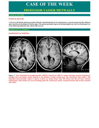
  • 1. CASE OF THE WEEK PROFESSOR YASSER METWALLY CLINICAL PICTURE CLINICAL PICTURE A 18 years old female patient presented clinically with disturbed level of consciousness, recurrent grand mal fits, bilateral optic neuritis and meningeal irritation signs. The patient gradually improved during hospital stay and was discharged two weeks following admission after almost full recovery. RADIOLOGICAL FINDINGS RADIOLOGICAL FINDINGS   Figure 1. Acute disseminated encephalomyelitis (ADEM). Postcontrast MRI T1 images showings scattered hypointense rounded and oval lesions, mostly situated at the junction of deep cortical gray and subcortical white matter. The immediate periventricular region is spared. Lesions are mostly larger than MS lesions and did not show contrast enhancement. Some lesions are seen encroaching upon the cortical grey matter. In general lesions did not have marked mass effect.
  • 2. Figure 2. Acute disseminated encephalomyelitis (ADEM). Postcontrast MRI T1 images showings scattered hypointense rounded and oval lesions, mostly situated at the junction of deep cortical gray and subcortical white matter. The immediate periventricular region is spared. Lesions are mostly larger than MS lesions and did not show contrast enhancement. Some lesions are seen encroaching upon the cortical grey matter. In general lesions did not have marked mass effect. Figure 3. A case of acute disseminated encephalomyelitis. Notice that the multifocal cortical / subcortical hyperintense foci are sparing the periventricular region, and this is the classic pattern in ADEM. The foci, although of large size they have mild mass effect.
  • 3. Figure 4. Acute disseminated encephalomyelitis (ADEM). Notice that the multifocal cortical / subcortical lesions are sparing the periventricular region. The ADEM lesions are hypointense on MRI T1 images and hyperintense on MRI T2 and FLAIR images. ADEM lesions, though large, exert mild mass effect. Figure 5. Acute disseminated encephalomyelitis (ADEM). Notice that the multifocal cortical / subcortical lesions are sparing the periventricular region. The ADEM lesions are hypointense on MRI T1 images and hyperintense on MRI T2 and FLAIR images. ADEM lesions, though large, exert mild mass effect. DIAGNOSIS: DIAGNOSIS: ACUTE DISSEMINATED ENCEPHALOMYELITIS (ADEM)
  • 4. DISCUSSION DISCUSSION Acute disseminated encephalomyelitis (ADEM) presents as an acute-onset neurological dysfunction following a triggering event such as an infection or vaccination. Patients present with polysymptomatic neurological dysfunction, and imaging shows multifocal white matter lesions in the brain and spinal cord. Clinical evaluation, magnetic resonance imaging, and cerebrospinal fluid study are most useful in establishing the diagnosis and ruling out important differential diagnoses. Corticosteroids are the mainstay of treatment and the role of other modalities of treatment, such as plasma exchange and intravenous immunoglobulin, require further study. Prognosis is generally good. The recently proposed consensus definitions are likely to facilitate delineation of ADEM from other acquired demyelinating disorders. Acute disseminated encephalomyelitis (ADEM) has evolved considerably since the first case description of a patient with post-measles neurological dysfunction by Lucas in 1790. [1] Histopathologic and immunologic studies have provided considerable insight into the mechanism of the disease, and the advent of magnetic resonance imaging (MRI) has facilitated early diagnosis. With the recent introduction of consensus definitions, it is now established as a well-delineated entity. ADEM is a monophasic, inflammatory demyelinating disorder of the central nervous system (CNS). Clinical examination reveals polysymptomatic neurological involvement and imaging shows multifocal demyelinating lesions in the brain and spinal cord. It is an immune-mediated disorder that usually follows infections or vaccinations. [2,3] Though previously described as an illness with poor prognosis, recent studies have demonstrated improved outcomes following the introduction of effective immunomodulation. [4,5,6,7] This article will focus on the recent trends in the diagnosis and management of ADEM.  Epidemiology The incidence of ADEM has been estimated to be 0.4 per 100,000 population per year [8] and it may represent 30% of all childhood encephalitic illnesses. [9] ADEM more commonly affects children and young adults, probably because of exposure to frequent vaccinations and exanthematous and upper respiratory tract infections in this age-group. [6,7,10,11,12,66] The mean age of onset in children ranges from 5 to 8 years. [6,12,13] ADEM generally does not show a predilection for any one sex, though two recent studies have shown a male predominance among the victims. [13,14] ADEM occurs throughout the year, but there is a significant increase in incidence during winter and spring. [6,7]  Triggering Events ADEM typically follows an antigenic challenge, such as infection or vaccination, which activates the immune system. Exanthematous fevers such as measles, mumps, and rubella are the usual triggering infections. Recently, there has been a distinct change in the trend and, in regions with extensive immunization coverage, nonspecific upper respiratory tract infections are the most common triggering events. [11] However, ADEM following vaccine-preventable diseases is still prevalent in Egypt. [15] The association with infection may be difficult to establish and only 70% of ADEM patients report a triggering event. Though extensive microbiological investigations have led to the inclusion of a wide spectrum of infections in the list of those alleged to cause ADEM [Table - 1], they may fail to identify a cause in the majority of cases. [6,7,10] Table 1. Infections alleged to cause ADEM Viral Measles, Mumps, Varicella, Rubella, Influenza, A,B Hepatitis, A,B Coxsackie, Epstein-Barr, Dengue [16] and HIV [17] Bacterial Mycoplasma pneumoniae, Borrelia burgdorferi, Mycobacterium tuberculosis, Brucella, Chlamydia, Legionella, Salmonella typhi, and Leptospira, Campylobacter, Streptococcus pyogenes Vaccination Rabies, Measles, Rubella, Smallpox, Diphtheria, Mumps, Tetanus, antitoxin, Pertussis, Japanese encephalitis, Polio, Hepatitis B, Influenza, and Meningococcal A, and C Drugs Gold, Arsenical compounds, Sulfonamides, streptomycin/PAS Miscellaneous Allogenic bone marrow transplantation [19], Heart-lung transplantation [19] Herbal extracts [20], Ventriculo-atrial shunts, [21] Stings, Leprosy type I reaction Post-immunization ADEM occurs most frequently following measles, rubella, or mumps vaccination. Many vaccines have been implicated in the causation of ADEM [Table - 1]. In countries where neural tissue-based vaccines are still used, antirabies immunization with either BPL (betapropionolactone inactivated) or Semple (phenol inactivated) vaccines are important causes for ADEM. [4]  Clinical Features The spectrum of neurological symptoms and signs in ADEM is broad, reflecting widespread central and peripheral nervous system (PNS) involvement. There may be a prodrome consisting of fever, headache, vomiting, and malaise.
  • 5. Neurological dysfunction generally occurs within 3-6 weeks of the triggering event and may appear abruptly or may progress over several days. [6,7] Generally, ADEM is a monophasic illness and its severity may range from mild disease, with headache, subtle drowsiness, and irritability, to fulminant disease characterized by coma, decerebration, and respiratory failure. The distribution of the lesions in the nervous system determines the clinical presentation, and the commonest features are altered sensorium, pyramidal dysfunction, cerebellar ataxia, optic neuritis, and/or myelitis. Retention of urine, urinary frequency, urgency, or incontinence may occur during the acute stage and lower urinary tract dysfunction may persist even after disappearance of other neurological deficits. [23] Seizures are not uncommon and may be of focal or generalized type . [24] There may be additional features such as fever, headache, and meningismus, with the disease mimicking meningitis. Children under 3 years generally present only with encephalopathy, probably because they have immature myelin. [2,7] ADEM may have various atypical presentations. Behavioral disturbances may occasionally be the sole symptom. Presence of flaccidity and areflexia in an otherwise typical case of ADEM betrays additional PNS involvement, which is most commonly at the level of the spinal roots; [25] this picture is frequently seen in antirabies vaccination-related ADEM. [4] Combined CNS and PNS demyelination may suggest the possibility of shared pathological epitopes. [26] There is evidence to suggest that central (ADEM) and peripheral (acute and chronic inflammatory demyelinating polyradiculoneuropathy) demyelinating disorders represent two ends of a spectrum and overlap of clinical features may occur. [27] Extrapyramidal manifestations such as chorea and dystonia are rare but may be prominent in ADEM following group A streptococcal infection. [28] Occasional patients may present with focal deficits, clinical features of raised intracranial pressure, and imaging results suggesting a mass lesion. Such a presentation of ADEM, tumefactive demyelination , may be mistaken for neoplasm till histopathology establishes the diagnosis. The pattern of neurological dysfunction may be influenced by the type of triggering event. For example, post-mumps demyelination commonly presents as myelitis, post-varicella demyelination has cerebellar ataxia as its hallmark, and rubella-associated ADEM often has an explosive onset of symptoms, seizures and mild pyramidal dysfunction. [29] Acute hemorrhagic leukoencephalitis (AHLE) and acute necrotizing hemorrhagic leukoencephalitis (ANHLE) of Weston Hurst represent the hyperacute, fulminant form of postinfectious demyelination [2] [Table - 2]. Table 2. ADEM versus Acute hemorrhagic leukoencephalitis (AHLE) Feature ADEM AHLE Age group Children Young adult Triggering events Viral exanthema, respiratory tract infection, respiratory tract infection vaccination Onset Acute Hyperacute, fulminant Blood picture Normal Leukocytosis ESR/acute phase Normal Elevated reactant Urine Normal Proteinuria CSF cellularity Mononuclear Neutrophils, red blood cells Neuroimaging Multifocal white matter lesions Large lesions, mass effect, hemorrhage, necrosis Pathology Periventricular demyelination, inflammation Fibrinoid necrosis, hemorrhage, necrosis, demyelination Prognosis Good Bad  Consensus Definition for ADEM Clinical research in ADEM has always been hampered by the lack of a uniform case definition, resulting in heterogeneous, incomparable study populations. The recent introduction of consensus definitions for children by the International Pediatric MS Study Group is likely to facilitate the differentiation of monophasic ADEM from other acquired demyelinating conditions [Table - 3]. [30] According to the consensus definition patients should have an acute or subacute onset of the first clinical event of multifocal CNS affection, characterized by polysymptomatic neurological dysfunction. Encephalopathy is an essential clinical feature; it is defined as either behavioral changes, alteration in consciousness, or both. MRI brain must show focal or multifocal lesions greater than 1-2 cm in size, which must be predominantly in the white matter, though it may also be present in the grey matter such as the basal ganglia or thalamus. In addition, the spinal cord may show confluent lesions. The clinical event must have a presumed inflammatory or demyelinating basis, without other etiologies. It should be followed by improvement, either clinically or on MRI, though there may be residual deficits. New or fluctuating symptoms and signs or MRI findings within three months of the initial event are considered part of the initial acute event. Table 3. Monophasic ADEM and other acquired demyelinating conditions Monophasic multifocal disease
  • 6. Acute disseminated encephalomyelitis  Acute hemorrhagic leukoencephalitis Clinically isolated syndromes  Transverse myelitis  Optic neuritis  Cerebellitis  Brain stem dysfunction Multiphasic and recurrent acute disseminated encephalomyelitis  Is ADEM always a monophasic disease? Though ADEM is classically described as a monophasic illness, a biphasic temporal profile has been documented in 0-20% of patients. [6,7,13,14] Such patients do not proceed to develop multiple sclerosis (MS). [7] It is now possible to define patients of ADEM who develop relapses, i.e., multiphasic or recurrent ADEM. [30] A new clinical event should have occurred that meets the case definition of ADEM. The new event should occur three or more months after the initial episode. The event should occur when the patient is not on steroids and at least one month after completion of therapy. In multiphasic ADEM , clinical examination and MRI should demonstrate involvement of new areas of the CNS during the relapse. In recurrent ADEM , clinical findings and MRI should suggest involvement of the same areas that had been involved in the initial ADEM episode.  Investigations  Cerebrospinal fluid Cerebrospinal fluid (CSF) should be analyzed to exclude the differential diagnoses of ADEM, especially the CNS infections. CSF is abnormal in nearly three-fourth of ADEM patients and is characterized by pleocytosis, elevated proteins, and normal sugar values. [6,7] Cellular response is usually lymphocytic and counts are moderately elevated. Intrathecal oligoclonal immunoglobulin synthesis is rare and usually disappears after clinical recovery. [7,13]  Electrophysiology Generalized slowing is the most common finding in electroencephalography and is nonspecific. Patients with spinal cord, brain stem, or optic nerve involvement may have abnormal somatosensory, brain stem auditory, or visual evoked potentials, respectively. [2]  Imaging Computed tomography (CT) may demonstrate hypodense lesions in the white matter and basal ganglia. However, initial CT imaging may be normal in 40% of cases, as lesions may appear only 5-14 days after the onset of clinical signs. [2,31,66] MRI forms the cornerstone for diagnosis of ADEM and is more sensitive than CT in detecting lesions. Abnormalities are best appreciated in T2-weighted and fluid-attenuated inversion recovery (FLAIR) sequences as patchy, multifocal, poorly marginated lesions of increased signal intensity. White matter lesions are usually asymmetric and most frequently situated subcortically, in the cerebellum, brain stem, and spinal cord. [32,33] Thalamic and basal ganglia lesions are seen in nearly one-third of cases and may be symmetric. Corpus callosum may be affected when involvement is extensive. [3] The spinal cord becomes swollen and may show increased signal intensities, most commonly in the thoracic region. Enhancement after contrast administration is variable and occurs in acute lesions due to disruption of the blood-brain barrier. [32,33] Lesions may show complete ring, incomplete ring ('open-ring sign'), nodular, gyral, or spotty patterns of enhancement. [33,34] Tumefactive demyelination appears as a large white matter lesion with mass effect. Though clinically a monophasic illness, new lesions may appear serially over several weeks and hence, may appear to be of varying ages on MRI. [33] Occasionally, initial MR images may be normal and lesions may appear in images repeated later in the course of the disease or even during the stage of clinical improvement. [35] Though lesions generally resolve with treatment, hyperintensities may persist in MRI long after clinical recovery and is due to astrocytic hyperplasia, gliosis, or cystic changes. [36,37] Resolution of MRI abnormalities within six months of the demyelinating episode favors the diagnosis of ADEM. [38]
  • 7. Figure 6. Acute disseminated encephalomyelitis. Notice the contrast enhancement which is characteristic of acute lesions. Also notice that many lesions are situated at the junction of deep cortical gray and subcortical white matter which is characteristic of ADEM Figure 7. In acute disseminated encephalomyelitis tumefactive demyelination appears as a large white matter lesion with mass effect.  Newer modalities Quantitative proton MR spectroscopic imaging has shown low N-acetylaspartate (NAA) and high lactate levels in acute lesions, which normalize after recovery. [39] Further studies are required to assess the role of MR imaging techniques such as magnetization transfer and diffusion tensor imaging in detecting early or small lesions. [40] Abnormalities in 99m Tc- HMPAO SPECT appear as areas of hypoperfusion, which are more extensive than the abnormalities seen in MRI and may parallel the time course of the disease more accurately. [41] Notably, in patients with residual cognitive deficits, SPECT with acetazolamide is able to detect persistent abnormalities from areas where lesions had apparently resolved on MRI. [42,43]  Pathogenesis  Immunology The lesions of ADEM are due to autoimmune-mediated inflammation of the CNS, and the absence of viral or bacterial antigens in the CNS is nearly universal. [5,7] T-cells have been shown to play an important role, possibly through molecular mimicry or by nonspecific activation of autoreactive T-cell clones. [3] Interleukin-6 may be associated with proliferation of B-lymphocytes and immunoglobulin G synthesis. [44] Anti-basal ganglia antibodies have been demonstrated in children with classical features of ADEM following streptococcal infection. [28] A complex interplay between cytokines and adhesion molecules is responsible for the cellular events of inflammatory encephalomyelitis and oligodendrocyte death. An association has been established between ADEM and certain class II HLA alleles, indicating
  • 8. that genetic factors may play a role in immunoregulation and progression from infection/vaccination to ADEM. [29,66]  Pathology Histopathology studies have demonstrated perivenous cuffing with inflammatory cells, especially lymphocytes and macrophages, and loss of myelin. Most of the changes are seen in white matter. However, grey matter also shows changes, including chromatolysis and neuronal loss, especially in the thalami, deep cortical lamina, and hypothalamus. The meninges may also show inflammatory reaction. [2,45] Figure 8. Histopathology studies In ADEM have demonstrated perivenous cuffing with inflammatory cells, especially lymphocytes and macrophages, and loss of myelin  Differential Diagnosis In the absence of specific biological markers, the diagnosis of ADEM is based upon clinical and imaging features. Clinical evaluation, neuroimaging, and blood and CSF analysis can help to distinguish ADEM from other conditions. [46] Herpes simplex encephalitis commonly presents with abnormal behavior and focal/secondary generalized seizures, which are more frequent and difficult to treat than in ADEM. MRI, EEG, and CSF polymerase chain reaction for Herpes simplex virus help in confirmation of the diagnosis. Japanese encephalitis presents with acute encephalopathy. MRI may show bilateral thalamic lesions, akin to ADEM. History, EEG findings, and CSF evidence of antibodies to the virus are useful for differentiation. Other infections such as bacterial meningitis and brain abscess can be ruled out by relevant investigations such as imaging and lumbar puncture. Complicated tuberculous meningitis may sometimes mimic ADEM and can be excluded if CSF sugar is normal and cultures are sterile. Neuromyelitis optica is characterized by optic neuritis and myelitis, with spinal lesions extending over three or more segments, features that may also be seen in ADEM. It may be distinguished from ADEM by the relative paucity of white matter lesions in MRI of the brain and presence of antibodies to aquaporin 4 (NMO-IgG). [47] Patients with Behcet's disease may present with multifocal neurological signs due to brain and spinal cord involvement. Clinical and imaging features may resemble ADEM, and the history of recurrent mucocutaneous ulcers of the mouth and genitalia will be essential to establish the diagnosis. Antiphospholipid antibody syndrome may also mimic the clinical and MRI features of ADEM and should be ruled out by measuring the specific antibodies. Immune-mediated disorders such as systemic lupus erythematosus, Sjögren syndrome, and sarcoidosis may present with neurological dysfunction and multifocal white matter changes and can be diagnosed by history and relevant blood tests. Susac's syndrome may present with subacute encephalopathy, with MRI of the brain showing multiple white matter lesions; however, it can be differentiated from ADEM based on additional features such as the presence of headache, visual impairment due to retinal artery branch occlusion, sensorineural hearing loss, and specific involvement of central corpus callosum in MRI. [48,66] The diagnosis of recurrent ADEM should be made only after excluding other lesions such as arteriovenous malformations, neoplasms, and vasculitis.
  • 9. Metabolic leukoencephalopathies such as metachromatic leukodystrophy (MLD), adrenoleukodystrophy, and MELAS (mitochondrial myopathy, encephalopathy, lactic acidosis, and stroke-like episodes) may mimic childhood ADEM. Leukodystrophies may have a stepladder clinical course, with the exacerbations associated with febrile illnesses. History may reveal prior neurological dysfunction. MRI may show symmetric, confluent white matter lesions, hallmarks of inherited rather than acquired leukoencephalopathies. Specific investigations such as serum aryl sulfatase for MLD and very long-chain fatty acids (VLCFA) levels for adrenoleukodystrophy will confirm the diagnosis. The clinical features of mitochondrial cytopathies such as MELAS may be similar to that of ADEM and lesions seen on MRI may be transient. Serum and CSF lactate levels may be elevated during the acute episode and MR spectroscopic imaging may show elevated lactate in the affected tissues.  ADEM or multiple sclerosis? In a patient presenting with neurological dysfunction and MRI showing multiple white matter lesions, the most important differential diagnosis is MS. Distinguishing between ADEM and MS is a diagnostic challenge and has important therapeutic and prognostic implications. There are several clinical, imaging, and laboratory parameters that may be useful to distinguish between the two [Table - 4]. CSF electrophoresis has shown a significant reduction in the beta-1 globulin fraction in patients with MS as compared to those with ADEM and this may be a potential CSF marker. [49] Features that strongly favor ADEM include a history of preceding infection, polysymptomatic neurological dysfunction, encephalopathy, grey matter involvement on MRI, and absence of oligoclonal bands in CSF. [6,7] Often, distinction between these two conditions cannot be made with certainty and follow-up with serial MRI may be necessary to establish the diagnosis. [3,36,66] Table 4. Differences between ADEM and multiple sclerosis Feature ADEM MS Onset Abrupt Subacute Triggering events Preceding infection or vaccination in 70 Uncommon % of cases Age group More common in children More common in young adult Temporal profile Monophasic, rarely relapsing Relapsing Clinical features Altered sensorium More common Rare Seizures More common Rare Neurological deficit Multifocal Usually single deficit Optic neuritis Bilateral Unilateral Myelitis Complete, transverse myelitis Partial myelopathy Lower motor neuron signs More common Rare Headache More common Rare Meningismus More common Rare Neuroimaging findings Distribution of the lesions Bilateral extensive lesions Scattered asymmetric lesions White matter lesions Confluent, ill-defined periventricular and Well-defined, with periventricular subcortical white matter lesions preponderance Corpus callosum lesions Rare Common Grey matter involvement Thalamic and basal ganglionic lesions are Uncommon common Edema and mass effect May be present Uncommon Follow-up MRI No new lesions New lesions with dissemination in time and place Cerebrospinal fluid Cell count Mild to moderate pleocytosis Normal to mild pleocytosis Protein Increased Normal Oligoclonal bands Uncommon, transient Common and persistent Mortality 10-25 Uncommon  Treatment
  • 10. Public health initiatives are important for minimizing exposure to the triggering events of ADEM. These include implementing effective vaccination programs and avoiding vaccines containing neural tissues or enveloped viruses. [2] Early initiation of antibiotics for infections may not prevent the immune trigger that culminates in ADEM. [50] Treatment of ADEM involves immunomodulation that aims to counter autoimmune-mediated inflammation. Though spontaneous resolution has been described, recovery is usually incomplete and hence patients presenting in the acute stage with significant, progressive neurological deficits should receive treatment. [51] In view of the rarity of ADEM, treatment regimes for children and adults are based on anecdotal reports and lack validation by randomized controlled trials. High- dose corticosteroids have become the first line of management and are associated with rapid recovery when given early in the disease. [6,7,13,14,52] Earlier studies used ACTH, but nowadays either intravenous methylprednisolone (10-30 mg/kg/day, up to maximum of 1 gm/day) or dexamethasone (1 mg/kg/day) for 3 to 5 days are being used. [6,7,13,14] Full recovery was reported in 50-80% of patients. Methylprednisolone treated patients have significantly better outcome with respect to disability compared to those treated with dexamethasone. [13] In addition to immunosuppression, steroids also have anti-inflammatory and antiedema actions, and the immediate improvement following administration of steroids is likely to be due to reduction in cerebral edema. [53] Oral corticosteroids are continued and gradually tapered over six weeks to minimize the risk of recurrence. [7] The role of corticosteroids in patients presenting late in the course of the disease is questionable. [54] Vaccinations should be avoided during the first six months after recovery. [7] Plasma exchange is another therapeutic modality, and its role in the management of demyelinating disorders that are nonresponsive to corticosteroids has been established in one randomized control trial. [55] In most studies, 1-1.4 plasma volumes were exchanged by continuous flow centrifugation and 70% of volume was replaced by 5% serum albumin, fresh- frozen plasma, or hydroxyethyl starch. Patients received 2-20 exchanges, depending on the severity of illness and clinical response. It is likely to be effective in patients of ADEM not responding to corticosteroids, especially when given early in the course of the disease. [56,57] Intravenous immunoglobulin is yet another option and the results so far are impressive, especially in corticosteroid nonresponders. In most studies, 0.4 gm/kg/day was given over 5 days and improvement was noted within 2 to 3 days. [58,59,60] However, in most reported cases, patients had received prior corticosteroids and the delayed effects of these drugs could not be ruled out as the reason for the improvement. At present, the evidence is insufficient to design a treatment algorithm for ADEM. However, based on the available reports, plasma exchange and intravenous immunoglobulin are likely to be second-line therapies when corticosteroids fail. The relative efficacies of the two have not been evaluated. The choice of second-line therapy should be individualized, depending upon the type of CNS involvement, complications, and comorbidities. For example, autonomic dysfunction and hypotension would preclude the use of plasma exchange. Anecdotal reports suggest that intravenous immunoglobulin may be more effective in patients with who also have PNS involvement [25] and plasma exchange in patients with tumefactive demyelination. [61] Combination therapy with methylprednisolone and immunoglobulin has been reported to be successful in atypical cases of ADEM. [62] Cyclophosphamide [52] and hypothermia [63] have been successfully tried in fulminant ADEM. Hemicraniectomy has been reported to be lifesaving in patients with massive life-threatening cerebral edema refractory to conventional medical management. [64,65]  Outcome ADEM is usually a monophasic illness lasting 2-4 weeks. [2] However recovery may continue for weeks or months. While earlier studies reported mortality of 20% and significant morbidity amongst survivors, recent reports suggest excellent outcomes, especially in children. [7,10,13,14] Visual impairment, weakness, lower urinary tract dysfunction, cognitive impairment, and behavioral changes may persist at follow-up. [7,13,23] Children thought to have made a full recovery may show subtle neurocognitive and behavioral changes on formal assessment, though these may be less severe than the changes seen in children with MS. [43] Patients having progressive neurological dysfunction , altered sensorium, and seizures have poor long-term outcomes. [24] SUMMARY SUMMARY ADEM presents as an acute onset neurological dysfunction following infection or vaccination. Clinical evaluation, MRI, and CSF study are most useful to establish the diagnosis and rule out important differential diagnoses. The recently proposed consensus definitions are likely to facilitate delineation of ADEM from other acquired demyelinating disorders. Corticosteroids are the most important treatment modality, while the role of other agents requires further study. Prognosis is generally good.
  • 11. Addendum  A new version of this PDF file (with a new case) is uploaded in my web site every week (every Saturday and remains available till Friday.)  To download the current version follow the link quot;http://pdf.yassermetwally.com/case.pdfquot;.  You can also download the current version from my web site at quot;http://yassermetwally.comquot;.  To download the software version of the publication (crow.exe) follow the link: http://neurology.yassermetwally.com/crow.zip  The case is also presented as a short case in PDF format, to download the short case follow the link: http://pdf.yassermetwally.com/short.pdf  At the end of each year, all the publications are compiled on a single CD-ROM, please contact the author to know more details.  Screen resolution is better set at 1024*768 pixel screen area for optimum display REFERENCES References 1. Lucas J. An account of uncommon symptoms succeeding the measles: With additional remarks on the infection of measles and smallpox. London Med J 1790;11:325-31. 2. Coyle PK. Post infectious encephalomyelitis. In : Kennedy PGE, Davis LE, editors. Infectious Diseases of the Nervous System. Butterworth-Heinemann: Oxford; 2000. p. 83-108. 3. Garg RK. Acute disseminated encephalomyelitis. Postgrad Med J 2003;79:11-7. 4. Swamy HS, Shankar SK, Chandra PS, Aroor SR, Krishna AS, Perumal VG. Neurological complications due to beta- propiolactone (BPL)-inactivated antirabies vaccination. Clinical, electrophysiological and therapeutic aspects. J Neurol Sci 1984;63:111-28. 5. Johnson RT, Griffin DE, Hirsch RL, Wolinsky JS, Roedenbeck S, de Soriano IL, et al . Measles encephalomyelitis- clinical and immunologic studies. N Engl J Med 1984;310:137-41. 6. Hynson JL, Kornberg AJ, Coleman LT, Shield L, Harvey AS, Kean MJ. Clinical and neuroradiologic features of acute disseminated encephalomyelitis in children. Neurology 2001;56:1308-12. 7. Dale RC, de Sousa C, Chong WK, Cox TC, Harding B, Neville BG. Acute disseminated encephalomyelitis, multiphasic disseminated encephalomyelitis and multiple sclerosis in children. Brain 2000;123:2407-22. 8. Leake JA, Albani S, Kao AS, Senac MO, Billman GF, Nespeca MP, et al . Acute disseminated encephalomyelitis in childhood: Epidemiologic, clinical and laboratory features. Pediatr Infect Dis J 2004;23:756-64. 9. Kennard C, Swash M. Acute viral encephalitis: Its diagnosis and outcome. Brain 1981;104:129-48. 10. Murthy SN, Faden HS, Cohen ME, Bakshi R. Acute disseminated encephalomyelitis in children. Pediatrics 2002;110:e1-7. 11. Menge T, Hemmer B, Nessler S, Wiendl H, Neuhaus O, Hartung HP, et al . Acute disseminated encephalomyelitis: An update. Arch Neurol 2005;62:1673-80. 12. Anlar B, Basaran C, Kose G, Guven A, Haspolat S, Yakut A, et al . Acute disseminated Encephalomyelitis in Children: Outcome and Prognosis. Neuropediatrics 2003;34:194-9. 13. Tenembaum S, Chamoles N, Fejerman N. Acute disseminated encephalomyelitis: A long-term follow-up study of 84 pediatric patients. Neurology 2002;59:1224-31. 14. Singhi PD, Ray M, Singhi S, Khandelwal NK. Acute disseminated encephalomyelitis in North Indian children: Clinical profile and follow-up. J Child Neurol 2006;21:851-7. 15. Murthy JM, Yangala R, Meena AK, Jaganmohan Reddy J. Acute disseminated encephalomyelitis: clinical and MRI Study from South India. J Neurol Sci 1999;165:133-8. 16. Yamamoto Y, Takasaki T, Yamada K, Kimura M, Washizaki K, Yoshikawa K, et al . Acute disseminated encephalomyelitis following dengue fever. J Infect Chemother 2002;8:175-7.
  • 12. 17. Narciso P, Galgani S, Del Grosso B, De Marco M, De Santis A, Balestra P, et al . Acute disseminated encephalomyelitis as manifestation of primary HIV infection. Neurology 2001;57:1493-6. 18. Tomonari A, Tojo A, Adachi D, Iseki T, Ooi J, Shirafuji N, et al . Acute disseminated encephalomyelitis (ADEM) after allogeneic bone marrow transplantation for acute myeloid leukemia. Ann Hematol 2003;82:37-40. 19. Horowitz MB, Comey C, Hirsch W, Marion D, Griffith B, Martinez J. Acute disseminated encephalomyelitis (ADEM) or ADEM-like inflammatory changes in a heart-lung transplant recipient: A case report. Neuroradiology 1995;37:434-7. 20. Schwarz S, Knauth M, Schwad S, Walter-Sack I, Bonmann E, Storch-Hagenlocher B. Acute disseminated encephalomyelitis after parenteral therapy with herbal extracts: A report of two cases. J Neurol Neurosurg Psychiatry 2000;69:516-8. 21. Boz C, Velioglu S, Ozmenoglu M. Acute disseminated encephalomyelitis after bee sting. Neurol Sci 2003;23:313-5. 22. Jacob J, Alexander M, Aaron S, Pulimood S, Walter N, Gnanamuthu C. Acute disseminated encephalomyelitis in Hansen's disease. Ann Indian Acad Neurol 2006;9:166-8. 23. Sakakibara R, Hattori T, Yasuda K, Yamanishi T. Micturitional disturbance in acute disseminated encephalomyelitis (ADEM). J Auton Nerv Syst 1996;60:200-5. 24. Panicker JN. Acute Disseminated Encephalomyelitis: Clinical Profile and Predictors of Outcome (dissertation). Bangalore: National Institute of Mental Health and Neurosciences, NIMHANS (Deemed University); 2004. 25. Marchioni E, Ravaglia S, Piccolo G, Furione M, Zardini E, Franciotta D, et al . Postinfectious inflammatory disorders: Subgroups based on prospective follow-up. Neurology 2005;65:1057-65. 26. Kinoshita A, Hayashi M, Miyamoto K, Oda M, Tanabe H. Inflammatory demyelinating polyradiculitis in a patient with acute disseminated encephalomyelitis (ADEM). J Neurol Neurosurg Psychiatry 1996;60:87-90. 27. Krivickas LS, Hochberg FH, Freeman S. Chronic inflammatory demyelinating polyradiculoneuropathy with tumefactive central demyelination. Muscle Nerve 2006;33:283-8. 28. Dale RC, Church AJ, Cardoso F, Goddard E, Cox TC, Chong WK, et al . Post streptococcal acute disseminated encephalomyelitis with basal ganglia involvement and auto-reactive antibasal ganglia antibodies. Ann Neurol 2001;50:588- 95. 29. Idrissova ZR, Boldyreva MN, Dekonenko EP, Malishev NA, Leontyeva IY, Martinenko IN, et al . Acute disseminated encephalomyelitis in children: Clinical features and HLA-DR linkage. Eur J Neurol 2003;10:537-46. 30. Krupp LB, Banwell B, Tenembaum S; International Pediatric MS Study Group. Consensus definitions proposed for pediatric multiple sclerosis and related disorders. Neurology 2007;68:S7-12. 31. Lukes SA, Norman D. Computed tomography in acute disseminated encephalomyelitis. Ann Neurol 1983;13:567-72. 32. Mader I, Stock KW, Ettlin T, Probst A. Acute disseminated encephalomyelitis: MR and CT features. AJNR Am J Neuroradiol 1996;17:104-9. 33. Caldemeyer KS, Smith RR, Harris TM, Edwards MK. MRI in acute disseminated encephalomyelitis. Neuroradiology 1994;36:216-20. 34. van der Meyden CH, de Villiers JF, Middlecote BD, Terblanche J. Gadolinium ring enhancement and mass effect in acute disseminated encephalomyelitis. Neuroradiology 1994;36:221-3. 35. Honkaniemi J, Dastidar P, Kdhdrd V, Haapasalo H. Delayed MR imaging changes in acute disseminated encephalomyelitis. AJNR Am J Neuroradiol 2001;22:1117-24. 36. Kesselring J, Miller DH, Robb SA, Kendall BE, Moseley IF, Kingsley D, et al . Acute disseminated encephalomyelitis. MRI findings and the distinction from multiple sclerosis. Brain 1990;113:291-302. 37. Go T, Imai T. A residual cystic lesion in acute disseminated encephalomyelitis. Neuroradiology 2000;42:682-4. 38. Richer LR, Sinclair DB, Bhargava R. Neuroimaging features of acute disseminated encephalomyelitis in childhood. Pediatr Neurol 2005;32:30-6. 39. Mader I, Wolff M, Nagele T, Niemann G, Grodd W, Kuker W. MRI and proton MR spectroscopy in acute disseminated encephalomyelitis. Childs Nerv Syst 2005;21:566-72.
  • 13. 40. Inglese M, Salvi F, Iannucci G, Mancardi GL, Mascalchi M, Filippi M. Magnetization transfer and Diffusion tensor MR imaging of acute disseminated encephalomyelitis. AJNR Am J Neuroradiol 2002;23:267-72. 41. Itti E, Huff K, Cornford ME, Itti L, Poruri K, Mishkin FS. Postinfectious encephalitis. A co registered SPECT and magnetic resonance imaging study. Clin Nucl Med 2002;27:129-30. 42. Okamoto M, Ashida KI, Imaizumi M. Hypoperfusion following encephalitis: SPECT with acetazolamide. Eur J Neurol 2001;8:471-4. 43. Hahn CD, Miles BS, MacGregor DL, Blaser SI, Banwell BL, Hetherington CR. Neurocognitive outcome after acute disseminated encephalomyelitis. Pediatr Neurol 2003;29:117-23. 44. Dale RC, Morovat A. Interleukin-6 and oligoclonal IgG synthesis in children with acute disseminated encephalomyelitis. Neuropediatrics 2003;34:141-5. 45. Kumar A, Swamy HS, Santhosh V, Taly AB, Arunodaya GR, Shankar SK. Pathology of allergic encephalomyelitis following Semple type antirabies vaccine from India. Neurol Infect Epidemiol 1997;2:239-48. 46. Hahn JS, Pohl D, Rensel M, Rao S; International Pediatric MS Study Group. Differential diagnosis and evaluation in pediatric multiple sclerosis. Neurology 2007;68:S13-22. 47. Wingerchuk DM, Lennon VA, Pittock SJ, Lucchinetti CF, Weinshenker BG. Revised diagnostic criteria for neuromyelitis optica. Neurology 2006;66:1485-9. 48. Susac JO. Susac syndrome: The triad of microangiopathy of the brain and retina with hearing loss in women. Neurology 1994;44:591-3. 49. Chopra B, Abraham R, Abraham A. CSF beta-1 Globulin- a potential marker in differentiating multiple sclerosis and acute disseminated encephalomyelitis: A preliminary study. Neurol India 2002;50:41-4. 50. Pellegrini M, O'Brien TJ, Hoy J, Sedal L. Mycoplasma pneumoniae infection associated with an acute brainstem syndrome. Acta Neurol Scand 1996;93:203-6. 51. Rust RS, Dodson W, Prensky A, Chun R, DeVivo D, Dodge P, et al . Classification and outcome of acute disseminated encephalomyelitis. Ann Neurol 1997;42:491. 52. Schwarz S, Mohr A, Knauth M, Wildemann B, Storch-Hagenlocher B. Acute disseminated encephalomyelitis: A follow- up study of 40 adult patients. Neurology 2001;56:1313-8. 53. Barkhof F, Hommes OR, Scheltens P, Valk J. Quantitative MRI changes in gadolinium-DTPA enhancement after high dose intravenous methyl prednisolone in multiple sclerosis. Neurology 1991;41:1219-22. 54. Straub J, Chofflon M, Delavelle J. Early high-dose intravenous methylprednisolone in acute disseminated encephalomyelitis: A successful recovery. Neurology 1997;49:1145-7. 55. Weinshenker BG, O'Brien PC, Petterson TM, Noseworthy JH, Lucchinetti CF, Dodick DW, et al . A randomized trial of plasma exchange in acute central nervous system inflammatory demyelinating disease. Ann Neurol 1999;46:878-86. 56. Lin CH, Jeng JS, Yip PK. Plasmapheresis in acute disseminated encephalomyelitis. J Clin Apheresis 2004;19:154-9. 57. Keegan M, Pineda AA, McClelland RL, Darby CH, Rodriguez M, Weinshenker BG. Plasma exchange for severe attacks of CNS demyelination: Predictors of response. Neurology 2002;58:143-6. 58. Pradhan S, Gupta RP, Shashank S, Pandey N. Intravenous immunoglobulin therapy in acute disseminated encephalomyelitis. J Neurol Sci 1999;165:56-61. 59. Sahlas DJ, Miller SP, Guerin M, Veilleux M, Francis G. Treatment of acute disseminated encephalomyelitis with intravenous immunoglobulin. Neurology 2000;54:1370-2. 60. Kleiman M, Brunquell P. Acute disseminated encephalomyelitis: Response to intravenous immunoglobulin? J Child Neurol 1995;10:481-3. 61. Mao-Draayer Y, Braff S, Pendlebury W, Panitch H. Treatment of steroid-unresponsive tumefactive demyelinating disease with plasma exchange. Neurology 2002;59:1074-7. 62. Straussberg R, Schonfeld T, Weitz R, Karmazyn B, Harel L. Improvement of atypical acute disseminated encephalomyelitis with steroids and intravenous immunoglobulins. Pediatr Neurol 2001;24:139-43.
  • 14. 63. Takata T, Hirakawa M, Sakurai M, Kanazawa I. Fulminant form of acute disseminated encephalomyelitis: Successful treatment with hypothermia. J Neurol Sci 1999;165:94-7. 64. von Stuckrad-Barre S, Klippel E, Foerch C, Lang JM, du Mesnil de Rochemont R, Sitzer M. Hemicraniectomy as a successful treatment of mass effect in acute disseminated encephalomyelitis. Neurology 2003;61:420-1. 65. Refai D, Lee MC, Goldenberg FD, Frank JI. Decompressive hemicraniectomy for acute disseminated encephalomyelitis: Case report. Neurosurgery 2005;56:E872. 66. Metwally, MYM: Textbook of neurimaging, A CD-ROM publication, (Metwally, MYM editor) WEB-CD agency for electronic publishing, version 9.1a January 2008