SlideShare una empresa de Scribd logo
1 de 12
Descargar para leer sin conexión
CASE OF THE WEEK
                   PROFESSOR YASSER METWALLY
CLINICAL PICTURE

CLINICAL PICTURE:

65 years old male patient with vascular risk factors (diabetis, hypertension, increased blood viscosity etc) presented
clinically with a mainly motor clinical presentation in the form of atrophy of small muscles of the hand and evidence
of upper motor neuron manifestations in the lower limbs and with a clinical picture characterized by a painless
onset with a chronic fluctuating course. Meticulous clinical examination reveled mild segmental sensory
manifestations at C5,C6 spinal segments. In this patient, increased whole blood viscosity was in the form of
increased fibrinogen level, increased platelet aggregability, relative increase in the hematocrit value, reduced RBCs
derformability...etc. Evidence of spinal cord claudication was present in the form of aggravation of weakness in the
lower limbs when walking and a sensation of hotness felt in the hands when the patient walks for a distance.

RADIOLOGICAL FINDINGS

RADIOLOGICAL FINDINGS:


Figure 1. CT myelography at C4,C6,C7 cervical spinal segments showing evidence of spondylitic changes in the
form of posterior osteophytes, cervical canal stenosis, luschka joint hypertrophy, and segmental spinal cord atrophy
at C5,C6 spnal segments
Figure 2. CT myelography at C4,C6,C7 cervical spinal segments showing evidence of spondylitic changes in the
form of posterior osteophytes, cervical canal stenosis, luschka joint hypertrophy, and segmental spinal cord atrophy
at C5,C6 spnal segments. Notice the calcified soft disc herniation.


Figure 3. MRI T2 images showing evidence of cervical disc degeneration and cervical canal stenosis

In the above reported case evidence of spondylitic cervical canal stenosis, with posterior osteophytes, calcified hard
disc herniation and C5,C6 segmental spinal cord atrophy was demonstrated. The clinical picture was in the form of
a mainly motor clinical manifestations and a remitting course with evidence of spinal cord claudication.

Because the vascular spondylitic myelopathy has a sudden painless onset and a fluctuating course with remission
and exacerbation, it was frequently misdiagnosed as multiple sclerosis. However major differences are present
between myelopathy due to disc disease and that due to multiple sclerosis as follows

       Unlike multiple sclerosis, myelopathy due to cervical spondylosis had a sudden onset with the clinical picture
   
       developing over just a few hours.

       Unlike multiple sclerosis, the duration of relapses in myelopathy due to cervical spondylosis is very short ( on
   
       the average few hours to one or two days).

       Unlike multiple sclerosis, relapses of myelopathy due to cervical spondylosis shared a similar clinical
   
       presentation in every single patient i.e. the disease was disseminated only in time and never in place. And
       although signs and symptoms might be severer on recurrent episodes (mainly due to the cumulative effect of
       structural damage and/or the functional disturbances caused by each ischaemic episode), however the disease
       used to recur in the same anatomical site (dorso-lumber spinal segments) and is never disseminated in place.

       Unlike multiple sclerosis, the clinical picture of myelopathy due to cervical spondylosis is mainly motor ( in
   
       the form of weakness and atrophy) and sensory manifestations, though definite, are detected only by careful
       examination.

In fact the quot;mainly motor clinical picturequot; was occasionally a potential source for anther misdiagnosis which is
motor neuron disease or motor neuropathy. However myelopathy due to cervical spondylosis can easily be
differentiated from motor neuron disease because of the relapsing remitting course, and because of the existence of
definite, though subtle, sensory manifestations. Also the existence of impotence, bladder disturbances and
occasional back pain are points against the diagnosis of primary motor neuron disease.

The predominance of motor manifestations in myelopathy due to cervical spondylosis is in fact anther point
favouring its ischemic aetiology. It is clear that when ischaemia occurs, the most vulnerable region of the spinal
cord is the grey matter because its metabolic rate is three to five times greater than the metabolic rate of the white
matter. This would account for the many cases reported in literature of paraparesis with little sensory
manifestations and for instances of lower motor neuron syndromes of an ischaemic basis.

In cervical spondylotic myelopathy patients the motor weakness is characteristically increased by walking and
relieved by rest and this is anther point favouring the ischemic aetiology of myelopathy due to degenerative disc
disease. Normally walking is associated with marked increase of blood flow to the spinal cord and cauda roots to
meet the increased metabolic rate of these neural structures, physiologically the spinal cord microvascular bed will
dilate to accommodate the increased blood flow. Cervical canal stenosis (induced by disc disease) and the associated
segmental arteriosclerosis will hinder this normal physiological quot;exertion induced hyperaemiaquot; of the neural
structures resulting in a temporary spinal cord quot;ischaemic dysfunction on exertionquot;.

Although the prognosis following a single ischaemic episode is good , however repetition of the ischaemic episodes
will ultimately result in spinal cord atrophy with irreversible neurological deficits The patient, following admission,
received medical treatment for diabetes, hypertension, antiplatelet medications and medications that improve RBCs
deformability, reduce whole blood viscosity and fibrinogen level (like pentoxifylline, bezafibrate etc) and he was
referred to surgery once diagnosed radiologically.

DIAGNOSIS:

DIAGNOSIS: CERVICAL SPONDYLOTIC VASCULAR MYELOPATHY

DISCUSSION

DISCUSSION:

Patients with spondylitic cervical myelopathy are classified, clinically, into two main groups. The painless
myelopathy group (due to vascular aetiology) and the painful myelopathy group (due to compression of the spinal
cord). Clinically the painless myelopathy group is characterized by a fluctuant course with remission and
exacerbations and a mainly motor clinical presentation.

                                                   In the painless myelopathy group, the natural history of the
                                                   disease is determined by the interaction of three main pathogenic
                                                   factors. Spondylitic factor, vascular factor and haemorheological
                                                   factor associated with increased whole blood viscosity (increased
                                                   haematocrit value and serum fibrinogen).


                                                   Figure 4. The painless cervical spondylitic myelopathy is the
                                                   interaction between three main pathogenic factors


The spondylitic factor ultimately results in bony and soft tissue hypertrophy that causes cervical canal stenosis, and
encroach upon the subarachnoid space, reducing its volume. Lack of the CSF cushioning effect will cause
embarrassment of the spinal circulation at the level of cervical enlargement since optimum blood supply to the
spinal cord needs an optimum CSF cushioning effect.

The second factor is the vascular arteriolar factor. Apparently the painless myelopathy group comprises a group of
patients where the incidence of vascular risk factors, (such as essential hypertension, NIDDM, hyperlipidaemia
(Type IV), hyperfibrinogenemia and increased platelet aggregation...the metabolic syndrome) is very high. The
incidence of arteriosclerosis is known to be high among patients with vascular risk factors. 4

This is consistent with the necropsy findings of Manen, 1966 2 and Jillinger 19671. The authors reported
arteriolosclerosis, lipohyalinosis and fibrosis of the perforating intramedullary vessels and the fine vessels lying on
the surface of the spinal cord in patients with cervical spondylitic vascular myelopathy.4

These changes were maximum in the cervical enlargement and the overlapping zone in the cervico-dorsal region,
making the spinal cord especially vulnerable at this zone to vesico-circulatory disorders, mainly of extra-medullary
origin, which cause critical decrease in spinal cord flow. It should be mentioned that the area of the spinal cord
between C4 and DI is a watershed area with marginal blood supply. This last field zone is most likely to suffer from
insufficiency of blood and has been shown to be a preferential zone for vascular damage. 4

According to the necropsy results of Jillinger, 19671 the arteriosclerotic changes in the overlapping cervico-dorsal
region were isolated findings. They did not depend on age and were negatively correlated with arteriosclerosis in
the rest of the body. A finding that probably denotes that the injurious effect of the spondylitic changes accelerate
the arteriosclerotic changes in the region of the cervical enlargement.

However it should be noted that both cervical spondylosis and arteriosclerosis are slowly progressive pathology and
they can not be held responsible for the sudden onset of the clinical symptomatology seen in the painless myelopathy
group. No compressing agents (like soft disc herniation, or osteophytes) are demonstrated radiologically, in this
group of patients, that can explain the clinical symptomatology in terms of compression of the spinal cord and/or an
important radicular artery. In short both cervical spondylosis and arteriosclerosis serve by furnishing the
background for the ultimate determinant of the clinical symptomatology.

Cervical spondylosis will result in canal stenosis, loss of the CSF cushioning effect and embarrassment of the spinal
circulation in the region of the cervical enlargement. Arteriosclerosis will result in reduction of the caliber of the
radicular and the perforating intramedullary arterioles with loss of the auto-regulatory physiological process. Flow
in the perforating arteries is dependent on the auto-regulatory process of the penetrating intramedullary arterioles
on one hand and the whole blood viscosity on the other hand. Loss of the auto-regulatory process secondary to
advanced arteriosclerosis will simply mean that the spinal cord perfusion, in the vulnerable region of the cervical
enlargement, will fluctuate with fluctuation of the whole blood viscosity, (the ultimate determinant of the vascular
myelopathy).4

Whole blood viscosity is a collective terminology that reflects the influence of various factors that include mainly the
corpuscular and the plasmatic components of the blood. Whole blood viscosity is usually increased in patients with
the vascular spondylitic myelopathy.

Blood viscosity is mainly determined by the hematocrit value and the plasma viscosity is mainly determined by the
plasma fibrinogen level. High values of lipid have also be found to increase whole blood viscosity. 4

Increase platelet aggregation also increases whole blood viscosity. The behavior of the red blood cells was also found
to affect the blood viscosity. Increased red cell aggregation and reduced red cell deformability increase whole blood
viscosity. The RBCs deformability is invariably reduced and the aggregability in invariably increased in the
presence of high fibrinogen level and high haematocrit values. Fibrinogen in particular is a strong RBCs aggregant
agent. Inverse correlation is present between the red cell deformability and the haematocrit value and serum
fibrinogen level.

Increase of the whole blood viscosity is a common finding in essential hypertension and NIDDM. The vascular
resistance of the perforating blood vessels of the spinal cord and the brain is dependent upon the ratio between the
whole blood viscosity over the caliber of the blood vessel. Increase of the whole blood viscosity results in high
vascular resistance to blood flow and subsequently low perfusion pressure and neuronal tissue ischaemia. Stenosis
of the perforating blood vessel secondary to arteriosclerosis further aggravates the problem. Inverse correlation is
present between the neuronal tissue blood flow and serum fibrinogen level and the haematocrit value. 4

Hyperfibrinogenemia and increased RBCS, and platelet aggregation reflect a hypercoagulable state with increased
thrombotic tendency that selectively affects the small perforating blood vessels and the microcirculation of the brain
and spinal cord. Microvascular occlusion can occur either by local aggregation of hyperaggragable platelets or by
red cell aggregation with impaction of rigid red blood cells in the microcirculation. This is more likely to occur with
the existence of high red cell mass (Hch value) that can displace the hypersensitive platelets Towards the arteriolar
wall resulting in platelet aggregation and thrombus formation. Thrombus formation is enhanced if the arteriolar
wall is abnormal (arteriolosclerosed).4

The high blood viscosity observed in the painless myelopathy patients should simply mean, especially when coupled
with arteriosclerosis of the small perforating blood vessels, that the blood flow to the spinal cord at the level of
cervical enlargement is subjected to high vascular resistance that could ultimately results in low perfusion pressure
and chronic ischaemia. The increased thrombotic tendency observed in those patients should mean that the chronic
ischaemia state could be interrupted by acute thrombotic microvascular occlusion that can result, pathologically, in
spinal cord lacunar infarction at the level of cervical-enlargement and clinically in lower cervical painless
myelopathy of sudden onset and regressive course. The acute thrombo-occlusive episodes are responsible for the
intramedullary cavitations observed in the painless myelopathy group. 4

NEUROIMAGING FINDINGS IN THE PAINLESS MYELOPATHY GROUP

In general two pathological subtypes are observed in this group, spinal cord atrophy and spinal cord cavitations,
both are very segmental and both are the end result of spinal cord ischemia.
 Two types of spinal cord of cavitations are observed in the painless myelopathy group
   


CAVITATIONS        1-Pencil shaped necrosis: the cavitation is maximum at the level of C5,C6 spinal segments and
                   extends one ore two segments above and/or below
CAVITATIONS
                   2-Central gray matter cavitation: bilateral ,symmetrical,rounded cavitations, localized in the
CAVITATIONS
                   bilateral anterior horns of spinal segments C5,C6

                                           Pencil shaped necrosis and central gray matter cavitations, most
                                           probably represent spinal lacunar infarctions. The spinal cord atrophy
                                           observed in those patients is very segmental and exclusively localized to
                                           the level of cervical enlargement between C4- C8 with maximum changes
                                           at C5,C6 segment. The spinal cord above C4 and below C8 is often
                                           normal. The segmental spinal cord atrophy observed in these patients is
                                           the result of long standing chronic ischaemia interrupted by recurrent
                                           thrombo-occlusive episodes.4

                                        All the atrophic and cystic changes are exclusively limited to the spinal
                                        cord area between C4 and C7. The C5-C6 segments are most frequently
                                        involved. The C5-C6 segments are the most vulnerable to vascular
damage as they represent a watershed area with higher incidence of segmental arteriosclerosis. 1


Figure 5. In cervical spondylitic myelopathy, both cystic and atrophic changes are exclusively localized to the level
of cervical enlargement with maximum changes at C5,C6 spinal segments


Figure 6. In cervical spondylitic myelopathy, both cystic [left image] and atrophic [right image] changes are
exclusively localized to the level of cervical enlargement with maximum changes at C5,C6 spinal segments

The central gray matter cavitations demonstrated in the painless group group represent lacunar infarctions
involving the bilateral anterior horns. It results clinically in a purely LMN picture ( proximal muscles
amyetrophy).1
Figure 7. CT myelography showing central gray matter
                                                                 cavitations as bilateral symmetrical,well defined rounded
                                                                 zones of intramedullary contrast accumulations in the
                                                                 presumed anatomical areas of anterior horns [right image is a
                                                                 schematic representation]


                                    The association between spondylitic myelopathy and spinal cord atrophy and/or
                                    cavitation is well known. The ischaemic aetiopathogenesis of myelopathy in this
                                    group of patients is further substantiated by the observation that relapses of
                                    myelopathy are intimately coupled temporally with rise of whole blood viscosity
                                    and thrombotic tendency. Relapses occurs more frequently in the summer time.
                                    Dehydration is more common in summer time, it results in contraction of the
                                    plasma volume and rise of the haematocrit value and subsequently blood
                                    viscosity.

                                    Figure 8. MRI T1,T2 images showing pencil-shaped necrosis


Figure 9. CT myelography showing normal spinal cord at the level of cervical enlargement (A) and spinal cord
atrophy in cervical spondylitic myelopathy (B,C), notice that the atrophic segments are flattened, collapsed, with
irregular contour and wide subarachnoid spaces

The ischaemic aetiology of myelopathy in the painless myelopathy group is consistent with the necropsy findings of
Jellinger, 1967 1. The author reported, in spondylitic myelopathy patients, white matter ischaemic demyelination,
neuronal degeneration and a diffuse lacunar state similar to those seen in the basal ganglion in the hypertensive
small vessel disease of the brain. It should also be mentioned that increased whole blood viscosity is also the ultimate
aetiopathogenic factor in hypertensive microvascular brain disease (diffuse lacunar state, leukoaraiosis, etc.).4

The haemorheological profile of vascular spondylitic myelopathy is similar to the haemorheological profile of
hypertensive micro-vascular brain disease (Lacunar infarction, leukoaraiosis etc.) previously reported by many
authors4. The haemorheological parameters tended to drop down to the normal levels following the acute phase in
myelopathy patients and this has also been reported in ischaemic brain disease.

The hypertensive micro-vascular brain disease is found to be similar in many ways to the spondylitic vascular
myelopathy regarding vascular risk factors, the vascular arteriolar pathology, parenchymatous pathology and the
haemorheological profile. Similarities are listed in in the following table.

Vascular risk factors          NIDDM,hypertension,hyperlipidaemia ,old age,LVH,are common in both diseases
Vascular arteriolar            Lipohyalinosis of the fine penetrating arterioles is common in both diseases
pathology
Pathological findings          Neuronal degeneration, ischaemic demyelination and a diffuse lacunar state are
                               common in both diseases
Haemorheological profile       Increased whole blood viscosity and thrombotic tendency of the blood are common
                               in both diseases

Because the vascular spondylitic myelopathy has a sudden painless onset and a fluctuating course with remission
and exacerbation, it was frequently misdiagnosed as multiple sclerosis or transverse myelitis. The demonstration of
haemorheological abnormalities in those patients should suggest important lines of treatment. Antiplatelet
medications, drugs that improve RBCs deformability, reduce whole blood viscosity and fibrinogen level (like
pentoxifylline, bezafibrate etc) will be of great value in those patient. Also maintenance of good body hydration is of
paramount importance especially in the older age group. Control of risk factors like hypertension and NIDDM is
essential.

Although the prognosis following a single ischaemic episode is good. however repetition of the ischaemic episodes
will ultimately result in spinal cord atrophy with irreversible neurological deficits ( So patients with the spondylitic
vascular myelopathy probably needs prophylactic treatment like cerebrovascular patients following the first acute
myelopathic episode).

In general the vascular aetiology is the most common cause of the spondylitic myelopathy. The spondylitic vascular
myelopathy is present mainly in males and is characterized, clinically, by a painless clinical picture and
radiologically by the presence of segmental spinal cord atrophy or cavitations. Incidence of vascular risk factors is
high among patients with vascular myelopathy with frequent haemorheological abnormalities denoting increased
whole blood viscosity. Although central disc herniation can cause a painless clinical picture, but this can easily be
excluded radiologically. The roles played by the spondylitic process, the vascular pathology and the
haemorheological abnormalities in the pathogenesis of the spondylitic vascular myelopathy are summarized in
following table.

Pathological factor            Effect
                              Cervical canal stenosis with reduction of the CSF subarachnoid spaces,lack of the
Cervical spondylosis          CSF cushioning effect will cause embarrassment of the circulation at the level
                              cervical enlargement
                              Reduction of the caliber of the intramedullary arterioles with loss of the
Sclerosis of the              autoregulatory process, this increases the vascular resistance and results in low
intramedullary arterioles     perfusion pressure and chronic tissue ischaemia. Arteriolosclerosis is maximum at
                              the level of cervical enlargement
                              Increased fibrinogen level,haematocrit value,and platelet aggregability. This
                              increases whole blood viscosity and subsequently the vascular resistance and results
Increased whole blood
                              in low perfusion pressure and chronic tissue ischaemia, it also increases the
viscosity
                              thrombotic tendency of the blood and results in recurrent acute microvascular
                              thrombo-occlusive episodes

SUMMARY

CT SCAN IMAGING OF THE SPONDYLITIC VASCULAR MYELOPATHY
Intrathecal enhancement is necessary
             
CT
SCAN             In spinal cord atrophy, the spinal cord is deformed, with wide subarachnoid spaces
             

CT
                 In pencil shaped necrosis, intramedullary contrast accumulation occurs at C5,C6 with occasional
             
SCAN
                 extension of cavitation one or two segments Above or below
CT
                 In central gray matter cavitation there is bilateral,symmetrical, rounded and well-defined
SCAN         
                 accumulation of contrast material in the regions of the anterior horns.

CERVICAL SPONDYLOSIS WITH SIGNIFICANT NECK PAIN ASSOCIATED WITH MYELOPATHY
AND/OR RADICULOPATHY

        Neck pain in association with cervical spondylosis is either due to
   


                         1- Acute neck pain : Is commonly due to acute soft disc herniation, this results clinically in
                         radiculopathy and or myelopathy.

                         2- Chronic neck pain: This is commonly due to foraminal stenosis due to facet joint
                         hypertrophy and or luschka joint disease.


                         Figure 10. luschka joint hypertrophy resulting in marked foraminal stenosis


SUMMARY


SUMMARY

Cervical spondylotic myelopathy is the most common cause of spinal cord dysfunction in older persons. The aging
process results in degenerative changes in the cervical spine that, in advanced stages, can cause compression of the
spinal cord. Symptoms often develop insidiously and are characterized by neck stiffness, arm pain, numbness in the
hands, and weakness of the hands and legs. The differential diagnosis includes any condition that can result in
myelopathy, such as multiple sclerosis, amyotrophic lateral sclerosis and masses (such as metastatic tumors) that
press on the spinal cord. The diagnosis is confirmed by magnetic resonance imaging that shows narrowing of the
spinal canal caused by osteophytes, herniated discs and ligamentum flavum hypertrophy. Choice of treatment
remains controversial, surgical procedures designed to decompress the spinal cord and, in some cases, stabilize the
spine are successful in many patients.

Cervical spondylotic myelopathy (CSM) is the most common spinal cord disorder in persons more than 55 years of
age in North America and perhaps in the world. As the number of older persons in the United States increases, the
incidence of CSM will most likely increase. In a prospective study designed to more accurately define the incidence
of CSM, 23.6 percent of 585 patients with tetraparesis or paraparesis admitted to a United Kingdom regional
neuroscience center had CSM.5 The overall prevalence in this population is unknown.

Pathophysiology of CSM

Spondylosis refers to the degenerative changes that occur in the spine, including
degeneration of the joints, intervertebral discs, ligaments and connective tissue of
the cervical vertebrae. There are three important pathophysiologic factors in the
development of CSM: (1) static mechanical; (2) dynamic mechanical; and (3) spinal
cord ischemia. Static mechanical factors result in the reduction of spinal canal
diameter and spinal cord compression. With aging, the intervertebral discs dry out
resulting in loss of disc height. This process puts greater stress on the articular
cartilage of the vertebrae and their respective end plates. Osteophytic spurs
develop at the margins of these end plates (Figure 11). Osteophytes stabilize
adjacent vertebrae whose hypermobility is caused by the degeneration of the disc.

The disc also calcifies, further stabilizing the vertebrae. Osteophytes increase the Figure 11. Axial computerized
                                                                                      tomography scan showing ventral
weight-bearing surface of the end plates and, therefore, decrease the effective force osteophytes pressing into the spinal
being placed on them. In addition to osteophytic overgrowth, the ligamentum canal.
flavum may stiffen and buckle into the spinal cord dorsally. Osteophytic
overgrowth ventrally and, in some cases, buckling of the ligamentum flavum dorsally can cause direct compression
of the spinal cord resulting in myelopathy (clinically evident spinal cord dysfunction). Symptoms are believed to
develop when the spinal cord has been reduced by at least 30 percent.

Dynamic mechanical factors relate to the fact that the normal motion of the cervical spine may aggravate spinal
cord damage precipitated by direct mechanical static compression. During flexion, the spinal cord lengthens, thus
stretching over ventral osteophytic ridges. During extension, the ligamentum flavum may buckle into the spinal
cord causing a reduction of available space for the spinal cord (Figure 12).


                 Figure 12. Dynamic mechanical factors in cervical spondylotic myelopathy. (Left) During flexion, the
                 spinal cord is stretched over ventral osteophytic ridges. (Right) During extension, the ligamentum flavum
                 may buckle into the spinal cord reducing space for the cord.


Spinal cord ischemia probably plays a role in the development of CSM, particularly in later stages. Histopathologic
changes in the spinal cord consistent with ischemia have been observed in patients with CSM. However, the precise
mechanism for spinal cord ischemia is not completely understood. Other factors associated with the development of
spondylosis include heavy labor, posture and genetic predisposition. Also, 70 percent of patients with Down
syndrome have an increased incidence of spondylosis by 50 years of age.
Clinical History

Patients with CSM will generally have these symptoms: neck
stiffness; unilateral or bilateral deep, aching neck, arm and shoulder     Common complaints of patients with cervical
pain; and possibly stiffness or clumsiness while walking. CSM              spondylotic myelopathy include neck
usually develops insidiously. In the early stages of CSM, complaints       stiffness, crepitus in the neck with
of neck stiffness are common because of the presence of advanced           movement, brachialgia, a dull quot;achyquot; feeling
cervical spondylosis. Other common complaints include crepitus in          in the arm, and numbness or tingling in the
the neck with movement; brachialgia, which is characterized as a           hands.
stabbing pain in the pre- or postaxial border of the arm, elbow, wrist
or fingers; a dull quot;achyquot; feeling in the arm; and numbness or
tingling in the hands.

Pain following a stereotypical dermatomal distribution is referred to as a radiculopathy rather than a myelopathy.
For example, in patients with a disc herniation between the sixth and seventh vertebrae, pain radiates into the
shoulder, upper arm, elbow, and index and middle fingers. It is typically unilateral. Numbness and weakness follow
the same distribution. Some patients will exhibit signs and symptoms of radiculopathy and myelopathy.

The hallmark symptom of CSM is weakness or stiffness in the legs. Patients with CSM may also present with
unsteadiness of gait. Weakness or clumsiness of the hands in conjunction with the legs is also characteristic of CSM.
Symptoms may be asymmetric particularly in the legs. Loss of sphincter control or frank incontinence is rare;
however, some patients may complain of slight hesitancy on urination.


       Addendum
   


             A new version of this PDF file (with a new case) is uploaded in my web site every week (every Saturday and
         
             remains available till Friday.)

             To download the current version follow the link quot;http://pdf.yassermetwally.com/case.pdfquot;.
         
             You can also download the current version from my web site at quot;http://yassermetwally.comquot;.
         
             To download the software version of the publication (crow.exe) follow the link:
         
             http://neurology.yassermetwally.com/crow.zip
             The case is also presented as a short case in PDF format, to download the short case follow the link:
         
             http://pdf.yassermetwally.com/short.pdf
             At the end of each year, all the publications are compiled on a single CD-ROM, please contact the author to
         
             know more details.
             Screen resolution is better set at 1024*768 pixel screen area for optimum display.
         
             For an archive of the previously reported cases go to www.yassermetwally.net, then under pages in the right
         
             panel, scroll down and click on the text entry quot;downloadable case records in PDF formatquot;
             Also to view a list of the previously published case records follow the following link
         
             (http://wordpress.com/tag/case-record/) or click on it if it appears as a link in your PDF reader


REFERENCES

References

1-Jellinger K : Spinal cord arteriosclerosis and progressive vascular myelopathy. J Neurol Neurosurg Psychiat
30:195-206, 1967
2-Manen T :Vascular lesions in the spinal cord in the elderly patients. Geriatrics 4:151-160, 1966

3-Metwally MYM :Value of ct scan in the evaluation of spinal cord lesions. Md thesis, Ain Shams university, cairo
egypt (department of neurology), 1991

4-Metwally MYM : Cervical spondylitic myelopathy, clinico-radiological approach,with correlation with the
haemorheological pararameters and vascular risk factors. Ain-Shams medical journal, vol 46, number 4,5,6 , 721-
750, 1995

5- Moore AP, Blumhardt LD. A prospective survey of the causes of nontraumatic spastic parapesis and a
tetraparapesis in 585 patients. Spinal Cord 1997; 35:361-7.

6- Metwally, MYM: Textbook of neuroimaging, A CD-ROM publication, (Metwally, MYM editor) WEB-CD
agency for electronic publication, version 10.1a January 2009

Más contenido relacionado

La actualidad más candente

Mi 3
Mi 3Mi 3
Mi 3carlo
 
LEFT VENTRICULAR NON COMPACTION (LVNC)
LEFT VENTRICULAR NON COMPACTION (LVNC)LEFT VENTRICULAR NON COMPACTION (LVNC)
LEFT VENTRICULAR NON COMPACTION (LVNC)Malleswara rao Dangeti
 
Cvs aneurysms&dissection-csbrp
Cvs aneurysms&dissection-csbrpCvs aneurysms&dissection-csbrp
Cvs aneurysms&dissection-csbrpPrasad CSBR
 
Left ventricular noncompaction
Left ventricular noncompactionLeft ventricular noncompaction
Left ventricular noncompactionRamachandra Barik
 
Topic of the month...The ischemic microvascular brain disease
Topic of the month...The ischemic microvascular brain diseaseTopic of the month...The ischemic microvascular brain disease
Topic of the month...The ischemic microvascular brain diseaseProfessor Yasser Metwally
 
Avascular necrosis femoral head by DR RAJAT MALOT (MS,DNB, MNAMS,FELLLOWSHIP ...
Avascular necrosis femoral head by DR RAJAT MALOT (MS,DNB, MNAMS,FELLLOWSHIP ...Avascular necrosis femoral head by DR RAJAT MALOT (MS,DNB, MNAMS,FELLLOWSHIP ...
Avascular necrosis femoral head by DR RAJAT MALOT (MS,DNB, MNAMS,FELLLOWSHIP ...chitrapandey
 
Issues in radiological pathology: Radiological pathology of watershed infarct...
Issues in radiological pathology: Radiological pathology of watershed infarct...Issues in radiological pathology: Radiological pathology of watershed infarct...
Issues in radiological pathology: Radiological pathology of watershed infarct...Professor Yasser Metwally
 
Radiological pathology of spontaneous cerebral hemorrhage
Radiological pathology of spontaneous cerebral hemorrhageRadiological pathology of spontaneous cerebral hemorrhage
Radiological pathology of spontaneous cerebral hemorrhageProfessor Yasser Metwally
 
Acute compartment syndrome pra bedah dasar
Acute compartment syndrome   pra bedah dasarAcute compartment syndrome   pra bedah dasar
Acute compartment syndrome pra bedah dasarYudiNug1
 
Presentation1, radiological imaging of amyloidosis.
Presentation1, radiological imaging of amyloidosis.Presentation1, radiological imaging of amyloidosis.
Presentation1, radiological imaging of amyloidosis.Abdellah Nazeer
 
Cerebellar cyst a case on mri
Cerebellar cyst a case on mriCerebellar cyst a case on mri
Cerebellar cyst a case on mriREKHAKHARE
 
Radiological pathology of cortical laminar necrosis
Radiological pathology of cortical laminar necrosisRadiological pathology of cortical laminar necrosis
Radiological pathology of cortical laminar necrosisProfessor Yasser Metwally
 
Radiological pathology of cerebral amyloid angiography
Radiological pathology of cerebral amyloid angiographyRadiological pathology of cerebral amyloid angiography
Radiological pathology of cerebral amyloid angiographyProfessor Yasser Metwally
 
Idiopathic intracranial hypertension, Looking for the black cat in the dark r...
Idiopathic intracranial hypertension, Looking for the black cat in the dark r...Idiopathic intracranial hypertension, Looking for the black cat in the dark r...
Idiopathic intracranial hypertension, Looking for the black cat in the dark r...Wafik Bahnasy
 
Presentation2, radiological imaging of neurodegenerative and dementai disease...
Presentation2, radiological imaging of neurodegenerative and dementai disease...Presentation2, radiological imaging of neurodegenerative and dementai disease...
Presentation2, radiological imaging of neurodegenerative and dementai disease...Abdellah Nazeer
 

La actualidad más candente (20)

Mi 3
Mi 3Mi 3
Mi 3
 
LEFT VENTRICULAR NON COMPACTION (LVNC)
LEFT VENTRICULAR NON COMPACTION (LVNC)LEFT VENTRICULAR NON COMPACTION (LVNC)
LEFT VENTRICULAR NON COMPACTION (LVNC)
 
Cvs aneurysms&dissection-csbrp
Cvs aneurysms&dissection-csbrpCvs aneurysms&dissection-csbrp
Cvs aneurysms&dissection-csbrp
 
Left ventricular noncompaction
Left ventricular noncompactionLeft ventricular noncompaction
Left ventricular noncompaction
 
Marfan syndrome
Marfan syndromeMarfan syndrome
Marfan syndrome
 
Topic of the month...The ischemic microvascular brain disease
Topic of the month...The ischemic microvascular brain diseaseTopic of the month...The ischemic microvascular brain disease
Topic of the month...The ischemic microvascular brain disease
 
Avascular necrosis femoral head by DR RAJAT MALOT (MS,DNB, MNAMS,FELLLOWSHIP ...
Avascular necrosis femoral head by DR RAJAT MALOT (MS,DNB, MNAMS,FELLLOWSHIP ...Avascular necrosis femoral head by DR RAJAT MALOT (MS,DNB, MNAMS,FELLLOWSHIP ...
Avascular necrosis femoral head by DR RAJAT MALOT (MS,DNB, MNAMS,FELLLOWSHIP ...
 
Issues in radiological pathology: Radiological pathology of watershed infarct...
Issues in radiological pathology: Radiological pathology of watershed infarct...Issues in radiological pathology: Radiological pathology of watershed infarct...
Issues in radiological pathology: Radiological pathology of watershed infarct...
 
Radiological pathology of spontaneous cerebral hemorrhage
Radiological pathology of spontaneous cerebral hemorrhageRadiological pathology of spontaneous cerebral hemorrhage
Radiological pathology of spontaneous cerebral hemorrhage
 
Acute compartment syndrome pra bedah dasar
Acute compartment syndrome   pra bedah dasarAcute compartment syndrome   pra bedah dasar
Acute compartment syndrome pra bedah dasar
 
Presentation1, radiological imaging of amyloidosis.
Presentation1, radiological imaging of amyloidosis.Presentation1, radiological imaging of amyloidosis.
Presentation1, radiological imaging of amyloidosis.
 
Cerebellar cyst a case on mri
Cerebellar cyst a case on mriCerebellar cyst a case on mri
Cerebellar cyst a case on mri
 
A case of LV Non Compaction
A case of LV Non CompactionA case of LV Non Compaction
A case of LV Non Compaction
 
Radiological pathology of cortical laminar necrosis
Radiological pathology of cortical laminar necrosisRadiological pathology of cortical laminar necrosis
Radiological pathology of cortical laminar necrosis
 
Radiological pathology of cerebral amyloid angiography
Radiological pathology of cerebral amyloid angiographyRadiological pathology of cerebral amyloid angiography
Radiological pathology of cerebral amyloid angiography
 
Idiopathic intracranial hypertension, Looking for the black cat in the dark r...
Idiopathic intracranial hypertension, Looking for the black cat in the dark r...Idiopathic intracranial hypertension, Looking for the black cat in the dark r...
Idiopathic intracranial hypertension, Looking for the black cat in the dark r...
 
Presentation2, radiological imaging of neurodegenerative and dementai disease...
Presentation2, radiological imaging of neurodegenerative and dementai disease...Presentation2, radiological imaging of neurodegenerative and dementai disease...
Presentation2, radiological imaging of neurodegenerative and dementai disease...
 
Progressive multifocal leukoencephalopathy
Progressive multifocal leukoencephalopathyProgressive multifocal leukoencephalopathy
Progressive multifocal leukoencephalopathy
 
Genetics of stroke
Genetics of strokeGenetics of stroke
Genetics of stroke
 
Aneurysms & dissection 7
Aneurysms & dissection 7Aneurysms & dissection 7
Aneurysms & dissection 7
 

Destacado

Issues in brainmapping...Age dependant epileptic syndromes
Issues in brainmapping...Age dependant epileptic syndromesIssues in brainmapping...Age dependant epileptic syndromes
Issues in brainmapping...Age dependant epileptic syndromesProfessor Yasser Metwally
 
Topic of the month.... The role of gamma knife in the management of benign br...
Topic of the month.... The role of gamma knife in the management of benign br...Topic of the month.... The role of gamma knife in the management of benign br...
Topic of the month.... The role of gamma knife in the management of benign br...Professor Yasser Metwally
 
Sodium channelopathy in neurological disorders
Sodium channelopathy in neurological disordersSodium channelopathy in neurological disorders
Sodium channelopathy in neurological disordersProfessor Yasser Metwally
 
Issues in brainmapping...EEG in dementias and hereditary encephalopathies
Issues in brainmapping...EEG in dementias and hereditary encephalopathiesIssues in brainmapping...EEG in dementias and hereditary encephalopathies
Issues in brainmapping...EEG in dementias and hereditary encephalopathiesProfessor Yasser Metwally
 
Topic of the month...Oxidative stress and parkinson disease
Topic of the month...Oxidative stress and parkinson diseaseTopic of the month...Oxidative stress and parkinson disease
Topic of the month...Oxidative stress and parkinson diseaseProfessor Yasser Metwally
 
Short case...Heavily calcified dorsal disc herniation at D11,D2 disc level
Short case...Heavily calcified dorsal disc herniation at D11,D2 disc levelShort case...Heavily calcified dorsal disc herniation at D11,D2 disc level
Short case...Heavily calcified dorsal disc herniation at D11,D2 disc levelProfessor Yasser Metwally
 
Preparing food, eating and drinking in ancient Egypt
Preparing food, eating and drinking in ancient EgyptPreparing food, eating and drinking in ancient Egypt
Preparing food, eating and drinking in ancient EgyptProfessor Yasser Metwally
 
Topic of the month: Radiological pathology of tuberous sclerosis
Topic of the month: Radiological pathology of tuberous sclerosisTopic of the month: Radiological pathology of tuberous sclerosis
Topic of the month: Radiological pathology of tuberous sclerosisProfessor Yasser Metwally
 
Topic of the month: Radiological pathology of neurosarcoidosis
Topic of the month: Radiological pathology of neurosarcoidosisTopic of the month: Radiological pathology of neurosarcoidosis
Topic of the month: Radiological pathology of neurosarcoidosisProfessor Yasser Metwally
 
Research section. Cervical spondylitic myelopathy, Clinico-radiological appro...
Research section. Cervical spondylitic myelopathy, Clinico-radiological appro...Research section. Cervical spondylitic myelopathy, Clinico-radiological appro...
Research section. Cervical spondylitic myelopathy, Clinico-radiological appro...Professor Yasser Metwally
 
Case record...Paraneoplastic leukoencephalopathy
Case record...Paraneoplastic leukoencephalopathyCase record...Paraneoplastic leukoencephalopathy
Case record...Paraneoplastic leukoencephalopathyProfessor Yasser Metwally
 

Destacado (20)

Short case...Lymphomatous leptomeningitis
Short case...Lymphomatous leptomeningitisShort case...Lymphomatous leptomeningitis
Short case...Lymphomatous leptomeningitis
 
Short case...brain stem glioma
Short case...brain stem gliomaShort case...brain stem glioma
Short case...brain stem glioma
 
Issues in brainmapping...Age dependant epileptic syndromes
Issues in brainmapping...Age dependant epileptic syndromesIssues in brainmapping...Age dependant epileptic syndromes
Issues in brainmapping...Age dependant epileptic syndromes
 
Short case...Thalamic glioma
Short case...Thalamic gliomaShort case...Thalamic glioma
Short case...Thalamic glioma
 
Topic of the month.... The role of gamma knife in the management of benign br...
Topic of the month.... The role of gamma knife in the management of benign br...Topic of the month.... The role of gamma knife in the management of benign br...
Topic of the month.... The role of gamma knife in the management of benign br...
 
Sodium channelopathy in neurological disorders
Sodium channelopathy in neurological disordersSodium channelopathy in neurological disorders
Sodium channelopathy in neurological disorders
 
Issues in brainmapping...EEG in dementias and hereditary encephalopathies
Issues in brainmapping...EEG in dementias and hereditary encephalopathiesIssues in brainmapping...EEG in dementias and hereditary encephalopathies
Issues in brainmapping...EEG in dementias and hereditary encephalopathies
 
Aneurysm
AneurysmAneurysm
Aneurysm
 
Topic of the month...Oxidative stress and parkinson disease
Topic of the month...Oxidative stress and parkinson diseaseTopic of the month...Oxidative stress and parkinson disease
Topic of the month...Oxidative stress and parkinson disease
 
Short case...Heavily calcified dorsal disc herniation at D11,D2 disc level
Short case...Heavily calcified dorsal disc herniation at D11,D2 disc levelShort case...Heavily calcified dorsal disc herniation at D11,D2 disc level
Short case...Heavily calcified dorsal disc herniation at D11,D2 disc level
 
Preparing food, eating and drinking in ancient Egypt
Preparing food, eating and drinking in ancient EgyptPreparing food, eating and drinking in ancient Egypt
Preparing food, eating and drinking in ancient Egypt
 
Topic of the month: Radiological pathology of tuberous sclerosis
Topic of the month: Radiological pathology of tuberous sclerosisTopic of the month: Radiological pathology of tuberous sclerosis
Topic of the month: Radiological pathology of tuberous sclerosis
 
Tutankhamen treasury
Tutankhamen treasuryTutankhamen treasury
Tutankhamen treasury
 
Topic of the month: Radiological pathology of neurosarcoidosis
Topic of the month: Radiological pathology of neurosarcoidosisTopic of the month: Radiological pathology of neurosarcoidosis
Topic of the month: Radiological pathology of neurosarcoidosis
 
The Egyptian Desert
The Egyptian DesertThe Egyptian Desert
The Egyptian Desert
 
Case record...Neuromyelitis optica
Case record...Neuromyelitis opticaCase record...Neuromyelitis optica
Case record...Neuromyelitis optica
 
Research section. Cervical spondylitic myelopathy, Clinico-radiological appro...
Research section. Cervical spondylitic myelopathy, Clinico-radiological appro...Research section. Cervical spondylitic myelopathy, Clinico-radiological appro...
Research section. Cervical spondylitic myelopathy, Clinico-radiological appro...
 
Case record...Paraneoplastic leukoencephalopathy
Case record...Paraneoplastic leukoencephalopathyCase record...Paraneoplastic leukoencephalopathy
Case record...Paraneoplastic leukoencephalopathy
 
Schizophrenia in mentally retarded patients
Schizophrenia in mentally retarded patientsSchizophrenia in mentally retarded patients
Schizophrenia in mentally retarded patients
 
Neurological lectures...Headaches
Neurological lectures...HeadachesNeurological lectures...Headaches
Neurological lectures...Headaches
 

Similar a Case record...Cervical vascular spondylotic myelopathy

Avascular necross
Avascular necrossAvascular necross
Avascular necrossramarawand
 
Presentation1.pptx,radiological imaging of cord myelopathy.
Presentation1.pptx,radiological imaging of cord myelopathy.Presentation1.pptx,radiological imaging of cord myelopathy.
Presentation1.pptx,radiological imaging of cord myelopathy.Abdellah Nazeer
 
AVN 2' to SCA sample CASE REPORT
AVN 2' to SCA sample CASE REPORTAVN 2' to SCA sample CASE REPORT
AVN 2' to SCA sample CASE REPORTLutfi Abdallah
 
Arterial_thrombosis_and_embolism-1.pdf
Arterial_thrombosis_and_embolism-1.pdfArterial_thrombosis_and_embolism-1.pdf
Arterial_thrombosis_and_embolism-1.pdfssuser886f40
 
Ccsvi and ms ameds centrum
Ccsvi and ms ameds centrumCcsvi and ms ameds centrum
Ccsvi and ms ameds centrumAmeds Centrum
 
Avascular Necrosis of the Femoral Head
Avascular Necrosis of the Femoral HeadAvascular Necrosis of the Femoral Head
Avascular Necrosis of the Femoral HeadQazi Manaan
 
Avascular necrosis femoral head by DR RAJAT MALOT (MS,DNB,Fellowship paediatr...
Avascular necrosis femoral head by DR RAJAT MALOT (MS,DNB,Fellowship paediatr...Avascular necrosis femoral head by DR RAJAT MALOT (MS,DNB,Fellowship paediatr...
Avascular necrosis femoral head by DR RAJAT MALOT (MS,DNB,Fellowship paediatr...chitrapandey
 
avn-131221141653-phpapp02 (1).pdf
avn-131221141653-phpapp02 (1).pdfavn-131221141653-phpapp02 (1).pdf
avn-131221141653-phpapp02 (1).pdfMohammedTauheed5
 
Meadows Syndrome as Manifested by Displaced Ischemia of the Lower Limb
Meadows Syndrome as Manifested by Displaced Ischemia of the Lower LimbMeadows Syndrome as Manifested by Displaced Ischemia of the Lower Limb
Meadows Syndrome as Manifested by Displaced Ischemia of the Lower Limbsemualkaira
 
Meadows Syndrome as Manifested by Displaced Ischemia of the Lower Limb
Meadows Syndrome as Manifested by Displaced Ischemia of the Lower LimbMeadows Syndrome as Manifested by Displaced Ischemia of the Lower Limb
Meadows Syndrome as Manifested by Displaced Ischemia of the Lower Limbsemualkaira
 
Meadows Syndrome as Manifested by Displaced Ischemia of the Lower Limb
Meadows Syndrome as Manifested by Displaced Ischemia of the Lower LimbMeadows Syndrome as Manifested by Displaced Ischemia of the Lower Limb
Meadows Syndrome as Manifested by Displaced Ischemia of the Lower Limbsemualkaira
 
Meadows Syndrome as Manifested by Displaced Ischemia of the Lower Limb
Meadows Syndrome as Manifested by Displaced Ischemia of the Lower LimbMeadows Syndrome as Manifested by Displaced Ischemia of the Lower Limb
Meadows Syndrome as Manifested by Displaced Ischemia of the Lower Limbsemualkaira
 
Meadows Syndrome as Manifested by Displaced Ischemia of the Lower Limb
Meadows Syndrome as Manifested by Displaced Ischemia of the Lower LimbMeadows Syndrome as Manifested by Displaced Ischemia of the Lower Limb
Meadows Syndrome as Manifested by Displaced Ischemia of the Lower Limbsemualkaira
 

Similar a Case record...Cervical vascular spondylotic myelopathy (20)

AVN
AVNAVN
AVN
 
Avn
AvnAvn
Avn
 
Avascular necross
Avascular necrossAvascular necross
Avascular necross
 
Avascular necross
Avascular necrossAvascular necross
Avascular necross
 
Ankylos ing spondylitis
Ankylos ing spondylitisAnkylos ing spondylitis
Ankylos ing spondylitis
 
Presentation1.pptx,radiological imaging of cord myelopathy.
Presentation1.pptx,radiological imaging of cord myelopathy.Presentation1.pptx,radiological imaging of cord myelopathy.
Presentation1.pptx,radiological imaging of cord myelopathy.
 
AVN 2' to SCA sample CASE REPORT
AVN 2' to SCA sample CASE REPORTAVN 2' to SCA sample CASE REPORT
AVN 2' to SCA sample CASE REPORT
 
Arterial_thrombosis_and_embolism-1.pdf
Arterial_thrombosis_and_embolism-1.pdfArterial_thrombosis_and_embolism-1.pdf
Arterial_thrombosis_and_embolism-1.pdf
 
Ccsvi and ms ameds centrum
Ccsvi and ms ameds centrumCcsvi and ms ameds centrum
Ccsvi and ms ameds centrum
 
Vein of Galen Malformation
Vein of Galen MalformationVein of Galen Malformation
Vein of Galen Malformation
 
Avascular Necrosis of the Femoral Head
Avascular Necrosis of the Femoral HeadAvascular Necrosis of the Femoral Head
Avascular Necrosis of the Femoral Head
 
DDX Paraplegia
DDX ParaplegiaDDX Paraplegia
DDX Paraplegia
 
C o m p a r t m e n t
C o m p a r t m e n tC o m p a r t m e n t
C o m p a r t m e n t
 
Avascular necrosis femoral head by DR RAJAT MALOT (MS,DNB,Fellowship paediatr...
Avascular necrosis femoral head by DR RAJAT MALOT (MS,DNB,Fellowship paediatr...Avascular necrosis femoral head by DR RAJAT MALOT (MS,DNB,Fellowship paediatr...
Avascular necrosis femoral head by DR RAJAT MALOT (MS,DNB,Fellowship paediatr...
 
avn-131221141653-phpapp02 (1).pdf
avn-131221141653-phpapp02 (1).pdfavn-131221141653-phpapp02 (1).pdf
avn-131221141653-phpapp02 (1).pdf
 
Meadows Syndrome as Manifested by Displaced Ischemia of the Lower Limb
Meadows Syndrome as Manifested by Displaced Ischemia of the Lower LimbMeadows Syndrome as Manifested by Displaced Ischemia of the Lower Limb
Meadows Syndrome as Manifested by Displaced Ischemia of the Lower Limb
 
Meadows Syndrome as Manifested by Displaced Ischemia of the Lower Limb
Meadows Syndrome as Manifested by Displaced Ischemia of the Lower LimbMeadows Syndrome as Manifested by Displaced Ischemia of the Lower Limb
Meadows Syndrome as Manifested by Displaced Ischemia of the Lower Limb
 
Meadows Syndrome as Manifested by Displaced Ischemia of the Lower Limb
Meadows Syndrome as Manifested by Displaced Ischemia of the Lower LimbMeadows Syndrome as Manifested by Displaced Ischemia of the Lower Limb
Meadows Syndrome as Manifested by Displaced Ischemia of the Lower Limb
 
Meadows Syndrome as Manifested by Displaced Ischemia of the Lower Limb
Meadows Syndrome as Manifested by Displaced Ischemia of the Lower LimbMeadows Syndrome as Manifested by Displaced Ischemia of the Lower Limb
Meadows Syndrome as Manifested by Displaced Ischemia of the Lower Limb
 
Meadows Syndrome as Manifested by Displaced Ischemia of the Lower Limb
Meadows Syndrome as Manifested by Displaced Ischemia of the Lower LimbMeadows Syndrome as Manifested by Displaced Ischemia of the Lower Limb
Meadows Syndrome as Manifested by Displaced Ischemia of the Lower Limb
 

Más de Professor Yasser Metwally

The Snake, the Scorpion, the turtle in Egypt
The Snake, the Scorpion, the turtle in EgyptThe Snake, the Scorpion, the turtle in Egypt
The Snake, the Scorpion, the turtle in EgyptProfessor Yasser Metwally
 
Radiological pathology of epileptic disorders
Radiological pathology of epileptic disordersRadiological pathology of epileptic disorders
Radiological pathology of epileptic disordersProfessor Yasser Metwally
 
Radiological pathology of cerebrovascular disorders
Radiological pathology of cerebrovascular disordersRadiological pathology of cerebrovascular disorders
Radiological pathology of cerebrovascular disordersProfessor Yasser Metwally
 
Radiological pathology of cerebral microbleeds
Radiological pathology of cerebral microbleedsRadiological pathology of cerebral microbleeds
Radiological pathology of cerebral microbleedsProfessor Yasser Metwally
 
Microvascular Diseases of the Brain: An Update
Microvascular Diseases of the Brain: An UpdateMicrovascular Diseases of the Brain: An Update
Microvascular Diseases of the Brain: An UpdateProfessor Yasser Metwally
 
Pathology/pathogenesis of lumbar disc degeneration
Pathology/pathogenesis of lumbar disc degenerationPathology/pathogenesis of lumbar disc degeneration
Pathology/pathogenesis of lumbar disc degenerationProfessor Yasser Metwally
 

Más de Professor Yasser Metwally (20)

The Egyptian Zoo in Cairo 2015
The Egyptian Zoo in Cairo 2015The Egyptian Zoo in Cairo 2015
The Egyptian Zoo in Cairo 2015
 
End of the great nile river in Ras Elbar
End of the great nile river in Ras ElbarEnd of the great nile river in Ras Elbar
End of the great nile river in Ras Elbar
 
The Lion and The tiger in Egypt
The Lion and The tiger in EgyptThe Lion and The tiger in Egypt
The Lion and The tiger in Egypt
 
The monkeys in Egypt
The monkeys in EgyptThe monkeys in Egypt
The monkeys in Egypt
 
The Snake, the Scorpion, the turtle in Egypt
The Snake, the Scorpion, the turtle in EgyptThe Snake, the Scorpion, the turtle in Egypt
The Snake, the Scorpion, the turtle in Egypt
 
The Egyptian Parrot
The Egyptian ParrotThe Egyptian Parrot
The Egyptian Parrot
 
The Egyptian Deer
The Egyptian DeerThe Egyptian Deer
The Egyptian Deer
 
The Egyptian Pelican
The Egyptian PelicanThe Egyptian Pelican
The Egyptian Pelican
 
The Flamingo bird in Egypt
The Flamingo bird in EgyptThe Flamingo bird in Egypt
The Flamingo bird in Egypt
 
Egyptian Cats
Egyptian CatsEgyptian Cats
Egyptian Cats
 
Radiological pathology of epileptic disorders
Radiological pathology of epileptic disordersRadiological pathology of epileptic disorders
Radiological pathology of epileptic disorders
 
Radiological pathology of cerebrovascular disorders
Radiological pathology of cerebrovascular disordersRadiological pathology of cerebrovascular disorders
Radiological pathology of cerebrovascular disorders
 
Radiological pathology of cerebral microbleeds
Radiological pathology of cerebral microbleedsRadiological pathology of cerebral microbleeds
Radiological pathology of cerebral microbleeds
 
The Egyptian Zoo in Cairo
The Egyptian Zoo in CairoThe Egyptian Zoo in Cairo
The Egyptian Zoo in Cairo
 
Progressive multifocal leukoencephalopathy
Progressive multifocal leukoencephalopathyProgressive multifocal leukoencephalopathy
Progressive multifocal leukoencephalopathy
 
Neurological examination PDF manual
Neurological examination  PDF manualNeurological examination  PDF manual
Neurological examination PDF manual
 
Fundus examination
Fundus examinationFundus examination
Fundus examination
 
Microvascular Diseases of the Brain: An Update
Microvascular Diseases of the Brain: An UpdateMicrovascular Diseases of the Brain: An Update
Microvascular Diseases of the Brain: An Update
 
Pathology/pathogenesis of lumbar disc degeneration
Pathology/pathogenesis of lumbar disc degenerationPathology/pathogenesis of lumbar disc degeneration
Pathology/pathogenesis of lumbar disc degeneration
 
Colossi of Memnon...Luxor city
Colossi of Memnon...Luxor cityColossi of Memnon...Luxor city
Colossi of Memnon...Luxor city
 

Último

ROLES IN A STAGE PRODUCTION in arts.pptx
ROLES IN A STAGE PRODUCTION in arts.pptxROLES IN A STAGE PRODUCTION in arts.pptx
ROLES IN A STAGE PRODUCTION in arts.pptxVanesaIglesias10
 
Oppenheimer Film Discussion for Philosophy and Film
Oppenheimer Film Discussion for Philosophy and FilmOppenheimer Film Discussion for Philosophy and Film
Oppenheimer Film Discussion for Philosophy and FilmStan Meyer
 
Active Learning Strategies (in short ALS).pdf
Active Learning Strategies (in short ALS).pdfActive Learning Strategies (in short ALS).pdf
Active Learning Strategies (in short ALS).pdfPatidar M
 
4.11.24 Poverty and Inequality in America.pptx
4.11.24 Poverty and Inequality in America.pptx4.11.24 Poverty and Inequality in America.pptx
4.11.24 Poverty and Inequality in America.pptxmary850239
 
week 1 cookery 8 fourth - quarter .pptx
week 1 cookery 8  fourth  -  quarter .pptxweek 1 cookery 8  fourth  -  quarter .pptx
week 1 cookery 8 fourth - quarter .pptxJonalynLegaspi2
 
4.16.24 Poverty and Precarity--Desmond.pptx
4.16.24 Poverty and Precarity--Desmond.pptx4.16.24 Poverty and Precarity--Desmond.pptx
4.16.24 Poverty and Precarity--Desmond.pptxmary850239
 
Beauty Amidst the Bytes_ Unearthing Unexpected Advantages of the Digital Wast...
Beauty Amidst the Bytes_ Unearthing Unexpected Advantages of the Digital Wast...Beauty Amidst the Bytes_ Unearthing Unexpected Advantages of the Digital Wast...
Beauty Amidst the Bytes_ Unearthing Unexpected Advantages of the Digital Wast...DhatriParmar
 
ESP 4-EDITED.pdfmmcncncncmcmmnmnmncnmncmnnjvnnv
ESP 4-EDITED.pdfmmcncncncmcmmnmnmncnmncmnnjvnnvESP 4-EDITED.pdfmmcncncncmcmmnmnmncnmncmnnjvnnv
ESP 4-EDITED.pdfmmcncncncmcmmnmnmncnmncmnnjvnnvRicaMaeCastro1
 
Q4-PPT-Music9_Lesson-1-Romantic-Opera.pptx
Q4-PPT-Music9_Lesson-1-Romantic-Opera.pptxQ4-PPT-Music9_Lesson-1-Romantic-Opera.pptx
Q4-PPT-Music9_Lesson-1-Romantic-Opera.pptxlancelewisportillo
 
ClimART Action | eTwinning Project
ClimART Action    |    eTwinning ProjectClimART Action    |    eTwinning Project
ClimART Action | eTwinning Projectjordimapav
 
Mythology Quiz-4th April 2024, Quiz Club NITW
Mythology Quiz-4th April 2024, Quiz Club NITWMythology Quiz-4th April 2024, Quiz Club NITW
Mythology Quiz-4th April 2024, Quiz Club NITWQuiz Club NITW
 
Visit to a blind student's school🧑‍🦯🧑‍🦯(community medicine)
Visit to a blind student's school🧑‍🦯🧑‍🦯(community medicine)Visit to a blind student's school🧑‍🦯🧑‍🦯(community medicine)
Visit to a blind student's school🧑‍🦯🧑‍🦯(community medicine)lakshayb543
 
MS4 level being good citizen -imperative- (1) (1).pdf
MS4 level   being good citizen -imperative- (1) (1).pdfMS4 level   being good citizen -imperative- (1) (1).pdf
MS4 level being good citizen -imperative- (1) (1).pdfMr Bounab Samir
 
Multi Domain Alias In the Odoo 17 ERP Module
Multi Domain Alias In the Odoo 17 ERP ModuleMulti Domain Alias In the Odoo 17 ERP Module
Multi Domain Alias In the Odoo 17 ERP ModuleCeline George
 
31 ĐỀ THI THỬ VÀO LỚP 10 - TIẾNG ANH - FORM MỚI 2025 - 40 CÂU HỎI - BÙI VĂN V...
31 ĐỀ THI THỬ VÀO LỚP 10 - TIẾNG ANH - FORM MỚI 2025 - 40 CÂU HỎI - BÙI VĂN V...31 ĐỀ THI THỬ VÀO LỚP 10 - TIẾNG ANH - FORM MỚI 2025 - 40 CÂU HỎI - BÙI VĂN V...
31 ĐỀ THI THỬ VÀO LỚP 10 - TIẾNG ANH - FORM MỚI 2025 - 40 CÂU HỎI - BÙI VĂN V...Nguyen Thanh Tu Collection
 
Congestive Cardiac Failure..presentation
Congestive Cardiac Failure..presentationCongestive Cardiac Failure..presentation
Congestive Cardiac Failure..presentationdeepaannamalai16
 
Using Grammatical Signals Suitable to Patterns of Idea Development
Using Grammatical Signals Suitable to Patterns of Idea DevelopmentUsing Grammatical Signals Suitable to Patterns of Idea Development
Using Grammatical Signals Suitable to Patterns of Idea Developmentchesterberbo7
 
Narcotic and Non Narcotic Analgesic..pdf
Narcotic and Non Narcotic Analgesic..pdfNarcotic and Non Narcotic Analgesic..pdf
Narcotic and Non Narcotic Analgesic..pdfPrerana Jadhav
 

Último (20)

ROLES IN A STAGE PRODUCTION in arts.pptx
ROLES IN A STAGE PRODUCTION in arts.pptxROLES IN A STAGE PRODUCTION in arts.pptx
ROLES IN A STAGE PRODUCTION in arts.pptx
 
Oppenheimer Film Discussion for Philosophy and Film
Oppenheimer Film Discussion for Philosophy and FilmOppenheimer Film Discussion for Philosophy and Film
Oppenheimer Film Discussion for Philosophy and Film
 
Active Learning Strategies (in short ALS).pdf
Active Learning Strategies (in short ALS).pdfActive Learning Strategies (in short ALS).pdf
Active Learning Strategies (in short ALS).pdf
 
4.11.24 Poverty and Inequality in America.pptx
4.11.24 Poverty and Inequality in America.pptx4.11.24 Poverty and Inequality in America.pptx
4.11.24 Poverty and Inequality in America.pptx
 
week 1 cookery 8 fourth - quarter .pptx
week 1 cookery 8  fourth  -  quarter .pptxweek 1 cookery 8  fourth  -  quarter .pptx
week 1 cookery 8 fourth - quarter .pptx
 
4.16.24 Poverty and Precarity--Desmond.pptx
4.16.24 Poverty and Precarity--Desmond.pptx4.16.24 Poverty and Precarity--Desmond.pptx
4.16.24 Poverty and Precarity--Desmond.pptx
 
Beauty Amidst the Bytes_ Unearthing Unexpected Advantages of the Digital Wast...
Beauty Amidst the Bytes_ Unearthing Unexpected Advantages of the Digital Wast...Beauty Amidst the Bytes_ Unearthing Unexpected Advantages of the Digital Wast...
Beauty Amidst the Bytes_ Unearthing Unexpected Advantages of the Digital Wast...
 
Paradigm shift in nursing research by RS MEHTA
Paradigm shift in nursing research by RS MEHTAParadigm shift in nursing research by RS MEHTA
Paradigm shift in nursing research by RS MEHTA
 
ESP 4-EDITED.pdfmmcncncncmcmmnmnmncnmncmnnjvnnv
ESP 4-EDITED.pdfmmcncncncmcmmnmnmncnmncmnnjvnnvESP 4-EDITED.pdfmmcncncncmcmmnmnmncnmncmnnjvnnv
ESP 4-EDITED.pdfmmcncncncmcmmnmnmncnmncmnnjvnnv
 
Q4-PPT-Music9_Lesson-1-Romantic-Opera.pptx
Q4-PPT-Music9_Lesson-1-Romantic-Opera.pptxQ4-PPT-Music9_Lesson-1-Romantic-Opera.pptx
Q4-PPT-Music9_Lesson-1-Romantic-Opera.pptx
 
ClimART Action | eTwinning Project
ClimART Action    |    eTwinning ProjectClimART Action    |    eTwinning Project
ClimART Action | eTwinning Project
 
Mythology Quiz-4th April 2024, Quiz Club NITW
Mythology Quiz-4th April 2024, Quiz Club NITWMythology Quiz-4th April 2024, Quiz Club NITW
Mythology Quiz-4th April 2024, Quiz Club NITW
 
Visit to a blind student's school🧑‍🦯🧑‍🦯(community medicine)
Visit to a blind student's school🧑‍🦯🧑‍🦯(community medicine)Visit to a blind student's school🧑‍🦯🧑‍🦯(community medicine)
Visit to a blind student's school🧑‍🦯🧑‍🦯(community medicine)
 
MS4 level being good citizen -imperative- (1) (1).pdf
MS4 level   being good citizen -imperative- (1) (1).pdfMS4 level   being good citizen -imperative- (1) (1).pdf
MS4 level being good citizen -imperative- (1) (1).pdf
 
Multi Domain Alias In the Odoo 17 ERP Module
Multi Domain Alias In the Odoo 17 ERP ModuleMulti Domain Alias In the Odoo 17 ERP Module
Multi Domain Alias In the Odoo 17 ERP Module
 
31 ĐỀ THI THỬ VÀO LỚP 10 - TIẾNG ANH - FORM MỚI 2025 - 40 CÂU HỎI - BÙI VĂN V...
31 ĐỀ THI THỬ VÀO LỚP 10 - TIẾNG ANH - FORM MỚI 2025 - 40 CÂU HỎI - BÙI VĂN V...31 ĐỀ THI THỬ VÀO LỚP 10 - TIẾNG ANH - FORM MỚI 2025 - 40 CÂU HỎI - BÙI VĂN V...
31 ĐỀ THI THỬ VÀO LỚP 10 - TIẾNG ANH - FORM MỚI 2025 - 40 CÂU HỎI - BÙI VĂN V...
 
Congestive Cardiac Failure..presentation
Congestive Cardiac Failure..presentationCongestive Cardiac Failure..presentation
Congestive Cardiac Failure..presentation
 
Faculty Profile prashantha K EEE dept Sri Sairam college of Engineering
Faculty Profile prashantha K EEE dept Sri Sairam college of EngineeringFaculty Profile prashantha K EEE dept Sri Sairam college of Engineering
Faculty Profile prashantha K EEE dept Sri Sairam college of Engineering
 
Using Grammatical Signals Suitable to Patterns of Idea Development
Using Grammatical Signals Suitable to Patterns of Idea DevelopmentUsing Grammatical Signals Suitable to Patterns of Idea Development
Using Grammatical Signals Suitable to Patterns of Idea Development
 
Narcotic and Non Narcotic Analgesic..pdf
Narcotic and Non Narcotic Analgesic..pdfNarcotic and Non Narcotic Analgesic..pdf
Narcotic and Non Narcotic Analgesic..pdf
 

Case record...Cervical vascular spondylotic myelopathy

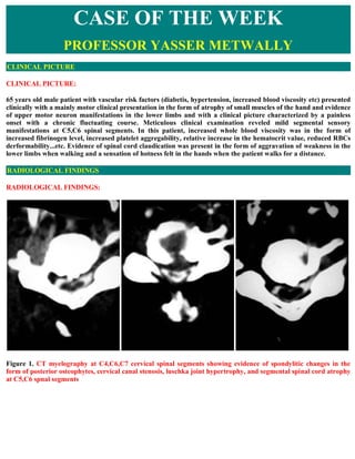
  • 1. CASE OF THE WEEK PROFESSOR YASSER METWALLY CLINICAL PICTURE CLINICAL PICTURE: 65 years old male patient with vascular risk factors (diabetis, hypertension, increased blood viscosity etc) presented clinically with a mainly motor clinical presentation in the form of atrophy of small muscles of the hand and evidence of upper motor neuron manifestations in the lower limbs and with a clinical picture characterized by a painless onset with a chronic fluctuating course. Meticulous clinical examination reveled mild segmental sensory manifestations at C5,C6 spinal segments. In this patient, increased whole blood viscosity was in the form of increased fibrinogen level, increased platelet aggregability, relative increase in the hematocrit value, reduced RBCs derformability...etc. Evidence of spinal cord claudication was present in the form of aggravation of weakness in the lower limbs when walking and a sensation of hotness felt in the hands when the patient walks for a distance. RADIOLOGICAL FINDINGS RADIOLOGICAL FINDINGS: Figure 1. CT myelography at C4,C6,C7 cervical spinal segments showing evidence of spondylitic changes in the form of posterior osteophytes, cervical canal stenosis, luschka joint hypertrophy, and segmental spinal cord atrophy at C5,C6 spnal segments
  • 2. Figure 2. CT myelography at C4,C6,C7 cervical spinal segments showing evidence of spondylitic changes in the form of posterior osteophytes, cervical canal stenosis, luschka joint hypertrophy, and segmental spinal cord atrophy at C5,C6 spnal segments. Notice the calcified soft disc herniation. Figure 3. MRI T2 images showing evidence of cervical disc degeneration and cervical canal stenosis In the above reported case evidence of spondylitic cervical canal stenosis, with posterior osteophytes, calcified hard disc herniation and C5,C6 segmental spinal cord atrophy was demonstrated. The clinical picture was in the form of a mainly motor clinical manifestations and a remitting course with evidence of spinal cord claudication. Because the vascular spondylitic myelopathy has a sudden painless onset and a fluctuating course with remission and exacerbation, it was frequently misdiagnosed as multiple sclerosis. However major differences are present
  • 3. between myelopathy due to disc disease and that due to multiple sclerosis as follows Unlike multiple sclerosis, myelopathy due to cervical spondylosis had a sudden onset with the clinical picture  developing over just a few hours. Unlike multiple sclerosis, the duration of relapses in myelopathy due to cervical spondylosis is very short ( on  the average few hours to one or two days). Unlike multiple sclerosis, relapses of myelopathy due to cervical spondylosis shared a similar clinical  presentation in every single patient i.e. the disease was disseminated only in time and never in place. And although signs and symptoms might be severer on recurrent episodes (mainly due to the cumulative effect of structural damage and/or the functional disturbances caused by each ischaemic episode), however the disease used to recur in the same anatomical site (dorso-lumber spinal segments) and is never disseminated in place. Unlike multiple sclerosis, the clinical picture of myelopathy due to cervical spondylosis is mainly motor ( in  the form of weakness and atrophy) and sensory manifestations, though definite, are detected only by careful examination. In fact the quot;mainly motor clinical picturequot; was occasionally a potential source for anther misdiagnosis which is motor neuron disease or motor neuropathy. However myelopathy due to cervical spondylosis can easily be differentiated from motor neuron disease because of the relapsing remitting course, and because of the existence of definite, though subtle, sensory manifestations. Also the existence of impotence, bladder disturbances and occasional back pain are points against the diagnosis of primary motor neuron disease. The predominance of motor manifestations in myelopathy due to cervical spondylosis is in fact anther point favouring its ischemic aetiology. It is clear that when ischaemia occurs, the most vulnerable region of the spinal cord is the grey matter because its metabolic rate is three to five times greater than the metabolic rate of the white matter. This would account for the many cases reported in literature of paraparesis with little sensory manifestations and for instances of lower motor neuron syndromes of an ischaemic basis. In cervical spondylotic myelopathy patients the motor weakness is characteristically increased by walking and relieved by rest and this is anther point favouring the ischemic aetiology of myelopathy due to degenerative disc disease. Normally walking is associated with marked increase of blood flow to the spinal cord and cauda roots to meet the increased metabolic rate of these neural structures, physiologically the spinal cord microvascular bed will dilate to accommodate the increased blood flow. Cervical canal stenosis (induced by disc disease) and the associated segmental arteriosclerosis will hinder this normal physiological quot;exertion induced hyperaemiaquot; of the neural structures resulting in a temporary spinal cord quot;ischaemic dysfunction on exertionquot;. Although the prognosis following a single ischaemic episode is good , however repetition of the ischaemic episodes will ultimately result in spinal cord atrophy with irreversible neurological deficits The patient, following admission, received medical treatment for diabetes, hypertension, antiplatelet medications and medications that improve RBCs deformability, reduce whole blood viscosity and fibrinogen level (like pentoxifylline, bezafibrate etc) and he was referred to surgery once diagnosed radiologically. DIAGNOSIS: DIAGNOSIS: CERVICAL SPONDYLOTIC VASCULAR MYELOPATHY DISCUSSION DISCUSSION: Patients with spondylitic cervical myelopathy are classified, clinically, into two main groups. The painless
  • 4. myelopathy group (due to vascular aetiology) and the painful myelopathy group (due to compression of the spinal cord). Clinically the painless myelopathy group is characterized by a fluctuant course with remission and exacerbations and a mainly motor clinical presentation. In the painless myelopathy group, the natural history of the disease is determined by the interaction of three main pathogenic factors. Spondylitic factor, vascular factor and haemorheological factor associated with increased whole blood viscosity (increased haematocrit value and serum fibrinogen).   Figure 4. The painless cervical spondylitic myelopathy is the interaction between three main pathogenic factors The spondylitic factor ultimately results in bony and soft tissue hypertrophy that causes cervical canal stenosis, and encroach upon the subarachnoid space, reducing its volume. Lack of the CSF cushioning effect will cause embarrassment of the spinal circulation at the level of cervical enlargement since optimum blood supply to the spinal cord needs an optimum CSF cushioning effect. The second factor is the vascular arteriolar factor. Apparently the painless myelopathy group comprises a group of patients where the incidence of vascular risk factors, (such as essential hypertension, NIDDM, hyperlipidaemia (Type IV), hyperfibrinogenemia and increased platelet aggregation...the metabolic syndrome) is very high. The incidence of arteriosclerosis is known to be high among patients with vascular risk factors. 4 This is consistent with the necropsy findings of Manen, 1966 2 and Jillinger 19671. The authors reported arteriolosclerosis, lipohyalinosis and fibrosis of the perforating intramedullary vessels and the fine vessels lying on the surface of the spinal cord in patients with cervical spondylitic vascular myelopathy.4 These changes were maximum in the cervical enlargement and the overlapping zone in the cervico-dorsal region, making the spinal cord especially vulnerable at this zone to vesico-circulatory disorders, mainly of extra-medullary origin, which cause critical decrease in spinal cord flow. It should be mentioned that the area of the spinal cord between C4 and DI is a watershed area with marginal blood supply. This last field zone is most likely to suffer from insufficiency of blood and has been shown to be a preferential zone for vascular damage. 4 According to the necropsy results of Jillinger, 19671 the arteriosclerotic changes in the overlapping cervico-dorsal region were isolated findings. They did not depend on age and were negatively correlated with arteriosclerosis in the rest of the body. A finding that probably denotes that the injurious effect of the spondylitic changes accelerate the arteriosclerotic changes in the region of the cervical enlargement. However it should be noted that both cervical spondylosis and arteriosclerosis are slowly progressive pathology and they can not be held responsible for the sudden onset of the clinical symptomatology seen in the painless myelopathy group. No compressing agents (like soft disc herniation, or osteophytes) are demonstrated radiologically, in this group of patients, that can explain the clinical symptomatology in terms of compression of the spinal cord and/or an important radicular artery. In short both cervical spondylosis and arteriosclerosis serve by furnishing the
  • 5. background for the ultimate determinant of the clinical symptomatology. Cervical spondylosis will result in canal stenosis, loss of the CSF cushioning effect and embarrassment of the spinal circulation in the region of the cervical enlargement. Arteriosclerosis will result in reduction of the caliber of the radicular and the perforating intramedullary arterioles with loss of the auto-regulatory physiological process. Flow in the perforating arteries is dependent on the auto-regulatory process of the penetrating intramedullary arterioles on one hand and the whole blood viscosity on the other hand. Loss of the auto-regulatory process secondary to advanced arteriosclerosis will simply mean that the spinal cord perfusion, in the vulnerable region of the cervical enlargement, will fluctuate with fluctuation of the whole blood viscosity, (the ultimate determinant of the vascular myelopathy).4 Whole blood viscosity is a collective terminology that reflects the influence of various factors that include mainly the corpuscular and the plasmatic components of the blood. Whole blood viscosity is usually increased in patients with the vascular spondylitic myelopathy. Blood viscosity is mainly determined by the hematocrit value and the plasma viscosity is mainly determined by the plasma fibrinogen level. High values of lipid have also be found to increase whole blood viscosity. 4 Increase platelet aggregation also increases whole blood viscosity. The behavior of the red blood cells was also found to affect the blood viscosity. Increased red cell aggregation and reduced red cell deformability increase whole blood viscosity. The RBCs deformability is invariably reduced and the aggregability in invariably increased in the presence of high fibrinogen level and high haematocrit values. Fibrinogen in particular is a strong RBCs aggregant agent. Inverse correlation is present between the red cell deformability and the haematocrit value and serum fibrinogen level. Increase of the whole blood viscosity is a common finding in essential hypertension and NIDDM. The vascular resistance of the perforating blood vessels of the spinal cord and the brain is dependent upon the ratio between the whole blood viscosity over the caliber of the blood vessel. Increase of the whole blood viscosity results in high vascular resistance to blood flow and subsequently low perfusion pressure and neuronal tissue ischaemia. Stenosis of the perforating blood vessel secondary to arteriosclerosis further aggravates the problem. Inverse correlation is present between the neuronal tissue blood flow and serum fibrinogen level and the haematocrit value. 4 Hyperfibrinogenemia and increased RBCS, and platelet aggregation reflect a hypercoagulable state with increased thrombotic tendency that selectively affects the small perforating blood vessels and the microcirculation of the brain and spinal cord. Microvascular occlusion can occur either by local aggregation of hyperaggragable platelets or by red cell aggregation with impaction of rigid red blood cells in the microcirculation. This is more likely to occur with the existence of high red cell mass (Hch value) that can displace the hypersensitive platelets Towards the arteriolar wall resulting in platelet aggregation and thrombus formation. Thrombus formation is enhanced if the arteriolar wall is abnormal (arteriolosclerosed).4 The high blood viscosity observed in the painless myelopathy patients should simply mean, especially when coupled with arteriosclerosis of the small perforating blood vessels, that the blood flow to the spinal cord at the level of cervical enlargement is subjected to high vascular resistance that could ultimately results in low perfusion pressure and chronic ischaemia. The increased thrombotic tendency observed in those patients should mean that the chronic ischaemia state could be interrupted by acute thrombotic microvascular occlusion that can result, pathologically, in spinal cord lacunar infarction at the level of cervical-enlargement and clinically in lower cervical painless myelopathy of sudden onset and regressive course. The acute thrombo-occlusive episodes are responsible for the intramedullary cavitations observed in the painless myelopathy group. 4 NEUROIMAGING FINDINGS IN THE PAINLESS MYELOPATHY GROUP In general two pathological subtypes are observed in this group, spinal cord atrophy and spinal cord cavitations, both are very segmental and both are the end result of spinal cord ischemia.
  • 6.  Two types of spinal cord of cavitations are observed in the painless myelopathy group  CAVITATIONS 1-Pencil shaped necrosis: the cavitation is maximum at the level of C5,C6 spinal segments and extends one ore two segments above and/or below CAVITATIONS 2-Central gray matter cavitation: bilateral ,symmetrical,rounded cavitations, localized in the CAVITATIONS bilateral anterior horns of spinal segments C5,C6 Pencil shaped necrosis and central gray matter cavitations, most probably represent spinal lacunar infarctions. The spinal cord atrophy observed in those patients is very segmental and exclusively localized to the level of cervical enlargement between C4- C8 with maximum changes at C5,C6 segment. The spinal cord above C4 and below C8 is often normal. The segmental spinal cord atrophy observed in these patients is the result of long standing chronic ischaemia interrupted by recurrent thrombo-occlusive episodes.4 All the atrophic and cystic changes are exclusively limited to the spinal cord area between C4 and C7. The C5-C6 segments are most frequently involved. The C5-C6 segments are the most vulnerable to vascular damage as they represent a watershed area with higher incidence of segmental arteriosclerosis. 1 Figure 5. In cervical spondylitic myelopathy, both cystic and atrophic changes are exclusively localized to the level of cervical enlargement with maximum changes at C5,C6 spinal segments Figure 6. In cervical spondylitic myelopathy, both cystic [left image] and atrophic [right image] changes are exclusively localized to the level of cervical enlargement with maximum changes at C5,C6 spinal segments The central gray matter cavitations demonstrated in the painless group group represent lacunar infarctions involving the bilateral anterior horns. It results clinically in a purely LMN picture ( proximal muscles amyetrophy).1
  • 7. Figure 7. CT myelography showing central gray matter cavitations as bilateral symmetrical,well defined rounded zones of intramedullary contrast accumulations in the presumed anatomical areas of anterior horns [right image is a schematic representation] The association between spondylitic myelopathy and spinal cord atrophy and/or cavitation is well known. The ischaemic aetiopathogenesis of myelopathy in this group of patients is further substantiated by the observation that relapses of myelopathy are intimately coupled temporally with rise of whole blood viscosity and thrombotic tendency. Relapses occurs more frequently in the summer time. Dehydration is more common in summer time, it results in contraction of the plasma volume and rise of the haematocrit value and subsequently blood viscosity. Figure 8. MRI T1,T2 images showing pencil-shaped necrosis Figure 9. CT myelography showing normal spinal cord at the level of cervical enlargement (A) and spinal cord atrophy in cervical spondylitic myelopathy (B,C), notice that the atrophic segments are flattened, collapsed, with irregular contour and wide subarachnoid spaces The ischaemic aetiology of myelopathy in the painless myelopathy group is consistent with the necropsy findings of Jellinger, 1967 1. The author reported, in spondylitic myelopathy patients, white matter ischaemic demyelination, neuronal degeneration and a diffuse lacunar state similar to those seen in the basal ganglion in the hypertensive small vessel disease of the brain. It should also be mentioned that increased whole blood viscosity is also the ultimate aetiopathogenic factor in hypertensive microvascular brain disease (diffuse lacunar state, leukoaraiosis, etc.).4 The haemorheological profile of vascular spondylitic myelopathy is similar to the haemorheological profile of hypertensive micro-vascular brain disease (Lacunar infarction, leukoaraiosis etc.) previously reported by many authors4. The haemorheological parameters tended to drop down to the normal levels following the acute phase in myelopathy patients and this has also been reported in ischaemic brain disease. The hypertensive micro-vascular brain disease is found to be similar in many ways to the spondylitic vascular
  • 8. myelopathy regarding vascular risk factors, the vascular arteriolar pathology, parenchymatous pathology and the haemorheological profile. Similarities are listed in in the following table. Vascular risk factors NIDDM,hypertension,hyperlipidaemia ,old age,LVH,are common in both diseases Vascular arteriolar Lipohyalinosis of the fine penetrating arterioles is common in both diseases pathology Pathological findings Neuronal degeneration, ischaemic demyelination and a diffuse lacunar state are common in both diseases Haemorheological profile Increased whole blood viscosity and thrombotic tendency of the blood are common in both diseases Because the vascular spondylitic myelopathy has a sudden painless onset and a fluctuating course with remission and exacerbation, it was frequently misdiagnosed as multiple sclerosis or transverse myelitis. The demonstration of haemorheological abnormalities in those patients should suggest important lines of treatment. Antiplatelet medications, drugs that improve RBCs deformability, reduce whole blood viscosity and fibrinogen level (like pentoxifylline, bezafibrate etc) will be of great value in those patient. Also maintenance of good body hydration is of paramount importance especially in the older age group. Control of risk factors like hypertension and NIDDM is essential. Although the prognosis following a single ischaemic episode is good. however repetition of the ischaemic episodes will ultimately result in spinal cord atrophy with irreversible neurological deficits ( So patients with the spondylitic vascular myelopathy probably needs prophylactic treatment like cerebrovascular patients following the first acute myelopathic episode). In general the vascular aetiology is the most common cause of the spondylitic myelopathy. The spondylitic vascular myelopathy is present mainly in males and is characterized, clinically, by a painless clinical picture and radiologically by the presence of segmental spinal cord atrophy or cavitations. Incidence of vascular risk factors is high among patients with vascular myelopathy with frequent haemorheological abnormalities denoting increased whole blood viscosity. Although central disc herniation can cause a painless clinical picture, but this can easily be excluded radiologically. The roles played by the spondylitic process, the vascular pathology and the haemorheological abnormalities in the pathogenesis of the spondylitic vascular myelopathy are summarized in following table. Pathological factor  Effect Cervical canal stenosis with reduction of the CSF subarachnoid spaces,lack of the Cervical spondylosis CSF cushioning effect will cause embarrassment of the circulation at the level cervical enlargement Reduction of the caliber of the intramedullary arterioles with loss of the Sclerosis of the autoregulatory process, this increases the vascular resistance and results in low intramedullary arterioles perfusion pressure and chronic tissue ischaemia. Arteriolosclerosis is maximum at the level of cervical enlargement Increased fibrinogen level,haematocrit value,and platelet aggregability. This increases whole blood viscosity and subsequently the vascular resistance and results Increased whole blood in low perfusion pressure and chronic tissue ischaemia, it also increases the viscosity thrombotic tendency of the blood and results in recurrent acute microvascular thrombo-occlusive episodes SUMMARY CT SCAN IMAGING OF THE SPONDYLITIC VASCULAR MYELOPATHY
  • 9. Intrathecal enhancement is necessary  CT SCAN In spinal cord atrophy, the spinal cord is deformed, with wide subarachnoid spaces  CT In pencil shaped necrosis, intramedullary contrast accumulation occurs at C5,C6 with occasional  SCAN extension of cavitation one or two segments Above or below CT In central gray matter cavitation there is bilateral,symmetrical, rounded and well-defined SCAN  accumulation of contrast material in the regions of the anterior horns. CERVICAL SPONDYLOSIS WITH SIGNIFICANT NECK PAIN ASSOCIATED WITH MYELOPATHY AND/OR RADICULOPATHY  Neck pain in association with cervical spondylosis is either due to  1- Acute neck pain : Is commonly due to acute soft disc herniation, this results clinically in radiculopathy and or myelopathy. 2- Chronic neck pain: This is commonly due to foraminal stenosis due to facet joint hypertrophy and or luschka joint disease.   Figure 10. luschka joint hypertrophy resulting in marked foraminal stenosis SUMMARY SUMMARY Cervical spondylotic myelopathy is the most common cause of spinal cord dysfunction in older persons. The aging process results in degenerative changes in the cervical spine that, in advanced stages, can cause compression of the spinal cord. Symptoms often develop insidiously and are characterized by neck stiffness, arm pain, numbness in the hands, and weakness of the hands and legs. The differential diagnosis includes any condition that can result in myelopathy, such as multiple sclerosis, amyotrophic lateral sclerosis and masses (such as metastatic tumors) that press on the spinal cord. The diagnosis is confirmed by magnetic resonance imaging that shows narrowing of the spinal canal caused by osteophytes, herniated discs and ligamentum flavum hypertrophy. Choice of treatment remains controversial, surgical procedures designed to decompress the spinal cord and, in some cases, stabilize the spine are successful in many patients. Cervical spondylotic myelopathy (CSM) is the most common spinal cord disorder in persons more than 55 years of age in North America and perhaps in the world. As the number of older persons in the United States increases, the incidence of CSM will most likely increase. In a prospective study designed to more accurately define the incidence
  • 10. of CSM, 23.6 percent of 585 patients with tetraparesis or paraparesis admitted to a United Kingdom regional neuroscience center had CSM.5 The overall prevalence in this population is unknown. Pathophysiology of CSM Spondylosis refers to the degenerative changes that occur in the spine, including degeneration of the joints, intervertebral discs, ligaments and connective tissue of the cervical vertebrae. There are three important pathophysiologic factors in the development of CSM: (1) static mechanical; (2) dynamic mechanical; and (3) spinal cord ischemia. Static mechanical factors result in the reduction of spinal canal diameter and spinal cord compression. With aging, the intervertebral discs dry out resulting in loss of disc height. This process puts greater stress on the articular cartilage of the vertebrae and their respective end plates. Osteophytic spurs develop at the margins of these end plates (Figure 11). Osteophytes stabilize adjacent vertebrae whose hypermobility is caused by the degeneration of the disc. The disc also calcifies, further stabilizing the vertebrae. Osteophytes increase the Figure 11. Axial computerized tomography scan showing ventral weight-bearing surface of the end plates and, therefore, decrease the effective force osteophytes pressing into the spinal being placed on them. In addition to osteophytic overgrowth, the ligamentum canal. flavum may stiffen and buckle into the spinal cord dorsally. Osteophytic overgrowth ventrally and, in some cases, buckling of the ligamentum flavum dorsally can cause direct compression of the spinal cord resulting in myelopathy (clinically evident spinal cord dysfunction). Symptoms are believed to develop when the spinal cord has been reduced by at least 30 percent. Dynamic mechanical factors relate to the fact that the normal motion of the cervical spine may aggravate spinal cord damage precipitated by direct mechanical static compression. During flexion, the spinal cord lengthens, thus stretching over ventral osteophytic ridges. During extension, the ligamentum flavum may buckle into the spinal cord causing a reduction of available space for the spinal cord (Figure 12). Figure 12. Dynamic mechanical factors in cervical spondylotic myelopathy. (Left) During flexion, the spinal cord is stretched over ventral osteophytic ridges. (Right) During extension, the ligamentum flavum may buckle into the spinal cord reducing space for the cord. Spinal cord ischemia probably plays a role in the development of CSM, particularly in later stages. Histopathologic changes in the spinal cord consistent with ischemia have been observed in patients with CSM. However, the precise mechanism for spinal cord ischemia is not completely understood. Other factors associated with the development of spondylosis include heavy labor, posture and genetic predisposition. Also, 70 percent of patients with Down syndrome have an increased incidence of spondylosis by 50 years of age.
  • 11. Clinical History Patients with CSM will generally have these symptoms: neck stiffness; unilateral or bilateral deep, aching neck, arm and shoulder Common complaints of patients with cervical pain; and possibly stiffness or clumsiness while walking. CSM spondylotic myelopathy include neck usually develops insidiously. In the early stages of CSM, complaints stiffness, crepitus in the neck with of neck stiffness are common because of the presence of advanced movement, brachialgia, a dull quot;achyquot; feeling cervical spondylosis. Other common complaints include crepitus in in the arm, and numbness or tingling in the the neck with movement; brachialgia, which is characterized as a hands. stabbing pain in the pre- or postaxial border of the arm, elbow, wrist or fingers; a dull quot;achyquot; feeling in the arm; and numbness or tingling in the hands. Pain following a stereotypical dermatomal distribution is referred to as a radiculopathy rather than a myelopathy. For example, in patients with a disc herniation between the sixth and seventh vertebrae, pain radiates into the shoulder, upper arm, elbow, and index and middle fingers. It is typically unilateral. Numbness and weakness follow the same distribution. Some patients will exhibit signs and symptoms of radiculopathy and myelopathy. The hallmark symptom of CSM is weakness or stiffness in the legs. Patients with CSM may also present with unsteadiness of gait. Weakness or clumsiness of the hands in conjunction with the legs is also characteristic of CSM. Symptoms may be asymmetric particularly in the legs. Loss of sphincter control or frank incontinence is rare; however, some patients may complain of slight hesitancy on urination. Addendum  A new version of this PDF file (with a new case) is uploaded in my web site every week (every Saturday and  remains available till Friday.) To download the current version follow the link quot;http://pdf.yassermetwally.com/case.pdfquot;.  You can also download the current version from my web site at quot;http://yassermetwally.comquot;.  To download the software version of the publication (crow.exe) follow the link:  http://neurology.yassermetwally.com/crow.zip The case is also presented as a short case in PDF format, to download the short case follow the link:  http://pdf.yassermetwally.com/short.pdf At the end of each year, all the publications are compiled on a single CD-ROM, please contact the author to  know more details. Screen resolution is better set at 1024*768 pixel screen area for optimum display.  For an archive of the previously reported cases go to www.yassermetwally.net, then under pages in the right  panel, scroll down and click on the text entry quot;downloadable case records in PDF formatquot; Also to view a list of the previously published case records follow the following link  (http://wordpress.com/tag/case-record/) or click on it if it appears as a link in your PDF reader REFERENCES References 1-Jellinger K : Spinal cord arteriosclerosis and progressive vascular myelopathy. J Neurol Neurosurg Psychiat 30:195-206, 1967
  • 12. 2-Manen T :Vascular lesions in the spinal cord in the elderly patients. Geriatrics 4:151-160, 1966 3-Metwally MYM :Value of ct scan in the evaluation of spinal cord lesions. Md thesis, Ain Shams university, cairo egypt (department of neurology), 1991 4-Metwally MYM : Cervical spondylitic myelopathy, clinico-radiological approach,with correlation with the haemorheological pararameters and vascular risk factors. Ain-Shams medical journal, vol 46, number 4,5,6 , 721- 750, 1995 5- Moore AP, Blumhardt LD. A prospective survey of the causes of nontraumatic spastic parapesis and a tetraparapesis in 585 patients. Spinal Cord 1997; 35:361-7. 6- Metwally, MYM: Textbook of neuroimaging, A CD-ROM publication, (Metwally, MYM editor) WEB-CD agency for electronic publication, version 10.1a January 2009