Case record...Hypertensive cerebral hemorrhage
•
6 recomendaciones•1,566 vistas

Hypertensive cerebral hemorrhage

Recomendados
Recomendados
Más contenido relacionado
Similar a Case record...Hypertensive cerebral hemorrhage
Similar a Case record...Hypertensive cerebral hemorrhage (20)
Más de Professor Yasser Metwally
Más de Professor Yasser Metwally (20)
Último
Último (20)
Case record...Hypertensive cerebral hemorrhage
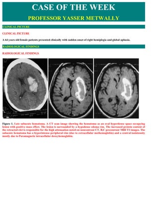
- 1. CASE OF THE WEEK PROFESSOR YASSER METWALLY CLINICAL PICTURE CLINICAL PICTURE A 64 years old female patients presented clinically with sudden onset of right hemiplegia and global aphasia. RADIOLOGICAL FINDINGS RADIOLOGICAL FINDINGS Figure 1. Late subacute hematoma. A CT scan image showing the hematoma as an oval hyperdense space occupying lesion with positive mass effect. The lesion is surrounded by a hypodense edema rim. The increased protein content of the retracted clot is responsible for the high attenuation noted on noncontrast CT. B,C precontrast MRI T1 images. The subacute hematoma has a hyperintense peripheral rim (due to extracellular methemoglobin) and a central isointensity mostly due to Paramagnetic intracellular deoxyhemoglobin.
- 2. Figure 2. Late subacute hematoma. MRI T2 images. The subacute hematoma has a hyperintense peripheral rim (due to extracellular methemoglobin) and a central hypointensity mostly due to Paramagnetic intracellular deoxyhemoglobin. Edema could also contribute to the peripheral T2 hyperintensity. Figure 3. Late subacute hematoma. MRI FLAIR images. The subacute hematoma has a hyperintense peripheral rim (due to extracellular methemoglobin) and a central iso to hypointensity mostly due to deoxyhemoglobin. In this patient the appearance of the subacute hematoma on FLAIR images is similar to that seen on MRI T2 images. Edema could also contribute to the peripheral T2 hyperintensity.
- 3. Figure 4. Diffusion weighted images. The appearance of hemorrhage on DW MR images is complex and involves many factors, including the relative amounts of different hemorrhagic products and the pulse sequence used. Oxyhemoglobin is hyperintense on DW images and has a lower ADC than does normal brain tissue; this may indicate the relative restriction of water movement inside the red blood cell (101). Extracellular methemoglobin has a higher ADC than does normal brain tissue, which indicates that the mobility of water in the extracellular space is increased. The prolongation of the T2 component of fluid with extracellular methemoglobin results in hyperintensity on DW images. Hemorrhage containing deoxyhemoglobin, intracellular methemoglobin, and hemosiderin are hypointense on DW images because of magnetic susceptibility effects. Because these products of hemorrhage have very low signal intensity on T2-weighted images, ADCs cannot be reliably calculated for them. Figure 5. For comparison, A view showing the hematoma on CT scan, precontrast MRI T1 image, MRI T2, FLAIR and diffusion weighted images
- 4. Figure 6. For comparison, A view showing the hematoma on CT scan, precontrast MRI T1 image, MRI T2, FLAIR and diffusion weighted images DIAGNOSIS: DIAGNOSIS: HYPERTENSIVE PUTAMENO-CAPSULAR HEMORRHAGE DISCUSSION DISCUSSION Haemorrhagic microvascular brain disease constitutes the other facet of the bad coin (the microvascular brain disease) the first facet of which is the ischemic microvascular brain disease. Both the haemorrhagic and the ischaemic microvascular brain disease share common haemorheological, metabolic endocrinal abnormalities and cardiac changes (LVH). In microvascular brain disease, the small penetrating arterioles of the subependymal and the pial microvascular systems tend to become stenosed and undergo lipohyalinosis or they may dilate to form microaneurysms. From the pathological point of view both Lipohyalinosis and microaneurysms, almost invariably, coexist in the same individual, thus making the patient Liable to develop either the ischaemic or the haemorrhagic microvascular brain diseases.
- 5. Figure 7. Microaneurysms of the small penetrating arterioles Microaneurysmal formation occurs predominantly in the territory of the subependymal microvascular system,thus making the incidence of the haemorrhagic microvascular events much more frequent in the periventricular gray matter (thalamus, basal ganglia and the internal capsule) or the immediate periventricular white matter. The coexistence of lipohyalinosis and microaneurysms in the periventricular regions will explain the propensity of the diseased microvascular system either to thrombose (resulting in lacunar infarctions) or to rupture and leak resulting in periventricular haematoma formation. Lacunar infarctions and hypertensive cerebral haemorrhages are two facets of one and the same bad coin (the microvascular brain disease). Figure 8. Microaneurysms are predominately distributed in the immediate periventricular region Microaneurysmal formation should weaken the arteriolar wall so that rupture and leakage can occur even in normotensive states. When microaneurysmal rupture occurs, the bleeding will result in haematoma formation. The bleeding will then be arrested by occlusive thrombosis of the bleeding microaneurysms. Following microaneurysmal rupture and bleeding, the size of the resulting haematoma will be determined by the bleeding time. The bleeding time is a function of the whole blood viscosity in general and the platelet aggregability in particular. Should microaneurysmal bleeding occurs during periods of higher blood viscosity, the bleeding time will be shorter and
- 6. subsequently the size of the resulting haematoma will be smaller. In fact during high blood viscosity the bleeding is not infrequently arrested before forming haemorrhages adequate to give rise to immediate clinical sequelae. Patients with higher blood viscosity and thrombotic tendency, although less likely to develop serious haemorrhagic microvascular events, they are particularly liable to develop serious ischaemic microvascular events. During periods of lower blood viscosity and thrombotic tendency of the blood, microaneurysmal bleeding might result in huge haematoma formation that may split along the planes of the white matter forming a substantial space occupying clot, or may rupture into the ventricular system resulting in massive ventricular haemorrhage. In general inverse correlation is present between the haematoma size and the current blood viscosity at the time of microaneurysmal bleeding. Patients with microvascular brain disease might have recurrent events which could be purely haemorrhagic or purely ischaemic, however, it is not uncommon for some patients to fluctuate between the haemorrhagic and the ischaemic events, developing haemorrhagic events at certain times and ischaemic events at other times. In general ischaemic microvascular events are much more common and much more frequent than the haemorrhagic events. PATHOGENESIS OF HYPERTENSIVE CEREBRAL HEMORRHAGE Hypertension causes fibrinoid necrosis of these penetrating arterioles. The massive intracerebral hemorrhage which is a complication of hypertension, arises from rupture of a necrotic arteriole or from rupture of a minute quot;miliaryquot; aneurysm formed at the site of necrosis. These aneurysms were first described by CHARCOT and BOUCHARD. The frequency of fibrinoid necrosis and miliary aneurysm formation in vessels within basal ganglia and thalamus accounts for the frequency of intracerebral hemorrhage in those locations. Fibrinoid is identified by its structureless or sometimes granular red appearance on H&E stain and by the fact that , unlike hyalinized smooth muscle which is also eosinophilic, the fibrinoid areas stain with stains for fibrin such as PTAH or Putz stain or with certain trichrome stains. The fibrinoid change in these vessels was called lipohyalinosis by Miller-Fisher in a very influential series of articles. However that term is confusing because hyalinized arteries are arteries whose media has undergone a pathologic change which is not fibrinoid necrosis and which by itself does not lead to rupture. Indeed hyalinized arterioles are common in hypertension. The term lipohyalinosis stresses the presence of fat in the degenerate arteriolar wall but again this change is not the hallmark of the arterioles that are in danger of rupturing or forming miliary aneurysms. The fibrinoid change is the critical change in these diseased arteriolar segments looks and stains just like the fibrinoid seen in renal and other arterioles in malignant hypertension. The important point to remember is that, for unknown reasons, the brain arterioles can undergo fibrinoid necrosis even in so-called benign hypertension--that is in patents with only modest blood pressure elevation. For that reason it is important to treat even benign hypertension. The series figures below illustrates the pathologic processes that can lead to rupture. Figure 9. A, The figure shows the wall of an arteriole stained with H&E. The amorphous pink [eosinophilic] material in
- 7. the wall could be either fibrinoid or amyloid. To prove that it is firbrinpoid the section or its close neighbor should be stained with any one of several techniques that stain fibrin [e.g. Putz stain-blue; or the PTAH stain-blue; or a trichrome stain such as the azo carmine stain; the azo carmine is particularly good because it distinguishes fibrinoid from garden variety hyalinization by staining fibrin/fibrinoid red while staining collagen or hyalinized collagen blue.]. B, This section was stained with azocarmine. An arteriole in the subarachnoid space has an amorphous red material occupying a good portion of its wall. This is fibrinoid. Fibrinoid is frequently segmental in distribution so that the entire circumference may not be involved and other areas along the length of the vessel may also be spared. C, This figure was also stained with azocarmine. The arteriole wall is replaced by red fibrinoid and displays aneurysmal dilation. Figure 10. A,B Sometimes a miliary aneurysm thrombosis rather than ruptures. It then appears as a fibrous ball which may be separated from the parent vessel due to the plane at which the section has been cut. If the section is close to the parent arteriole there will be elastic tissue at the margin of the ball. This elastic tissue stains black with the VVG stain in (B) Figure 11. The pathologist got lucky when this section was taken. Here a miliary aneurysm that has neen converted to a fibrous ball or globe, shown in this longitudinal section, still connected to the parent arteriole by a thin neck. PATHOLOGY Cerebral Haematomas occur much more frequently at the putameno-capsular and the thalamic regions and may rupture into the ventricular system. Less common sites include the cortical and the immediate subcortical white matter, especially in the parietal region, the pons and the cerebellum. The resulting haematoma is dark red in colour due to the existence of deoxyhaemoglobin inside the intact RBCS. During the subacute stage (3 days - one month) the dark red colour of the haematoma is replaced by a brownish discoloration, which starts at the periphery of the haematoma and then extends to its center. This brownish discoloration occurs due to the replacement of deoxyhaemoglobin by the oxidized methemoglobin. Acute hematoma usually spreads between white matter tracts resulting in island of viable brain tissues within the hematoma itself. Bleeding usually stops shortly after the initial ictus, however in a substantial minority of patients the hematoma continues to expand usually within the first hour after the presentation. Expansion after one hour is unusual.
- 8. Once hematoma forms, vasogenic edema forms around the clot as osmotically active serum proteins are released from the hematoma. Edema peaks at about 48 hours and usually begins to resolve after 5 days. Whether the brain tissues surrounding the acute hematoma is ischemic -due to vascular compression- or not is controversial. Functional suppression (diaschisis) of brain activity rather than ischemia is more probable. Risk of Hematoma Enlargement In nearly one quarter of initially alert patients presenting with spontaneous intracerebral hemorrhage, secondary deterioration in level of consciousness occurs within the first 24 hours after onset. Hematoma expansion and edema formation are believed to be the major factors involved In several large prospective and retrospective studies, investigators have evaluated the rate of hematoma enlargement after initial presentation and report rates ranging from 14 to 38% within the first 24 hours of admission.[27,28] In their review of 627 patients with spontaneous intracerebral hemorrhage Fujii, et al..[27] reported that CT scanning within 24 hours of admission demonstrated enlargement of the hematoma in 14% of patients. Five factors were found to be associated with enlargement: admission shortly after onset of symptoms, heavy alcohol consumption, irregularly shaped hematoma, reduced level of consciousness, and low level of fibrinogen. Figure 12. Cerebral (A) and pontine (B) acute haemorrhage, C, acute cerebellar hemorrhage Gradually the haematoma is surrounded by reactive gliosis and macrophages laden with haemosiderin granules (Ferric hydroxide). The clot is gradually absorbed starting with its periphery and is replaced by a yellow fluid, this is called an apoplectic cyst. Reactive gliosis progressively increases and ultimately transforms the haematoma into a slit-like scar. Figure 13. A, acute putameno-capsular & intraventricular hemorrhage, B, apoplectic cyst
- 9. Figure 14. A, Subacute caudate hemorrhage, B, apoplectic cyst, C, Hypertensive hemorrhage into basal ganglia region (specifically: internal capsule). Pathologically the brains of patients with cerebral haemorrhages very frequently show evidence of past microvascular ischaemic events such as lacunar infarctions, leukoaraiosis,etc. INCIDENCE OF COMMON ANATOMICAL SITES IN HYPERTENSIVE INTRACEREBRAL HAEMORRHAGE Figure 15. Incidence (in % ) of the common anatomical sites in hypertensive intracerebral haemorrhage 28 STRUCTURAL NEUROIMAGING OF MICROVASCULAR CEREBRAL HAEMORRHAGE CT imaging of haematoma. A cerebral haematoma, in the acute stage, has higher attenuation values on precontrast scan (hyperdense). The higher attenuation values of fresh blood is due to the existence of packed haemoglobin in the haematoma. In particular the globin component of the haemoglobin is responsible for the increased CT density on precontrast scan. With progressive absorption of haemoglobin, (this usually starts from the periphery of the haematoma) the attenuation value of the haematoma gradually decreases until the high density haematoma is replaced by a low density space occupying cyst.
- 10. Figure 16. A, Acute haematoma, B, an apoplectic cyst and C, an old haematoma (slit-like scar) The evolution of the haematoma from a high density clot to a low density cyst usually takes a period that ranges between one month to three months. The walls of this cyst might enhance and the haematoma at this stage might be mixed with abscess or glioma. History is of paramount significance at this stage. Very old haematoma appears by CT scan as a slit- like hypodense area with negative mass effect. In general Haematomas are space-occupying with positive mass effect and are commonly surrounded by a hypodense oedema area. The most common sites are the putameno-capsular and the thalamic sites and either of them might rupture intraventricularly. Less common sites includes the parietal lobe, pons and cerebellum. The diagnosis of acute ICH is virtually 100% reliable with non-contrast CT due to the characteristic mass of blood of high attenuation value, due to the presence of the globin component of the haemoglobin molecule. Under exceptional circumstances, patients with profound anaemia, with a haematocrit of 20% or less have presented with an acute haematoma which was isointense to brain on account of the low haemoglobin contents of the fresh haematoma. Fresh blood has an attenuation value of 55-85 Hounsfield units, the high attenuation (50-70 Hounsfield units) is from high protein concentration within intact red blood cells and not iron content 1. As the fresh clot starts to retract after 24-48 hours from onset, there is serum extrusion around its periphery, resulting in a ring of hypointensity that surrounds the haematoma . In the subacute stage, the haematoma maintains its mass effect but becomes progressively less dense, from the periphery toward the center, until reaching isointensity with the adjacent brain parenchyma. The infusion of intravenous contrast at this stage can demonstrate an area of ring enhancement at the periphery of the haematoma. In the chronic stage, the mass effect of the haematoma is no longer present, post-contrast enhancement has disappeared after about 6 weeks from onset , and the residual is a hypointense cavity, at times in the form of a slit that can be indistinguishable from an area of old cavitated infarction. More detailed description of the CT scan appearance of brain hemorrhage The CT appearance of hemorrhage is determined by the degree of attenuation of the x-ray beam, which is proportional to the density of hemoglobin protein (relative to plasma concentration) within the hematoma. Immediately following vessel rupture, the hematoma consists of a collection of red blood cells, white blood cells, platelet clumps, and protein-rich serum that has a heterogeneous appearance on CT with attenuation in the range of 30–60 Hounsfield units (HU), depending on the degree of plasma extrusion [20]. In this hyperacute phase, hemorrhage may be difficult to distinguish from normal cortex because of similar attenuation. Over minutes to hours, a fibrin clot forms with an increase in attenuation to 60–80 HU (Fig. 11) [20]. Clot retraction and extrusion of serum can further increase attenuation to as high as 80–100 HU in the center of the hematoma. The degree of attenuation may be reduced in patients with severe anemia [21], impaired clot formation due to coagulopathy, or volume averaging with adjacent tissue. Vasogenic edema evolves around the hematoma within hours and may continue to increase for up to 2 weeks after hemorrhage onset [22].
- 11. Figure 17. CT appearance of hemorrhage. Serial CT scans of right thalamic hematoma. (A) Acute ICH in the right thalamus with mean attenuation 65 HU. (B) CT performed 8 days later than (A); the periphery of the hematoma is now isodense to the brain while the center of the hematoma has mean attenuation 45 HU. (C) CT performed 13 days later than (A) shows continued evolution of the hematoma with decreasing attenuation. (D) CT performed 5 months later than (A) shows a small area of encephalomalacia in the location of the previous hemorrhage. Over the following days, cells and protein are broken down and scavenged by macrophages, leading to slowly decreasing attenuation, with the greatest decrease at the periphery of the hematoma and more gradual evolution toward the center (Fig. 11) [23]. Within 4 to 9 days, the hematoma attenuation decreases to that of normal cortex, and within 2 to 3 weeks to that of normal white matter [20]. The CT recognition of subacute intracerebral hematoma can be challenging because the attenuation is similar to that of normal brain tissue, although mass effect may still be present. MR imaging can confirm subacute hematoma. As time goes on, attenuation continues to decrease to levels below that of the normal brain. Eventually, the hematoma resolves into a fluid-filled or slit-like cavity that may be difficult to visualize on CT (Fig. 11). Contrast enhancement is not present in the initial days following ICH but may develop at the periphery in weeks to months [24], sometimes leading to diagnostic confusion with brain tumor or abscess. A blood-fluid level may be seen in medium to large ICH within the first hours after onset; the dependent portion displays higher attenuation (Fig. 12) due to sedimentation of cellular elements [25]. This finding may be more common in ICH caused by anticoagulation [26], but it is not specific and has also been described in ICH due to hypertension, trauma, tumor, or arterial-venous malformation. The association with shorter time interval from ICH onset, and in some cases with anticoagulation, has led to speculation that incomplete clotting is required for blood-fluid level formation. Figure 18. CT with blood-fluid level. A 77-year-old woman was admitted with coma of 4 hours' duration. CT scan shows massive left hemispheric hematoma with blood-fluid level. No history of anticoagulation or coagulopathy. Box 1. As the hemorrhage evolves, different characteristic appearances can be identified on CT, depending on the age of the bleed. CT findings over time are as follows: After 7-10 days, the high density of blood begins to decrease, starting from the periphery of the lesion. From 1-6 weeks, peripheral enhancement can be seen, mimicking the appearance of an abscess, possibly related
- 12. to hypervascularity at the periphery of a resolving hematoma or disruption of the blood-brain barrier. By 2-4 months, decreased density indicates cavity formation. A residual cavity is the final stage, which is reached after complete absorption of necrotic and hemorrhagic tissue. MRI Imaging of cerebral haematoma Imaging of haematoma by MRI is time dependent as follow: The hyperacute stage (0 - 12 hour) The acute hematoma less than 12 hours old is composed mostly of intracellular oxyhemoglobin with the edematous brain undergoing necrosis. 1 On T2-weighted MR images, hyperacute hematoma will exhibit inhomogeneous signal due to hypointense deoxyhemoglobin and hyperintense, edematous cortical tissue. MR is less sensitive than CT in the hyperacute stage because diamagnetic intra- cellular oxyhemoglobin lacks unpaired electrons and thus clot signal is close to normal brain parenchyma- normal to slightly lower signal on TI-weighted images and slightly higher signal on T2-weighted images 2,3. Repeat imaging is indicated to monitor the size of the hemorrhage and the development of delayed hemorrhage and vasogenic edema. The acute stage (12 Hr - 3 days) Due to the presence of the magnetically susceptible deoxyhaemoglobin. The T2 relaxation time will be markedly shortened, so that fresh blood appears hypointense (black) on the T2 weighted MRI images. This hypointensity is commonly surrounded by a wider hyperintense area that represents oedema. On the T1 weighted images fresh blood appears isointense or slightly hyperintense. Acute hematoma one to three days old are composed mostly of paramagnetic intracellular deoxyhemoglobin. The deoxyhemoglobin is formed by the dissociation of oxygen from hemoglobin, a process that begins within several hours. Because the deoxyhemoglobin within intact, clotted hypoxic red blood cells does not cause T1 shortening, the hematoma will have normal to slightly lower signal on TI-weighted MR images. The concentration of red blood cells with clot and the concentration of fibrin cause T2 shortening, with areas of very low signal on T2-weighted spin echo and T2 * - weighted gradient echo images 3. Figure 19. A 62-year-old female with hypertension presented with acute-onset ataxia and confusion. Noncontrast CT exam of the head [left image] showed a large, right cerebellar hemorrhage, which was evacuated to relieve the mass effect on the brainstem and fourth ventricle. The cerebellar hemorrhage is seen hypointense on the T2 image due to Deoxyhemoglobin [right image]. Figure 20. The concentration of red blood cells with clot and the concentration of fibrin cause T2 shortening, with areas of very low signal on T2-weighted spin echo and T2 * -weighted gradient echo images The subacute stage (3 days - one month)
- 13. The picture of hematoma is determined by the oxidation of deoxyhemoglobin to methemoglobin and its shift from the intracellular to the extracellular compartment. The picture of haematoma, during this period is governed by the progressive reduction in the concentration of deoxyhaemoglobin and the progressive increase in the concentration of the oxidized methemoglobin. These changes take place from the periphery of the haematoma to its center. Intracellular oxidized methemoglobin induces shorting of T2 relaxation time while extracellular oxidized methemoglobin induces prolongation of T2 relaxation time Progressive reduction in the concentration of deoxyhaemoglobin and shift of oxidized methemoglobin from the intracellular to the extracellular compartment, due to lyse of RBCs, results in progressive disappearance of the T2 hypointensity observed in the acute stage. Absence of the deoxyhaemoglobin and appearance extracellular oxidized methemoglobin will result in progressive prolongation of the T2 relaxation time that starts from the periphery of the haematoma to its center, this results in progressive increase of the T2 signal intensity (it becomes brighter); At first the periphery of the haematoma becomes brighter on the T2 weighted images, and this brightness progressively extends to the center. Within a few days, the subacute hematoma start to undergo liquefaction with development of vasogenic edema. As the edema increases over the first week, it may be great enough to cause herniation. The edema has fluid or water characteristics: iso- to hypointense on TI-weighted images, and hyperintense on T2-weighted images. With oxidation of deoxyhemoglobin to strongly paramagnetic intracellular methemoglobin, proton-electron dipole-dipole interactions between hydrogen atoms and the paramagnetic centers of methemoglobin will cause marked TI shortening and very high signal intensity on TI-weighted images 4 within the periphery of the hematoma. The intracellular methemoglobin will cause T2 shortening and very low signal on T2-weighted images. After erythrocyte membrane breakdown and extracellular migration of methemoglobin, there is neovascularization with removal of blood components and debris by macrophages. The new blood vessels at the periphery of the lesion lack the tight endothelial junctions of an intact blood brain barrier, and so there is intense enhancement of the margins on both contrast CT and MR 1. The fragile granulation tissue vessels predispose the patient to additional episodes of acute hemorrhage. CT will show a decrease in the density of the hemorrhage and decrease in the mass effect, the latter due to a decrease in edema. MR will exhibit the persistent high signal of extracellular methemoglobin on TI - and T2-weighted images 4 for up to a year. The peripheral rim of hemosiderin and ferritin has slightly low signal on Tl- and marked low signal on T2-weighted images [201 from the susceptibility effect of hemosiderin within macrophage lysosomes. Figure 21. MRI T2 image (A) and proton density image (B) showing a subacute haematoma, notice the peripheral hypointense hemosiderin ring Because the extracellular oxidized methemoglobin has a paramagnetic quality it results in shortening of the T1 relaxation time, so that the haematoma in the subacute stage appears hyperintense (bright) on the T1 weighted MRI images. This again starts from the periphery of the haematoma and progresses to its center, because as mentioned before methemoglobin starts to appear at the periphery of the haematoma, this results initially in ring hyperintensity on the T1 images.
- 14. Figure 22. Early subacute hemorrhagic contusion in a 78-year- old male. Sagittal TI-weighted image demonstrates high signal intensity at the periphery of the hematoma, consistent with intracellular methemoglobin. The haemosiderin pigmentation that surrounds the haematoma in the subacute and chronic stages is responsible for the rim of hypointensity that surrounds the haematoma on the T2 weighted and proton density images. Figure 23. The hypointense hemosiderin ring of subacute haematoma Chronic stage (one month to 3 months) Due to complete absorption of the deoxyhaemoglobin and diffuse and homogeneous increase of the oxidized methemoglobin within the haematoma; it appears diffusely hyperintense (bright) on both the T1 and T2 weighted images. Clot resorption begins from the periphery inward, and depending on the size of the hematoma, may vary from one to six weeks in duration. Necrotic tissue is sloughed and cystic cavities are formed over the next 6 to 12 months. Focal atrophy is characterized by a decrease in the size of cortical gyri, with compensatory enlargement of cerebrospinal fluid spaces and dilatation of the adjacent ventricle. Cystic cavities are surrounded by gliosis and hemosiderin scarring. The hematoma biochemical stages
- 15. Table 1. The MRI biochemical stages of cerebral hematomas Biochemical substance MRI changes Oxyhemoglobin Oxyhemoglobin lacks unpaired electrons and thus clot signal is close to normal brain parenchyma- normal to slightly lower signal on TI-weighted images and slightly higher signal on T2-weighted images Paramagnetic intracellular Because the deoxyhemoglobin within intact, clotted hypoxic red blood cells does deoxyhemoglobin. not cause T1 shortening, the hematoma will have normal to slightly lower signal on TI-weighted MR images. The concentration of red blood cells with clot and the concentration of fibrin cause T2 shortening, with areas of very low signal on T2-weighted spin echo and T2 * -weighted gradient echo images Paramagnetic intracellular Proton-electron dipole-dipole interactions between hydrogen atoms and the methemoglobin. paramagnetic centers of methemoglobin will cause marked TI shortening and very high signal intensity on TI-weighted images within the periphery of the hematoma. The intracellular methemoglobin will cause T2 shortening and very low signal on T2-weighted images. Extracellular migration of MR will exhibit the persistent high signal of extracellular methemoglobin on TI methemoglobin. - and T2-weighted images for up to a year. The peripheral rim of hemosiderin and ferritin has slightly low signal on Tl- and marked low signal on T2- weighted images [20] from the susceptibility effect of hemosiderin within macrophage lysosomes. Clot resorption begins from the Focal atrophy is characterized by a decrease in the size of cortical gyri, with periphery inward, and depending on compensatory enlargement of cerebrospinal fluid spaces and dilatation of the the size of the hematoma, may vary adjacent ventricle. Cystic cavities are surrounded by gliosis and hemosiderin from one to six weeks in duration. scarring. Necrotic tissue is sloughed and cystic cavities are formed over the next 6 to 12 months. Table 2. The biochemical stages of cerebral hematomas Hyperacute stage [0-12 Immediately after an intracerebral bleed, the liquefied mass in the brain substance Hr] contains oxyhemoglobin but no paramagnetic substances. Therefore, it looks like any other proteinaceous fluid collection. Reduction in oxygen tension in the hematoma results in the formation of intracellular deoxyhemolobin and methemoglobin in intact red cells. These substances have a Acute stage [4Hr -3 days] paramagnetic effect that produces T2 shortening. A thin rim of increased signal surrounding the hematoma on T2-weighted images represents edema. As red blood cells lyse, redistribution of methemoglobin into the extracellular space changes the effect of this paramagnetic substance to one of predominantly T1 shortening. Subacute stage [3days-3 The longer T2 results from(1) a combination of red blood cell lysis (T2 shortening weeks] disappears), (2) osmotic effects that draw fluid into the hematoma, and (3) the repetition times (TR) that are in general use for T2-weighted sequences, which are not sufficiently long to eliminate T1 contrast effects in the image. Phagocytic cells invade the hematoma (starting at the outer rim and working inward), Chronic stage[3 weeks-3 metabolizing the hemoglobin breakdown products and storing the iron as months] superparamagnetic hemosiderin and ferritin. Table 3. Effect of blood products on the MRI signal T1 T2 lacks unpaired electrons and thus clot signal is close to Hyperacute stage [0-12 Hr] Oxyhemoglobin normal brain parenchyma- normal to slightly lower signal on TI-weighted images and slightly higher signal on T2- weighted images
- 16. T2 shortening, with areas of Deoxyhemoglobin within very low signal on T2- Acute stage [4Hr -3 days] intact, clotted hypoxic red No effect weighted spin echo and T2 * blood -weighted gradient echo images The intracellular TI shortening and very high Strongly paramagnetic methemoglobin will cause T2 Early subacute stage [3days- signal intensity on TI- intracellular shortening and very low 3 weeks] weighted images within the methemoglobin, signal on T2-weighted periphery of the hematoma images MR will exhibit the persistent high signal of extracellular Late subacute stage [3days-3 extracellular migration of methemoglobin on TI - and T2-weighted images for up to a weeks] ethemoglobin year Focal atrophy is characterized by a decrease in the size of cortical gyri, with Chronic stage[3 weeks-3 compensatory enlargement of cerebrospinal fluid spaces and dilatation of the adjacent months] ventricle. Cystic cavities are surrounded by gliosis and hemosiderin scarring. Table 4. Effect of blood products on the MRI signal Phase Time Hemoglobin T1 T2 Hyperacute <24 hours Oxyhemoglobin Iso or hypo Hyper (intracellular) Acute 1-3 days Deoxyhemoglobin Iso or hypo Hypo (intracellular) Early subacute >3 days Methemoglobin Hyper Hypo (intracellular) Late subacute >7 days Methemoglobin Hyper Hyper (extracellular) Chronic >14 days Hemosiderin (extracellular) Iso or hypo Hypo SUMMARY SUMMARY Hyperacute Hyperacute hemorrhage refers to a collection of blood in which oxyhemoglobin predominates. Hemorrhage (0 TO This type of hemorrhage is rarely encountered in the clinical setting because several hours 12 Hours) commonly elapse between the onset of hemorrhage and a patient being referred for MR imaging. A hyperacute hematoma may have a nonspecific appearance on MR imaging because oxyhemoglobin is not paramagnetic. Hyperacute hemorrhages can behave similar to other lesions with increased water content and are typically isointense or low signal on Tl-weighted images and high signal on T2- weighted images.With clot retraction in the hyperacute phase, the increased protein content of the hemorrhage results in the lesion being isointense to hyperintense on Tl- weighted images, which can facilitate differentiation from other mass lesions. A hypointense rim along the periphery of a hyperacute hemorrhage was noted, and this may provide an additional clue to the diagnosis. Hyperacute hemorrhage, however, is more readily identified on CT, which
- 17. remains the initial examination of choice for its evaluation. Acute Hemorrhage In the acute setting, there is clot retraction and resorption of serum. As the hematoma becomes (12 Hours to 3 isolated from the normal cerebral circulation, oxyhemoglobin rapidly deoxygenates to yield Days) deoxyhemoglobin. The increased protein content of the retracted clot, which is responsible for the high attenuation noted on noncontrast CT, causes the hematoma to be slightly hyperintense relative to low-intensity cerebrospinal fluid (CSF) and slightly hypointense to brain parenchyma on Tl-weighted images. In contrast to CT in the acute setting, which reliably demonstrates high attenuation characteristic of acute hemorrhage, MR imaging findings on Tl-weighted images are relatively nonspecific. T2-weighted images demonstrate a marked decrease in signal intensity because of magnetic susceptibility effects. The signal loss is due to local field inhomogeneity related to paramagnetic deoxyhemoglobin within intact red blood cells. Gradient echo images exaggerate this effect and are often useful for the diagnosis of acute hemorrhage Subacute In the early subacute stage, oxidation of deoxyhemoglobin to methemoglobin occurs within intact Hemorrhage: red blood cells. Methemoglobin is paramagnetic, which results in marked increased signal Early (3 Days to I intensity on Tl-weighted images. Typically the increased signal begins at the periphery of the clot Week) and progresses inward. The differential diagnosis of lesions that demonstrate increased signal intensity on noncontrast TI-weighted images is relatively limited. Fat; substances with elevated protein content (mucus or fluid within certain intracranial tumors, i.e., craniopharyngioma); and paramagnetic moieties, such as melanin, free radicals (within the wall of parenchymal abscesses), and ions including calcium, manganese, and copper, can exhibit high signal intensity on Tl- weighted images. Correlation with the clinical history, morphology, and location of the lesion as well as appearance on other imaging sequences (i.e., T2, gradient echo, and fat suppression) typically allows differentiation from subacute hemorrhage. On T2-weighted images, paramagnetic methemoglobin in intact red blood cells results in low signal intensity similar to that of deoxyhemoglobin. The combination of bright signal intensity on Tl and markedly decreased signal intensity on T2 is relatively specific for paramagnetic substances and, with the appropriate morphology, is highly suggestive of intracranial hemorrhage Subacute The late subacute stage is characterized by lysis of the blood cells. Concurrently, there is dilution Hemorrhage: Late of extracellular methemoglobin and breakdown of the proteinaceous clot. High signal intensity (1 Week to persists on TI-weighted images because of methemoglobin; however, lysis of the red blood cells Months) and the decrease in protein content result in increased signal intensity on T2-weighted images. Bright signal intensity on both Tl-weighted and T2- weighted images is highly specific for hemorrhage. Furthermore, at the periphery of the hemorrhage, early accumulation of hemosiderin and ferritin within macrophages causes a low signal intensity ring, most prominent on T2-weighted images. Chronic stage The hallmark of chronic hemorrhage is low signal intensity on T2-weighted images because of (Months to Years) ferritin and hemosiderin within macrophages, which are the final breakdown products of hemoglobin. The resulting low intensity first appears during the late subacute stage of hematoma evolution at the margin of the lesion. With time, the rim thickens. A collapsed cavity with peripheral areas of low signal intensity on T2-weighted images is the residuum of an uncomplicated intraparenchymal hemorrhage. This appearance can persist indefinitely. Addendum A new version of this PDF file (with a new case) is uploaded in my web site every week (every Saturday and remains available till Friday.) To download the current version follow the link quot;http://pdf.yassermetwally.com/case.pdfquot;. You can also download the current version from my web site at quot;http://yassermetwally.comquot;. To download the software version of the publication (crow.exe) follow the link: http://neurology.yassermetwally.com/crow.zip The case is also presented as a short case in PDF format, to download the short case follow the link: http://pdf.yassermetwally.com/short.pdf At the end of each year, all the publications are compiled on a single CD-ROM, please contact the author to know
- 18. more details. Screen resolution is better set at 1024*768 pixel screen area for optimum display. For an archive of the previously reported cases go to www.yassermetwally.net, then under pages in the right panel, scroll down and click on the text entry quot;downloadable case records in PDF formatquot; REFERENCES References 1. New PF, Aronow S. Attenuation measurements of whole blood and blood fractions in computed tomography. Radiology 1976;121:635-40. 2. Atlas SW, Thulbom KR. MR detection of hyperacute parenchymal hemorrhage of the brain. Am J Neuroradiol 1998;19:1471-507. 3. Gomori JM, Grossman RI, Goldberg HI, et al. Intracranial hematomas: imaging by high-field MR. Radiology 1985;157:87-93. 4. Wilberger JE, Rothfus WE, Tabas J, et al. Acute tissue tear hemorrhages of the brain: computed tomography and clinicopathological correlations. Neurosurgery 1990;27:208-13. 5. Barnett HJM, Yatsu FM, Mohr JP, Stein BM, eds.: Stroke: Pathophysiology, Diagnosis, and Management. 3rd ed. Churchill Livingstone; 1998. 6. Bradley WG Jr: MR appearance of hemorrhage in the brain. Radiology 1993 Oct; 189(1): 15-26. 7. Broderick JP, Brott T, Tomsick T: Intracerebral hemorrhage more than twice as common as subarachnoid hemorrhage. J Neurosurg 1993 Feb; 78(2): 188-91. 8. roderick JP, Brott TG, Duldner JE: Volume of intracerebral hemorrhage. A powerful and easy-to-use predictor of 30- day mortality. Stroke 1993 Jul; 24(7): 987-93. 9. Challa VR, Moody DM, Bell MA: The Charcot-Bouchard aneurysm controversy: impact of a new histologic technique. J Neuropathol Exp Neurol 1992 May; 51(3): 264-71. 10. Chan S, Kartha K, Yoon SS: Multifocal hypointense cerebral lesions on gradient-echo MR are associated with chronic hypertension. AJNR Am J Neuroradiol 1996 Nov-Dec; 17(10): 1821-7. 11. Fazekas F, Kleinert R, Roob G: Histopathologic analysis of foci of signal loss on gradient-echo T2*- weighted MR images in patients with spontaneous intracerebral hemorrhage: evidence of microangiopathy-related microbleeds. AJNR Am J Neuroradiol 1999 Apr; 20(4): 637-42. 12. Gokaslan ZL, Narayan RK: Intracranial Hemorrhage in the Hypertensive Patient. Neuroimaging Clinics of North America 1992; 2: 171-86. 13. Gomori JM, Grossman RI: Mechanisms responsible for the MR appearance and evolution of intracranial hemorrhage. Radiographics 1988 May; 8(3): 427-40. 14. Nelson JS, Parisi JE, Schochet SS Jr: Principles and Practise of Neuropathology. Mosby - Year Book, Inc. St. Louis, MO; 1993. 15. Robertson CS, Contant CF, Gokaslan ZL: Cerebral blood flow, arteriovenous oxygen difference, and outcome in head injured patients. J Neurol Neurosurg Psychiatry 1992 Jul; 55(7): 594-603. 16. Ruscalleda J, Peiro A: Prognostic factors in intraparenchymatous hematoma with ventricular hemorrhage. Neuroradiology 1986; 28(1): 34-7. 17. Spangler KM, Challa VR, Moody DM: Arteriolar tortuosity of the white matter in aging and hypertension. A
- 19. microradiographic study. J Neuropathol Exp Neurol 1994 Jan; 53(1): 22-6. 18. Taveras JM, Pile-Spellman J: Neuroradiology. 3rd ed. Williams & Wilkins; 1996. 19. Welch KMA, Caplan LR, Reis DJ, Weir B, Siesjo BK, eds.: Primer on Cerebrovascular Diseases. Morgan Kaufmann; 1997. 20. Bergstrom M, Ericson K, Levander B, et al.. Variation with time of the attenuation values of intracranial hematomas. J Comput Assist Tomogr. 1977;1(1):57–63. 21. Kasdon DL, Scott RM, Adelman LS, et al.. Cerebellar hemorrhage with decreased absorption values on computed tomography: a case report. Neuroradiology. 1977;13(5):265–266. 22. Inaji M, Tomita H, Tone O, et al.. Chronological changes of perihematomal edema of human intracerebral hematoma. Acta Neurochir Suppl. 2003;86:445–448. 23.Messina AV. Computed tomography: contrast enhancement in resolving intracerebral hemorrhage. AJR Am J Roentgenol. 1976;127(6):1050–1052. 24. Ichikawa K, Yanagihara C. Sedimentation level in acute intracerebral hematoma in a patient receiving anticoagulation therapy: an autopsy study. Neuroradiology. 1998;40(6):380–382. 25. Pfleger MJ, Hardee EP, Contant CF, et al.. Sensitivity and specificity of fluid-blood levels for coagulopathy in acute intracerebral hematomas. AJNR Am J Neuroradiol. 1994;15(2):217–223. 26. Dolinskas CA, Bilaniuk LT, Zimmerman RA, et al.. Computed tomography of intracerebral hematomas. I. Transmission CT observations on hematoma resolution. AJR Am J Roentgenol. 1977;129(4):681–688. 27. Fujii Y, Takeuchi S, Sasaki O, et al: Multivariate analysis of predictors of hematoma enlargement in spontaneous intracerebral hemorrhage. Stroke 29:1160–1166, 1998 27. Kazui S, Naritomi H, Yamamoto H, et al: Enlargement of spontaneous intracerebral hemorrhage. Incidence and time course. Stroke 27:1783–1787, 1996 28. Metwally, MYM: Textbook of neurimaging, A CD-ROM publication, (Metwally, MYM editor) WEB-CD agency for electronic publishing, version 9.1a January 2008