Case record...Spinal dural arteriovenous fistula
•
4 recomendaciones•1,930 vistas

Case record...Spinal dural arteriovenous fistula http://yassermetwally.com http://yassermetwally.net

Recomendados
Recomendados
Más contenido relacionado
La actualidad más candente
La actualidad más candente (20)
Destacado
Destacado (13)
Similar a Case record...Spinal dural arteriovenous fistula
Similar a Case record...Spinal dural arteriovenous fistula (20)
Más de Professor Yasser Metwally
Más de Professor Yasser Metwally (20)
Último
Último (20)
Case record...Spinal dural arteriovenous fistula
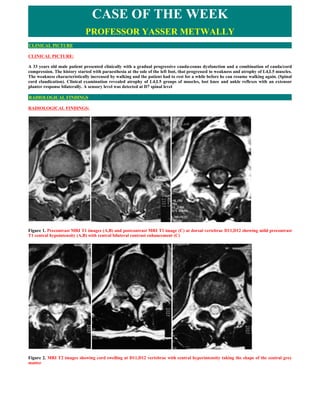
- 1. CASE OF THE WEEK PROFESSOR YASSER METWALLY CLINICAL PICTURE CLINICAL PICTURE: A 33 years old male patient presented clinically with a gradual progressive cauda-conus dysfunction and a combination of cauda/cord compression. The history started with paraesthesia at the sole of the left foot, that progressed to weakness and atrophy of L4,L5 muscles. The weakness characteristically increased by walking and the patient had to rest for a while before he can resume walking again. (Spinal cord claudication). Clinical examination revealed atrophy of L4,L5 groups of muscles, lost knee and ankle reflexes with an extensor planter response bilaterally. A sensory level was detected at D7 spinal level RADIOLOGICAL FINDINGS RADIOLOGICAL FINDINGS: Figure 1. Precontrast MRI T1 images (A,B) and postcontrast MRI T1 image (C) at dorsal vertebrae D11,D12 showing mild precontrast T1 central hypointensity (A,B) with central bilateral contrast enhancement (C) Figure 2. MRI T2 images showing cord swelling at D11,D12 vertebrae with central hyperintensity taking the shape of the central grey matter
- 2. Figure 3. Precontrast MRI T1 image (A) and postcontrast MRI T1 images (B,C). Before contrast (A) the spinal cord is enlarged and hypointese. After contrast injestion, dilated enhanced perimedullary veins were observed on the surface of the spinal cord (B,C) Figure 4. MRI T2 imaging showing pencil-shape multisegmental central spinal cord hyperintensity extending from D6 to D12 vertebrae with cording swelling Figure 5. Pre-embolization angiography showing arteriovenous fistula. The arteriovenous fistula is located in the dura itself. The feeding vessels are nonradicular branches of the spinal arteries, small, tortuous arterioles that originate from the dura. The feeding arteries are generally normal in caliber and flow through these lesions is exceptionally slow. A single draining radicular vein is often at the level of the spinal root foramina. The vein is dilated many times the size of the artery and flows retrograde into the anterior and posterior medullary veins and coronal venous plexus surrounding the spinal cord. Chronic venous hypertension and stagnation result in chronic
- 3. medullary ischemia. Figure 5. Pre-embolization angiography (A) and Postembolization angiography (B) of both internal iliac arteries and feeding vessels demonstrates no further filling of the fistula. Summary of the case 1. The onset in middle age suggests that SDAVF is an acquired condition, in contrast to intradural ventral fistulas or AVMs, which are assumed to be congenital abnormalities 2. An SDAVF is never located within the spinal parenchyma, in contrast to AVMs. 3. Patients with SDAVF very rarely present with spinal haemorrhage in contrast to patients with AVMs. 4. Associated vascular lesions are seen in AVMs, not in SDAVF. 5. Intradural AVMs occur much more often in the cervical region than SDAVF. 6. The increased pressure causes the venous system to ‘arterialize’, that is, the walls of intramedullary veins become thickened and also tortuous. The radicular feeding artery is often a dural branch and in a minority, the medullary artery. The shunt results in venous hypertension in the spinal cord, because the intramedullary veins and the radicular vein share a common venous outflow. The reduced arteriovenous pressure gradient (with the resultant of venous hypertension) results initially in congestive myelopathy (cord edema, swelling and cord petechial hemorrhages) that is responsible for the spinal cord swelling and central T2 hyperintensity, T1 central hypointensity. The central T2 hyperintensity took the shape of the central grey matter denoting the cord edema is primarily located in the central grey matter in congestive myelopathy due to spinal arteriovenous fistula probably because the central grey matter has the highest blood supply and the highest venous return. On long term basis (and with persistence of venous hypertension) a decrease in tissue perfusion might occur and might eventually result in venous infarction and spinal cord atrophy. DIAGNOSIS: DIAGNOSIS: SPINAL DURAL ARTERIOVENOUS FISTULA DISCUSSION DISCUSSION: Spinal cord vascular malformations are often a cause of spinal myelopathy. Endovascular surgical approaches to spinal cord vascular malformations have become an important adjunct and often the primary treatment of these disorders. Spinal cord vascular malformations may be divided into several categories based on their anatomy and location. They may present with a wide range of symptoms far removed from the vascular pathology. Early recognition and treatment can effect a better outcome and often reverse presenting neurologic deficits. Emerging magnetic resonance imaging techniques allow visualization of flow voids, some vascular anatomy, and intramedullary pathology and are a good screening modality. However, selective diagnostic spinal angiography remains the study of choice to diagnose and elaborate on the anatomy and potential treatment protocols. Rapidly evolving endovascular techniques and technology are revolutionizing the treatment of vascular pathology in the central nervous system, either as an adjunct to traditional neurosurgical techniques or as definitive therapy. We review the diagnosis and treatment options for spinal cord vascular malformations. Vascular malformations of the spinal cord are a major cause of spinal cord myelopathy. These are complex disorders with complex management. Endovascular surgical approaches to spinal cord vascular malformations have become an important adjunct and often the primary treatment of these disorders. Patients present with a wide range of signs and symptoms, often distal to the site of the pathology, and early recognition, diagnosis, and treatment offer the best chance of neurologic recovery.[1] Selective spinal angiography remains the "gold standard" for visualization of the intricate vascular anatomy; embolization, pioneered by Rene Djindjian and colleagues,[2] provides the endovascular means of
- 4. therapy. We will classify, discuss, and delineate the endovascular therapy of spinal cord vascular malformations. CLASSIFICATION Spinal cord vascular malformations may be divided into intramedullary arteriovenous malformations, perimedullary arteriovenous malformations, spinal-dural arteriovenous fistulas, epidural arteriovenous malformations, paravertebral vascular malformations, vertebral hemangiomas, and complex angiomatosis, including metameric angiomatosis (Cobb's syndrome) and disseminated angiodysplasias (Osler-Weber-Rendu syndrome). Spinal cord cavernomas, telangiectasias, and venous angiomas complete the list, yet are not amenable to endovascular therapy ( Table 1 ). Table 1. Spinal Cord Vascular Malformations Amendable to Endovascular Surgical Intervention Not Amenable to Endovascular Surgical Intervention Simple Intramedullary arteriovenous malformations Cavernomas Perimedullary arteriovenous malformations Telangiectasias Spinal-dural arteriovenous fistulas Venous angiomas Epidural arteriovenous malformations Paravertebral vascular malformations Vertebral hemangiomas Complex Metameric angiomatosis (Cobb's syndrome) Disseminated angiodysplasias (Osler-Weber-Rendu syndrome) Intramedullary Arteriovenous Malformations Intramedullary arteriovenous malformations (AVMs) demonstrate an intervening nidus between an artery and vein, partially or totally, within the spinal cord parenchyma. The anterior spinal and posterolateral spinal arteries supply nearly all intramedullary AVMs, and they drain into engorged medullary spinal veins. Most are found in the cervical or thoracic segments of the spinal cord. If the nidus is compact, the AVM is referred to as a glomus type (or a type II spinal AVM[3]). If the nidus is diffusely infiltrating the spinal cord, it is referred to as a juvenile type (or type III spinal AVM[3,4]). Spinal artery aneurysms or venous spinal aneurysms occurred in 20% to 40% of patients with intramedullary AVMs, and the presence of a spinal artery aneurysm was associated with a statistically significant increase in the risk of bleeding.[4,5] Metameric angiomatosis was found in 6 of 14 patients (43%) with spinal artery aneurysms.[5,6] Spinal subarachnoid hemorrhage or acute medullary syndrome is usually the presentation during childhood or adolescence.[4,7-9] The clinical course is progressive with deterioration of spinal cord function and recurrent hemorrhage. When the hemorrhage occurs within the spinal parenchyma, it can result in acute-onset neurologic decline or a functionally complete spinal cord transection. Magnetic resonance imaging (MRI) is the screening modality of choice. Decreased signals on T1- and T2-weighted images, described as "flow voids," delineate the AVM nidus within the spinal cord substance. The myelographic effect of T2-weighted images also demonstrates engorged draining medullary veins and possibly feeding arteries within the cerebrospinal fluid. Intraparenchymal hemorrhage, myelomalacia, and edema are also better delineated by MRI than by any other diagnostic modality. Computed tomography (CT) is clearly inferior. The complete evaluation of a patient suspected of harboring a spinal AVM then includes selective spinal angiography. Multiple spinal arteries need to be catheterized and studied to evaluate the entire extent of the malformation. Biplane angiography is useful to demonstrate the relationship of the AVM to the spinal cord and canal. Rapid filming (five exposures per second) is imperative to identify feeding artery aneurysms and draining vein aneurysms. Feeding arteries are generally hypertrophied and tortuous. Branches of the vertebral arteries, thyrocervical trunk, costocervical trunk, supreme intercostal, and thoracic intercostal arteries may supply AVMs in the cervical spinal cord.[10] All may need to be catheterized individually. Drainage occurs through dilated medullary veins to radicular veins. Therapeutic alternatives include endovascular embolization/obliteration, open surgery, or a combination of both. AVMs medially located over the posterior aspect of the spinal cord are more easily accessible following laminotomy than AVMs located lateral and anterior to the spinal cord and supplied by the anterior spinal artery. Surgery may be difficult because of the intramedullary location and the difficulty in distinguishing arteries from arterialized veins. However, over 60% of spinal cord AVMs were removed successfully in one early series with stabilization of the patient's condition or clinical improvement.[11] Intraoperative monitoring with somatosensory evoked potentials may improve clinical outcomes.[12] Endovascular surgical embolization may be performed in combination with open surgery or as the sole treatment if the feeding arteries are of sufficient caliber.[12-15] The embolic agent must pass through the anterior and posterior spinal arteries to reach the malformation itself while sparing perforating arteries and draining veins. Embolization can be performed with polyvinyl alcohol (PVA) [13] or biospheres[16,17] if temporary or liquid acrylic agent for permanence. The normal anterior spinal artery diameter is in the range 100 to 340 µm and the diameter of the normal central spinal artery varies between 60 and 72 µm.[18] Therefore, PVA with diameters of 150 to 250 µm should pass through a normal anterior spinal artery without lodging in the central spinal arteries. Often the anterior spinal artery is dilated, allowing the use of larger particles. PVA allows long-term recanalization, however, and necessitates repeated embolization or surgical excision. Embolization with liquid adhesives such as N-butyl cyanoacrylate (NBCA) has the advantage of
- 5. achieving permanent occlusion with, however, a higher risk of cord infarct. Occasionally, one can reach the nidus itself or feeding artery aneurysm, which can be obliterated with coils.[6] The majority of these procedures are now performed under general anesthesia for clarity of the angiographic images, although evoked potentials can be performed. Figure 1 demonstrates the MRI and angiographic images of a 28-year-old man presenting with spastic hyperreflexic paraparesis from myelopathy associated with cord edema secondary to a T9 AVM. Figure 1A demonstrates the T1 signal weighted MRI with gadolinium, axial projection. We note high signal within the cord parenchyma representing blood products or edema, or both. Signal voids surrounding the spinal cord represent engorged medullary veins. Figure 1B, the lateral T1-weighted MRI image, better demonstrates the intramedullary serpiginous flow voids highly suspicious for a spinal cord vascular malformation. Spinal angiography (Fig. 1C) demonstrates the main supply derived from a posterolateral spinal artery from the right T10 intercostal artery. This artery was embolized with embospheres, resulting in obliteration of the nidus (Fig. 1D). This was followed by surgical resection. The patient had transient worsening of his preoperative paraparesis and then returned to baseline. Figure 1. (A) T1 signal weighted MRI with gadolinium, axial projection, of a 28-year-old man presenting with spastic paraparesis. High signal within the cord parenchyma represents blood products and/or edema. Signal voids surrounding the spinal cord represent engorged medullary veins. (B) Lateral T1-weighted MRI image demonstrating the intramedullary serpiginous flow voids posterior to the T9 vertebral body highly suspicious for a spinal cord vascular malformation. Intramedullary high signal again represents blood products and/or edema. (C) Microcatheter injection of the right T10 radiculomedullary artery, demonstrating a spinal AVM supplied by the posterolateral spinal artery. This artery was embolized with embospheres for occlusion. (D) A right T10 intercostal injection after embolization demonstrating no flow or early shunting within the previously enlarged posterolateral spinal artery (the anterior spinal artery filled by the left T11 intercostal artery). Perimedullary Arteriovenous Malformations Perimedullary AVMs are direct fistulas between a spinal artery and a medullary vein. Most are located on the surface of the spinal cord, are fed by the anterior or posterolateral spinal arteries, and occur near the conus medullaris. These are thought to be congenital lesions. Patients present with progressive paraparesis secondary to venous hypertension or acute deterioration due to rupture of a feeding artery aneurysm.[10] Although most perimedullary AVMs occur in adults, they must be considered as a cause of slow progressive paraparesis in children.[19] Three types of perimedullary AVMs can be defined on the basis of angiographic findings. In type I the fistula is small and barely detectable. The fistula itself is at the point where there is a change in the vascular caliber; venous drainage frequently ascends over the dorsal cervical cord and is minimally dilated. In type II there is dilatation of the feeding artery, the shunt site, and the draining vein. In type III we see a giant fistula with multiple feeding arteries and a giant feeding vein.[4,20-22] Perimedullary AVMs are also known as type IV spinal cord arteriovenous malformations in the literature.[23] Type I perimedullary AVMs may be difficult to see with MRI because of their small size. Phase contrast MRI[24] or dynamic gadolinium-enhanced MRI[25] may increase detection. An intramedullary increased signal on T2-weighted images is nonspecific and due to venous hypertension. Selective spinal angiography is mandatory to confirm the diagnosis and demonstrate the feeding arteries
- 6. and veins. Surgical clipping of the fistula site is the best therapy for type I lesions if the feeding vessel is too small for selective catheterization. Type II fistulas can be approached by endovascular means and cured with NBCA injection into the fistula site. If a complete cure with a glue injection is not possible, particulate embolization can be followed by surgical excision of a more manageable lesion. Type III lesions can be attacked with a combination of detachable or pushable coils and balloons or NBCA injection with adjunctive surgery if necessary. Endovascular intra-arterial injection of indigo carmine dye has also been described as an adjunct to aid with orientation of the vascular anatomy during surgery.[26] Neurologic morbidity remains higher with these lesions. Ten patients with giant intradural spinal arteriovenous fistulas (perimedullary types II and III) have been described.[27] Three patients were treated with embolization alone or in combination with surgery (seven patients). Eight patients were classified with perimedullary type III and two with perimedullary type II. Seven patients had their fistula cured (as demonstrated by angiography); two patients had 5% residual filling and were scheduled for future therapy. One complication was related to embolization-rupture of the anterior spinal artery by a detachable balloon, resulting in transient worsening of paraplegia with recovery to baseline. Transient worsening of symptoms after endovascular surgery was common, but all patients returned to baseline or better. Dramatic improvement was observed in four patients. Figure 2 demonstrates the case of a 31-year-old physician with bladder dysfunction, progressive left leg weakness, and difficulty walking occurring over 5 years. MRI reveals a large signal void adjacent to the conus producing bony erosion of the vertebral body (Fig. 2A). Spinal angiography of a left L2 lumbar artery reveals multiple tortuous spinal arteries entering a varix (arrow) with washout of contrast at this point from other feeding vessels entering separately (Fig. 2B). The multiplicity and tortuosity of the feeding arteries suggested the option of a transvenous approach. A microcatheter was navigated from the femoral vein through the lumbar epidural plexus and into draining medullary veins and finally into the giant varix (Fig. 2C). Multiple platinum coils were deposited into the varix for a significant reduction in shunting. The patient developed back pain without neurologic deficit and was given heparin for 48 hours with resolution of pain. Two months later, a second transvenous procedure with the addition of coils and silk suture was accomplished. Again the procedure was followed by back pain, this time treated with glucocorticoids and morphine. Ten days later the patient developed decreased sensory and left leg motor function. An angiogram showed complete thrombosis of the fistula (Fig. 2D). His motor and sensory function improved over the next several weeks to better than baseline. He was able to walk several miles and climb stairs without difficulty. His bladder function remained unchanged. Four years later, he developed a slight increase in leg weakness and reexamination disclosed a small fistula fed now by internal iliac collaterals too small to permit endovascular therapy. Laminectomy and excision of the small fistula and thrombosed varix were accomplished without further neurologic morbidity.
- 7. Figure 2. (A) Sagittal T1-weighted MRI demonstrates a large signal void adjacent to the conus producing bony erosion of the vertebral body. (B) Left L2 lumbar arterial injection, anteroposterior projection, reveals multiple tortuous spinal arteries entering a varix (arrow) with washout of contrast at this point from other feeding vessels entering separately. (C) A microcatheter was navigated from the femoral vein through the lumbar epidural plexus and draining medullary veins into the giant varix, and contrast material was injected in the anteroposterior projection. (D) Left L2 lumbar arterial injection, anteroposterior projection, status after stage 2 transvenous embolization with multiple platinum coils and silk suture deposited into the varix. Complete thrombosis of the fistula was achieved. Spinal-Dural Arteriovenous Fistulas Spinal-dural arteriovenous fistulas are the most common variety of spinal cord AVM.[1] Spinal-dural arteriovenous fistulas are thought to be acquired lesions,[4] occur mainly in older adults (mean age of 51 years), and are found more often in men than women. Patients present with slowly progressive myelopathy and radiculopathy,[21,28] which, if left untreated, can progress to paraparesis or quadriparesis. The arteriovenous fistula is located in the dura itself. The feeding vessels are nonradicular branches of the spinal arteries, small, tortuous arterioles that originate from the dura. The feeding arteries are generally normal in caliber and flow through these lesions is exceptionally slow. A single draining radicular vein is often at the level of the spinal root foramina. The vein is dilated many times the size of the artery and flows retrograde into the anterior and posterior medullary veins and coronal venous plexus surrounding the spinal cord. Chronic venous hypertension and stagnation result in chronic medullary ischemia.[29] Although phase contrast MRI[24] or dynamic gadolinium-enhanced MRI[25] may increase detection, in our experience these small connections can be easily missed even on excellent quality MRI images. Serpiginous flow voids around the cord may represent flow in dilated medullary veins. The spinal cord may also be enlarged, and intramedullary increased signal on T2-weighted images may represent edema or ischemia secondary to venous hypertension. Contrast myelography in both the supine position, which can best demonstrate the retromedullary veins, and the prone position reveals dilated and tortuous veins over the dorsum of the cord. If dilated veins are observed, complete spinal angiography is indicated. Image quality must be the best possible to delineate the origin of the shunt. Filming should continue into the late venous phase up to several seconds after the injection. The majority of patients with the fistula in the thoracic and lumbar region have arterial supply independent of the supply to the spinal cord. Internal iliac artery supply was observed in 12.5% of cases.[30] Endovascular embolization with liquid adhesive can frequently cure these lesions. Initial apparently successful embolization was achieved in 90% of 20 patients in one study; the fistula recurrence rate (failure to occlude the draining vein) for NBCA was 15% (3 patients). All patients who underwent embolization had either improved (55%) or unchanged (45%) gait disability at last follow-up.[31] Not infrequently, complete clinical cure can be achieved in cases presenting with a nonfixed, moderate, neurologic deficit. Recanalization has occurred in cases in which PVA was used as the primary therapy.[32] Open surgery is recommended if embolization fails to occlude the dural arteriovenous fistula. Figure 3 represents the case of a 58-year-old man with a 4-year history of loss of sensation and proprioception and pain in both feet that progressed to include legs and hip regions as well as bilateral leg weakness. By admission he had developed urinary retention requiring catheterization and bowel retention requiring manual evacuation. On examination he demonstrated 2/5 strength in his lower extremities and 5/5 in the upper extremities. He was wheelchair bound, had a T12 sensory level, and had loss of deep tendon responses in the lower extremities.
- 8. Figure 3. (A) Sagittal first and second echoes of the T2-weighted MRI of the lower thoracic spine demonstrate intramedullary hyperintensity within the conus without cord enlargement and a serpiginous flow voids anterior to the cord (white arrow). (B) Axial T1- weighted MRI of the thoracic spine before (left) and after (right) gadolinium demonstrates cord enhancement. (C) Selective right internal iliac angiogram demonstrates a nidus of AV fistula (between arrows), supplied by the lateral sacral artery and draining by an ascending perimedullary vein (arrowhead). (D) Unsubtracted fluoroscopic image of the pelvis demonstrates the NBCA glue cast at the site of the previous fistula (between arrows). MRI of the thoracic spine revealed hyperintensity in the spinal cord on T2-weighted images from T11 to the conus without focal cord enlargement and enhancement of the cord on axial T1-weighted MRI (Figs. 3A and 3B). A selective right internal iliac angiogram demonstrates a nidus of AV fistula (Fig. 3C) supplied by the lateral sacral artery and draining by an ascending perimedullary vein. Subselective catheterization using a microcatheter was performed and endovascular surgical obliteration achieved using NBCA (Fig. 3D). Postembolization angiography of both internal iliac arteries and feeding vessels demonstrates no further filling of the fistula. Epidural Arteriovenous Malformations Epidural AVMs are primarily epidural and drain into the epidural venous plexus or medullary veins.[20,33-36] Patients present with spontaneous cervical epidural hemorrhage,[33] radiculopathy, or subarachnoid hemorrhage.[34] Arterial supply may be derived from the vertebral, thyrocervical, or costocervical arteries. Endovascular embolization can result in complete closure of the fistula and cure. [36] Paravertebral Arteriovenous Malformations Paravertebral AVMs exhibit intercostal, paramedian, extraspinal, and even medullary venous drainage. Complete spinal angiography demonstrates the extension of the malformation through the nerve root foramina and rules out the possibility of an association with an intramedullary AVM. This category includes extra- and intraspinal single hole fistulas, the presence of a well-defined AVM nidus, or a combination of both.[22] Endovascular embolization may be achieved with NBCA liquid adhesive and should be attempted first. Particle embolization may be used if open surgery is planned. Metameric Angiomatosis (Cobb's Syndrome) and Disseminated Angiomatosis (Osler-Weber-Rendu Syndrome) In cases of metameric angiomatosis there is a well-defined spinal AVM with involvement of the dura, vertebrae, muscular wall, skin, and even viscera. Cobb's syndrome presents in a number of ways: (1) flat, metameric angiomas involving the thoracic spine and chest or cervical or lumbar spine and extremities; (2) flat, metameric angiomas associated with subarachnoid hemorrhage; and (3) superficial arteriovenous angiomatosis of the trunk or extremities associated with subarachnoid hemorrhage.[22] It is difficult to achieve complete cure. Embolization, surgery, and percutaneous vertebroplasty may be used in combination or alone, depending on the symptomatology. In disseminated angiomatosis, vascular telangiectasias may involve the skin of the face and/or hands or mucous membranes of the respiratory and/or gastrointestinal tract. Often the main presenting feature is epistaxis. Pulmonary angiomatosis is less common but may present with septic emboli, paradoxical emboli, or pulmonary fistula. Capillary telangiectasias, arteriovenous fistulas, AVMs,
- 9. capillary angiomas, and intercranial aneurysms may also be found in the brain or spinal cord, or both. Spinal Hemangiomas Spinal hemangiomas are the most common benign spinal neoplasms, often located in the thoracic and lumbar spine, with a peak incidence of occurrence in the fourth to sixth decades. They are relatively common, found in 11% of autopsy series.[37] Sixty percent are asymptomatic without gender preference.[38] Symptomatic hemangiomas are more common in women and present with back pain, radicular pain, or spinal cord compression. Acute symptoms occur from compression fracture, epidural extension and sudden mass effect, and hemorrhage. MRI demonstrates a hyperintense lesion in the vertebral body seen on T1- and T2-weighted images.[39] MRI may demonstrate expansion to include the posterior elements of the spinal column, invasion of the spinal canal, and encroachment of the spinal cord. [40,41] Plain radiographs exhibit a coarse-striated appearance of the vertebral body, and CT reflects a "polka dot" appearance. Radiation therapy appears highly beneficial and safe in the treatment of pain caused by vertebral hemangiomas and even for epidural extension to some degree.[42-46] Percutaneous injection of ethanol under fluoroscopic guidance has also been efficacious in the treatment of pain. Encouraging results were seen in 86% of patients in one study,[47] provided the dose was less than 15 mL[48] and provided that pretreatment test injection of contrast medium was retained by the hemangioma.[49] Preoperative embolization with methyl methacrylate, followed by laminectomy and/or vertebrectomy for mass effect and stabilization, is another treatment option.[50] Conclusion Endovascular surgical techniques continue to evolve and now play a large role in the treatment of most spinal cord vascular malformations. Early diagnosis improves outcomes, and spinal angiography remains the gold standard to define the vascular anatomy. MRI is an excellent screening modality and is also a rapidly evolving technology. Patients need to be evaluated on an individual basis from both a traditional neurosurgical and endovascular points of view and with regard to the expertise available. This combined approach offers the best hope for patients with often challenging spinal cord vascular pathology. SUMMARY SUMMARY Addendum A new version of this PDF file (with a new case) is uploaded in my web site every week (every Saturday and remains available till Friday.) To download the current version follow the link "http://pdf.yassermetwally.com/case.pdf". You can also download the current version from my web site at "http://yassermetwally.com". To download the software version of the publication (crow.exe) follow the link: http://neurology.yassermetwally.com/crow.zip The case is also presented as a short case in PDF format, to download the short case follow the link: http://pdf.yassermetwally.com/short.pdf At the end of each year, all the publications are compiled on a single CD-ROM, please contact the author to know more details. Screen resolution is better set at 1024*768 pixel screen area for optimum display. For an archive of the previously reported cases go to www.yassermetwally.net, then under pages in the right panel, scroll down and click on the text entry "downloadable case records in PDF format" Also to view a list of the previously published case records follow the following link (http://wordpress.com/tag/case-record/) or click on it if it appears as a link in your PDF reader REFERENCES References 1. Muraszko KM, Oldfield EH. Vascular malformations of the spinal cord and dura. Neurosurg Clin North Am 1990;1: 631-652 2. Djindjian R. Angiography of the Spinal Cord. Baltimore: University Park Press; 1970 3. Anson A, Spetzler RF. Spinal dural arteriovenous malformations. In: Awad IA, Barrow DL, eds. Dural Arteriovenous Malformations. Park Ridge, IL: American Association of Neurological Surgeons; 1993:175-192 4. Rosenblum B, Oldfield EH, Doppman JL, Di Chiro G. Spinal arteriovenous malformations: a comparison of dural arteriovenous fistulas and intradural AVM's in 81 patients. J Neurosurg 1987;67:795-802
- 10. 5. Biondi A, Merland JJ, Hodes JE, Pruvo JP, Reizine D. Aneurysms of spinal arteries associated with intramedullary arteriovenous malformations: I. Angiographic and clinical aspects. AJNR Am J Neuroradiol 1992;13:913-922 6. Biondi A, Merland JJ, Hodes JE, Aymard A, Reizine D. Aneurysms of spinal arteries associated with intramedullary arteriovenous malformations: II. Results of AVM endovascular treatment and hemodynamic considerations. AJNR Am J Neuroradiol 1992;13:923-931 7. Djindjian A. Arteriovenous malformations of the spinal cord: clinical, anatomical and therapeutic consideration -- a series of 150 cases. Prog Neurol Surg 1978;9:238-266 8. Eldridge PR, Holland IM, Punt JA. Spinal arteriovenous malformations in children. Br J Neurosurg 1989;3:393-397 9. Shephard RH. Spinal arteriovenous malformations and subarachnoid haemorrhage. Br J Neurosurg 1992;6:5-12 10. Larson D, Halbach V. Vascular pathology and endovascular therapy. In: Lee RR, ed. Spinal Imaging. Vol. 9. Philadelphia: Hanley and Belfus; 1995:245-259 11. Yasargil MG, Symon L, Teddy PJ. Arteriovenous malformations of the spinal cord. Adv Tech Stand Neurosurg 1984; 11:61-102 12. Owen MP, Brown RH, Spetzler RF, Nash CL Jr, Brodkey JS, Nulsen FE. Excision of intramedullary arteriovenous malformation using intraoperative spinal cord monitoring. Surg Neurol 1979;12:271-276 13. Latchaw RE, Harris RD, Chou SN, Gold LH. Combined embolization and operation in the treatment of cervical arteriovenous malformations. Neurosurgery 1980;6:131-137 14. Horton JA, Latchaw RE, Gold LH, Pang D. Embolization of intramedullary arteriovenous malformations of the spinal cord. AJNR Am J Neuroradiol 1986;7:113-118 15. Riche MC, Melki JP, Merland JJ. Embolization of spinal cord vascular malformations via the anterior spinal artery. AJNR Am J Neuroradiol 1983;4:378-381 16. Beaujeux R, Laurent A, Wassef M, et al. Trisacryl gelatin microspheres for therapeutic embolization, II: preliminary clinical evaluation in tumors and arteriovenous malformations. AJNR Am J Neuroradiol 1996;17:541-548 17. Laurent A, Beaujeux R, Wassef M, Rufenacht D, Boschetti E, Merland JJ. Trisacryl gelatin microspheres for therapeutic embolization, I: development and in vitro evaluation. AJNR Am J Neuroradiol 1996;17:533-540 18. Suh TH. Vascular system of the human spinal cord. Arch Neurol Psychol 1939;41:659-677 19. Sure U, Wakat JP, Gatscher S, Becker R, Bien S, Bertalanffy H. Spinal type IV arteriovenous malformations (perimedullary fistulas) in children. Childs Nerv Syst 2000;16:508- 515 20. Kendall B, Logue V. Spinal epidural angiomatous malformations draining into intrathecal veins. Neuroradiology 1977; 13:181-189 21. Merland JJ, Riche MC, Chiras J. Intraspinal extramedullary arterio-venous fistulae draining into the medullar veins. J Neuroradiol 1980;7:271-320 22. Merland JJ, Laurent A, Khayata MH, Casasco A, Aymard A, Gobin P. Embolization of spinal cord vascular lesions. In: Vinuela F, Dion JE, eds. Interventional Neuroradiology: Endovascular Therapy of the Central Nervous System. New York: Raven Press; 1992:153-165 23. Barrow DL, Colohan AR, Dawson R. Intradural perimedullary arteriovenous fistulas (type IV spinal cord arteriovenous malformations). J Neurosurg 1994;81:221-229 24. Mourier KL, Gelbert F, Reizine D, et al. Phase contrast magnetic resonance of the spinal cord preliminary results in spinal cord arterio-venous malformations. Acta Neurochir (Wien) 1993;123:57-63 25. Thorpe JW, Kendall BE, MacManus DG, McDonald WI, Miller DH. Dynamic gadolinium-enhanced MRI in the detection of spinal arteriovenous malformations. Neuroradiology 1994;36:522-529 26. Tani S, Ikeuchi S, Hata Y, Abe T. Vascular orientation by intra-arterial dye injection during spinal arteriovenous malformation surgery. Neurosurgery 2001;48:240-242 27. Halbach VV, Higashida RT, Dowd CF, Fraser KW, Edwards MS, Barnwell SL. Treatment of giant intradural (perimedullary) arteriovenous fistulas. Neurosurgery 1993;33: 972-979; discussion 979-980 28. Koenig E, Thron A, Schrader V, Dichgans J. Spinal arteriovenous malformations and fistulae: clinical, neuroradiological and neurophysiological findings. J Neurol 1989;236:260-266 29. Kataoka H, Miyamoto S, Nagata I, Ueba T, Hashimoto N. Venous congestion is a major cause of neurological deterioration in spinal arteriovenous malformations. Neurosurgery 2001;48:1224-1229; discussion 1229-1230 30. Larsen DW, Halbach VV, Teitelbaum GP, et al. Spinal dural arteriovenous fistulas supplied by branches of the internal iliac arteries. Surg Neurol 1995;43:35-40; discussion 40-41
- 11. 31. Song JK, Gobin YP, Duckwiler GR, et al. N-Butyl 2-cyanoacrylate embolization of spinal dural arteriovenous fistulae. AJNR Am J Neuroradiol 2001;22:40-47 32. Nichols DA, Rufenacht DA, Jack CR Jr, Forbes GS. Embolization of spinal dural arteriovenous fistula with polyvinyl alcohol particles: experience in 14 patients. AJNR Am J Neuroradiol 1992;13:933-940 33. Foo D, Chang YC, Rossier AB. Spontaneous cervical epidural hemorrhage, anterior cord syndrome, and familial vascular malformation. Neurology 1980;30:1253-1254 34. Halbach VV, Higashida RT, Hieshima GB. Treatment of vertebral arteriovenous fistulas. AJR Am J Roentgenol 1988;150: 405- 412 35. Janda J, Mracek Z. [Angioreticuloma of the brain stem: simultaneous occurrence of an arteriovenous malformation in the epidural space of the cervical spinal cord]. Cesk Neurol Neurochir 1978;41:397-399 36. Willinsky R, terBrugge K, Montanera W, Wallace MC, Gentili F. Spinal epidural arteriovenous fistulas: arterial and venous approaches to embolization. AJNR Am J Neuroradiol 1993;14:812-817 37. Schmorl G. Tumors and tumor metastases. In: Schmorl G, Besemann EF, Junghanns H, eds. The Human Spine in Health and Disease. New York: Grune & Stratton; 1971: 325-327 38. Fox MW, Onofrio BM. The natural history and management of symptomatic and asymptomatic vertebral hemangiomas. J Neurosurg 1993;78:36-45 39. Ross JS, Masaryk TJ, Modic MT, Carter JR, Mapstone T, Dengel FH. Vertebral hemangiomas: MR imaging. Radiology 1987;165:165-169 40. Lee S, Hadlow AT. Extraosseous extension of vertebral hemangioma, a rare cause of spinal cord compression. Spine 1999;24:2111- 2114 41. Nassar SI, Hanbali FS, Haddad MC, Fahl MH. Thoracic vertebral hemangioma with extradural extension and spinal cord compression: case report. Clin Imaging 1998;22: 65-68 42. Miszczyk L, Ficek K, Trela K, Spindel J. The efficacy of radiotherapy for vertebral hemangiomas. Neoplasma 2001;48:82-84 43. Heyd R, Strassmann G, Filipowicz I, Borowsky K, Martin T, Zamboglou N. [Radiotherapy in vertebral hemangioma]. Rontgenpraxis 2001;53:208-220 44. Brackrock S, Krull A, Schwarz R, Alberti W. [Results of radiotherapy for vertebral hemangioma]. Strahlenther Onkol 1999;175:405-408 45. Winkler C, Dornfeld S, Baumann M, Christen N, Herrmann T, Eberhardt HJ. [The efficacy of radiotherapy in vertebral hemangiomas]. Strahlenther Onkol 1996;172:681-684 46. Bremnes RM, Hauge HN, Sagsveen R. Radiotherapy in the treatment of symptomatic vertebral hemangiomas: technical case report. Neurosurgery 1996;39:1054-1058 47. Goyal M, Mishra NK, Sharma A, Gaikwad SB, Mohanty BK, Sharma S. Alcohol ablation of symptomatic vertebral hemangiomas. AJNR Am J Neuroradiol 1999;20:1091-1096 48. Doppman JL, Oldfield EH, Heiss JD. Symptomatic vertebral hemangiomas: treatment by means of direct intralesional injection of ethanol. Radiology 2000;214:341-348 49. Bas T, Aparisi F, Bas JL. Efficacy and safety of ethanol injections in 18 cases of vertebral hemangioma: a mean follow-up of 2 years. Spine 2001;26:1577-1582 50. Nguyen JP, Djindjian M, Pavlovitch JM, Badiane S. [Vertebral hemangioma with neurologic signs: therapeutic results. Survey of the French Society of Neurosurgery]. Neurochirurgie 1989; 35:299-303, 305-308 51. Metwally, MYM: Textbook of neuroimaging, A CD-ROM publication, (Metwally, MYM editor) WEB-CD agency for electronic publication, version 11.1a. January 2010