SlideShare una empresa de Scribd logo
1 de 21
Descargar para leer sin conexión
Professor Yasser Metwally
www.yassermetwally.com

CASE OF THE WEEK
PROFESSOR YASSER METWALLY
CLINICAL PICTURE:
A 57 years old male patient with a purely motor clinical manifestations in the form of left
lower limb pyramidal manifestations of three months duration. The clinical picture has an
insidious onset and a rapidly progressive course.(To inspect the patient's full radiological
study, click on the attachment icon (The paper clip icon in the left pane) of the acrobat
reader then double click on the attached file) (Click here to download the attached file)
RADIOLOGICAL FINDINGS
RADIOLOGICAL FINDINGS:

Figure 1. Precontrast TI images (A,B) and postcontrast (C) showing bilateral, more or less
symmetrical, posterior parieto-occipital white matter hypointensities with lateral scalloped
margins. The TI hypointensities is coalescent and periventricular/ subcortical in location.
The scalloped lateral margin of the posterior parieto-occipital white matter TI signal
changes reflects the strictly subcortical nature of the lesion that apparently did not involve
the cortical grey matter. Notice absence of contrast enhancement.

www.yassermetwally.com
Professor Yasser Metwally
www.yassermetwally.com

Figure 2. MRI T2 images showing bilateral, more or less symmetrical, posterior parietooccipital white matter hyperintensity with lateral scalloped margins. The T2 hyperintensity
is coalescent and periventricular/ subcortical in location. The scalloped lateral margin of
the posterior parieto-occipital white matter T2 signal changes reflects the strictly
subcortical nature of the lesion that apparently did not involve the cortical grey matter.
The coalescent posterior parieto-occipital white matter T2 signal changes is surrounded by
a continuous hypointense rim that, most probably, reflects the inflammatory nature of the
lesion. The mechanism of the T2 hypointensity is not certain. Proposals in this respect
include relative lack of water in the periphery of inflammatory lesion, the presence of blood
products in the periphery of the lesion (Deoxyhaemoglobin/hemosiderin induce T2
hypointensity) or the presence of paramagnetic free radicals within the phagocytosing
macrophages which are heterogeneously distributed in the periphery of inflammatory
lesions. (Paramagnetic free radicals induce T2 hypointensity).

www.yassermetwally.com
Professor Yasser Metwally
www.yassermetwally.com

Figure 3. MRI T2 images showing bilateral, more or less symmetrical, posterior parietooccipital white matter hyperintensity with lateral scalloped margins. The T2 hyperintensity
is coalescent and periventricular/ subcortical in location. The scalloped lateral margin of
the posterior parieto-occipital white matter T2 signal changes reflects the strictly
subcortical nature of the lesion that apparently did not involve the cortical grey matter.
The coalescent posterior parieto-occipital white matter T2 signal changes is surrounded by
a continuous hypointense rim that, most probably, reflects the inflammatory nature of the
lesion. The mechanism of the T2 hypointensity is not certain. Proposals in this respect
include relative lack of water in the periphery of inflammatory lesion, the presence of blood
products in the periphery of the lesion (Deoxyhaemoglobin/hemosiderin induce T2
hypointensity) or the presence of paramagnetic free radicals within the phagocytosing
macrophages which are heterogeneously distributed in the periphery of inflammatory
lesions. (Paramagnetic free radicals induce T2 hypointensity). Notice the presence popcornlike lesions in (B) that probably reflect the existence of some vascular/venous anomalies.
The findings of vascular blush and arteriovenous shunting on angiography, in progressive
multifocal leukoencephalopathy, are nonspecific and was reported before. (14) Notice
absence of mass effect.

www.yassermetwally.com
Professor Yasser Metwally
www.yassermetwally.com

Figure 4. The coalescent posterior parieto-occipital white matter T2 signal changes is
surrounded by a continuous hypointense rim that, most, probably, reflects the
inflammatory nature of the lesion. The mechanism of the T2 hypointensity is not certain.
Proposals in this respect include relative lack of water in the periphery of inflammatory
lesion, the presence of blood products in the periphery of the lesion
(deoxyhaemoglobin/hemosiderin induce T2 hypointensity) or the presence of paramagnetic
free radicals within the phagocytosing macrophages which are heterogeneously distributed
in the periphery of inflammatory lesions. (Paramagnetic free radicals induce T2
hypointensity). Multiple foci of acute hemorrhage and abundant petechial hemorrhages in
and at the perphery of the posterior parieto-occipital white matte lesions was occaionally
reported in progressive multifocal leukoencephalopathy. (14) Notice the popcorn-like
lesions in this image, that probably reflect the existence of some vascular/venous anomalies.
The findings of vascular blush and arteriovenous shunting on angiography, in progressive
multifocal leukoencephalopathy, are nonspecific and was reported before. (14) Notice
absence of mass effect.

www.yassermetwally.com
Professor Yasser Metwally
www.yassermetwally.com

Figure 5. FLAIR MRI images showing the bilateral, more or less symmetrical, posterior
parieto-occipital white matter hyperintensity with lateral scalloped margins.

www.yassermetwally.com
Professor Yasser Metwally
www.yassermetwally.com

Figure 6. Chest Plain X ray showing an
upper quadrant lung lesion. The lesion
wan not biopsied in this patient.

Progressive multifocal leukoencephalopathy (PML) is a subacute opportunistic infection
caused by the unenveloped DNA virus JC Polyomavirus (JCV). By the age of 65 years,
50% to 70% of individuals have antibodies to the virus. (26) After entering, the virus
persists in the renal tubular epithelial cells. With reactivation of latent JCV in the kidneys
and subsequent viremia, the JCV enters the brain and causes Progressive multifocal
leukoencephalopathy. (26)
The incidence of progressive multifocal leukoencephalopathy has greatly increased as a
result of the AIDS epidemic, and 0.7% to 11% of patients who have HIV develop
progressive multifocal leukoencephalopathy during the course of their illness. The
histopathologic hallmark of progressive multifocal leukoencephalopathy is demyelination
with enlarged oligodendroglial nuclei and bizarre astrocytes. The disease is usually
multifocal, and the lesions may occur in any location in the white matter, most often in the
parieto-occipital region. Thalamic lesions are frequently present, and the cerebellum and
brain stem can also be involved. Posterior fossa lesions are usually present simultaneously
with supratentorial lesions, and, in rare cases, progressive multifocal leukoencephalopathy
may be limited to the posterior fossa. Although there are no significant pathologic
differences between patients who have and do not have AIDS, in the AIDS population,
more extensive disease with a necrotizing character was observed. (26)

www.yassermetwally.com
Professor Yasser Metwally
www.yassermetwally.com

The findings on MR imaging correlate well with macroscopic changes. On T2-weighted
MR imaging, progressive multifocal leukoencephalopathy lesions are patchy, scalloped,
high signal intensity lesions located in the white matter, with extension along the white
fibers. Subcortical arcuate fibers are involved, mass effect is mild or absent, and peripheral
faint enhancement is a rare feature. On T1-weighted images, the progressive multifocal
leukoencephalopathy lesions have low signal, in contrast to isointense HIV-associated
lesions. (26)
The coalescent posterior parieto-occipital white matter T2 signal changes is sometimes
surrounded by a continuous hypointense rim that, most, probably, reflects the
inflammatory nature of the lesion. The mechanism of the T2 hypointensity is not certain.
Proposals in this respect include relative lack of water in the periphery of inflammatory
lesion, the presence of blood products in the periphery of the lesion
(deoxyhaemoglobin/hemosiderin induce T2 hypointensity) or the presence of paramagnetic
free radicals within the phagocytosing macrophages which are heterogeneously distributed
in the periphery of inflammatory lesions. (Paramagnetic free radicals induce T2
hypointensity). Multiple foci of acute hemorrhage and abundant petechial hemorrhages in
and at the perphery of the posterior parieto-occipital white matte lesions was occaionally
reported in progressive multifocal leukoencephalopathy. (14) A popcorn-like lesions are
sometimes reported in PML, and probably reflect the existence of some vascular/venous
anomalies in PML. The findings of vascular blush and arteriovenous shunting on
angiography, in progressive multifocal leukoencephalopathy, are nonspecific and was
reported before. (14)
DIAGNOSIS:
DIAGNOSIS: PROGRESSIVE MULTIFOCAL LEUKOENCEPHALOPATHY
DISCUSSION
DISCUSSION:
Progressive multifocal leukoencephalopathy (PML) is a fatal subacute progressive
demyelinating disease seen in persons with impaired cell-mediated immune response.
Progressive multifocal leukoencephalopathy predominantly occurs in patients with AIDS.
Before the AIDS epidemic, Progressive multifocal leukoencephalopathy was rare and
associated with immunocompromised conditions, such as leukemia, lymphoma, systemic
lupus erythematosus (SLE), organ transplantation, Wiskott-Aldrich syndrome, and severe
combined immunodeficiency (SCID).
At present, Progressive multifocal leukoencephalopathy develops in as many as 5% of all
patients with AIDS. This demyelinating disease results from infection with the JC virus,
which belongs to the genus Polyomavirus of the Papovaviridae family of viruses.
Anstrom et al first described Progressive multifocal leukoencephalopathy in 1958, and
Zurhein and Chou initially demonstrated the association of Progressive multifocal

www.yassermetwally.com
Professor Yasser Metwally
www.yassermetwally.com

leukoencephalopathy with a viral infection. In 1971, Padgett et al confirmed the viral
etiology by isolating a virus from the brain of a patient who had died from Progressive
multifocal leukoencephalopathy. The patient's initials were J.C.; hence, the virus is known
as the JC virus.
Progressive multifocal leukoencephalopathy is characterized by 3 cardinal histopathologic
features: demyelination, enlarged nuclei of oligodendrocytes, and bizarre astrocytes.
The JC virus is believed to produce infection after it enters the tonsillar tissue during an
upper respiratory tract infection. After infection, the virus becomes latent in the spleen, the
reticuloendothelial system, and the medulla of the kidney. The JC virus is thought to be
undetectable in the brain tissue.
Antibodies (immunoglobulin G [IgG]) to the JC virus are common in most Western
populations. Seroconversion is seen in 10% of the children by the age of 5 years, in 40-60%
by 10 years, and in up to 90% of young adults. Acute infection is usually not noticed. After
several years of latency, reactivation occurs under appropriate conditions of
immunocompromise.
Approximately 50-80% of all Progressive multifocal leukoencephalopathy cases occur in
patients with HIV infection, whereas cases are rare in patients with organ transplantation.
This difference may indicate the need for an interaction between the JC virus and HIV for
Progressive multifocal leukoencephalopathy to develop rather than just an underlying
setting of decreased immune function. Although reactivation of JC virus may be necessary,
this itself is insufficient to cause Progressive multifocal leukoencephalopathy. A specific
deficiency in cellular immune response to the JC viral antigen is probably required in
addition to the general cellular immunodeficiency in persons with Progressive multifocal
leukoencephalopathy.
Reactivation of the virus occurs in the kidney and bone marrow, usually in the setting of
immunosuppression. Infected lymphocytes (B cells) then cross the blood-brain barrier and
pass infection to astrocytes at the border of vessels. The infection may then be augmented
by multiplication and eventual infection of adjacent oligodendrocytes. Oligodendrocytes
are responsible for forming and maintaining the myelin sheath. Infection of the
oligodendrocytes causes destruction of the cells and loss of the myelin sheath. The axons
are usually spared.


Histopathology

On microscopy, the cardinal feature of Progressive multifocal leukoencephalopathy is
demyelination, which is usually multifocal. The lesions may occur in any location in the
white matter, and they range from 1 mm to several centimeters in size.
The histopathologic hallmarks of Progressive multifocal leukoencephalopathy include a
triad of multifocal demyelination, hyperchromatic enlarged oligodendroglial nuclei, and
enlarged bizarre astrocytes with lobulated and hyperchromatic nuclei. Electron

www.yassermetwally.com
Professor Yasser Metwally
www.yassermetwally.com

microscopy reveals JC virus in the oligodendroglial cells. Gross examination reveals gray
or brown discoloration of the affected brain as a result of loss of myelin.
In the US: The prevalence of Progressive multifocal leukoencephalopathy in patients with
HIV infection is 1-5% in clinical studies, though postmortem data show a rate of up to 8%.
The prevalence of this disease initially increased with the rising incidence of HIV infection,
but more recently, the incidence has decreased with the widespread use of highly active
antiretroviral therapy (HAART), which has reversed immunosuppression in many patients
with AIDS (see subsection on Prognostic utility of MRI in Progressive multifocal
leukoencephalopathy). For unknown reasons, Progressive multifocal leukoencephalopathy
rarely affects children with HIV infection.
The median survival of patients with Progressive multifocal leukoencephalopathy as a
complication of AIDS is 6 months. In 10% of patients, survival exceeds 12 months. The
longest reported survival is 92 months from the onset of illness. Progressive multifocal
leukoencephalopathy is an AIDS-defining illness. Patients whose MRIs show enhancement,
which is rare, and those with an increased CD4 count appear to have a better prognosis
than that of other patients; these findings probably represent their relatively good immune
status. Before the AIDS epidemic, men and women were affected in a male-to female ratio
of 3:2, and lymphoproliferative disease was the most common cause. At present, HIV
infection is most common cause, with a male-to-female ratio of 7:1 Progressive multifocal
leukoencephalopathy chiefly affects homosexual or bisexual men aged 25-50 years. For
unknown reasons, Progressive multifocal leukoencephalopathy rarely affects children with
HIV infection. The white matter of the brain is usually involved. Lesions of Progressive
multifocal leukoencephalopathy may occur anywhere in brain, but the frontal lobes and
parieto-occipital regions are commonly affected. Isolated involvement of basal ganglia,
external capsule, and posterior-fossa structures may be seen.
As of now, radiographically guided intervention has no role in the treatment of Progressive
multifocal leukoencephalopathy. An antiretroviral regimen containing protease inhibitors
has been successful in prolonging the survival of patients with HIV infection and
Progressive multifocal leukoencephalopathy. Topotecan and cidofovir are promising drugs
presently under evaluation. The role of splenectomy remains unproven, though reports
have described remission in association with HAART.


Clinical Details
o Symptoms

Progressive focal neurologic deficit is the clinical hallmark. Weakness and disturbance of
speech are most common symptoms. Other symptoms include cognitive abnormalities,
headaches, gait disorders, visual impairment, and sensory loss.
Headaches are most common in the HIV-infected population, and visual disturbances are
most common in those without HIV infection. About 10% of patients have seizures.
Cognitive deficits do not persist in isolation for long and distinguish Progressive multifocal
leukoencephalopathy from HIV dementia. Progressive multifocal leukoencephalopathy

www.yassermetwally.com
Professor Yasser Metwally
www.yassermetwally.com

seems to have a more aggressive course in persons with HIV disease than in persons with
other predisposing conditions.
o

Signs

Most common physical sign is limb weakness, which occurs in more than 50% of patients.
Cognitive disturbances and gait disorders affect 25-33%, and diplopia affects 9%. Opticnerve disease does not occur with Progressive multifocal leukoencephalopathy, and spinalcord involvement is rare.


Laboratory findings

Severe cellular immunosuppression, as defined by a CD4 lymphocyte count of 200 cells/L,
is observed in most patients. The mean CD4 count is 84-104 cells/L.
Results of CSF examination are usually normal or show slightly elevated protein levels.
CSF study is not helpful in diagnosis except that it may help in excluding other diagnoses.
Cell counts are usually less than 20 cells/L.
Several studies demonstrated that polymerase chain reaction (PCR) study of the CSF has
high sensitivity and specificity for JC virus in Progressive multifocal leukoencephalopathy,
with some investigators reporting 95% sensitivity and 100% specificity.
An additional test of the CSF involves measurement of an antibody to the major structural
protein of the JC virus known as VPI. The criterion standard for the diagnosis remains
histologic confirmation by means of tissue biopsy. However, with a characteristic clinical
and MRI pattern and a positive PCR result for JC virus in the CSF, brain biopsy is often
avoided.
NEUROIMAGING
Radiographic imaging strongly supports the diagnosis of Progressive multifocal
leukoencephalopathy in the appropriate clinical context. MRI is sensitive to white matter
lesions and shows hyperintense lesions on T2-weighted (T2W) images in affected regions.
Therefore, MRI is the preferred form of imaging. MRI is the preferred diagnostic test.
Because of its superior contrast resolution, it can be used to detect subtle white matter
abnormalities, whereas CT depicts the lesions at an advanced stage. Lesions are found at
the gray matter–white matter interface and tend to involve the subcortical white matter.
This predilection accounts for the scalloped margins of the lesions. Lesions are initially
multiple and discrete, but they eventually may coalesce into large lesions. The lesions may
occur anywhere, but are most often seen in the parieto-occipital and frontal lobes.
CT is often the first neuroimaging technique used, but is not as sensitive as MRI in the
detection of white-matter lesions. MRI offers superior sensitivity in the detection of whitematter lesions of Progressive multifocal leukoencephalopathy, but it is contraindicated in

www.yassermetwally.com
Professor Yasser Metwally
www.yassermetwally.com

patients with a cardiac pacemaker, in those with MRI-incompatible implants, and those
with intraocular metallic foreign bodies.


CT scan

CT scans usually show several bilateral, asymmetric hypoattenuating foci of various sizes
without mass effect or enhancement. The lesions may involve the periventricular white
matter, subcortical white matter, or both. Subcortical U-fiber involvement results in lesions
having a lateral scalloped margin that follows the gray matter–white matter junction.
Although lesions may be seen on CT scanning, MRI offers superior sensitivity in the
detection and characterization of the lesions. The diagnosis may be suspected with CT, but
MRI is needed for a more confident exclusion of other differential considerations.
Artifacts; chronic, ischemic white-matter changes; and chronic infarcts may mimic true
white-matter lesions of Progressive multifocal leukoencephalopathy.


MRI
o

Findings on T1- and T2-weighted and fluid-attenuated inversion recovery
images

MRI has far greater sensitivity than other studies in detecting the lesions of Progressive
multifocal leukoencephalopathy and in defining their extent of involvement. On T2W
images, lesions appear hyperintense and typically involve the periventricular and
subcortical white matter, having a characteristic scalloped lateral margin when they
involve the subcortical white matter. Lesions are more conspicuously visualized on fluidattenuated inversion recovery (FLAIR) images, appearing hyperintense against a
background of suppressed CSF signal intensity.
Lesions appear hypointense and well demarcated on T1-weighted (T1W) images, though
they may be isointense in the initial phase of the disease. Although the disease may involve
any part of the brain, lesions typically occur in the parieto-occipital lobes. Lesions appear
to start in the subcortical white matter before extending to the periventricular white
matter. Mass effect is infrequently described and usually minimal and correlated with
shorter survival when seen on initial studies.
o

Distribution of lesions

A multifocal distribution pattern is seen. This pattern may be unilateral, but more often, it
is bilateral and asymmetric. Lesions may start small, but they eventually enlarge and
coalesce. Posterior-fossa involvement is common and is seen in up to one third of patients.
Synchronous lesions are usually seen in the supratentorial compartment, though in 10% of
patients, lesions are confined to the structures of the posterior fossa. Progressive multifocal
leukoencephalopathy may also appear to involve the basal ganglia and deep gray-matter
nuclei because of white-matter fibers in these structures. When present, involvement of the
gray matter is a secondary finding. In rare cases, lesions are single and can occur anywhere
in the brain, including the brainstem.

www.yassermetwally.com
Professor Yasser Metwally
www.yassermetwally.com

o

Findings on contrast-enhanced images

The lesions typically do not enhance and do not have mass effect; however, some reports
describe lesions with faint peripheral enhancement or diffuse enhancement with mass
effect, especially in the early stages. Enhancement could suggest a relatively good immune
response and hence an improved prognosis.
o

Findings on diffusion-weighted images

On diffusion-weighted images (DWI), lesions can show restricted diffusion, though this is
uncommon. The extent of abnormal diffusion appears to be correlated with the speed of
clinical progression. Areas with DWI abnormality may correspond to areas that are
actively infected by the virus at the time of imaging, but this has been uncommonly
reported.
o

Prognostic utility of MRI in Progressive multifocal leukoencephalopathy

Post et al (1999) studied the prognostic utility of MRI in Progressive multifocal
leukoencephalopathy. None of the MRI variables was predictive of patient survival, with
the exception of mass effect, which was usually minimal, infrequent, and associated with
decreased survival. Serial MRI studies showed progression of disease in 1-24 weeks, and a
more rapid change was seen in many patients in just 9 weeks.
The authors suggested that findings of increasing atrophy, increasing confluence and
extent of white-matter lesions, spread of disease across the corpus callosum, and increasing
hypointensity of the lesions on follow-up T1W MRIs may be poor prognostic indicators or
as failure of response to therapy. Stabilization or a decrease in the size of the lesion, clinical
improvement, and loss JC viral detection on CSF PCR testing may indicate a response to
therapy. However, improvements on MRI can lag clinical improvement by 2-6 months or
even indicate temporary worsening.
In Progressive multifocal leukoencephalopathy patients undergoing HAART, MRIs can
show initial worsening in the first few months followed by stabilization and regression by
12 months. HAART consists of a combination of 3 or 4 anti-HIV drugs from the following
classes: nucleoside reverse transcriptase inhibitors, non-nucleoside reverse transcriptase
inhibitors, and protease inhibitors.
Thurnher et al (2001) reported 2 patients with long-term responses to HAART, apparent
increased mass effect and enhancement before eventual improvement, and atrophy of the
involved brain parenchyma. T1W MRIs but not FLAIR MRIs showed hypointensity. Two
nonresponders initially had extensive changes, without enhancement or mass effect after
the start of therapy. Their T1W MRIs also showed hypointense areas that were
hyperintense on FLAIR images.
This increasing hypointensity on T1W MRIs might have been due to increasing
demyelination, which appears hyperintense on FLAIR images. This finding might indicate

www.yassermetwally.com
Professor Yasser Metwally
www.yassermetwally.com

a worsened prognosis. Increased T1W hypointensity in responders was due to gliosis and
necrosis seen in burned-out lesions; this finding is hypointense on FLAIR images.
Berger et al (Ann Neurol, 1998) found enhancement in 8.9% of short-term survivors in
contrast to 50% in long-term survivors. Enhancement suggests an improved immune
response and hence an improved prognosis.
In a study by Collazos et al (1999), contrast enhancement was seen on images in patients
with Progressive multifocal leukoencephalopathy treated with HAART only when their
CD4+ count increased. In these patients, the Progressive multifocal leukoencephalopathy
lesions changed from enhancing to nonenhancing on follow-up MRI after 3-6 months of
therapy. Larger studies than this are needed to clearly identify the MRI changes after
therapy and to confirm the utility of MRI in assessing the patient's prognosis and response
to therapy.
o

Magnetic resonance spectroscopy

Evaluation of the Progressive multifocal leukoencephalopathy lesions with magnetic
resonance (MR) spectroscopy reveals reduced N-acetylaspartate (NAA) and creatine levels,
increased choline levels, and an excess of lipids and sometimes of myo-inositol. In some
cases, lactate is present. Cell membrane and myelin breakdown is the presumed cause of
elevated choline levels, neuronal loss leads to decreased NAA concentrations, and glial-cells
proliferation elevates myo-inositol values. The mild elevation of lactate and lipid levels may
be due to the activity of macrophages and the breakdown products of myelin.
The elevation of choline and myo-inositol values is seen in the early phase of the disease. In
the later phase all the metabolites are decreased. These metabolic abnormalities are not
specific for Progressive multifocal leukoencephalopathy and may be similar in other lesions
complicating HIV disease.
o

Magnetization transfer imaging

Progressive multifocal leukoencephalopathy lesions appear to have strongly reduced
magnetization transfer ratios. According to Ernst et al (1999), these features may help in
distinguishing lesions from white-matter lesions of HIV leukoencephalopathy. Large
prospective studies are needed to assess the utility of newer MR techniques, such as MR
spectroscopy and magnetization transfer imaging, in the diagnosis and follow-up of
Progressive multifocal leukoencephalopathy.
Degree of Confidence: MRI has far greater sensitivity than that of other studies in
depicting the lesions of Progressive multifocal leukoencephalopathy and in defining their
extent of involvement. Although MRI results may suggest the diagnosis of Progressive
multifocal leukoencephalopathy in the appropriate clinical setting, typical features of
Progressive multifocal leukoencephalopathy are often nonspecific, and other differential
considerations, such as ischemic changes, HIV leukoencephalopathy, and gliotic changes
from previous trauma, must be considered. When the lesions of Progressive multifocal

www.yassermetwally.com
Professor Yasser Metwally
www.yassermetwally.com

leukoencephalopathy are atypically enhancing or when they have mass effect, other lesions,
such as those due to toxoplasmosis, lymphoma, or other intracranial masses should be
considered.
The presence of multiple pathologies in immunocompromised conditions adds to the
complexity of image interpretation. Infection coexisting with other opportunistic infection
may be responsible for some atypical manifestations of the disease and for an apparent
response to some form of therapy in some patients.
It is unusual for a normal variant to mimic Progressive multifocal leukoencephalopathy
lesions on MRI. Progressive multifocal leukoencephalopathy must be distinguished from
HIV leukoencephalopathy. Progressive multifocal leukoencephalopathy tends to be
multifocal, with bilateral, asymmetric, and predominantly subcortical involvement. In
comparison, HIV leukoencephalopathy tends to produce lesions that are usually diffuse,
bilateral, and symmetric; these predominantly affect the periventricular white matter.
Diffuse cortical atrophy and ventricular dilatation are not predominant findings of
Progressive multifocal leukoencephalopathy and may be helpful in distinguishing from
HIV leukoencephalopathy, which is usually associated with substantial atrophy. Lesions
appear well defined and hypointense on T1W images, whereas they appear isointense and
poorly defined in HIV leukoencephalopathy. Clinical correlation may be helpful, as
patients with Progressive multifocal leukoencephalopathy have progressive focal motor
and sensory neurologic deficits, whereas those with HIV leukoencephalopathy present with
global cognitive changes and dementia.

Figure 7. T2-weighted MRI shows left occipital hyperintense white matter changes with the
lesion margin reaching the cortex., This sliced fixed brain shows multiple isolated or
confluent gray demyelinative foci. Atrophy may be present., Microscopically, multiple
demyelinative foci are detected. The microscopic hallmark of the disease is intranuclear
basophilic or eosinophilic inclusions within the swollen nuclei of oligodendrocytes, often at
the periphery of lesions. Large, occasionally multinucleated astrocytes with prominent
processes are another characteristic feature.

www.yassermetwally.com
Professor Yasser Metwally
www.yassermetwally.com

Figure 8. Progressive multifocal leukoencephalopathy, A, Axial PD (A) and T2W (B)
images show large confluent area of hyperintensity (*) of left frontotemporal region. While
there is some sulcal and ventricular effacement secondary to edema, the amount of edema
and mass effect present is disproportionately small, given the large area of hyperintensity.
Pre-contrast axial T1W (C) image shows corresponding. B, Axial PD (A) and T2W (B)
images show large confluent area of hyperintensity (*) of left frontotemporal region. While
there is some sulcal and ventricular effacement secondary to edema, the amount of edema
and mass effect present is disproportionately small, given the large area of hyperintensity.
Pre-contrast axial T1W (C) image shows corresponding region of hypointensity . C, Axial
PD (A) and T2W (B) images show large confluent area of hyperintensity (*) of left
frontotemporal region. While there is some sulcal and ventricular effacement secondary to
edema, the amount of edema and mass effect present is disproportionately small, given the
large area of hyperintensity. Pre-contrast axial T1W (C) image shows corresponding region
of hypointensity . D, Post-contrast axial (D) and coronal (E) T1W images show minimal

www.yassermetwally.com
Professor Yasser Metwally
www.yassermetwally.com

enhancement along lateral margins of mass (*). The cortical surface is spared. E, Postcontrast axial (D) and coronal (E) T1W images show minimal enhancement along lateral
margins of mass (*). The cortical surface is spared. F, Coronal autopsy specimen shows
large area (arrows) of demyelination of left frontal and temporal lobes with sparing of the
cortical surface.

Figure 9. Progressive multifocal leukoencephalopathy, A,
Axial PDW images show large confluent areas of
hyperintensity of both centra semiovale, the posterior
white matter, the left basal ganglia, and the splenium of
the corpus collosum. There is relatively little mass effect
for the size of these lesions. The cortex is predominantly
spared. B, Axial PDW images show large confluent areas
of hyperintensity of both centra semiovale, the posterior
white matter, the left basal ganglia, and the splenium of
the corpus collosum. There is relatively little mass effect
for the size of these lesions. The cortex is predominantly
spared. C, Axial autopsy specimen show extensive
demyelination of the white matter bilaterally
(arrowheads). D, Axial autopsy specimen at different level
than (C) shows demyelination of the splenium of the
corpus collosum (arrowheads) and the posterior white
matter (arrow). Microscopically, there was marked myelin
loss and variable axonal loss of the white matter with
numerous gemistocytic astrocytes, some of which had
enlarged nuclei. In addition, foamy macrophages, multiple
round oligodendrocytic type nuclei, and multinucleated
cells were present. Chronic inflammatory reactions were
infrequently noted.

www.yassermetwally.com
Professor Yasser Metwally
www.yassermetwally.com



Nuclear medicine

Nuclear medicine studies do not play a major role in the diagnosis of Progressive multifocal
leukoencephalopathy. However, Port et al (1999) described a case of Progressive multifocal
leukoencephalopathy in a patient with AIDS in whom MRIs of the lesion showed
enhancement and mass effect, with increased thallium-201 uptake on single photon
emission CT (SPECT). O'Mally et al (1994) had reported a case of Progressive multifocal
leukoencephalopathy with no uptake on 201Th SPECT.
On thallium SPECT, photopenic lesions can be caused by any previous insult to the brain
parenchyma, and hence, the differential considerations include a wide variety of causes.
The uncommon observation of increased radiotracer uptake may be seen with lymphoma
and is atypically found in some infections. No anatomic variants have been described to
mimic the lesions of Progressive multifocal leukoencephalopathy, but cold lesions (eg,
arachnoid cyst, porencephalic cyst) can conceivably appear as photopenic lesions similar to
the lesions of Progressive multifocal leukoencephalopathy.


Angiography

Angiography has no role in the diagnosis of Progressive multifocal leukoencephalopathy.
However, Nelson et al (1999) reported angiographic findings in 6 patients with Progressive
multifocal leukoencephalopathy. In 4, angiograms showed abnormal parenchymal blush of
the affected region in the early to mid arterial phase and persisting into the venous phase.
Associated arteriovenous shunting was also present. In only 1 of the 4 patients did MRI
demonstrate enhancement.
On pathologic evaluation, intense perivascular inflammatory cellular infiltrates,
angiogenesis, and gliosis were found in the patients with angiographic abnormalities,
whereas changes were minimal in patients who did not have angiographic abnormalities.
The findings of vascular blush and arteriovenous shunting on angiography are nonspecific
and can be seen with infectious, neoplastic, vascular, and ischemic etiologies.

SUMMARY

SUMMARY
Progressive multifocal leukoencephalopathy is an uncommon progressive fatal
demyelinating disease that affects immunocompromised patients. The cause is a
papovavirus—the Creutzfeldt-Jakob virus. The lesions are usually multifocal and
asymmetric, most commonly affecting the subcortical white matter and corpus callosum. In
the corpus callosum, focal lesions can occur that enlarge and become confluent as the
disease progresses. The lesions are hyperintense on long-TR sequences and hypointense on

www.yassermetwally.com
Professor Yasser Metwally
www.yassermetwally.com

short-TR/TE sequences. The lesions usually do not enhance, although they may enhance
faintly at the periphery. Progressive multifocal leukoencephalopathy should be considered
in the differential diagnosis of space-occupying lesions in HIV patients. The lack of
enhancement and mass effect can act as features differentiating this entity from others such
as lymphoma or glioblastoma.
Progressive multifocal leukoencephalopathy (PML) is a demyelinating disease caused by an
infection of papovavirus and for many years was known as a rare complication in
immunocompromised patients with lymphoma, sarcoidosis, or following transplant
surgery.
Progressive multifocal leukoencephalopathy has become a relatively common complication
of acquired immunodeficiency syndrome (AIDS) patients. 10,21 Apparently, Progressive
multifocal leukoencephalopathy lesions in AIDS patients are more destructive, and show a
higher density of papovavirus-infected cells. On CT, the Progressive multifocal
leukoencephalopathy lesions are multifocal, initially involving subcortical white matter,
progressing to involve deeper white matter, and typically are of low density without
significant enhancement, mass effect, or edema. The MR imaging diagnosis of Progressive
multifocal leukoencephalopathy is facilitated by knowledge of the patient's
immunocompromised state. Geographic or maplike regions of increased water content are
seen on MR images as decreased signal on Tl-weighted images, and increased signal on
proton density and T2-weighted images. Peripheral enhancement and hemorrhage are rare
but have been described.
The brain may appear completely normal on external examination. On sectioning,
however, typical multifocal demyelinating lesions of Progressive multifocal
leukoencephalopathy usually are found in the subcortical white matter. In long-lasting
cases, large confluent areas of demyelination may extend into deep white matter,
accompanied by many small satellite foci. In the well-fixed brain, smaller foci of
demyelination usually are discolored gray, whereas the surface of larger lesions may be
granular, with a moth-eaten appearance. Extensively involved white matter may be
discolored yellow, necrotic, or cavitated. The cerebrum is commonly involved; however,
lesions in the cerebellum and brainstem, with or without cerebral involvement, have been
reported. The histology of the lesions varies considerably depending on the stage of the
disease process. In very early stages, the changes may be subtle, and scattered reactive
astrocytes without any obvious foci of demyelination may be the only changes seen in the
white matter. Within typical small foci of demyelination are many reactive astrocytes, some
showing bizarre nuclei suggestive of neoplastic astrocytes. Reactive microglia/
macrophages also are present. Oligodendrocytes usually are found in the peripheral zone
of these foci of demyelination. Their nuclei often are enlarged, glassy, and homogeneously
amphophilic to mildly basophilic. The nuclei of these oligodendrocytes are immunostained
with the antibody to the papovavirus and harbor virions, as detected with the electron
microscope. Viral antigen also can be detected in the nuclei with in situ hybridization. In
large confluent lesions, it is common to find packed reactive astrocytes and foamy, lipidcontaining macrophages occupying most of the lesion.

www.yassermetwally.com
Professor Yasser Metwally
www.yassermetwally.com



Addendum
o A new version of this PDF file (with a new case) is uploaded in my web site every
week (every Saturday and remains available till Friday.)
o To
download
the
current
version
follow
the
link
"http://pdf.yassermetwally.com/case.pdf".
o You can also download the current version from my web site at
"http://yassermetwally.com".
o To download the software version of the publication (crow.exe) follow the link:
http://neurology.yassermetwally.com/crow.zip
o The case is also presented as a short case in PDF format, to download the short
case follow the link: http://pdf.yassermetwally.com/short.pdf
o At the end of each year, all the publications are compiled on a single CD-ROM,
please contact the author to know more details.
o Screen resolution is better set at 1024*768 pixel screen area for optimum display.
o Also to view a list of the previously published case records follow the following
link (http://wordpress.com/tag/case-record/) or click on it if it appears as a link in
your PDF reader
o To inspect the patient's full radiological study, click on the attachment icon (The
paper clip icon in the left pane) of the acrobat reader then double click on the
attached file.
o Click here to download the short case version of this case record in PDF format

REFERENCES
References
1. Anstrom KE, Mancall EL, Richardson EP Jr: Progressive multifocal
leukoencephalopathy. Brain 1958; 81: 93-127.
2. Berger JR, Pall L, Lanska D, Whiteman M: Progressive multifocal
leukoencephalopathy in patients with HIV infection. J Neurovirol 1998; 4(1): 59-68.
3. Berger JR, Levy RM, Flomenhoft D, Dobbs M: Predictive factors for prolonged
survival in acquired immunodeficiency syndrome-associated progressive multifocal
leukoencephalopathy. Ann Neurol 1998 Sep; 44(3): 341-9.
4. Berger JR, Major EO: Progressive multifocal leukoencephalopathy. Semin Neurol
1999; 19(2): 193-200.
5. Chang L, Ernst T, Tornatore C, et al: Metabolite abnormalities in progressive
multifocal leukoencephalopathy by proton magnetic resonance spectroscopy.
Neurology 1997 Apr; 48(4): 836-45.
6. Collazos J, Mayo J, MartÃnez E, et al: Contrast-enhancing progressive multifocal
leukoencephalopathy as an immune reconstitution event in AIDS patients. AIDS
1999 Jul 30; 13(11): 1426-8.
7. Dworkin MS: A review of progressive multifocal leukoencephalopathy in persons
with and without AIDS. Curr Clin Top Infect Dis 2002; 22: 181-95.

www.yassermetwally.com
Professor Yasser Metwally
www.yassermetwally.com

8. Ernst T, Chang L, Witt M, et al: Progressive multifocal leukoencephalopathy and
human immunodeficiency virus-associated white matter lesions in AIDS:
magnetization transfer MR imaging. Radiology 1999 Feb; 210(2): 539-43.
9. Iranzo A, Moreno A, Pujol J, et al: Proton magnetic resonance spectroscopy pattern
of progressive multifocal leukoencephalopathy in AIDS. J Neurol Neurosurg
Psychiatry 1999 Apr; 66(4): 520-3.
10. Kastrup O, Maschke M, Diener HC, et al: Progressive multifocal
leukoencephalopathy limited to the brain stem. Neuroradiology 2002 Mar; 44(3):
227-9.
11. Kotecha N, George MJ, Smith TW, et al: Enhancing progressive multifocal
leukoencephalopathy: an indicator of improved immune status?. Am J Med 1998
Dec; 105(6): 541-3.
12. Mader I, Herrlinger U, Klose U, et al: Progressive multifocal leukoencephalopathy:
analysis of lesion development with diffusion-weighted MRI. Neuroradiology 2003
Oct; 45(10): 717-21.
13. Mayo J, Collazos J, MartÃnez E: Progressive multifocal leukoencephalopathy
following initiation of highly active antiretroviral therapy. AIDS 1998 Sep 10;
12(13): 1720-2.
14. Nelson PK, Masters LT, Zagzag D, et al: Angiographic abnormalities in progressive
multifocal leukoencephalopathy: an explanation based on neuropathologic findings.
AJNR Am J Neuroradiol 1999 Mar; 20(3): 487-94.
15. Nicoli F, Chave B, Peragut JC, Gastaut JL: Efficacy of cytarabine in progressive
multifocal leucoencephalopathy in AIDS. Lancet 1992; 339(8788): 306.
16. O'Malley JP, Ziessman HA, Kumar PN, et al: Diagnosis of intracranial lymphoma
in patients with AIDS: value of 201TI single-photon emission computed
tomography. AJR Am J Roentgenol 1994 Aug; 163(2): 417-21.
17. Ohta K, Obara K, Sakauchi M, et al: Lesion extension detected by diffusionweighted
magnetic
resonance
imaging
in
progressive
multifocal
leukoencephalopathy. J Neurol 2001; 248(9): 809-811.
18. Padgett BL, Walker DL, ZuRhein GM, et al: Cultivation of papova-like virus from
human brain with progressive multifocal leucoencephalopathy. Lancet 1971;
1(7712): 1257-1260.
19. Port JD, Miseljic S, Lee RR, et al: Progressive multifocal leukoencephalopathy
demonstrating contrast enhancement on MRI and uptake of thallium-201: a case
report. Neuroradiology 1999; 41 (12): 895-898.
20. Portegies P, Algra PR, Hollak CE, et al: Response to cytarabine in progressive
multifocal leucoencephalopathy in AIDS. Lancet 1991; 337(8742): 680-681.
21. Post MJ, Yiannoutsos C, Simpson D, et al: Progressive multifocal
leukoencephalopathy in AIDS: are there any MR findings useful to patient
management and predictive of patient survival? AIDS Clinical Trials Group, 243
Team. AJNR Am J Neuroradiol 1999; 20(10): 1896-1906.
22. Sadler M, Nelson MR: Progressive multifocal leukoencephalopathy in HIV. Int J
STD AIDS 1997; 8(6): 351-357.
23. Simone IL, Federico F, Tortorella C, et al: Localized 1H-MR spectroscopy for
metabolic characterisation of diffuse and focal brain lesions in patients infected with
HIV. J Neurol Neurosurg Psychiatry 1998 Apr; 64(4): 516-23.

www.yassermetwally.com
Professor Yasser Metwally
www.yassermetwally.com

24. Thurnher MM, Post MJ, Rieger A, et al: Initial and follow-up MR imaging findings
in AIDS-related progressive multifocal leukoencephalopathy treated with highly
active antiretroviral therapy. AJNR Am J Neuroradiol 2001; 22(5): 977-84.
25. ZuRhein GM: Association of papovavirus with a human demyelinating disease
(progressive multifocal leukoencephalopathy). Prog Med Virol 1969; 11: 185-247.
26. Metwally, MYM: Textbook of neuroimaging, A CD-ROM publication, (Metwally,
MYM editor) WEB-CD agency for electronic publication, version 11.4a October
2010

www.yassermetwally.com

Más contenido relacionado

La actualidad más candente

Microvascular Diseases of the Brain: An Update
Microvascular Diseases of the Brain: An UpdateMicrovascular Diseases of the Brain: An Update
Microvascular Diseases of the Brain: An UpdateProfessor Yasser Metwally
 
COMPUTATIONAL ASSESSMENT OF INTRA-CRANIAL ANEURYSM
COMPUTATIONAL ASSESSMENT OF INTRA-CRANIAL ANEURYSMCOMPUTATIONAL ASSESSMENT OF INTRA-CRANIAL ANEURYSM
COMPUTATIONAL ASSESSMENT OF INTRA-CRANIAL ANEURYSMguest629cef
 
Topic of the month...The ischemic microvascular brain disease
Topic of the month...The ischemic microvascular brain diseaseTopic of the month...The ischemic microvascular brain disease
Topic of the month...The ischemic microvascular brain diseaseProfessor Yasser Metwally
 
Radiological pathology of ischemic microvascular brain disease…An update
Radiological pathology of ischemic microvascular brain disease…An updateRadiological pathology of ischemic microvascular brain disease…An update
Radiological pathology of ischemic microvascular brain disease…An updateProfessor Yasser Metwally
 
Case record...Multiple dural arteriovenous fistulas
Case record...Multiple dural arteriovenous fistulasCase record...Multiple dural arteriovenous fistulas
Case record...Multiple dural arteriovenous fistulasProfessor Yasser Metwally
 
Radiological pathology of hemorrhagic infarctions
Radiological pathology of hemorrhagic infarctionsRadiological pathology of hemorrhagic infarctions
Radiological pathology of hemorrhagic infarctionsProfessor Yasser Metwally
 
2D CFD simulation of intracranial aneurysm
2D CFD simulation of intracranial aneurysm2D CFD simulation of intracranial aneurysm
2D CFD simulation of intracranial aneurysmwalshb88
 
Endovascular Coil Embolization of Dissecting distal MCA Aneurysm with ICH in ...
Endovascular Coil Embolization of Dissecting distal MCA Aneurysm with ICH in ...Endovascular Coil Embolization of Dissecting distal MCA Aneurysm with ICH in ...
Endovascular Coil Embolization of Dissecting distal MCA Aneurysm with ICH in ...Dr. Shahnawaz Alam
 
Presentation1.pptx, brain film reading, lecture 1.
Presentation1.pptx, brain film reading, lecture 1.Presentation1.pptx, brain film reading, lecture 1.
Presentation1.pptx, brain film reading, lecture 1.Abdellah Nazeer
 
Degenerative diseases of brain (1)
Degenerative diseases of brain (1)Degenerative diseases of brain (1)
Degenerative diseases of brain (1)Khalaf Saba
 
Non traumatic haemorrhage
Non traumatic haemorrhageNon traumatic haemorrhage
Non traumatic haemorrhagenidhi aggarwal
 
Presentation1.pptx, intra cranial vascular malformation.
Presentation1.pptx, intra cranial vascular malformation.Presentation1.pptx, intra cranial vascular malformation.
Presentation1.pptx, intra cranial vascular malformation.Abdellah Nazeer
 
Cerebro vascular lesions
Cerebro vascular lesionsCerebro vascular lesions
Cerebro vascular lesionsAbdellah Nazeer
 
Presentation1.pptx, radiological imaging of intra cranial calcification.
Presentation1.pptx, radiological imaging of intra cranial calcification.Presentation1.pptx, radiological imaging of intra cranial calcification.
Presentation1.pptx, radiological imaging of intra cranial calcification.Abdellah Nazeer
 
Radiological pathology of cerebral venous & dural sinus thrombosis
Radiological pathology of cerebral venous & dural sinus thrombosisRadiological pathology of cerebral venous & dural sinus thrombosis
Radiological pathology of cerebral venous & dural sinus thrombosisProfessor Yasser Metwally
 
Presentation2, radiological imaging of neurodegenerative and dementai disease...
Presentation2, radiological imaging of neurodegenerative and dementai disease...Presentation2, radiological imaging of neurodegenerative and dementai disease...
Presentation2, radiological imaging of neurodegenerative and dementai disease...Abdellah Nazeer
 

La actualidad más candente (20)

Microvascular Diseases of the Brain: An Update
Microvascular Diseases of the Brain: An UpdateMicrovascular Diseases of the Brain: An Update
Microvascular Diseases of the Brain: An Update
 
COMPUTATIONAL ASSESSMENT OF INTRA-CRANIAL ANEURYSM
COMPUTATIONAL ASSESSMENT OF INTRA-CRANIAL ANEURYSMCOMPUTATIONAL ASSESSMENT OF INTRA-CRANIAL ANEURYSM
COMPUTATIONAL ASSESSMENT OF INTRA-CRANIAL ANEURYSM
 
Topic of the month...The ischemic microvascular brain disease
Topic of the month...The ischemic microvascular brain diseaseTopic of the month...The ischemic microvascular brain disease
Topic of the month...The ischemic microvascular brain disease
 
Radiological pathology of ischemic microvascular brain disease…An update
Radiological pathology of ischemic microvascular brain disease…An updateRadiological pathology of ischemic microvascular brain disease…An update
Radiological pathology of ischemic microvascular brain disease…An update
 
Case record...Multiple dural arteriovenous fistulas
Case record...Multiple dural arteriovenous fistulasCase record...Multiple dural arteriovenous fistulas
Case record...Multiple dural arteriovenous fistulas
 
Genetics of stroke
Genetics of strokeGenetics of stroke
Genetics of stroke
 
Radiological pathology of hemorrhagic infarctions
Radiological pathology of hemorrhagic infarctionsRadiological pathology of hemorrhagic infarctions
Radiological pathology of hemorrhagic infarctions
 
2D CFD simulation of intracranial aneurysm
2D CFD simulation of intracranial aneurysm2D CFD simulation of intracranial aneurysm
2D CFD simulation of intracranial aneurysm
 
Endovascular Coil Embolization of Dissecting distal MCA Aneurysm with ICH in ...
Endovascular Coil Embolization of Dissecting distal MCA Aneurysm with ICH in ...Endovascular Coil Embolization of Dissecting distal MCA Aneurysm with ICH in ...
Endovascular Coil Embolization of Dissecting distal MCA Aneurysm with ICH in ...
 
Presentation1.pptx, brain film reading, lecture 1.
Presentation1.pptx, brain film reading, lecture 1.Presentation1.pptx, brain film reading, lecture 1.
Presentation1.pptx, brain film reading, lecture 1.
 
Degenerative diseases of brain (1)
Degenerative diseases of brain (1)Degenerative diseases of brain (1)
Degenerative diseases of brain (1)
 
intracerebral calcification
intracerebral calcificationintracerebral calcification
intracerebral calcification
 
Non traumatic haemorrhage
Non traumatic haemorrhageNon traumatic haemorrhage
Non traumatic haemorrhage
 
Presentation1.pptx, intra cranial vascular malformation.
Presentation1.pptx, intra cranial vascular malformation.Presentation1.pptx, intra cranial vascular malformation.
Presentation1.pptx, intra cranial vascular malformation.
 
Cerebro vascular lesions
Cerebro vascular lesionsCerebro vascular lesions
Cerebro vascular lesions
 
Presentation1.pptx, radiological imaging of intra cranial calcification.
Presentation1.pptx, radiological imaging of intra cranial calcification.Presentation1.pptx, radiological imaging of intra cranial calcification.
Presentation1.pptx, radiological imaging of intra cranial calcification.
 
Stroke lancet 2020
Stroke lancet 2020Stroke lancet 2020
Stroke lancet 2020
 
Radiological pathology of cerebral venous & dural sinus thrombosis
Radiological pathology of cerebral venous & dural sinus thrombosisRadiological pathology of cerebral venous & dural sinus thrombosis
Radiological pathology of cerebral venous & dural sinus thrombosis
 
Presentation2, radiological imaging of neurodegenerative and dementai disease...
Presentation2, radiological imaging of neurodegenerative and dementai disease...Presentation2, radiological imaging of neurodegenerative and dementai disease...
Presentation2, radiological imaging of neurodegenerative and dementai disease...
 
Textbook of neuroimaging: MRI approach
Textbook of neuroimaging: MRI approachTextbook of neuroimaging: MRI approach
Textbook of neuroimaging: MRI approach
 

Similar a Progressive multifocal leukoencephalopathy

Ultrasounds findings of acute male genitourinary pathology
Ultrasounds findings of acute male genitourinary pathologyUltrasounds findings of acute male genitourinary pathology
Ultrasounds findings of acute male genitourinary pathologyWCER 2021
 
Radiology Spots PPT- 3 by Dr Chandni Wadhwani
 Radiology Spots PPT- 3 by Dr Chandni Wadhwani Radiology Spots PPT- 3 by Dr Chandni Wadhwani
Radiology Spots PPT- 3 by Dr Chandni WadhwaniChandni Wadhwani
 
Ich imaging mbs kota
Ich imaging mbs kotaIch imaging mbs kota
Ich imaging mbs kotaNeurologyKota
 
IMAGING IN CEREBRAL VENOUS THROMBOSIS
IMAGING IN CEREBRAL VENOUS THROMBOSIS IMAGING IN CEREBRAL VENOUS THROMBOSIS
IMAGING IN CEREBRAL VENOUS THROMBOSIS Shivshankar Badole
 
SALIVARY GLAND RADIOLOGY
SALIVARY GLAND RADIOLOGYSALIVARY GLAND RADIOLOGY
SALIVARY GLAND RADIOLOGYIbrahim Amer
 
imaging of viral encephalitis
imaging of viral encephalitisimaging of viral encephalitis
imaging of viral encephalitisOsama Ragab
 
Cavernous Sinus Thrombosis:Current Therapy
Cavernous Sinus Thrombosis:Current TherapyCavernous Sinus Thrombosis:Current Therapy
Cavernous Sinus Thrombosis:Current TherapyDrKamini Dadsena
 
Pediatric vasculitides
Pediatric vasculitidesPediatric vasculitides
Pediatric vasculitidesdattasrisaila
 
Case record...Cervical vascular spondylotic myelopathy
Case record...Cervical vascular spondylotic myelopathyCase record...Cervical vascular spondylotic myelopathy
Case record...Cervical vascular spondylotic myelopathyProfessor Yasser Metwally
 
Case record...Cerebral amyloid angiopathy (CAA),
Case record...Cerebral amyloid angiopathy (CAA),Case record...Cerebral amyloid angiopathy (CAA),
Case record...Cerebral amyloid angiopathy (CAA),Professor Yasser Metwally
 
Neuroradiology case presentation
Neuroradiology case presentationNeuroradiology case presentation
Neuroradiology case presentationVamshi Medico
 

Similar a Progressive multifocal leukoencephalopathy (20)

Case record... Behcet disease
Case record... Behcet diseaseCase record... Behcet disease
Case record... Behcet disease
 
Moyamoya disease
Moyamoya diseaseMoyamoya disease
Moyamoya disease
 
Ultrasounds findings of acute male genitourinary pathology
Ultrasounds findings of acute male genitourinary pathologyUltrasounds findings of acute male genitourinary pathology
Ultrasounds findings of acute male genitourinary pathology
 
Case record...Neuromyelitis optica
Case record...Neuromyelitis opticaCase record...Neuromyelitis optica
Case record...Neuromyelitis optica
 
Radiology Spots PPT- 3 by Dr Chandni Wadhwani
 Radiology Spots PPT- 3 by Dr Chandni Wadhwani Radiology Spots PPT- 3 by Dr Chandni Wadhwani
Radiology Spots PPT- 3 by Dr Chandni Wadhwani
 
Mrcp Part 2 Witten Exam
Mrcp Part 2 Witten ExamMrcp Part 2 Witten Exam
Mrcp Part 2 Witten Exam
 
Ich imaging mbs kota
Ich imaging mbs kotaIch imaging mbs kota
Ich imaging mbs kota
 
IMAGING IN CEREBRAL VENOUS THROMBOSIS
IMAGING IN CEREBRAL VENOUS THROMBOSIS IMAGING IN CEREBRAL VENOUS THROMBOSIS
IMAGING IN CEREBRAL VENOUS THROMBOSIS
 
SALIVARY GLAND RADIOLOGY
SALIVARY GLAND RADIOLOGYSALIVARY GLAND RADIOLOGY
SALIVARY GLAND RADIOLOGY
 
imaging of viral encephalitis
imaging of viral encephalitisimaging of viral encephalitis
imaging of viral encephalitis
 
Case record... Behcet disease
Case record... Behcet diseaseCase record... Behcet disease
Case record... Behcet disease
 
Cavernous Sinus Thrombosis:Current Therapy
Cavernous Sinus Thrombosis:Current TherapyCavernous Sinus Thrombosis:Current Therapy
Cavernous Sinus Thrombosis:Current Therapy
 
Case record...Neuro-Behcet
Case record...Neuro-BehcetCase record...Neuro-Behcet
Case record...Neuro-Behcet
 
Pediatric vasculitides
Pediatric vasculitidesPediatric vasculitides
Pediatric vasculitides
 
DDX Paraplegia
DDX ParaplegiaDDX Paraplegia
DDX Paraplegia
 
Case record...Cervical vascular spondylotic myelopathy
Case record...Cervical vascular spondylotic myelopathyCase record...Cervical vascular spondylotic myelopathy
Case record...Cervical vascular spondylotic myelopathy
 
Case record...Cerebral amyloid angiopathy (CAA),
Case record...Cerebral amyloid angiopathy (CAA),Case record...Cerebral amyloid angiopathy (CAA),
Case record...Cerebral amyloid angiopathy (CAA),
 
Aortitis ppt
Aortitis pptAortitis ppt
Aortitis ppt
 
Neuroradiology case presentation
Neuroradiology case presentationNeuroradiology case presentation
Neuroradiology case presentation
 
Spots with keys
Spots with keysSpots with keys
Spots with keys
 

Más de Professor Yasser Metwally

The Snake, the Scorpion, the turtle in Egypt
The Snake, the Scorpion, the turtle in EgyptThe Snake, the Scorpion, the turtle in Egypt
The Snake, the Scorpion, the turtle in EgyptProfessor Yasser Metwally
 
Radiological pathology of epileptic disorders
Radiological pathology of epileptic disordersRadiological pathology of epileptic disorders
Radiological pathology of epileptic disordersProfessor Yasser Metwally
 
Pathology/pathogenesis of lumbar disc degeneration
Pathology/pathogenesis of lumbar disc degenerationPathology/pathogenesis of lumbar disc degeneration
Pathology/pathogenesis of lumbar disc degenerationProfessor Yasser Metwally
 

Más de Professor Yasser Metwally (20)

The Egyptian Zoo in Cairo 2015
The Egyptian Zoo in Cairo 2015The Egyptian Zoo in Cairo 2015
The Egyptian Zoo in Cairo 2015
 
End of the great nile river in Ras Elbar
End of the great nile river in Ras ElbarEnd of the great nile river in Ras Elbar
End of the great nile river in Ras Elbar
 
The Lion and The tiger in Egypt
The Lion and The tiger in EgyptThe Lion and The tiger in Egypt
The Lion and The tiger in Egypt
 
The monkeys in Egypt
The monkeys in EgyptThe monkeys in Egypt
The monkeys in Egypt
 
The Snake, the Scorpion, the turtle in Egypt
The Snake, the Scorpion, the turtle in EgyptThe Snake, the Scorpion, the turtle in Egypt
The Snake, the Scorpion, the turtle in Egypt
 
The Egyptian Parrot
The Egyptian ParrotThe Egyptian Parrot
The Egyptian Parrot
 
The Egyptian Deer
The Egyptian DeerThe Egyptian Deer
The Egyptian Deer
 
The Egyptian Pelican
The Egyptian PelicanThe Egyptian Pelican
The Egyptian Pelican
 
The Flamingo bird in Egypt
The Flamingo bird in EgyptThe Flamingo bird in Egypt
The Flamingo bird in Egypt
 
Egyptian Cats
Egyptian CatsEgyptian Cats
Egyptian Cats
 
Radiological pathology of epileptic disorders
Radiological pathology of epileptic disordersRadiological pathology of epileptic disorders
Radiological pathology of epileptic disorders
 
The Egyptian Zoo in Cairo
The Egyptian Zoo in CairoThe Egyptian Zoo in Cairo
The Egyptian Zoo in Cairo
 
Progressive multifocal leukoencephalopathy
Progressive multifocal leukoencephalopathyProgressive multifocal leukoencephalopathy
Progressive multifocal leukoencephalopathy
 
Neurological examination PDF manual
Neurological examination  PDF manualNeurological examination  PDF manual
Neurological examination PDF manual
 
Fundus examination
Fundus examinationFundus examination
Fundus examination
 
Pathology/pathogenesis of lumbar disc degeneration
Pathology/pathogenesis of lumbar disc degenerationPathology/pathogenesis of lumbar disc degeneration
Pathology/pathogenesis of lumbar disc degeneration
 
Colossi of Memnon...Luxor city
Colossi of Memnon...Luxor cityColossi of Memnon...Luxor city
Colossi of Memnon...Luxor city
 
Egyptian Girl
Egyptian GirlEgyptian Girl
Egyptian Girl
 
Battle of Kadish...Ramsis the great
Battle of Kadish...Ramsis the greatBattle of Kadish...Ramsis the great
Battle of Kadish...Ramsis the great
 
Husbands and wifes in ancient Egypt
Husbands and wifes in ancient EgyptHusbands and wifes in ancient Egypt
Husbands and wifes in ancient Egypt
 

Último

Call Girls Varanasi Just Call 9907093804 Top Class Call Girl Service Available
Call Girls Varanasi Just Call 9907093804 Top Class Call Girl Service AvailableCall Girls Varanasi Just Call 9907093804 Top Class Call Girl Service Available
Call Girls Varanasi Just Call 9907093804 Top Class Call Girl Service AvailableDipal Arora
 
Call Girls Cuttack Just Call 9907093804 Top Class Call Girl Service Available
Call Girls Cuttack Just Call 9907093804 Top Class Call Girl Service AvailableCall Girls Cuttack Just Call 9907093804 Top Class Call Girl Service Available
Call Girls Cuttack Just Call 9907093804 Top Class Call Girl Service AvailableDipal Arora
 
Top Rated Bangalore Call Girls Richmond Circle ⟟ 8250192130 ⟟ Call Me For Gen...
Top Rated Bangalore Call Girls Richmond Circle ⟟ 8250192130 ⟟ Call Me For Gen...Top Rated Bangalore Call Girls Richmond Circle ⟟ 8250192130 ⟟ Call Me For Gen...
Top Rated Bangalore Call Girls Richmond Circle ⟟ 8250192130 ⟟ Call Me For Gen...narwatsonia7
 
Best Rate (Guwahati ) Call Girls Guwahati ⟟ 8617370543 ⟟ High Class Call Girl...
Best Rate (Guwahati ) Call Girls Guwahati ⟟ 8617370543 ⟟ High Class Call Girl...Best Rate (Guwahati ) Call Girls Guwahati ⟟ 8617370543 ⟟ High Class Call Girl...
Best Rate (Guwahati ) Call Girls Guwahati ⟟ 8617370543 ⟟ High Class Call Girl...Dipal Arora
 
Call Girls Ludhiana Just Call 9907093804 Top Class Call Girl Service Available
Call Girls Ludhiana Just Call 9907093804 Top Class Call Girl Service AvailableCall Girls Ludhiana Just Call 9907093804 Top Class Call Girl Service Available
Call Girls Ludhiana Just Call 9907093804 Top Class Call Girl Service AvailableDipal Arora
 
Call Girls Service Surat Samaira ❤️🍑 8250192130 👄 Independent Escort Service ...
Call Girls Service Surat Samaira ❤️🍑 8250192130 👄 Independent Escort Service ...Call Girls Service Surat Samaira ❤️🍑 8250192130 👄 Independent Escort Service ...
Call Girls Service Surat Samaira ❤️🍑 8250192130 👄 Independent Escort Service ...CALL GIRLS
 
Call Girls Kochi Just Call 9907093804 Top Class Call Girl Service Available
Call Girls Kochi Just Call 9907093804 Top Class Call Girl Service AvailableCall Girls Kochi Just Call 9907093804 Top Class Call Girl Service Available
Call Girls Kochi Just Call 9907093804 Top Class Call Girl Service AvailableDipal Arora
 
Call Girls Ooty Just Call 9907093804 Top Class Call Girl Service Available
Call Girls Ooty Just Call 9907093804 Top Class Call Girl Service AvailableCall Girls Ooty Just Call 9907093804 Top Class Call Girl Service Available
Call Girls Ooty Just Call 9907093804 Top Class Call Girl Service AvailableDipal Arora
 
Vip Call Girls Anna Salai Chennai 👉 8250192130 ❣️💯 Top Class Girls Available
Vip Call Girls Anna Salai Chennai 👉 8250192130 ❣️💯 Top Class Girls AvailableVip Call Girls Anna Salai Chennai 👉 8250192130 ❣️💯 Top Class Girls Available
Vip Call Girls Anna Salai Chennai 👉 8250192130 ❣️💯 Top Class Girls AvailableNehru place Escorts
 
VIP Russian Call Girls in Varanasi Samaira 8250192130 Independent Escort Serv...
VIP Russian Call Girls in Varanasi Samaira 8250192130 Independent Escort Serv...VIP Russian Call Girls in Varanasi Samaira 8250192130 Independent Escort Serv...
VIP Russian Call Girls in Varanasi Samaira 8250192130 Independent Escort Serv...Neha Kaur
 
VIP Service Call Girls Sindhi Colony 📳 7877925207 For 18+ VIP Call Girl At Th...
VIP Service Call Girls Sindhi Colony 📳 7877925207 For 18+ VIP Call Girl At Th...VIP Service Call Girls Sindhi Colony 📳 7877925207 For 18+ VIP Call Girl At Th...
VIP Service Call Girls Sindhi Colony 📳 7877925207 For 18+ VIP Call Girl At Th...jageshsingh5554
 
Chandrapur Call girls 8617370543 Provides all area service COD available
Chandrapur Call girls 8617370543 Provides all area service COD availableChandrapur Call girls 8617370543 Provides all area service COD available
Chandrapur Call girls 8617370543 Provides all area service COD availableDipal Arora
 
Bangalore Call Girls Nelamangala Number 7001035870 Meetin With Bangalore Esc...
Bangalore Call Girls Nelamangala Number 7001035870  Meetin With Bangalore Esc...Bangalore Call Girls Nelamangala Number 7001035870  Meetin With Bangalore Esc...
Bangalore Call Girls Nelamangala Number 7001035870 Meetin With Bangalore Esc...narwatsonia7
 
Call Girls Bareilly Just Call 9907093804 Top Class Call Girl Service Available
Call Girls Bareilly Just Call 9907093804 Top Class Call Girl Service AvailableCall Girls Bareilly Just Call 9907093804 Top Class Call Girl Service Available
Call Girls Bareilly Just Call 9907093804 Top Class Call Girl Service AvailableDipal Arora
 
Premium Call Girls Cottonpet Whatsapp 7001035870 Independent Escort Service
Premium Call Girls Cottonpet Whatsapp 7001035870 Independent Escort ServicePremium Call Girls Cottonpet Whatsapp 7001035870 Independent Escort Service
Premium Call Girls Cottonpet Whatsapp 7001035870 Independent Escort Servicevidya singh
 
VIP Call Girls Indore Kirti 💚😋 9256729539 🚀 Indore Escorts
VIP Call Girls Indore Kirti 💚😋  9256729539 🚀 Indore EscortsVIP Call Girls Indore Kirti 💚😋  9256729539 🚀 Indore Escorts
VIP Call Girls Indore Kirti 💚😋 9256729539 🚀 Indore Escortsaditipandeya
 
Top Rated Hyderabad Call Girls Erragadda ⟟ 6297143586 ⟟ Call Me For Genuine ...
Top Rated  Hyderabad Call Girls Erragadda ⟟ 6297143586 ⟟ Call Me For Genuine ...Top Rated  Hyderabad Call Girls Erragadda ⟟ 6297143586 ⟟ Call Me For Genuine ...
Top Rated Hyderabad Call Girls Erragadda ⟟ 6297143586 ⟟ Call Me For Genuine ...chandars293
 
Call Girls Dehradun Just Call 9907093804 Top Class Call Girl Service Available
Call Girls Dehradun Just Call 9907093804 Top Class Call Girl Service AvailableCall Girls Dehradun Just Call 9907093804 Top Class Call Girl Service Available
Call Girls Dehradun Just Call 9907093804 Top Class Call Girl Service AvailableDipal Arora
 
Bangalore Call Girl Whatsapp Number 100% Complete Your Sexual Needs
Bangalore Call Girl Whatsapp Number 100% Complete Your Sexual NeedsBangalore Call Girl Whatsapp Number 100% Complete Your Sexual Needs
Bangalore Call Girl Whatsapp Number 100% Complete Your Sexual NeedsGfnyt
 
Lucknow Call girls - 8800925952 - 24x7 service with hotel room
Lucknow Call girls - 8800925952 - 24x7 service with hotel roomLucknow Call girls - 8800925952 - 24x7 service with hotel room
Lucknow Call girls - 8800925952 - 24x7 service with hotel roomdiscovermytutordmt
 

Último (20)

Call Girls Varanasi Just Call 9907093804 Top Class Call Girl Service Available
Call Girls Varanasi Just Call 9907093804 Top Class Call Girl Service AvailableCall Girls Varanasi Just Call 9907093804 Top Class Call Girl Service Available
Call Girls Varanasi Just Call 9907093804 Top Class Call Girl Service Available
 
Call Girls Cuttack Just Call 9907093804 Top Class Call Girl Service Available
Call Girls Cuttack Just Call 9907093804 Top Class Call Girl Service AvailableCall Girls Cuttack Just Call 9907093804 Top Class Call Girl Service Available
Call Girls Cuttack Just Call 9907093804 Top Class Call Girl Service Available
 
Top Rated Bangalore Call Girls Richmond Circle ⟟ 8250192130 ⟟ Call Me For Gen...
Top Rated Bangalore Call Girls Richmond Circle ⟟ 8250192130 ⟟ Call Me For Gen...Top Rated Bangalore Call Girls Richmond Circle ⟟ 8250192130 ⟟ Call Me For Gen...
Top Rated Bangalore Call Girls Richmond Circle ⟟ 8250192130 ⟟ Call Me For Gen...
 
Best Rate (Guwahati ) Call Girls Guwahati ⟟ 8617370543 ⟟ High Class Call Girl...
Best Rate (Guwahati ) Call Girls Guwahati ⟟ 8617370543 ⟟ High Class Call Girl...Best Rate (Guwahati ) Call Girls Guwahati ⟟ 8617370543 ⟟ High Class Call Girl...
Best Rate (Guwahati ) Call Girls Guwahati ⟟ 8617370543 ⟟ High Class Call Girl...
 
Call Girls Ludhiana Just Call 9907093804 Top Class Call Girl Service Available
Call Girls Ludhiana Just Call 9907093804 Top Class Call Girl Service AvailableCall Girls Ludhiana Just Call 9907093804 Top Class Call Girl Service Available
Call Girls Ludhiana Just Call 9907093804 Top Class Call Girl Service Available
 
Call Girls Service Surat Samaira ❤️🍑 8250192130 👄 Independent Escort Service ...
Call Girls Service Surat Samaira ❤️🍑 8250192130 👄 Independent Escort Service ...Call Girls Service Surat Samaira ❤️🍑 8250192130 👄 Independent Escort Service ...
Call Girls Service Surat Samaira ❤️🍑 8250192130 👄 Independent Escort Service ...
 
Call Girls Kochi Just Call 9907093804 Top Class Call Girl Service Available
Call Girls Kochi Just Call 9907093804 Top Class Call Girl Service AvailableCall Girls Kochi Just Call 9907093804 Top Class Call Girl Service Available
Call Girls Kochi Just Call 9907093804 Top Class Call Girl Service Available
 
Call Girls Ooty Just Call 9907093804 Top Class Call Girl Service Available
Call Girls Ooty Just Call 9907093804 Top Class Call Girl Service AvailableCall Girls Ooty Just Call 9907093804 Top Class Call Girl Service Available
Call Girls Ooty Just Call 9907093804 Top Class Call Girl Service Available
 
Vip Call Girls Anna Salai Chennai 👉 8250192130 ❣️💯 Top Class Girls Available
Vip Call Girls Anna Salai Chennai 👉 8250192130 ❣️💯 Top Class Girls AvailableVip Call Girls Anna Salai Chennai 👉 8250192130 ❣️💯 Top Class Girls Available
Vip Call Girls Anna Salai Chennai 👉 8250192130 ❣️💯 Top Class Girls Available
 
VIP Russian Call Girls in Varanasi Samaira 8250192130 Independent Escort Serv...
VIP Russian Call Girls in Varanasi Samaira 8250192130 Independent Escort Serv...VIP Russian Call Girls in Varanasi Samaira 8250192130 Independent Escort Serv...
VIP Russian Call Girls in Varanasi Samaira 8250192130 Independent Escort Serv...
 
VIP Service Call Girls Sindhi Colony 📳 7877925207 For 18+ VIP Call Girl At Th...
VIP Service Call Girls Sindhi Colony 📳 7877925207 For 18+ VIP Call Girl At Th...VIP Service Call Girls Sindhi Colony 📳 7877925207 For 18+ VIP Call Girl At Th...
VIP Service Call Girls Sindhi Colony 📳 7877925207 For 18+ VIP Call Girl At Th...
 
Chandrapur Call girls 8617370543 Provides all area service COD available
Chandrapur Call girls 8617370543 Provides all area service COD availableChandrapur Call girls 8617370543 Provides all area service COD available
Chandrapur Call girls 8617370543 Provides all area service COD available
 
Bangalore Call Girls Nelamangala Number 7001035870 Meetin With Bangalore Esc...
Bangalore Call Girls Nelamangala Number 7001035870  Meetin With Bangalore Esc...Bangalore Call Girls Nelamangala Number 7001035870  Meetin With Bangalore Esc...
Bangalore Call Girls Nelamangala Number 7001035870 Meetin With Bangalore Esc...
 
Call Girls Bareilly Just Call 9907093804 Top Class Call Girl Service Available
Call Girls Bareilly Just Call 9907093804 Top Class Call Girl Service AvailableCall Girls Bareilly Just Call 9907093804 Top Class Call Girl Service Available
Call Girls Bareilly Just Call 9907093804 Top Class Call Girl Service Available
 
Premium Call Girls Cottonpet Whatsapp 7001035870 Independent Escort Service
Premium Call Girls Cottonpet Whatsapp 7001035870 Independent Escort ServicePremium Call Girls Cottonpet Whatsapp 7001035870 Independent Escort Service
Premium Call Girls Cottonpet Whatsapp 7001035870 Independent Escort Service
 
VIP Call Girls Indore Kirti 💚😋 9256729539 🚀 Indore Escorts
VIP Call Girls Indore Kirti 💚😋  9256729539 🚀 Indore EscortsVIP Call Girls Indore Kirti 💚😋  9256729539 🚀 Indore Escorts
VIP Call Girls Indore Kirti 💚😋 9256729539 🚀 Indore Escorts
 
Top Rated Hyderabad Call Girls Erragadda ⟟ 6297143586 ⟟ Call Me For Genuine ...
Top Rated  Hyderabad Call Girls Erragadda ⟟ 6297143586 ⟟ Call Me For Genuine ...Top Rated  Hyderabad Call Girls Erragadda ⟟ 6297143586 ⟟ Call Me For Genuine ...
Top Rated Hyderabad Call Girls Erragadda ⟟ 6297143586 ⟟ Call Me For Genuine ...
 
Call Girls Dehradun Just Call 9907093804 Top Class Call Girl Service Available
Call Girls Dehradun Just Call 9907093804 Top Class Call Girl Service AvailableCall Girls Dehradun Just Call 9907093804 Top Class Call Girl Service Available
Call Girls Dehradun Just Call 9907093804 Top Class Call Girl Service Available
 
Bangalore Call Girl Whatsapp Number 100% Complete Your Sexual Needs
Bangalore Call Girl Whatsapp Number 100% Complete Your Sexual NeedsBangalore Call Girl Whatsapp Number 100% Complete Your Sexual Needs
Bangalore Call Girl Whatsapp Number 100% Complete Your Sexual Needs
 
Lucknow Call girls - 8800925952 - 24x7 service with hotel room
Lucknow Call girls - 8800925952 - 24x7 service with hotel roomLucknow Call girls - 8800925952 - 24x7 service with hotel room
Lucknow Call girls - 8800925952 - 24x7 service with hotel room
 

Progressive multifocal leukoencephalopathy

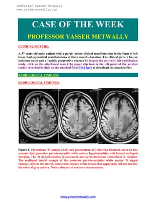
  • 1. Professor Yasser Metwally www.yassermetwally.com CASE OF THE WEEK PROFESSOR YASSER METWALLY CLINICAL PICTURE: A 57 years old male patient with a purely motor clinical manifestations in the form of left lower limb pyramidal manifestations of three months duration. The clinical picture has an insidious onset and a rapidly progressive course.(To inspect the patient's full radiological study, click on the attachment icon (The paper clip icon in the left pane) of the acrobat reader then double click on the attached file) (Click here to download the attached file) RADIOLOGICAL FINDINGS RADIOLOGICAL FINDINGS: Figure 1. Precontrast TI images (A,B) and postcontrast (C) showing bilateral, more or less symmetrical, posterior parieto-occipital white matter hypointensities with lateral scalloped margins. The TI hypointensities is coalescent and periventricular/ subcortical in location. The scalloped lateral margin of the posterior parieto-occipital white matter TI signal changes reflects the strictly subcortical nature of the lesion that apparently did not involve the cortical grey matter. Notice absence of contrast enhancement. www.yassermetwally.com
  • 2. Professor Yasser Metwally www.yassermetwally.com Figure 2. MRI T2 images showing bilateral, more or less symmetrical, posterior parietooccipital white matter hyperintensity with lateral scalloped margins. The T2 hyperintensity is coalescent and periventricular/ subcortical in location. The scalloped lateral margin of the posterior parieto-occipital white matter T2 signal changes reflects the strictly subcortical nature of the lesion that apparently did not involve the cortical grey matter. The coalescent posterior parieto-occipital white matter T2 signal changes is surrounded by a continuous hypointense rim that, most probably, reflects the inflammatory nature of the lesion. The mechanism of the T2 hypointensity is not certain. Proposals in this respect include relative lack of water in the periphery of inflammatory lesion, the presence of blood products in the periphery of the lesion (Deoxyhaemoglobin/hemosiderin induce T2 hypointensity) or the presence of paramagnetic free radicals within the phagocytosing macrophages which are heterogeneously distributed in the periphery of inflammatory lesions. (Paramagnetic free radicals induce T2 hypointensity). www.yassermetwally.com
  • 3. Professor Yasser Metwally www.yassermetwally.com Figure 3. MRI T2 images showing bilateral, more or less symmetrical, posterior parietooccipital white matter hyperintensity with lateral scalloped margins. The T2 hyperintensity is coalescent and periventricular/ subcortical in location. The scalloped lateral margin of the posterior parieto-occipital white matter T2 signal changes reflects the strictly subcortical nature of the lesion that apparently did not involve the cortical grey matter. The coalescent posterior parieto-occipital white matter T2 signal changes is surrounded by a continuous hypointense rim that, most probably, reflects the inflammatory nature of the lesion. The mechanism of the T2 hypointensity is not certain. Proposals in this respect include relative lack of water in the periphery of inflammatory lesion, the presence of blood products in the periphery of the lesion (Deoxyhaemoglobin/hemosiderin induce T2 hypointensity) or the presence of paramagnetic free radicals within the phagocytosing macrophages which are heterogeneously distributed in the periphery of inflammatory lesions. (Paramagnetic free radicals induce T2 hypointensity). Notice the presence popcornlike lesions in (B) that probably reflect the existence of some vascular/venous anomalies. The findings of vascular blush and arteriovenous shunting on angiography, in progressive multifocal leukoencephalopathy, are nonspecific and was reported before. (14) Notice absence of mass effect. www.yassermetwally.com
  • 4. Professor Yasser Metwally www.yassermetwally.com Figure 4. The coalescent posterior parieto-occipital white matter T2 signal changes is surrounded by a continuous hypointense rim that, most, probably, reflects the inflammatory nature of the lesion. The mechanism of the T2 hypointensity is not certain. Proposals in this respect include relative lack of water in the periphery of inflammatory lesion, the presence of blood products in the periphery of the lesion (deoxyhaemoglobin/hemosiderin induce T2 hypointensity) or the presence of paramagnetic free radicals within the phagocytosing macrophages which are heterogeneously distributed in the periphery of inflammatory lesions. (Paramagnetic free radicals induce T2 hypointensity). Multiple foci of acute hemorrhage and abundant petechial hemorrhages in and at the perphery of the posterior parieto-occipital white matte lesions was occaionally reported in progressive multifocal leukoencephalopathy. (14) Notice the popcorn-like lesions in this image, that probably reflect the existence of some vascular/venous anomalies. The findings of vascular blush and arteriovenous shunting on angiography, in progressive multifocal leukoencephalopathy, are nonspecific and was reported before. (14) Notice absence of mass effect. www.yassermetwally.com
  • 5. Professor Yasser Metwally www.yassermetwally.com Figure 5. FLAIR MRI images showing the bilateral, more or less symmetrical, posterior parieto-occipital white matter hyperintensity with lateral scalloped margins. www.yassermetwally.com
  • 6. Professor Yasser Metwally www.yassermetwally.com Figure 6. Chest Plain X ray showing an upper quadrant lung lesion. The lesion wan not biopsied in this patient. Progressive multifocal leukoencephalopathy (PML) is a subacute opportunistic infection caused by the unenveloped DNA virus JC Polyomavirus (JCV). By the age of 65 years, 50% to 70% of individuals have antibodies to the virus. (26) After entering, the virus persists in the renal tubular epithelial cells. With reactivation of latent JCV in the kidneys and subsequent viremia, the JCV enters the brain and causes Progressive multifocal leukoencephalopathy. (26) The incidence of progressive multifocal leukoencephalopathy has greatly increased as a result of the AIDS epidemic, and 0.7% to 11% of patients who have HIV develop progressive multifocal leukoencephalopathy during the course of their illness. The histopathologic hallmark of progressive multifocal leukoencephalopathy is demyelination with enlarged oligodendroglial nuclei and bizarre astrocytes. The disease is usually multifocal, and the lesions may occur in any location in the white matter, most often in the parieto-occipital region. Thalamic lesions are frequently present, and the cerebellum and brain stem can also be involved. Posterior fossa lesions are usually present simultaneously with supratentorial lesions, and, in rare cases, progressive multifocal leukoencephalopathy may be limited to the posterior fossa. Although there are no significant pathologic differences between patients who have and do not have AIDS, in the AIDS population, more extensive disease with a necrotizing character was observed. (26) www.yassermetwally.com
  • 7. Professor Yasser Metwally www.yassermetwally.com The findings on MR imaging correlate well with macroscopic changes. On T2-weighted MR imaging, progressive multifocal leukoencephalopathy lesions are patchy, scalloped, high signal intensity lesions located in the white matter, with extension along the white fibers. Subcortical arcuate fibers are involved, mass effect is mild or absent, and peripheral faint enhancement is a rare feature. On T1-weighted images, the progressive multifocal leukoencephalopathy lesions have low signal, in contrast to isointense HIV-associated lesions. (26) The coalescent posterior parieto-occipital white matter T2 signal changes is sometimes surrounded by a continuous hypointense rim that, most, probably, reflects the inflammatory nature of the lesion. The mechanism of the T2 hypointensity is not certain. Proposals in this respect include relative lack of water in the periphery of inflammatory lesion, the presence of blood products in the periphery of the lesion (deoxyhaemoglobin/hemosiderin induce T2 hypointensity) or the presence of paramagnetic free radicals within the phagocytosing macrophages which are heterogeneously distributed in the periphery of inflammatory lesions. (Paramagnetic free radicals induce T2 hypointensity). Multiple foci of acute hemorrhage and abundant petechial hemorrhages in and at the perphery of the posterior parieto-occipital white matte lesions was occaionally reported in progressive multifocal leukoencephalopathy. (14) A popcorn-like lesions are sometimes reported in PML, and probably reflect the existence of some vascular/venous anomalies in PML. The findings of vascular blush and arteriovenous shunting on angiography, in progressive multifocal leukoencephalopathy, are nonspecific and was reported before. (14) DIAGNOSIS: DIAGNOSIS: PROGRESSIVE MULTIFOCAL LEUKOENCEPHALOPATHY DISCUSSION DISCUSSION: Progressive multifocal leukoencephalopathy (PML) is a fatal subacute progressive demyelinating disease seen in persons with impaired cell-mediated immune response. Progressive multifocal leukoencephalopathy predominantly occurs in patients with AIDS. Before the AIDS epidemic, Progressive multifocal leukoencephalopathy was rare and associated with immunocompromised conditions, such as leukemia, lymphoma, systemic lupus erythematosus (SLE), organ transplantation, Wiskott-Aldrich syndrome, and severe combined immunodeficiency (SCID). At present, Progressive multifocal leukoencephalopathy develops in as many as 5% of all patients with AIDS. This demyelinating disease results from infection with the JC virus, which belongs to the genus Polyomavirus of the Papovaviridae family of viruses. Anstrom et al first described Progressive multifocal leukoencephalopathy in 1958, and Zurhein and Chou initially demonstrated the association of Progressive multifocal www.yassermetwally.com
  • 8. Professor Yasser Metwally www.yassermetwally.com leukoencephalopathy with a viral infection. In 1971, Padgett et al confirmed the viral etiology by isolating a virus from the brain of a patient who had died from Progressive multifocal leukoencephalopathy. The patient's initials were J.C.; hence, the virus is known as the JC virus. Progressive multifocal leukoencephalopathy is characterized by 3 cardinal histopathologic features: demyelination, enlarged nuclei of oligodendrocytes, and bizarre astrocytes. The JC virus is believed to produce infection after it enters the tonsillar tissue during an upper respiratory tract infection. After infection, the virus becomes latent in the spleen, the reticuloendothelial system, and the medulla of the kidney. The JC virus is thought to be undetectable in the brain tissue. Antibodies (immunoglobulin G [IgG]) to the JC virus are common in most Western populations. Seroconversion is seen in 10% of the children by the age of 5 years, in 40-60% by 10 years, and in up to 90% of young adults. Acute infection is usually not noticed. After several years of latency, reactivation occurs under appropriate conditions of immunocompromise. Approximately 50-80% of all Progressive multifocal leukoencephalopathy cases occur in patients with HIV infection, whereas cases are rare in patients with organ transplantation. This difference may indicate the need for an interaction between the JC virus and HIV for Progressive multifocal leukoencephalopathy to develop rather than just an underlying setting of decreased immune function. Although reactivation of JC virus may be necessary, this itself is insufficient to cause Progressive multifocal leukoencephalopathy. A specific deficiency in cellular immune response to the JC viral antigen is probably required in addition to the general cellular immunodeficiency in persons with Progressive multifocal leukoencephalopathy. Reactivation of the virus occurs in the kidney and bone marrow, usually in the setting of immunosuppression. Infected lymphocytes (B cells) then cross the blood-brain barrier and pass infection to astrocytes at the border of vessels. The infection may then be augmented by multiplication and eventual infection of adjacent oligodendrocytes. Oligodendrocytes are responsible for forming and maintaining the myelin sheath. Infection of the oligodendrocytes causes destruction of the cells and loss of the myelin sheath. The axons are usually spared.  Histopathology On microscopy, the cardinal feature of Progressive multifocal leukoencephalopathy is demyelination, which is usually multifocal. The lesions may occur in any location in the white matter, and they range from 1 mm to several centimeters in size. The histopathologic hallmarks of Progressive multifocal leukoencephalopathy include a triad of multifocal demyelination, hyperchromatic enlarged oligodendroglial nuclei, and enlarged bizarre astrocytes with lobulated and hyperchromatic nuclei. Electron www.yassermetwally.com
  • 9. Professor Yasser Metwally www.yassermetwally.com microscopy reveals JC virus in the oligodendroglial cells. Gross examination reveals gray or brown discoloration of the affected brain as a result of loss of myelin. In the US: The prevalence of Progressive multifocal leukoencephalopathy in patients with HIV infection is 1-5% in clinical studies, though postmortem data show a rate of up to 8%. The prevalence of this disease initially increased with the rising incidence of HIV infection, but more recently, the incidence has decreased with the widespread use of highly active antiretroviral therapy (HAART), which has reversed immunosuppression in many patients with AIDS (see subsection on Prognostic utility of MRI in Progressive multifocal leukoencephalopathy). For unknown reasons, Progressive multifocal leukoencephalopathy rarely affects children with HIV infection. The median survival of patients with Progressive multifocal leukoencephalopathy as a complication of AIDS is 6 months. In 10% of patients, survival exceeds 12 months. The longest reported survival is 92 months from the onset of illness. Progressive multifocal leukoencephalopathy is an AIDS-defining illness. Patients whose MRIs show enhancement, which is rare, and those with an increased CD4 count appear to have a better prognosis than that of other patients; these findings probably represent their relatively good immune status. Before the AIDS epidemic, men and women were affected in a male-to female ratio of 3:2, and lymphoproliferative disease was the most common cause. At present, HIV infection is most common cause, with a male-to-female ratio of 7:1 Progressive multifocal leukoencephalopathy chiefly affects homosexual or bisexual men aged 25-50 years. For unknown reasons, Progressive multifocal leukoencephalopathy rarely affects children with HIV infection. The white matter of the brain is usually involved. Lesions of Progressive multifocal leukoencephalopathy may occur anywhere in brain, but the frontal lobes and parieto-occipital regions are commonly affected. Isolated involvement of basal ganglia, external capsule, and posterior-fossa structures may be seen. As of now, radiographically guided intervention has no role in the treatment of Progressive multifocal leukoencephalopathy. An antiretroviral regimen containing protease inhibitors has been successful in prolonging the survival of patients with HIV infection and Progressive multifocal leukoencephalopathy. Topotecan and cidofovir are promising drugs presently under evaluation. The role of splenectomy remains unproven, though reports have described remission in association with HAART.  Clinical Details o Symptoms Progressive focal neurologic deficit is the clinical hallmark. Weakness and disturbance of speech are most common symptoms. Other symptoms include cognitive abnormalities, headaches, gait disorders, visual impairment, and sensory loss. Headaches are most common in the HIV-infected population, and visual disturbances are most common in those without HIV infection. About 10% of patients have seizures. Cognitive deficits do not persist in isolation for long and distinguish Progressive multifocal leukoencephalopathy from HIV dementia. Progressive multifocal leukoencephalopathy www.yassermetwally.com
  • 10. Professor Yasser Metwally www.yassermetwally.com seems to have a more aggressive course in persons with HIV disease than in persons with other predisposing conditions. o Signs Most common physical sign is limb weakness, which occurs in more than 50% of patients. Cognitive disturbances and gait disorders affect 25-33%, and diplopia affects 9%. Opticnerve disease does not occur with Progressive multifocal leukoencephalopathy, and spinalcord involvement is rare.  Laboratory findings Severe cellular immunosuppression, as defined by a CD4 lymphocyte count of 200 cells/L, is observed in most patients. The mean CD4 count is 84-104 cells/L. Results of CSF examination are usually normal or show slightly elevated protein levels. CSF study is not helpful in diagnosis except that it may help in excluding other diagnoses. Cell counts are usually less than 20 cells/L. Several studies demonstrated that polymerase chain reaction (PCR) study of the CSF has high sensitivity and specificity for JC virus in Progressive multifocal leukoencephalopathy, with some investigators reporting 95% sensitivity and 100% specificity. An additional test of the CSF involves measurement of an antibody to the major structural protein of the JC virus known as VPI. The criterion standard for the diagnosis remains histologic confirmation by means of tissue biopsy. However, with a characteristic clinical and MRI pattern and a positive PCR result for JC virus in the CSF, brain biopsy is often avoided. NEUROIMAGING Radiographic imaging strongly supports the diagnosis of Progressive multifocal leukoencephalopathy in the appropriate clinical context. MRI is sensitive to white matter lesions and shows hyperintense lesions on T2-weighted (T2W) images in affected regions. Therefore, MRI is the preferred form of imaging. MRI is the preferred diagnostic test. Because of its superior contrast resolution, it can be used to detect subtle white matter abnormalities, whereas CT depicts the lesions at an advanced stage. Lesions are found at the gray matter–white matter interface and tend to involve the subcortical white matter. This predilection accounts for the scalloped margins of the lesions. Lesions are initially multiple and discrete, but they eventually may coalesce into large lesions. The lesions may occur anywhere, but are most often seen in the parieto-occipital and frontal lobes. CT is often the first neuroimaging technique used, but is not as sensitive as MRI in the detection of white-matter lesions. MRI offers superior sensitivity in the detection of whitematter lesions of Progressive multifocal leukoencephalopathy, but it is contraindicated in www.yassermetwally.com
  • 11. Professor Yasser Metwally www.yassermetwally.com patients with a cardiac pacemaker, in those with MRI-incompatible implants, and those with intraocular metallic foreign bodies.  CT scan CT scans usually show several bilateral, asymmetric hypoattenuating foci of various sizes without mass effect or enhancement. The lesions may involve the periventricular white matter, subcortical white matter, or both. Subcortical U-fiber involvement results in lesions having a lateral scalloped margin that follows the gray matter–white matter junction. Although lesions may be seen on CT scanning, MRI offers superior sensitivity in the detection and characterization of the lesions. The diagnosis may be suspected with CT, but MRI is needed for a more confident exclusion of other differential considerations. Artifacts; chronic, ischemic white-matter changes; and chronic infarcts may mimic true white-matter lesions of Progressive multifocal leukoencephalopathy.  MRI o Findings on T1- and T2-weighted and fluid-attenuated inversion recovery images MRI has far greater sensitivity than other studies in detecting the lesions of Progressive multifocal leukoencephalopathy and in defining their extent of involvement. On T2W images, lesions appear hyperintense and typically involve the periventricular and subcortical white matter, having a characteristic scalloped lateral margin when they involve the subcortical white matter. Lesions are more conspicuously visualized on fluidattenuated inversion recovery (FLAIR) images, appearing hyperintense against a background of suppressed CSF signal intensity. Lesions appear hypointense and well demarcated on T1-weighted (T1W) images, though they may be isointense in the initial phase of the disease. Although the disease may involve any part of the brain, lesions typically occur in the parieto-occipital lobes. Lesions appear to start in the subcortical white matter before extending to the periventricular white matter. Mass effect is infrequently described and usually minimal and correlated with shorter survival when seen on initial studies. o Distribution of lesions A multifocal distribution pattern is seen. This pattern may be unilateral, but more often, it is bilateral and asymmetric. Lesions may start small, but they eventually enlarge and coalesce. Posterior-fossa involvement is common and is seen in up to one third of patients. Synchronous lesions are usually seen in the supratentorial compartment, though in 10% of patients, lesions are confined to the structures of the posterior fossa. Progressive multifocal leukoencephalopathy may also appear to involve the basal ganglia and deep gray-matter nuclei because of white-matter fibers in these structures. When present, involvement of the gray matter is a secondary finding. In rare cases, lesions are single and can occur anywhere in the brain, including the brainstem. www.yassermetwally.com
  • 12. Professor Yasser Metwally www.yassermetwally.com o Findings on contrast-enhanced images The lesions typically do not enhance and do not have mass effect; however, some reports describe lesions with faint peripheral enhancement or diffuse enhancement with mass effect, especially in the early stages. Enhancement could suggest a relatively good immune response and hence an improved prognosis. o Findings on diffusion-weighted images On diffusion-weighted images (DWI), lesions can show restricted diffusion, though this is uncommon. The extent of abnormal diffusion appears to be correlated with the speed of clinical progression. Areas with DWI abnormality may correspond to areas that are actively infected by the virus at the time of imaging, but this has been uncommonly reported. o Prognostic utility of MRI in Progressive multifocal leukoencephalopathy Post et al (1999) studied the prognostic utility of MRI in Progressive multifocal leukoencephalopathy. None of the MRI variables was predictive of patient survival, with the exception of mass effect, which was usually minimal, infrequent, and associated with decreased survival. Serial MRI studies showed progression of disease in 1-24 weeks, and a more rapid change was seen in many patients in just 9 weeks. The authors suggested that findings of increasing atrophy, increasing confluence and extent of white-matter lesions, spread of disease across the corpus callosum, and increasing hypointensity of the lesions on follow-up T1W MRIs may be poor prognostic indicators or as failure of response to therapy. Stabilization or a decrease in the size of the lesion, clinical improvement, and loss JC viral detection on CSF PCR testing may indicate a response to therapy. However, improvements on MRI can lag clinical improvement by 2-6 months or even indicate temporary worsening. In Progressive multifocal leukoencephalopathy patients undergoing HAART, MRIs can show initial worsening in the first few months followed by stabilization and regression by 12 months. HAART consists of a combination of 3 or 4 anti-HIV drugs from the following classes: nucleoside reverse transcriptase inhibitors, non-nucleoside reverse transcriptase inhibitors, and protease inhibitors. Thurnher et al (2001) reported 2 patients with long-term responses to HAART, apparent increased mass effect and enhancement before eventual improvement, and atrophy of the involved brain parenchyma. T1W MRIs but not FLAIR MRIs showed hypointensity. Two nonresponders initially had extensive changes, without enhancement or mass effect after the start of therapy. Their T1W MRIs also showed hypointense areas that were hyperintense on FLAIR images. This increasing hypointensity on T1W MRIs might have been due to increasing demyelination, which appears hyperintense on FLAIR images. This finding might indicate www.yassermetwally.com
  • 13. Professor Yasser Metwally www.yassermetwally.com a worsened prognosis. Increased T1W hypointensity in responders was due to gliosis and necrosis seen in burned-out lesions; this finding is hypointense on FLAIR images. Berger et al (Ann Neurol, 1998) found enhancement in 8.9% of short-term survivors in contrast to 50% in long-term survivors. Enhancement suggests an improved immune response and hence an improved prognosis. In a study by Collazos et al (1999), contrast enhancement was seen on images in patients with Progressive multifocal leukoencephalopathy treated with HAART only when their CD4+ count increased. In these patients, the Progressive multifocal leukoencephalopathy lesions changed from enhancing to nonenhancing on follow-up MRI after 3-6 months of therapy. Larger studies than this are needed to clearly identify the MRI changes after therapy and to confirm the utility of MRI in assessing the patient's prognosis and response to therapy. o Magnetic resonance spectroscopy Evaluation of the Progressive multifocal leukoencephalopathy lesions with magnetic resonance (MR) spectroscopy reveals reduced N-acetylaspartate (NAA) and creatine levels, increased choline levels, and an excess of lipids and sometimes of myo-inositol. In some cases, lactate is present. Cell membrane and myelin breakdown is the presumed cause of elevated choline levels, neuronal loss leads to decreased NAA concentrations, and glial-cells proliferation elevates myo-inositol values. The mild elevation of lactate and lipid levels may be due to the activity of macrophages and the breakdown products of myelin. The elevation of choline and myo-inositol values is seen in the early phase of the disease. In the later phase all the metabolites are decreased. These metabolic abnormalities are not specific for Progressive multifocal leukoencephalopathy and may be similar in other lesions complicating HIV disease. o Magnetization transfer imaging Progressive multifocal leukoencephalopathy lesions appear to have strongly reduced magnetization transfer ratios. According to Ernst et al (1999), these features may help in distinguishing lesions from white-matter lesions of HIV leukoencephalopathy. Large prospective studies are needed to assess the utility of newer MR techniques, such as MR spectroscopy and magnetization transfer imaging, in the diagnosis and follow-up of Progressive multifocal leukoencephalopathy. Degree of Confidence: MRI has far greater sensitivity than that of other studies in depicting the lesions of Progressive multifocal leukoencephalopathy and in defining their extent of involvement. Although MRI results may suggest the diagnosis of Progressive multifocal leukoencephalopathy in the appropriate clinical setting, typical features of Progressive multifocal leukoencephalopathy are often nonspecific, and other differential considerations, such as ischemic changes, HIV leukoencephalopathy, and gliotic changes from previous trauma, must be considered. When the lesions of Progressive multifocal www.yassermetwally.com
  • 14. Professor Yasser Metwally www.yassermetwally.com leukoencephalopathy are atypically enhancing or when they have mass effect, other lesions, such as those due to toxoplasmosis, lymphoma, or other intracranial masses should be considered. The presence of multiple pathologies in immunocompromised conditions adds to the complexity of image interpretation. Infection coexisting with other opportunistic infection may be responsible for some atypical manifestations of the disease and for an apparent response to some form of therapy in some patients. It is unusual for a normal variant to mimic Progressive multifocal leukoencephalopathy lesions on MRI. Progressive multifocal leukoencephalopathy must be distinguished from HIV leukoencephalopathy. Progressive multifocal leukoencephalopathy tends to be multifocal, with bilateral, asymmetric, and predominantly subcortical involvement. In comparison, HIV leukoencephalopathy tends to produce lesions that are usually diffuse, bilateral, and symmetric; these predominantly affect the periventricular white matter. Diffuse cortical atrophy and ventricular dilatation are not predominant findings of Progressive multifocal leukoencephalopathy and may be helpful in distinguishing from HIV leukoencephalopathy, which is usually associated with substantial atrophy. Lesions appear well defined and hypointense on T1W images, whereas they appear isointense and poorly defined in HIV leukoencephalopathy. Clinical correlation may be helpful, as patients with Progressive multifocal leukoencephalopathy have progressive focal motor and sensory neurologic deficits, whereas those with HIV leukoencephalopathy present with global cognitive changes and dementia. Figure 7. T2-weighted MRI shows left occipital hyperintense white matter changes with the lesion margin reaching the cortex., This sliced fixed brain shows multiple isolated or confluent gray demyelinative foci. Atrophy may be present., Microscopically, multiple demyelinative foci are detected. The microscopic hallmark of the disease is intranuclear basophilic or eosinophilic inclusions within the swollen nuclei of oligodendrocytes, often at the periphery of lesions. Large, occasionally multinucleated astrocytes with prominent processes are another characteristic feature. www.yassermetwally.com
  • 15. Professor Yasser Metwally www.yassermetwally.com Figure 8. Progressive multifocal leukoencephalopathy, A, Axial PD (A) and T2W (B) images show large confluent area of hyperintensity (*) of left frontotemporal region. While there is some sulcal and ventricular effacement secondary to edema, the amount of edema and mass effect present is disproportionately small, given the large area of hyperintensity. Pre-contrast axial T1W (C) image shows corresponding. B, Axial PD (A) and T2W (B) images show large confluent area of hyperintensity (*) of left frontotemporal region. While there is some sulcal and ventricular effacement secondary to edema, the amount of edema and mass effect present is disproportionately small, given the large area of hyperintensity. Pre-contrast axial T1W (C) image shows corresponding region of hypointensity . C, Axial PD (A) and T2W (B) images show large confluent area of hyperintensity (*) of left frontotemporal region. While there is some sulcal and ventricular effacement secondary to edema, the amount of edema and mass effect present is disproportionately small, given the large area of hyperintensity. Pre-contrast axial T1W (C) image shows corresponding region of hypointensity . D, Post-contrast axial (D) and coronal (E) T1W images show minimal www.yassermetwally.com
  • 16. Professor Yasser Metwally www.yassermetwally.com enhancement along lateral margins of mass (*). The cortical surface is spared. E, Postcontrast axial (D) and coronal (E) T1W images show minimal enhancement along lateral margins of mass (*). The cortical surface is spared. F, Coronal autopsy specimen shows large area (arrows) of demyelination of left frontal and temporal lobes with sparing of the cortical surface. Figure 9. Progressive multifocal leukoencephalopathy, A, Axial PDW images show large confluent areas of hyperintensity of both centra semiovale, the posterior white matter, the left basal ganglia, and the splenium of the corpus collosum. There is relatively little mass effect for the size of these lesions. The cortex is predominantly spared. B, Axial PDW images show large confluent areas of hyperintensity of both centra semiovale, the posterior white matter, the left basal ganglia, and the splenium of the corpus collosum. There is relatively little mass effect for the size of these lesions. The cortex is predominantly spared. C, Axial autopsy specimen show extensive demyelination of the white matter bilaterally (arrowheads). D, Axial autopsy specimen at different level than (C) shows demyelination of the splenium of the corpus collosum (arrowheads) and the posterior white matter (arrow). Microscopically, there was marked myelin loss and variable axonal loss of the white matter with numerous gemistocytic astrocytes, some of which had enlarged nuclei. In addition, foamy macrophages, multiple round oligodendrocytic type nuclei, and multinucleated cells were present. Chronic inflammatory reactions were infrequently noted. www.yassermetwally.com
  • 17. Professor Yasser Metwally www.yassermetwally.com  Nuclear medicine Nuclear medicine studies do not play a major role in the diagnosis of Progressive multifocal leukoencephalopathy. However, Port et al (1999) described a case of Progressive multifocal leukoencephalopathy in a patient with AIDS in whom MRIs of the lesion showed enhancement and mass effect, with increased thallium-201 uptake on single photon emission CT (SPECT). O'Mally et al (1994) had reported a case of Progressive multifocal leukoencephalopathy with no uptake on 201Th SPECT. On thallium SPECT, photopenic lesions can be caused by any previous insult to the brain parenchyma, and hence, the differential considerations include a wide variety of causes. The uncommon observation of increased radiotracer uptake may be seen with lymphoma and is atypically found in some infections. No anatomic variants have been described to mimic the lesions of Progressive multifocal leukoencephalopathy, but cold lesions (eg, arachnoid cyst, porencephalic cyst) can conceivably appear as photopenic lesions similar to the lesions of Progressive multifocal leukoencephalopathy.  Angiography Angiography has no role in the diagnosis of Progressive multifocal leukoencephalopathy. However, Nelson et al (1999) reported angiographic findings in 6 patients with Progressive multifocal leukoencephalopathy. In 4, angiograms showed abnormal parenchymal blush of the affected region in the early to mid arterial phase and persisting into the venous phase. Associated arteriovenous shunting was also present. In only 1 of the 4 patients did MRI demonstrate enhancement. On pathologic evaluation, intense perivascular inflammatory cellular infiltrates, angiogenesis, and gliosis were found in the patients with angiographic abnormalities, whereas changes were minimal in patients who did not have angiographic abnormalities. The findings of vascular blush and arteriovenous shunting on angiography are nonspecific and can be seen with infectious, neoplastic, vascular, and ischemic etiologies. SUMMARY SUMMARY Progressive multifocal leukoencephalopathy is an uncommon progressive fatal demyelinating disease that affects immunocompromised patients. The cause is a papovavirus—the Creutzfeldt-Jakob virus. The lesions are usually multifocal and asymmetric, most commonly affecting the subcortical white matter and corpus callosum. In the corpus callosum, focal lesions can occur that enlarge and become confluent as the disease progresses. The lesions are hyperintense on long-TR sequences and hypointense on www.yassermetwally.com
  • 18. Professor Yasser Metwally www.yassermetwally.com short-TR/TE sequences. The lesions usually do not enhance, although they may enhance faintly at the periphery. Progressive multifocal leukoencephalopathy should be considered in the differential diagnosis of space-occupying lesions in HIV patients. The lack of enhancement and mass effect can act as features differentiating this entity from others such as lymphoma or glioblastoma. Progressive multifocal leukoencephalopathy (PML) is a demyelinating disease caused by an infection of papovavirus and for many years was known as a rare complication in immunocompromised patients with lymphoma, sarcoidosis, or following transplant surgery. Progressive multifocal leukoencephalopathy has become a relatively common complication of acquired immunodeficiency syndrome (AIDS) patients. 10,21 Apparently, Progressive multifocal leukoencephalopathy lesions in AIDS patients are more destructive, and show a higher density of papovavirus-infected cells. On CT, the Progressive multifocal leukoencephalopathy lesions are multifocal, initially involving subcortical white matter, progressing to involve deeper white matter, and typically are of low density without significant enhancement, mass effect, or edema. The MR imaging diagnosis of Progressive multifocal leukoencephalopathy is facilitated by knowledge of the patient's immunocompromised state. Geographic or maplike regions of increased water content are seen on MR images as decreased signal on Tl-weighted images, and increased signal on proton density and T2-weighted images. Peripheral enhancement and hemorrhage are rare but have been described. The brain may appear completely normal on external examination. On sectioning, however, typical multifocal demyelinating lesions of Progressive multifocal leukoencephalopathy usually are found in the subcortical white matter. In long-lasting cases, large confluent areas of demyelination may extend into deep white matter, accompanied by many small satellite foci. In the well-fixed brain, smaller foci of demyelination usually are discolored gray, whereas the surface of larger lesions may be granular, with a moth-eaten appearance. Extensively involved white matter may be discolored yellow, necrotic, or cavitated. The cerebrum is commonly involved; however, lesions in the cerebellum and brainstem, with or without cerebral involvement, have been reported. The histology of the lesions varies considerably depending on the stage of the disease process. In very early stages, the changes may be subtle, and scattered reactive astrocytes without any obvious foci of demyelination may be the only changes seen in the white matter. Within typical small foci of demyelination are many reactive astrocytes, some showing bizarre nuclei suggestive of neoplastic astrocytes. Reactive microglia/ macrophages also are present. Oligodendrocytes usually are found in the peripheral zone of these foci of demyelination. Their nuclei often are enlarged, glassy, and homogeneously amphophilic to mildly basophilic. The nuclei of these oligodendrocytes are immunostained with the antibody to the papovavirus and harbor virions, as detected with the electron microscope. Viral antigen also can be detected in the nuclei with in situ hybridization. In large confluent lesions, it is common to find packed reactive astrocytes and foamy, lipidcontaining macrophages occupying most of the lesion. www.yassermetwally.com
  • 19. Professor Yasser Metwally www.yassermetwally.com  Addendum o A new version of this PDF file (with a new case) is uploaded in my web site every week (every Saturday and remains available till Friday.) o To download the current version follow the link "http://pdf.yassermetwally.com/case.pdf". o You can also download the current version from my web site at "http://yassermetwally.com". o To download the software version of the publication (crow.exe) follow the link: http://neurology.yassermetwally.com/crow.zip o The case is also presented as a short case in PDF format, to download the short case follow the link: http://pdf.yassermetwally.com/short.pdf o At the end of each year, all the publications are compiled on a single CD-ROM, please contact the author to know more details. o Screen resolution is better set at 1024*768 pixel screen area for optimum display. o Also to view a list of the previously published case records follow the following link (http://wordpress.com/tag/case-record/) or click on it if it appears as a link in your PDF reader o To inspect the patient's full radiological study, click on the attachment icon (The paper clip icon in the left pane) of the acrobat reader then double click on the attached file. o Click here to download the short case version of this case record in PDF format REFERENCES References 1. Anstrom KE, Mancall EL, Richardson EP Jr: Progressive multifocal leukoencephalopathy. Brain 1958; 81: 93-127. 2. Berger JR, Pall L, Lanska D, Whiteman M: Progressive multifocal leukoencephalopathy in patients with HIV infection. J Neurovirol 1998; 4(1): 59-68. 3. Berger JR, Levy RM, Flomenhoft D, Dobbs M: Predictive factors for prolonged survival in acquired immunodeficiency syndrome-associated progressive multifocal leukoencephalopathy. Ann Neurol 1998 Sep; 44(3): 341-9. 4. Berger JR, Major EO: Progressive multifocal leukoencephalopathy. Semin Neurol 1999; 19(2): 193-200. 5. Chang L, Ernst T, Tornatore C, et al: Metabolite abnormalities in progressive multifocal leukoencephalopathy by proton magnetic resonance spectroscopy. Neurology 1997 Apr; 48(4): 836-45. 6. Collazos J, Mayo J, MartÃnez E, et al: Contrast-enhancing progressive multifocal leukoencephalopathy as an immune reconstitution event in AIDS patients. AIDS 1999 Jul 30; 13(11): 1426-8. 7. Dworkin MS: A review of progressive multifocal leukoencephalopathy in persons with and without AIDS. Curr Clin Top Infect Dis 2002; 22: 181-95. www.yassermetwally.com
  • 20. Professor Yasser Metwally www.yassermetwally.com 8. Ernst T, Chang L, Witt M, et al: Progressive multifocal leukoencephalopathy and human immunodeficiency virus-associated white matter lesions in AIDS: magnetization transfer MR imaging. Radiology 1999 Feb; 210(2): 539-43. 9. Iranzo A, Moreno A, Pujol J, et al: Proton magnetic resonance spectroscopy pattern of progressive multifocal leukoencephalopathy in AIDS. J Neurol Neurosurg Psychiatry 1999 Apr; 66(4): 520-3. 10. Kastrup O, Maschke M, Diener HC, et al: Progressive multifocal leukoencephalopathy limited to the brain stem. Neuroradiology 2002 Mar; 44(3): 227-9. 11. Kotecha N, George MJ, Smith TW, et al: Enhancing progressive multifocal leukoencephalopathy: an indicator of improved immune status?. Am J Med 1998 Dec; 105(6): 541-3. 12. Mader I, Herrlinger U, Klose U, et al: Progressive multifocal leukoencephalopathy: analysis of lesion development with diffusion-weighted MRI. Neuroradiology 2003 Oct; 45(10): 717-21. 13. Mayo J, Collazos J, MartÃnez E: Progressive multifocal leukoencephalopathy following initiation of highly active antiretroviral therapy. AIDS 1998 Sep 10; 12(13): 1720-2. 14. Nelson PK, Masters LT, Zagzag D, et al: Angiographic abnormalities in progressive multifocal leukoencephalopathy: an explanation based on neuropathologic findings. AJNR Am J Neuroradiol 1999 Mar; 20(3): 487-94. 15. Nicoli F, Chave B, Peragut JC, Gastaut JL: Efficacy of cytarabine in progressive multifocal leucoencephalopathy in AIDS. Lancet 1992; 339(8788): 306. 16. O'Malley JP, Ziessman HA, Kumar PN, et al: Diagnosis of intracranial lymphoma in patients with AIDS: value of 201TI single-photon emission computed tomography. AJR Am J Roentgenol 1994 Aug; 163(2): 417-21. 17. Ohta K, Obara K, Sakauchi M, et al: Lesion extension detected by diffusionweighted magnetic resonance imaging in progressive multifocal leukoencephalopathy. J Neurol 2001; 248(9): 809-811. 18. Padgett BL, Walker DL, ZuRhein GM, et al: Cultivation of papova-like virus from human brain with progressive multifocal leucoencephalopathy. Lancet 1971; 1(7712): 1257-1260. 19. Port JD, Miseljic S, Lee RR, et al: Progressive multifocal leukoencephalopathy demonstrating contrast enhancement on MRI and uptake of thallium-201: a case report. Neuroradiology 1999; 41 (12): 895-898. 20. Portegies P, Algra PR, Hollak CE, et al: Response to cytarabine in progressive multifocal leucoencephalopathy in AIDS. Lancet 1991; 337(8742): 680-681. 21. Post MJ, Yiannoutsos C, Simpson D, et al: Progressive multifocal leukoencephalopathy in AIDS: are there any MR findings useful to patient management and predictive of patient survival? AIDS Clinical Trials Group, 243 Team. AJNR Am J Neuroradiol 1999; 20(10): 1896-1906. 22. Sadler M, Nelson MR: Progressive multifocal leukoencephalopathy in HIV. Int J STD AIDS 1997; 8(6): 351-357. 23. Simone IL, Federico F, Tortorella C, et al: Localized 1H-MR spectroscopy for metabolic characterisation of diffuse and focal brain lesions in patients infected with HIV. J Neurol Neurosurg Psychiatry 1998 Apr; 64(4): 516-23. www.yassermetwally.com
  • 21. Professor Yasser Metwally www.yassermetwally.com 24. Thurnher MM, Post MJ, Rieger A, et al: Initial and follow-up MR imaging findings in AIDS-related progressive multifocal leukoencephalopathy treated with highly active antiretroviral therapy. AJNR Am J Neuroradiol 2001; 22(5): 977-84. 25. ZuRhein GM: Association of papovavirus with a human demyelinating disease (progressive multifocal leukoencephalopathy). Prog Med Virol 1969; 11: 185-247. 26. Metwally, MYM: Textbook of neuroimaging, A CD-ROM publication, (Metwally, MYM editor) WEB-CD agency for electronic publication, version 11.4a October 2010 www.yassermetwally.com