
Recommended
More Related Content
What's hot
What's hot (17)
Viewers also liked
Viewers also liked (20)
Similar to Wayang kulit, no 1 a fundamentals
Similar to Wayang kulit, no 1 a fundamentals (20)
Recently uploaded
Recently uploaded (20)
Wayang kulit, no 1 a fundamentals
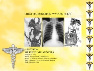
- 1. CHEST RADIOGRAPHS, WAYANG KULIT A REVISION OF THE FUNDAMENTALS Dr Ng Kian Seng MBBS (Singapore) MCGP (Malaysia) Master Of Medicine (Internal Medicine, Singapore) FAFP (Malaysia) Cert In Occupational Medicine Ph D (Theology, USA)
- 2. Hippocrates of Cos, Father of Medicine ANATOMY IN THE CHEST RADIOGRAPH
- 3. A Chest Radiograph, Not A Chest X-Ray A Normal Chest Radiograph Some examiners like you to call x ray films radiographs; strictly speaking you can’t actually see the x rays themselves.
- 4. Anatomy in the Chest Radiograph The right main bronchus is slightly larger than the left & comes off at a less acute angle than the left (hence septic material & foreign substances are more likely to be inhaled into the right lung than into the left).
- 5. Chest Radiograph, PA View, No 1 Apex Of Lung Carina Trachea Right para-tracheal stripe Aortic arch Main Pulmonary Artery Left Atrial appendage Descending thoracic aorta Left ventricle Gastric Air Bubble
- 6. Chest Radiograph, PA View, No 2 Right upper lobe pulmonary vein Horizontal fissure Right hilum Right lower lobe pulmonary artery Right atrium Right Cardiophrenic Angle Right Costophrenic Angle
- 7. Chest Radiograph, PA View, No 3 Spinous process Scapula Anterior Rib Clavicle Posterior Rib Right Bronchus Left Bronchus Diaphragm Breast Soft Tissue Lung Tissue Superimposed On diaphragm Retrocardiac vertebra
- 8. Chest Radiograph, PA View, No 4 Anatomy Of the Heart In The Chest Radiograph
- 9. THE MEDIASTINAL STRUCTURES IN THE C-XRAY
- 10. Aorto Pulmonary Window Aorto-pulmonary window. The aorto-pulmonary window lies between the arch of the aorta and the pulmonary arteries. It contains the ligamentum arteriosum, the recurrent laryngeal nerve, lymph nodes, and fatty tissue. ...
- 11. RIGHT PARA- TRACHEAL STRIPE From the level of the clavicles to the azygous vein the right edge of the trachea is seen as a thin white stripe. This appearance is created by air of low density (blacker) lying either side of the comparatively dense (whiter) tracheal wall. If this stripe is thickened (normally less than 5 mm) this may represent pathology such as a paratracheal mass or enlarged lymph node. The left side of the trachea is not so well defined because of the position of the aortic arch and great vessels.
- 12. Anatomy in the Lateral Chest X-ray 1. Ascending thoracic Aorta 2. Sternum 3. Right ventricle 4. Left ventricle 5. Left atrium 6. Gastric air bubble 7. Right Hemidiaphragm 8. Left Hemidiaphragm 9. Right upper lobe bronchus 10. Left upper lobe bronchus 11. Trachea.
- 13. NAME THE STRUCTURES IN THE LATERAL CHEST X-RAY 1.Trachea 2. Aortopulmonary window 3. Sternum 4. Right ventricle 5. Right Hemidiaphragm 6. Left Hemidiaphragm 7. Left atrium 8. Scapula 9. Right Upper Lobe Bronchus 10. Left upper Lobe Bronchus 9 10
- 14. THE MEDIASTINUM The mediastinum is divided by a plane passing from the sternal angle to T4-T5 into: Superior mediastinum and The inferior mediastinum The inferior mediastinum is further subdivided into three regions namely: Anterior mediastinum Middle mediastinum Posterior mediastinum These divisions are for descriptive purposes, they merge into each other imperceptibly. There are no distinct boundaries between them.
- 15. ZONES OF THE CHEST RADIOGRAPH Apex to a line drawn through The lower borders of the Anterior ends of the 2nd costal Cartilage. UPPER ZONE MIDDLE ZONE LOWER ZONE From the 1st line to one drawn Through the lower borders of the 4th costal cartilage & includes The Hila of the lungs From the 2nd line to the Bases of the lungs.
- 16. THE FISSURES OF THE LUNGS Oblique Fissure From 4 th Dorsal spine sweeping down Obliquely to the 6th rib in mid mammary line or the 6th costo Chondral junction, anteriorly. Horizontal Fissure. Runs from the 4th costo chondral junction To meet Oblique Fissure at the mid axillary line.
- 17. THE LOBES & FISSURES OF THE LUNGS Base of the Lung: 6th costochondral junction, obliquely to the 10th rib in anterior Axillary line, then horizontally to 12th thoracic vertebra
- 18. OBLIQUE FISSURE , HORIZONTAL FISSURE Oblique Fissure : From 4th Dorsal spine sweeping down Obliquely to the 6th rib in mid mammary line or the 6th Costochondral junction, anteriorly. Horizontal Fissure. Runs from the 4th costochondral Junction to meet Oblique Fissure at the mid axillary line.
- 19. THE RIGHT & LEFT OBLIQUE FISSURES From 4th Dorsal spine sweeping down Obliquely to the 6th rib in mid mammary line or the 6th Costochondral junction, anteriorly.
- 20. THE HORIZONTAL FISSURE Horizontal Fissure. Runs from the 4th costochondral junction to meet Oblique Fissure at the Mid Axillary Line.
- 21. WHAT IS THE ABNORMALITY HERE?
- 22. ACESSORY FISSURE, THE AZYGOS FISSURE . The azygos lobe appears starting in a teardrop shape at around the level of T5 to the right of the midline as a pale line curving outward and upward and then back in to meet the root of the neck, the line is the infolding of the pleura. Also described as a “curvilinear opacity, Inverted comma, tadpole.” (See Notes in “Companion”, J)
- 23. Louis Pasteur NORMAL VARIANTS IN THE CHEST RADIOGRAPH
- 24. NIPPLE SHADOWS
- 25. NIPPLE SHADOWS RIGHT NIPPLE LEFT NIPPLE Confirm these are indeed nipple Shadows by using metal markers!
- 26. ASYMMETRY OF THE BREASTS Breast asymmetry is very common, even to the extent that no breast tissue is visible on one side. It should not be assumed that the patient has had a mastectomy, unless this is known from the history.
- 27. BONE ISLAND IN THE RIB
- 28. DROMEDARY HUMP IN THE DIAPHRAGM
- 29. EXAMINE THE FIRST & SECOND RIBS ON BOTH SIDES
- 30. FUSION OF FIRST & SECOND RIB ON THE LEFT
- 31. PSEUDO-ARTHROSIS OF THE FIRST TWO RIBS ON THE LEFT
- 32. BIFURCATED RIB
- 33. Soft tissue fat This close-up demonstrates a normal fat plane between layers of muscle. Fat is less dense than muscle and so appears blacker. Note that the edge of fat is smooth. Irregular areas of black within the soft tissues may represent air tracking in the subcutaneous layers. This is known as surgical emphesyma
- 34. What is the bony abnormality in this patient? Chest radiograph is showing well developed bilateral cervical ribs.
- 35. Cervical rib is an extra rib that arises from the 7th cervical vertebrae. How do you know these are cervical ribs? And not the 1st thoracic ribs? Cervical Transverse Processes Points Downwards= CD Thoracic Transverse Processes Points Upwards = TU Look at the transverse processes that articulate with these ribs. Cervical transverse processes points down while thoracic transverse processes points up.
- 36. Edward Jenner THE BLACK & WHITE RADIOLOGICAL TERMS
- 37. RADIODENSITY SCALE Radiodensity : Physical quality of an object that determines how much radiation it absorbs from the X-Ray beam. Radiodensity is determined by composition ( atomic weight) and thickness RADIOLUCENT INCREASING RADIODENSITY DECREASING RADIODENSITY AIR FAT BLOOD MUSCLE BONE BARIUM RADIOPAQUE radioLucent = bLack radiopaquE = whitE
- 39. RADIODENSE VERSUS RADIOLUCENT RADIOLUCENT RADIODENSE RADIOPAQUE
- 40. Antonie von Leeuwenhoek “What is the student but a lover courting a fickle mistress who ever eludes his grasp?” Sir William Osler Contact Me At : plusultra.ng@gmail.com
Editor's Notes
- The Greek physician Hippocrates of Cos, born around 460 B.C. the Founding Father of Medicine
- Note that the lower zones reach below the diaphragm. This is because the lungs pass behind the dome of the diaphragm into the posterior sulcus of each hemithorax. Normal lung markings can be seen below the well defined edges of the diaphragm.
- costochondral junction
- LouisPasteur: The Chemist Who Transformed Medicine. He debunk the theory that microbes appear by “spontaneous generation”, and demonstrated that they are instead produced from other microbes. Pasteur’s discoveries helped develop the “germ theory” of disease and the process of pasteurization that is used to this day.
- This painting depicts the use of the first Western vaccine: the cowpox vaccine to protect against smallpox. Edward Jenner, a rural English doctor, is shown injecting his first patient, James Phipps, in 1796, using fluid obtained from scratches on the hand of dairymaid Sarah Nelmes. His observation that dairymaids seemed to gain immunity to smallpox from their exposure to cowpox led to Jenner’s experiments and the eventual widespread adoption of smallpox vaccination. In 1980, the disease of smallpox was declared eradicated from the world.
- Antonie von Leeuwenhoek is shown exploring the microscopic world through handmade lenses. The 17th-century Dutch scientist was the first to report seeing what we now know as protozoa and bacteria (which he called “animacules”), and to document blood flow in small vessels called capillaries. He is considered a forefather of modern microbiology, and a key contributor to the development of the microscope, having ground hundreds of lenses that were mounted in metal frames such as the one he holds in the painting.