
Recomendados
Más contenido relacionado
La actualidad más candente
La actualidad más candente (20)
Similar a AXILLA AND BREAST.pptx
Similar a AXILLA AND BREAST.pptx (20)
Más de AgabaSaphan
Último
Último (20)
AXILLA AND BREAST.pptx
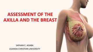
- 1. ASSESSMENT OF THE AXILLA AND THE BREAST SAPHAN C. AGABA UGANDA CHRISTIAN UNIVERSITY
- 2. LEARNING OBJECTIVES By the end of the unit, learners will be able to: • Discuss the history questions pertaining to male and female breast and Genitalia assessment. • Perform a breast examination including axillary nodes and interpret findings. • Discuss components of a genital exam on a male or female. • Review components of a comprehensive reproductive history. • Document findings. • List the changes in breast, male & female • genitalia that are characteristics of aging process.
- 3. FEATURES OF THE BREAST • Location-- chest wall • Axillary tail of Spence • Nipple and areola
- 4. Internal Features • Glandular tissue – Lobes, lobules, and alveoli – Lactiferous ducts and sinuses • Fibrous tissue – Suspensory ligaments or Cooper’s ligaments – Adipose tissue
- 5. THE 4 QUADRANTS The quadrants play an important role in localizing and examining the breasts
- 6. LYMPHATICS • Axillary nodes – Central • – Pectoral (anterior) – Subscapular (posterior) – Lateral Drainage patterns
- 7. HEALTH HISTORY/SUBJECTIVE DATA • History of Breast Disease and or Surgery • Lumps or thickening • Discharge • Rash • Swelling/Trauma • Pain • Does pt. perform self breast exam monthly • Axillary tenderness, lumps swelling, rash
- 8. PHYSICAL EXAM/OBJECTIVE DATA Inspection- • patient sitting, disrobed to waist, Arms aside • Note symmetry, size and shape • Skin normally smooth and even in color. • Observe the axillary and supra clavicular areas for any bulging, discoloration or edema • Nipples- symmetrical? Flat? Inverted? • Discharge? Bleeding?
- 9. Inspect with Arms Above the Head Inspect with Arms place on the hips Inspect with patient leaning forward SCREEN FOR RETRACTIONS
- 10. PALPATION OF THE BREASTS •Palpation is best performed when the breast tissue is flattened. •The patient should be supine. •Palpate the rectangular area extending from the clavicle to the inframammary fold or bra line, and from the midsternal line to the posterior axillary line and well into the axilla to ensure that you examine the tail of the breast.
- 11. PALPATION OF THE BREASTS • A thorough examination takes at least 3 minutes for each breast. • Use the pads of the 2nd, 3rd, and 4th fingers, keeping the fingers slightly flexed. • It is important to be systematic. • The vertical strip pattern is currently the best validated technique for detecting breast masses. • Palpate in small, concentric circles applying light, medium, and deep pressure at each examining point. • Press more firmly to reach the deeper tissues of a large breast. Examine the entire breast, including the periphery, tail, and axilla.
- 12. PALPATION OF THE BREASTS • Three Methods for systematic examination of the breast: 1. Vertical strips: • In this technique, you are breaking the breast into a series of vertical strips, each of which is evaluated sequentially, moving lateral to medial. • Start at the clavicle, adjacent to the axilla. • Move your hand down in a vertical line until you've reached the area below the breast. • Actual palpation technique is as described above.
- 13. PALPATION OF THE BREASTS Cont’d • Then move a bit more medially, and examine while traveling up towards the top of the breast. • When you reach the clavicle, move medially and repeat until you've evaluated the entire breast. • There is a "tail" of breast tissue that extends from the lateral aspect of the structure towards the axilla. Make sure that you palpate this region as well.
- 14. PALPATION OF THE BREASTS Cont’d • The breast is a comma shaped organ with its tail extending upwards into the axilla. • All of the breast tissue needs to be systematically palpated.
- 15. NOTE THE FOLLOWING AS YOU PALPATE • Consistency of the tissues. • Tenderness ---- may occur prior to menses. • Nodules. Palpate carefully for any lump or mass that is qualitatively different from or larger than the rest of the breast tissue. • This is sometimes called a dominant mass that may be pathologic when evaluated by mammogram, aspiration, or biopsy.
- 16. Inspect and Palpate Axillae •While patient is sitting, lift and support the arm so patient’s muscles are relaxed •Use the right hand to palpate left axillae •Reach fingers high into axillae •Move fingers firmly down in four directions: •– Down the chest wall, along the anterior and posterior borders of axillae and around the inner aspect of the arm •Move arm through ROM to have access to areas.
- 18. Bimanual Breast Palpation • For pendulous breasts • Patient sitting, leaning forward • Support inferior part of breast with one hand. • Use other hand to palpate breast tissue against supporting hand.
- 19. THE NIPPLES • Performed after breast palpation. • Palpate nipple, noting any indurations or mass. • Use thumb and forefinger to apply gentle pressure to note any discharge.
- 20. BREAST SELF EXAM (BSE) INSTRUCTIONS • LYING IN SUPINE 1. Lie down with a pillow under your right shoulder, Place your right arm behind your head. 2. Use the finger pads of the three middle fingers on your left hand to feel for lumps in the right breast. Make overlapping, dime-sized circular motions to feel the breast tissue.
- 21. BREAST SELF EXAM (BSE) INSTRUCTIONS 3. Apply three levels of pressure in each spot: light, medium, and firm, using firmer pressure for tissue closest to the chest and ribs. • A firm ridge in the lower curve of each breast is normal. • If you’re not sure how hard to press, talk with your health care provider, or try to copy the way the doctor or nurse does it
- 22. BREAST SELF EXAM (BSE) INSTRUCTIONS 4. Examine the breast in an up-and-down or “strip” pattern. Start at an imaginary straight line under the arm, moving up and down across the entire breast, from the ribs to the collarbone, until you reach the middle of the chest bone (the sternum). Remember how your breast feels from month to month. • 5. Repeat the examination on your left breast, using the finger pads of the right hand. • 6. If you find any masses, lumps, or skin changes, see your clinician right away.
- 23. BREAST SELF EXAM (BSE) INSTRUCTIONS • While Standing • While standing in front of a mirror with your hands pressing firmly down on your hips, look at your breasts for any changes of size, shape, contour, or dimpling, or redness or scaliness of the nipple or breast skin
- 24. BREAST SELF EXAM (BSE) INSTRUCTIONS • . The pressing down on the hips position contracts the chest wall muscles and enhances any breast changes. • Examine each underarm while sitting up or standing and with your arm only slightly raised so you can easily feel in this area. • Raising your arm straight up tightens the tissue in this area and makes it harder to examine
- 25. The Male Breast • Examination can be abbreviated but not omitted. • Inspect the chest wall noting skin surface and any lumps or swelling. • Palpate nipple area for lumps or enlargement. • Normal male breast has a flat disc of • undeveloped breast tissue beneath the • nipple. Should be even without any nodules
- 26. Documenting the Breasts and Axillae Examination • Breasts symmetric, firm and smooth without nodules or masses. Nipples without discharge. No axillary adenopathy or abnormal skin color