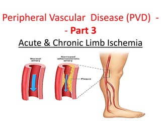
SlideShare a Scribd company logo
1 of 49
Peripheral Vascular Disease (PVD) -
- Part 3
Acute & Chronic Limb Ischemia
What is PVD?
• known as Peripheral Arterial
Disease or Peripheral Arterial
Occlusive Disease (PAD/PAOD).
• Occlusion at lower extremity.
• Most common causes:
o Atherothrombosis, arteritis,
aneurysm + embolism.
• Can be ACUTE or CHRONIC.
Pathophysiology:
• Arterial narrowing  Decreased
blood flow = Pain
• Pain results from an imbalance
between supply and demand of
blood flow that fails to satisfy
ongoing metabolic requirements.
The Facts:
• The prevalence: >55 years is 10%–25%
• 70%–80% of affected young individuals are asymptomatic
• Pt’s with PVD alone have the same relative risk of death from
cardiovascular causes as those CAD or CVD
• PVD pt’s = 4X more likely to die within 10 years than without.
• Patients with PAD require medical management to prevent
future coronary and cerebral vascular events.
• Prognosis at 1 yr in patient’s with Critical Limb Ischemia :
• Alive with two limbs — 50%
• Amputation — 25%
• Cardiovascular mortality 25%
Typical Patient at risk of PVD:
• Smoker (2.5-3x)
• Diabetic (3-4x)
• Hypertension
• Hx of Hypercholesterolemia/AF/IHD/CVA
Risk Factors:
• Age ≥ 70 years.
• Age 50 - 69 years with a history of smoking or diabetes.
• Age 40 - 49 with diabetes and at least one other risk factor
for atherosclerosis.
• Leg symptoms suggestive of claudication with exertion or
ischemic pain at rest.
• Abnormal lower extremity pulse examination.
• Known atherosclerosis at other sites (eg, coronary, carotid, or
renal artery disease).
Circulatory disorders
Peripheralvasculardisease:causedby
blockageofthe arteries,usuallyinthelegs
S/S:painorcramping inthelegsor
buttocks whilewalking
Tx:Rxtodecreasecholestrol, dietchange,improvecirculation
Chronic PVD History:
3. Critical Stenosis = >60%, impending acute ischemic limb:
- rest pain
- ischemic ulceration
- gangrene
2. Other Symptom/Signs:
• A burning or aching pain in the feet (especially at night)
• Cold skin/feet
• Increased occurrence of infection
• Non-healing Ulcers
• Asymptomatic
1. INTERMITTENT CLAUDICATION
• “Reproducible pain on exercise which is relieved by rest”
• Pain can also be reproduced by elevating the leg
• “my legs get sore at night and feel better when I hang
them over the edge of the bed”
Chronic PVD History:
Unfortunately, however, a PAD diagnosis can be missed
since nearly 50% of old patients are asymptomatic or
have atypical symptoms.
Thus, a high index of suspicion is necessary in patients
presenting with potential risk factors.
– Classic claudication — 10 to 35 percent
– Critical limb ischemia — 1 to 2 percent
– Cramp or tingling which recurs on walking the
same distance
60% Upper 2/3 Calf Claudication
— superficial femoral artery
Lower 1/3 Calf Claudication —
popliteal artery
Foot Claudication — tibial or
peroneal artery
30% Buttock , thigh & Hip
Claudication - aorto-iliac or
common femoral artery
DDx of Leg Pain
1. Vascular
a) DVT (as for risk factors)
b) PVD (claudication)
2. Neurospinal (neurogenic)
a) Disc Disease
b) Spinal Stenosis (Pseudoclaudication)
3. Neuropathic
a) Diabetes
b) Chronic EtOH (ethanol) abuse
4. Musculoskeletal
a) OA (variation w/weather + time of day)
b) Chronic compartment syndrome
DDx of Leg Pain or PVD
• Osteoarthritis of Hip or knee joints — OA can be distinguished
clinically from Aorto-iliac PVD because OA pain may not
disappear promptly after exercise & may be associated with
weather changes(usually worse in the morning or upon
wakening), and vary in intensity from day to day
• Neurogenic claudication (pseudo-claudication), pain due to
lumbar canal compression,(due to osteophytic narrowing).
• Unlike true claudication, which occurs with walking and relieved
by rest, pseudo-claudication causes pain with erect posture
(lumbar lordosis) and is relieved by sitting or lying down.
• Patients with pseudo-claudication may also find symptomatic
relief by leaning forward and straightening the spine (usually
done with pushing a shopping cart or leaning against a wall).
Physical Examination:
Examination: What do to:
Inspection
Expose the skin
and look for:
• Thick Shiny Skin
• Hair Loss
• Brittle Nails
• Colour Changes (pallor)
• Ulcers
• Muscle Wasting
Palpation • Temperature (cool, bilateral/unilateral)
• Pulses: ?Regular,
• Capillary Refill
• Sensation/Movement
Auscultation • Femoral Bruits
Ankle Brachial
Index (ABI)
= Systolic BP in ankle
Systolic BP in brachial artery
Buerger’s Test • Elevate the leg to 45° - and look for pallor
• Place the leg in a dependent position 90°& look for a
red flushed foot before returning to normal
• Pallor at <20° = severe PAD.
PVD Pictures:
ULCER
•associated with claudication + signs of
ischaemia
•occur on dorsum of foot + anterior skin
•↓ pulses, cold to touch, hairless skin
•Painful, punched out edge
What does the ABI mean?
CAUTION:
Patient’s with Diabetes + Renal Failure:
They have calcified arterial walls which can falsely elevate their
ABI.
• a simple, non-invasive bedside tool
for Dxing PAD
= SBP in Ankle/SBP in brachial artery
• Take the highest measurement in
both limbs
•
• low ABI predictive of an increased
risk of all-cause cardiovascular
mortality and CAD
• 95% sensitive in detecting
angiogram positive disease and
around 99% specific in identifying
supposedly healthy subjects
What does the ABI mean?
ABI Clinical Correlation
>0.9 Normal Limb
<0.9 = diagnostic for PAD
0.5-0.9 Intermittent Claudication (Mild PAD)
<0.4 Rest Pain (Moderate PAD)
<0.15 Gangrene (Severe PAD)
CAUTION:
Patient’s with Diabetes + Renal Failure:
They have calcified arterial walls which can falsely elevate
their ABI.
Investigations:
NON INVASIVE:
Duplex Ultrasound
 normal is triphasic  biphasic  monophasic  absent
BLOOD TESTS:
1. FBE/Homocysteine Levels
2. Coagulation Studies
3. Fasting Lipids and Fasting Glucose
4. HBA1C
WHEN TO IMAGE:
1. To image = to intervene
2. Pt’s with disabling symptoms where revascularisation is considered
3. To accurately depict anatomy of stenosis and plan for PCI or Surgery
4. Sometimes in pt’s with discrepancy in hx and clinical findings
ANGIOGRAPHY:
Non-invasive:
• CT Angiogram
• MR Angiogram
Invasive:
• Digital Subtraction Angiography
 Gold Standard
 Intervention at same time
CT Angiography Digital Subtraction
Angiography
Value of angiography
Localizes the obstruction
Visualize the arterial tree & distal
run-off
Can diagnose an embolus:
Sharp cutoff, reversed meniscus or clot
silhouette
Treatment:
1. RISK FACTOR MODIFICATION:
a) Smoking Cessation
b) Rigorous BSL control
c) BP reduction
d) Lipid Lowering Therapy
3. MEDICAL MANAGEMENT:
a) Antiplatelet therapy e.g. Aspirin/Clopidogrel
b) Phosphodiesterase Inhibitor e.g. Cilostazol
c) Foot Care
2. EXERCISE:
a) Claudication exercise rehabilitation program
b) 45-60mins 3x weekly for 12 weeks
c) 6 months later +6.5mins walking time
(before pain)
What are the features of an acute
ischemic limb?
REMEMBER THE 6 P’S:
PAIN
PALLOR
PULSELESNESS
PERISHING COLD
(POIKILOTHERMIA)
PARASTHESIAS
PARALYSIS
Fixed
mottling &
cyanosis
Circulatory disorders
• Transient ischemic attack (show signs of symptoms of
typical Stroke but px return tonormal in less than 24 hours
Hypertension: HBP
• High blood pressure
• Silent Killer, usually no symptoms
• Leads to strokes, heart attacks, kidney failure
• Higher in African Americans and post-menopausal
women
• Risk factors = smoking , overweight, stress, high fat diets,
family history
• Treatment relaxation, low fat diet exercise weight loss,
medication
Hypotension
• Low blood pressure
systolic <100
21
Circulatory disorders
Aneurysm: ballooning of
an artery, thinning and
weakening
23
Circulatory disorders
Arrhythmia
• Any change from normal heart
rate or rhythm
• BRADYCARDIA
– Slow heart rate (<60/minute)
• TACHYCARDIA
– Rapid heart rate (>100/minute)
Circulatory disorders
Murmurs:indicatesdefect
inheartvalves
– Gurglingorhissingsound
Bruit–Murmuroutsidetheheart
region
Circulatory disorders
Arteriosclerosis: Walls thicken, lose elasticity due to fatty
deposits in arteries
Coronary arterydisease:anarrowing ofthearteriesthatsupply oxygenandnutrient-filled
bloodtotheheart
MI/heartattackcausedbyLackofbloodsupply
(blockageofcoronaryarteryorbloodclot)topartof
myocardiumcausesitsdamage.
• Amountofdamagedependsonsizeofareadeprived
ofoxygen
• Immediatemedicalcareiscritical
• Rx:bedrest,oxygen,medication
• Morphineforpain,tpatodissolveclot
• Anticoagulanttherapytopreventfurtherclots
• Angioplastyandby-passsurgerymaybenecessary
Myocardialinfarction(MI):
Circulatory disorders
Heartfailure:occurs when theventricles
areunabletocontracteffectively and
bloodpoolsintheheart
Circulatory disorders
Varicoseveins:swollen,distendedveins
• heredityorduetoposture
• prolongedperiods ofstanding,physical exertion,ageandpregnancy
Prevention
• Wearbroad spectrum sunscreen that's a minimum of
SPF 30. Slather
• Elevatelegswhen resting
• Wearelasticstockings
• Eatingfiber diets
• Exercise
• Weightcontrol
Edema Facts
• Is an observable swelling due to fluid accumulation under
the skin in the spaces within the tissues.
• All body tissues are made up of cells and connective tissues
(interstitium) to hold them together.
• Most commonly occurs in feet and legs (peripheral edema).
• In various diseases, excess fluid can accumulate in either
one or both of these compartments.
• 99% of interstitial fluid is fixed to collagen,
mucopolysaccharide and hyaluronic acid (gel), (connective
tissue), which called fixed water outside the cells).
• 1% of interstitial fluid is free water (moving freely)
•
Edema facts
• The most common systemic diseases that cause edema
– Heart failure,
– cirrhosis of the liver, and
– nephrotic syndrome
• Edema that occurs in heart, liver, and kidneys is mainly caused by salt
retention, which holds the excess fluid.
• In certain liver and kidney diseases, low levels of albumin in the blood
can contribute to fluid retention.
• Fluid accumulation in the lungs is called Pulmonary edema.
• Fluid accumulation in the abdominal cavity is called Ascites.
• Edema of unknown cause occurs primarily in women.
• Varicose veins / thrombophlebitis (a blood clot in an inflamed vein) of
the deep veins in the legs causes edema that is localized to the legs.
1. Causes and pathogenesis of edema
Basic causes:
(1) Imbalance of exchange between intra-vascular
and extra-vascular fluid.
• 1) Increased capillary hydrostatic pressure (CHP)
• 2) Decreased plasma colloidal OP
• 3) Increased permeability of the capillary wall
4) Obstruction of lymphatic return
(2) Retention of fluid in the
body. (retention of water and sodium)
Under normal circumstances, the intake
and excretion of fluid is in dynamic
balance, in which the kidneys play
important role.
Normally the filtrate from
glomeruli is 180 liter/day. 60~70% of
filtrate is reabsorbed at proximal
tubules, finally 99%~99.5% of filtrate is
reabsorbed by renal tubules, which is
called glomerular-tubular balance.
(2) Classification of Edema
1) According to the distribution
of edematous fluid:
(a) Local edema:
Local edema occurs in one
organ or a part of the body.
(brain edema, pulmonary
edema, etc).
(b) Generalized edema:
Generalized edema occurs in
whole body (cardiac edema,
renal edema, etc).
• The body's organs have interstitial spaces where
fluid can accumulate. An accumulation of fluid in
the interstitial air spaces (alveoli) in the lungs occurs
in a disorder called pulmonary edema.
• Additionally, excess fluid sometimes collects in what
is called the third space, which includes cavities in
the abdomen (abdominal or peritoneal cavity -
called "ascites") or in the chest (lung or pleural
cavity - called "pleural effusion").
• Anasarca: Severe, widespread accumulation of fluid
in all tissues& cavities of the body at same time.
Pulmonary edema
Is the abnormal accumulation of fluid in pulmonary
interstitial space (interstitial pulmonary edema) and
alveoli (alveolar pulmonary edema).
Mechanism:The output of left ventricle is reduced, the
end-diastolic volume in left ventricle in increased.
Pressure of left atrium is increased. The pulmonary
venous pressure is increased leading to
accumulation of interstitial fluid.
2) According to the causes of edema:
• cardiac edema
• renal edema
• hepatic edema
• idiopathic edema
3) According to the gravity of edema:
• recessive (non-pitting) edema;
• frank (pitting) edema.
Cardiac edema
A) Manifestation:
Cardiac edema is caused by right
heart failure. Right heart failure means:
(1) the output of right ventricle is
reduced,
(2) the end-diastolic volume in right
ventricle in increased. Total venous
pressure is increased.
When the patient is ambulant, the
legs are firstly involved, the swelling of
ankles is often the first sign.
Liver engorgement Distention of jugular vein
B. Mechanism of cardiac edema -
Decreased cardiac output
↓myocardial contraction
•
• ↓ volume of blood pumped to the aorta
•
• ↓ renal blood supply
•
• ↑renin -angiotensin II -aldosterone system
•
• ↑reabsorption of sodium
•
• ↑ADH release the extracellular volume
• expands
• ↑water reabsorption
• Frank (pitting) edema:
• By pressing finger firmly against the tissue for
a few seconds, a dent can be produced. When
the finger is withdrawn, the dent may persist for
several minutes.
• Pitting edema can be demonstrated by applying
pressure to the swollen area by depressing the skin
with a finger.
• If the pressing causes an indentation that persists
for some time after the release of the pressure, the
edema is referred to as pitting edema. Any form of
pressure, such as from the elastic in socks, can
induce pitting with this type of edema.
• This type of edema may be normal depending on
the severity. Almost everyone wears socks all day
will have mild pitting edema by the end of the day.
• In non-pitting edema, which usually affects the
legs or arms, Non-pitting edema can occur in
lymphedema, which is a disturbance of the
lymphatic circulation that may occur after a
mastectomy, lymph node surgery, or
congenitally.
• Another cause of non-pitting edema of the legs is
called pretibial myxedema, which is a swelling
over the shin that occurs in some patients with
hypothyroidism.
C. Characteristics of edema
(1) Properties of edematous fluid
------------------------------------------------------------------------------------------
Edematous causes protein appearance specific
fluid concentration gravity
------------------------------------------------------------------------------------------
transudate ↑effective filtration 1~2g % clear low
pressure
exudates ↑permeability of 4g % muddy high
vascular wall
lymph obstruction of 4~5 % chyliform higher
lymphatic vessel
-----------------------------------------------------------------------------------------
D. Effects of edema on the body
(1) Beneficial effects
(2) Harmful effects
(1) Beneficial effect
I) Protective effects in inflammatory edema
(a) Dilute and neutralize the toxin by the
edematous fluid.
(b) Promote transportation of antibody and
drugs to the inflammatory area due to the
increased permeability of vascular wall.
(c) Form the fibrin net to prevent bacteria from
spreading to the peripheral area.
.
II) ‘Safety valve’ of circulatory system
• When the volume of plasma is rapidly
expanded (venous infusion), large amount of fluid
may transfer to the interstitial space.
• It is an important way to regulate the blood
volume.
(2) Harmful effects
I) Increase the distance between capillary and
cells.
1. The supplies of nutrients and oxygen will be
reduced.
2. The edematous tissue will be dystrophic.
3. The potential of anti-infection is reduced.
4. The wound will be not easy to heal.
II) Dysfunction of edematous organs and tissue
1. Dysfunction of edematous organs and tissue
depends on the rate and extent of
development of edema.
2. Edema of vital organs (lungs, brain, larynx)
will be dangerous for the life.
3. Laryngeal edema (asphyxia)
E. General Management of Edema
Consists of:
– Treating the underlying conditions (heart, kidney,
protein def.)
– Restricting salt and fluid intake,
– using diuretics (medicines to induce urination).
– Elevation of affected parts (legs & arms)
– Postural drainage for lungs
– Lymphatic drainage for lymphatic obstruction
– Compression therapy (stockings, Vasotrain)

More Related Content

Similar to 12 - cardiovascular diseases - Part 3.ppt

Approach to a Child with Congenital Heart Disese
Approach to a Child with Congenital Heart DiseseApproach to a Child with Congenital Heart Disese
Approach to a Child with Congenital Heart Disese
CSN Vittal
 
Congestive heart failure
Congestive heart failureCongestive heart failure
Congestive heart failure
cardilogy
 
approach to congenital cyanotic heart diseases
approach to congenital cyanotic heart diseasesapproach to congenital cyanotic heart diseases
approach to congenital cyanotic heart diseases
RyanKhan40
 
Approach to congenital cyanotic heart diseases
Approach to congenital cyanotic heart diseases Approach to congenital cyanotic heart diseases
Approach to congenital cyanotic heart diseases
Dr.Debasis Maity
 
Ischaemic heart disease.pptx
Ischaemic heart disease.pptxIschaemic heart disease.pptx
Ischaemic heart disease.pptx
Imtiyaz60
 

Similar to 12 - cardiovascular diseases - Part 3.ppt (20)

PVD neo
PVD neoPVD neo
PVD neo
 
Critical limb ischemia. povd . dr mnr
Critical  limb ischemia. povd . dr mnrCritical  limb ischemia. povd . dr mnr
Critical limb ischemia. povd . dr mnr
 
CONGENITAL HEART DISEASES PPT
CONGENITAL HEART DISEASES   PPT CONGENITAL HEART DISEASES   PPT
CONGENITAL HEART DISEASES PPT
 
PAD
PADPAD
PAD
 
Tutorial ischaemia of lower limbs
Tutorial ischaemia of lower limbsTutorial ischaemia of lower limbs
Tutorial ischaemia of lower limbs
 
Peripheral artery disease nikku
Peripheral artery disease nikkuPeripheral artery disease nikku
Peripheral artery disease nikku
 
Approach to a Child with Congenital Heart Disese
Approach to a Child with Congenital Heart DiseseApproach to a Child with Congenital Heart Disese
Approach to a Child with Congenital Heart Disese
 
Aortic stenosis 11 4
Aortic stenosis 11 4Aortic stenosis 11 4
Aortic stenosis 11 4
 
Congestive Cardiac Failure presentation and diagnosis
Congestive Cardiac Failure presentation and diagnosisCongestive Cardiac Failure presentation and diagnosis
Congestive Cardiac Failure presentation and diagnosis
 
Cardiovascular disease
Cardiovascular diseaseCardiovascular disease
Cardiovascular disease
 
Determining a vascular cause for leg pain and referrals
Determining a vascular cause for leg pain and referralsDetermining a vascular cause for leg pain and referrals
Determining a vascular cause for leg pain and referrals
 
New ppta.pptx n
New ppta.pptx nNew ppta.pptx n
New ppta.pptx n
 
peripheral vascular disease
peripheral vascular diseaseperipheral vascular disease
peripheral vascular disease
 
CHRONIC PERIPHERAL ARTERIAL OCCLUSIVE DISEASE
CHRONIC PERIPHERAL ARTERIAL OCCLUSIVE DISEASECHRONIC PERIPHERAL ARTERIAL OCCLUSIVE DISEASE
CHRONIC PERIPHERAL ARTERIAL OCCLUSIVE DISEASE
 
Congestive heart failure
Congestive heart failureCongestive heart failure
Congestive heart failure
 
approach to congenital cyanotic heart diseases
approach to congenital cyanotic heart diseasesapproach to congenital cyanotic heart diseases
approach to congenital cyanotic heart diseases
 
Approach to congenital cyanotic heart diseases
Approach to congenital cyanotic heart diseases Approach to congenital cyanotic heart diseases
Approach to congenital cyanotic heart diseases
 
Approach to congenital cyanotic heart diseases.pptx
Approach to congenital cyanotic heart diseases.pptxApproach to congenital cyanotic heart diseases.pptx
Approach to congenital cyanotic heart diseases.pptx
 
Peripheral arterial diseases
Peripheral arterial diseasesPeripheral arterial diseases
Peripheral arterial diseases
 
Ischaemic heart disease.pptx
Ischaemic heart disease.pptxIschaemic heart disease.pptx
Ischaemic heart disease.pptx
 

More from AhmedFaisal59561

More from AhmedFaisal59561 (6)

11- cardiovascular diseases - Part 2.ppt
11- cardiovascular diseases - Part 2.ppt11- cardiovascular diseases - Part 2.ppt
11- cardiovascular diseases - Part 2.ppt
 
2+3- Pathology-Cellular-Injury Adaptation-to-Stress Iinjury.ppt
2+3- Pathology-Cellular-Injury  Adaptation-to-Stress Iinjury.ppt2+3- Pathology-Cellular-Injury  Adaptation-to-Stress Iinjury.ppt
2+3- Pathology-Cellular-Injury Adaptation-to-Stress Iinjury.ppt
 
9- Immunity Hypersensitivity Reactions -Part 2.pptx
9- Immunity  Hypersensitivity Reactions -Part 2.pptx9- Immunity  Hypersensitivity Reactions -Part 2.pptx
9- Immunity Hypersensitivity Reactions -Part 2.pptx
 
10- cardiovascular diseases - Part 1.ppt
10- cardiovascular diseases - Part 1.ppt10- cardiovascular diseases - Part 1.ppt
10- cardiovascular diseases - Part 1.ppt
 
ch 3 Shoulder Elbow joint.pptx
ch 3 Shoulder Elbow joint.pptxch 3 Shoulder Elbow joint.pptx
ch 3 Shoulder Elbow joint.pptx
 
ch 4 t m hip joint part 3.pptx
ch 4 t  m hip joint part 3.pptxch 4 t  m hip joint part 3.pptx
ch 4 t m hip joint part 3.pptx
 

Recently uploaded

Russian Escorts Girls Nehru Place ZINATHI 🔝9711199012 ☪ 24/7 Call Girls Delhi
Russian Escorts Girls  Nehru Place ZINATHI 🔝9711199012 ☪ 24/7 Call Girls DelhiRussian Escorts Girls  Nehru Place ZINATHI 🔝9711199012 ☪ 24/7 Call Girls Delhi
Russian Escorts Girls Nehru Place ZINATHI 🔝9711199012 ☪ 24/7 Call Girls Delhi
AlinaDevecerski
 
Call Girls Bhubaneswar Just Call 9907093804 Top Class Call Girl Service Avail...
Call Girls Bhubaneswar Just Call 9907093804 Top Class Call Girl Service Avail...Call Girls Bhubaneswar Just Call 9907093804 Top Class Call Girl Service Avail...
Call Girls Bhubaneswar Just Call 9907093804 Top Class Call Girl Service Avail...
Dipal Arora
 

Recently uploaded (20)

Call Girls Kochi Just Call 8250077686 Top Class Call Girl Service Available
Call Girls Kochi Just Call 8250077686 Top Class Call Girl Service AvailableCall Girls Kochi Just Call 8250077686 Top Class Call Girl Service Available
Call Girls Kochi Just Call 8250077686 Top Class Call Girl Service Available
 
Call Girls Cuttack Just Call 9907093804 Top Class Call Girl Service Available
Call Girls Cuttack Just Call 9907093804 Top Class Call Girl Service AvailableCall Girls Cuttack Just Call 9907093804 Top Class Call Girl Service Available
Call Girls Cuttack Just Call 9907093804 Top Class Call Girl Service Available
 
(Low Rate RASHMI ) Rate Of Call Girls Jaipur ❣ 8445551418 ❣ Elite Models & Ce...
(Low Rate RASHMI ) Rate Of Call Girls Jaipur ❣ 8445551418 ❣ Elite Models & Ce...(Low Rate RASHMI ) Rate Of Call Girls Jaipur ❣ 8445551418 ❣ Elite Models & Ce...
(Low Rate RASHMI ) Rate Of Call Girls Jaipur ❣ 8445551418 ❣ Elite Models & Ce...
 
Call Girls Visakhapatnam Just Call 9907093804 Top Class Call Girl Service Ava...
Call Girls Visakhapatnam Just Call 9907093804 Top Class Call Girl Service Ava...Call Girls Visakhapatnam Just Call 9907093804 Top Class Call Girl Service Ava...
Call Girls Visakhapatnam Just Call 9907093804 Top Class Call Girl Service Ava...
 
Russian Escorts Girls Nehru Place ZINATHI 🔝9711199012 ☪ 24/7 Call Girls Delhi
Russian Escorts Girls  Nehru Place ZINATHI 🔝9711199012 ☪ 24/7 Call Girls DelhiRussian Escorts Girls  Nehru Place ZINATHI 🔝9711199012 ☪ 24/7 Call Girls Delhi
Russian Escorts Girls Nehru Place ZINATHI 🔝9711199012 ☪ 24/7 Call Girls Delhi
 
Top Rated Bangalore Call Girls Mg Road ⟟ 9332606886 ⟟ Call Me For Genuine S...
Top Rated Bangalore Call Girls Mg Road ⟟   9332606886 ⟟ Call Me For Genuine S...Top Rated Bangalore Call Girls Mg Road ⟟   9332606886 ⟟ Call Me For Genuine S...
Top Rated Bangalore Call Girls Mg Road ⟟ 9332606886 ⟟ Call Me For Genuine S...
 
Call Girls Haridwar Just Call 8250077686 Top Class Call Girl Service Available
Call Girls Haridwar Just Call 8250077686 Top Class Call Girl Service AvailableCall Girls Haridwar Just Call 8250077686 Top Class Call Girl Service Available
Call Girls Haridwar Just Call 8250077686 Top Class Call Girl Service Available
 
Top Rated Hyderabad Call Girls Erragadda ⟟ 6297143586 ⟟ Call Me For Genuine ...
Top Rated  Hyderabad Call Girls Erragadda ⟟ 6297143586 ⟟ Call Me For Genuine ...Top Rated  Hyderabad Call Girls Erragadda ⟟ 6297143586 ⟟ Call Me For Genuine ...
Top Rated Hyderabad Call Girls Erragadda ⟟ 6297143586 ⟟ Call Me For Genuine ...
 
Call Girls Horamavu WhatsApp Number 7001035870 Meeting With Bangalore Escorts
Call Girls Horamavu WhatsApp Number 7001035870 Meeting With Bangalore EscortsCall Girls Horamavu WhatsApp Number 7001035870 Meeting With Bangalore Escorts
Call Girls Horamavu WhatsApp Number 7001035870 Meeting With Bangalore Escorts
 
Night 7k to 12k Navi Mumbai Call Girl Photo 👉 BOOK NOW 9833363713 👈 ♀️ night ...
Night 7k to 12k Navi Mumbai Call Girl Photo 👉 BOOK NOW 9833363713 👈 ♀️ night ...Night 7k to 12k Navi Mumbai Call Girl Photo 👉 BOOK NOW 9833363713 👈 ♀️ night ...
Night 7k to 12k Navi Mumbai Call Girl Photo 👉 BOOK NOW 9833363713 👈 ♀️ night ...
 
Call Girls Gwalior Just Call 9907093804 Top Class Call Girl Service Available
Call Girls Gwalior Just Call 9907093804 Top Class Call Girl Service AvailableCall Girls Gwalior Just Call 9907093804 Top Class Call Girl Service Available
Call Girls Gwalior Just Call 9907093804 Top Class Call Girl Service Available
 
(👑VVIP ISHAAN ) Russian Call Girls Service Navi Mumbai🖕9920874524🖕Independent...
(👑VVIP ISHAAN ) Russian Call Girls Service Navi Mumbai🖕9920874524🖕Independent...(👑VVIP ISHAAN ) Russian Call Girls Service Navi Mumbai🖕9920874524🖕Independent...
(👑VVIP ISHAAN ) Russian Call Girls Service Navi Mumbai🖕9920874524🖕Independent...
 
Best Rate (Guwahati ) Call Girls Guwahati ⟟ 8617370543 ⟟ High Class Call Girl...
Best Rate (Guwahati ) Call Girls Guwahati ⟟ 8617370543 ⟟ High Class Call Girl...Best Rate (Guwahati ) Call Girls Guwahati ⟟ 8617370543 ⟟ High Class Call Girl...
Best Rate (Guwahati ) Call Girls Guwahati ⟟ 8617370543 ⟟ High Class Call Girl...
 
Call Girls Varanasi Just Call 9907093804 Top Class Call Girl Service Available
Call Girls Varanasi Just Call 9907093804 Top Class Call Girl Service AvailableCall Girls Varanasi Just Call 9907093804 Top Class Call Girl Service Available
Call Girls Varanasi Just Call 9907093804 Top Class Call Girl Service Available
 
Call Girls Bhubaneswar Just Call 9907093804 Top Class Call Girl Service Avail...
Call Girls Bhubaneswar Just Call 9907093804 Top Class Call Girl Service Avail...Call Girls Bhubaneswar Just Call 9907093804 Top Class Call Girl Service Avail...
Call Girls Bhubaneswar Just Call 9907093804 Top Class Call Girl Service Avail...
 
Call Girls Ooty Just Call 8250077686 Top Class Call Girl Service Available
Call Girls Ooty Just Call 8250077686 Top Class Call Girl Service AvailableCall Girls Ooty Just Call 8250077686 Top Class Call Girl Service Available
Call Girls Ooty Just Call 8250077686 Top Class Call Girl Service Available
 
Book Paid Powai Call Girls Mumbai 𖠋 9930245274 𖠋Low Budget Full Independent H...
Book Paid Powai Call Girls Mumbai 𖠋 9930245274 𖠋Low Budget Full Independent H...Book Paid Powai Call Girls Mumbai 𖠋 9930245274 𖠋Low Budget Full Independent H...
Book Paid Powai Call Girls Mumbai 𖠋 9930245274 𖠋Low Budget Full Independent H...
 
Lucknow Call girls - 8800925952 - 24x7 service with hotel room
Lucknow Call girls - 8800925952 - 24x7 service with hotel roomLucknow Call girls - 8800925952 - 24x7 service with hotel room
Lucknow Call girls - 8800925952 - 24x7 service with hotel room
 
Call Girls Coimbatore Just Call 9907093804 Top Class Call Girl Service Available
Call Girls Coimbatore Just Call 9907093804 Top Class Call Girl Service AvailableCall Girls Coimbatore Just Call 9907093804 Top Class Call Girl Service Available
Call Girls Coimbatore Just Call 9907093804 Top Class Call Girl Service Available
 
VIP Call Girls Indore Kirti 💚😋 9256729539 🚀 Indore Escorts
VIP Call Girls Indore Kirti 💚😋  9256729539 🚀 Indore EscortsVIP Call Girls Indore Kirti 💚😋  9256729539 🚀 Indore Escorts
VIP Call Girls Indore Kirti 💚😋 9256729539 🚀 Indore Escorts
 

12 - cardiovascular diseases - Part 3.ppt

  • 1. Peripheral Vascular Disease (PVD) - - Part 3 Acute & Chronic Limb Ischemia
  • 2. What is PVD? • known as Peripheral Arterial Disease or Peripheral Arterial Occlusive Disease (PAD/PAOD). • Occlusion at lower extremity. • Most common causes: o Atherothrombosis, arteritis, aneurysm + embolism. • Can be ACUTE or CHRONIC. Pathophysiology: • Arterial narrowing  Decreased blood flow = Pain • Pain results from an imbalance between supply and demand of blood flow that fails to satisfy ongoing metabolic requirements.
  • 3. The Facts: • The prevalence: >55 years is 10%–25% • 70%–80% of affected young individuals are asymptomatic • Pt’s with PVD alone have the same relative risk of death from cardiovascular causes as those CAD or CVD • PVD pt’s = 4X more likely to die within 10 years than without. • Patients with PAD require medical management to prevent future coronary and cerebral vascular events. • Prognosis at 1 yr in patient’s with Critical Limb Ischemia : • Alive with two limbs — 50% • Amputation — 25% • Cardiovascular mortality 25%
  • 4. Typical Patient at risk of PVD: • Smoker (2.5-3x) • Diabetic (3-4x) • Hypertension • Hx of Hypercholesterolemia/AF/IHD/CVA Risk Factors: • Age ≥ 70 years. • Age 50 - 69 years with a history of smoking or diabetes. • Age 40 - 49 with diabetes and at least one other risk factor for atherosclerosis. • Leg symptoms suggestive of claudication with exertion or ischemic pain at rest. • Abnormal lower extremity pulse examination. • Known atherosclerosis at other sites (eg, coronary, carotid, or renal artery disease).
  • 5. Circulatory disorders Peripheralvasculardisease:causedby blockageofthe arteries,usuallyinthelegs S/S:painorcramping inthelegsor buttocks whilewalking Tx:Rxtodecreasecholestrol, dietchange,improvecirculation
  • 6. Chronic PVD History: 3. Critical Stenosis = >60%, impending acute ischemic limb: - rest pain - ischemic ulceration - gangrene 2. Other Symptom/Signs: • A burning or aching pain in the feet (especially at night) • Cold skin/feet • Increased occurrence of infection • Non-healing Ulcers • Asymptomatic 1. INTERMITTENT CLAUDICATION • “Reproducible pain on exercise which is relieved by rest” • Pain can also be reproduced by elevating the leg • “my legs get sore at night and feel better when I hang them over the edge of the bed”
  • 7. Chronic PVD History: Unfortunately, however, a PAD diagnosis can be missed since nearly 50% of old patients are asymptomatic or have atypical symptoms. Thus, a high index of suspicion is necessary in patients presenting with potential risk factors. – Classic claudication — 10 to 35 percent – Critical limb ischemia — 1 to 2 percent – Cramp or tingling which recurs on walking the same distance
  • 8. 60% Upper 2/3 Calf Claudication — superficial femoral artery Lower 1/3 Calf Claudication — popliteal artery Foot Claudication — tibial or peroneal artery 30% Buttock , thigh & Hip Claudication - aorto-iliac or common femoral artery
  • 9. DDx of Leg Pain 1. Vascular a) DVT (as for risk factors) b) PVD (claudication) 2. Neurospinal (neurogenic) a) Disc Disease b) Spinal Stenosis (Pseudoclaudication) 3. Neuropathic a) Diabetes b) Chronic EtOH (ethanol) abuse 4. Musculoskeletal a) OA (variation w/weather + time of day) b) Chronic compartment syndrome
  • 10. DDx of Leg Pain or PVD • Osteoarthritis of Hip or knee joints — OA can be distinguished clinically from Aorto-iliac PVD because OA pain may not disappear promptly after exercise & may be associated with weather changes(usually worse in the morning or upon wakening), and vary in intensity from day to day • Neurogenic claudication (pseudo-claudication), pain due to lumbar canal compression,(due to osteophytic narrowing). • Unlike true claudication, which occurs with walking and relieved by rest, pseudo-claudication causes pain with erect posture (lumbar lordosis) and is relieved by sitting or lying down. • Patients with pseudo-claudication may also find symptomatic relief by leaning forward and straightening the spine (usually done with pushing a shopping cart or leaning against a wall).
  • 11. Physical Examination: Examination: What do to: Inspection Expose the skin and look for: • Thick Shiny Skin • Hair Loss • Brittle Nails • Colour Changes (pallor) • Ulcers • Muscle Wasting Palpation • Temperature (cool, bilateral/unilateral) • Pulses: ?Regular, • Capillary Refill • Sensation/Movement Auscultation • Femoral Bruits Ankle Brachial Index (ABI) = Systolic BP in ankle Systolic BP in brachial artery Buerger’s Test • Elevate the leg to 45° - and look for pallor • Place the leg in a dependent position 90°& look for a red flushed foot before returning to normal • Pallor at <20° = severe PAD.
  • 12. PVD Pictures: ULCER •associated with claudication + signs of ischaemia •occur on dorsum of foot + anterior skin •↓ pulses, cold to touch, hairless skin •Painful, punched out edge
  • 13. What does the ABI mean? CAUTION: Patient’s with Diabetes + Renal Failure: They have calcified arterial walls which can falsely elevate their ABI. • a simple, non-invasive bedside tool for Dxing PAD = SBP in Ankle/SBP in brachial artery • Take the highest measurement in both limbs • • low ABI predictive of an increased risk of all-cause cardiovascular mortality and CAD • 95% sensitive in detecting angiogram positive disease and around 99% specific in identifying supposedly healthy subjects
  • 14. What does the ABI mean? ABI Clinical Correlation >0.9 Normal Limb <0.9 = diagnostic for PAD 0.5-0.9 Intermittent Claudication (Mild PAD) <0.4 Rest Pain (Moderate PAD) <0.15 Gangrene (Severe PAD) CAUTION: Patient’s with Diabetes + Renal Failure: They have calcified arterial walls which can falsely elevate their ABI.
  • 15. Investigations: NON INVASIVE: Duplex Ultrasound  normal is triphasic  biphasic  monophasic  absent BLOOD TESTS: 1. FBE/Homocysteine Levels 2. Coagulation Studies 3. Fasting Lipids and Fasting Glucose 4. HBA1C WHEN TO IMAGE: 1. To image = to intervene 2. Pt’s with disabling symptoms where revascularisation is considered 3. To accurately depict anatomy of stenosis and plan for PCI or Surgery 4. Sometimes in pt’s with discrepancy in hx and clinical findings
  • 16. ANGIOGRAPHY: Non-invasive: • CT Angiogram • MR Angiogram Invasive: • Digital Subtraction Angiography  Gold Standard  Intervention at same time
  • 17. CT Angiography Digital Subtraction Angiography Value of angiography Localizes the obstruction Visualize the arterial tree & distal run-off Can diagnose an embolus: Sharp cutoff, reversed meniscus or clot silhouette
  • 18. Treatment: 1. RISK FACTOR MODIFICATION: a) Smoking Cessation b) Rigorous BSL control c) BP reduction d) Lipid Lowering Therapy 3. MEDICAL MANAGEMENT: a) Antiplatelet therapy e.g. Aspirin/Clopidogrel b) Phosphodiesterase Inhibitor e.g. Cilostazol c) Foot Care 2. EXERCISE: a) Claudication exercise rehabilitation program b) 45-60mins 3x weekly for 12 weeks c) 6 months later +6.5mins walking time (before pain)
  • 19. What are the features of an acute ischemic limb? REMEMBER THE 6 P’S: PAIN PALLOR PULSELESNESS PERISHING COLD (POIKILOTHERMIA) PARASTHESIAS PARALYSIS Fixed mottling & cyanosis
  • 20. Circulatory disorders • Transient ischemic attack (show signs of symptoms of typical Stroke but px return tonormal in less than 24 hours
  • 21. Hypertension: HBP • High blood pressure • Silent Killer, usually no symptoms • Leads to strokes, heart attacks, kidney failure • Higher in African Americans and post-menopausal women • Risk factors = smoking , overweight, stress, high fat diets, family history • Treatment relaxation, low fat diet exercise weight loss, medication Hypotension • Low blood pressure systolic <100 21
  • 22. Circulatory disorders Aneurysm: ballooning of an artery, thinning and weakening
  • 23. 23 Circulatory disorders Arrhythmia • Any change from normal heart rate or rhythm • BRADYCARDIA – Slow heart rate (<60/minute) • TACHYCARDIA – Rapid heart rate (>100/minute)
  • 25. Circulatory disorders Arteriosclerosis: Walls thicken, lose elasticity due to fatty deposits in arteries Coronary arterydisease:anarrowing ofthearteriesthatsupply oxygenandnutrient-filled bloodtotheheart
  • 26. MI/heartattackcausedbyLackofbloodsupply (blockageofcoronaryarteryorbloodclot)topartof myocardiumcausesitsdamage. • Amountofdamagedependsonsizeofareadeprived ofoxygen • Immediatemedicalcareiscritical • Rx:bedrest,oxygen,medication • Morphineforpain,tpatodissolveclot • Anticoagulanttherapytopreventfurtherclots • Angioplastyandby-passsurgerymaybenecessary Myocardialinfarction(MI):
  • 27. Circulatory disorders Heartfailure:occurs when theventricles areunabletocontracteffectively and bloodpoolsintheheart
  • 28. Circulatory disorders Varicoseveins:swollen,distendedveins • heredityorduetoposture • prolongedperiods ofstanding,physical exertion,ageandpregnancy Prevention • Wearbroad spectrum sunscreen that's a minimum of SPF 30. Slather • Elevatelegswhen resting • Wearelasticstockings • Eatingfiber diets • Exercise • Weightcontrol
  • 29. Edema Facts • Is an observable swelling due to fluid accumulation under the skin in the spaces within the tissues. • All body tissues are made up of cells and connective tissues (interstitium) to hold them together. • Most commonly occurs in feet and legs (peripheral edema). • In various diseases, excess fluid can accumulate in either one or both of these compartments. • 99% of interstitial fluid is fixed to collagen, mucopolysaccharide and hyaluronic acid (gel), (connective tissue), which called fixed water outside the cells). • 1% of interstitial fluid is free water (moving freely) •
  • 30. Edema facts • The most common systemic diseases that cause edema – Heart failure, – cirrhosis of the liver, and – nephrotic syndrome • Edema that occurs in heart, liver, and kidneys is mainly caused by salt retention, which holds the excess fluid. • In certain liver and kidney diseases, low levels of albumin in the blood can contribute to fluid retention. • Fluid accumulation in the lungs is called Pulmonary edema. • Fluid accumulation in the abdominal cavity is called Ascites. • Edema of unknown cause occurs primarily in women. • Varicose veins / thrombophlebitis (a blood clot in an inflamed vein) of the deep veins in the legs causes edema that is localized to the legs.
  • 31. 1. Causes and pathogenesis of edema Basic causes: (1) Imbalance of exchange between intra-vascular and extra-vascular fluid. • 1) Increased capillary hydrostatic pressure (CHP) • 2) Decreased plasma colloidal OP • 3) Increased permeability of the capillary wall 4) Obstruction of lymphatic return
  • 32. (2) Retention of fluid in the body. (retention of water and sodium) Under normal circumstances, the intake and excretion of fluid is in dynamic balance, in which the kidneys play important role. Normally the filtrate from glomeruli is 180 liter/day. 60~70% of filtrate is reabsorbed at proximal tubules, finally 99%~99.5% of filtrate is reabsorbed by renal tubules, which is called glomerular-tubular balance.
  • 33. (2) Classification of Edema 1) According to the distribution of edematous fluid: (a) Local edema: Local edema occurs in one organ or a part of the body. (brain edema, pulmonary edema, etc). (b) Generalized edema: Generalized edema occurs in whole body (cardiac edema, renal edema, etc).
  • 34. • The body's organs have interstitial spaces where fluid can accumulate. An accumulation of fluid in the interstitial air spaces (alveoli) in the lungs occurs in a disorder called pulmonary edema. • Additionally, excess fluid sometimes collects in what is called the third space, which includes cavities in the abdomen (abdominal or peritoneal cavity - called "ascites") or in the chest (lung or pleural cavity - called "pleural effusion"). • Anasarca: Severe, widespread accumulation of fluid in all tissues& cavities of the body at same time.
  • 35. Pulmonary edema Is the abnormal accumulation of fluid in pulmonary interstitial space (interstitial pulmonary edema) and alveoli (alveolar pulmonary edema). Mechanism:The output of left ventricle is reduced, the end-diastolic volume in left ventricle in increased. Pressure of left atrium is increased. The pulmonary venous pressure is increased leading to accumulation of interstitial fluid.
  • 36. 2) According to the causes of edema: • cardiac edema • renal edema • hepatic edema • idiopathic edema 3) According to the gravity of edema: • recessive (non-pitting) edema; • frank (pitting) edema.
  • 37. Cardiac edema A) Manifestation: Cardiac edema is caused by right heart failure. Right heart failure means: (1) the output of right ventricle is reduced, (2) the end-diastolic volume in right ventricle in increased. Total venous pressure is increased. When the patient is ambulant, the legs are firstly involved, the swelling of ankles is often the first sign.
  • 38. Liver engorgement Distention of jugular vein
  • 39. B. Mechanism of cardiac edema - Decreased cardiac output ↓myocardial contraction • • ↓ volume of blood pumped to the aorta • • ↓ renal blood supply • • ↑renin -angiotensin II -aldosterone system • • ↑reabsorption of sodium • • ↑ADH release the extracellular volume • expands • ↑water reabsorption
  • 40. • Frank (pitting) edema: • By pressing finger firmly against the tissue for a few seconds, a dent can be produced. When the finger is withdrawn, the dent may persist for several minutes.
  • 41. • Pitting edema can be demonstrated by applying pressure to the swollen area by depressing the skin with a finger. • If the pressing causes an indentation that persists for some time after the release of the pressure, the edema is referred to as pitting edema. Any form of pressure, such as from the elastic in socks, can induce pitting with this type of edema. • This type of edema may be normal depending on the severity. Almost everyone wears socks all day will have mild pitting edema by the end of the day.
  • 42. • In non-pitting edema, which usually affects the legs or arms, Non-pitting edema can occur in lymphedema, which is a disturbance of the lymphatic circulation that may occur after a mastectomy, lymph node surgery, or congenitally. • Another cause of non-pitting edema of the legs is called pretibial myxedema, which is a swelling over the shin that occurs in some patients with hypothyroidism.
  • 43. C. Characteristics of edema (1) Properties of edematous fluid ------------------------------------------------------------------------------------------ Edematous causes protein appearance specific fluid concentration gravity ------------------------------------------------------------------------------------------ transudate ↑effective filtration 1~2g % clear low pressure exudates ↑permeability of 4g % muddy high vascular wall lymph obstruction of 4~5 % chyliform higher lymphatic vessel -----------------------------------------------------------------------------------------
  • 44. D. Effects of edema on the body (1) Beneficial effects (2) Harmful effects
  • 45. (1) Beneficial effect I) Protective effects in inflammatory edema (a) Dilute and neutralize the toxin by the edematous fluid. (b) Promote transportation of antibody and drugs to the inflammatory area due to the increased permeability of vascular wall. (c) Form the fibrin net to prevent bacteria from spreading to the peripheral area. .
  • 46. II) ‘Safety valve’ of circulatory system • When the volume of plasma is rapidly expanded (venous infusion), large amount of fluid may transfer to the interstitial space. • It is an important way to regulate the blood volume.
  • 47. (2) Harmful effects I) Increase the distance between capillary and cells. 1. The supplies of nutrients and oxygen will be reduced. 2. The edematous tissue will be dystrophic. 3. The potential of anti-infection is reduced. 4. The wound will be not easy to heal.
  • 48. II) Dysfunction of edematous organs and tissue 1. Dysfunction of edematous organs and tissue depends on the rate and extent of development of edema. 2. Edema of vital organs (lungs, brain, larynx) will be dangerous for the life. 3. Laryngeal edema (asphyxia)
  • 49. E. General Management of Edema Consists of: – Treating the underlying conditions (heart, kidney, protein def.) – Restricting salt and fluid intake, – using diuretics (medicines to induce urination). – Elevation of affected parts (legs & arms) – Postural drainage for lungs – Lymphatic drainage for lymphatic obstruction – Compression therapy (stockings, Vasotrain)

Editor's Notes

  1. There are currently insufficient data to recommend routine population screening for asymptomatic PAD using the ABPI. - In recent years, it has become evident that PAD is an important predictor of substantial coronary and cerebral vascular risk Patients with symptomatic PAD have a 15-year accrued survival rate of about 22%, compared with a survival rate of 78% in patients without symptoms of PAD. Patients with critical leg ischaemia, who have the lowest ABPI values, have an annual mortality of 25%
  2. Patients at risk — Based in part upon the above observations, the 2005 American College of Cardiology/American Heart Association (ACC/AHA) guidelines on PAD, which were produced in collaboration with major vascular medicine, vascular surgery, and interventional radiology societies, identified the following groups at risk for lower extremity PAD Risk Factors: Atherosclerosis (same as RF’s for CAD and CVD) Smoking (2.5-3x) Diabetes 3-4x Hypertension, increased age >50, male and family history RARE: homocysteinuria
  3. Asymptomatic — 20 to 50 percent ** Unfortunately, however, a PAD diagnosis can be missed since nearly 50% of patients are asymptomatic or have atypical symptoms.  Thus, a high index of suspicion is necessary in patients presenting with potential risk factors. Classic claudication — 10 to 35 percent Critical limb ischemia — 1 to 2 percent Cramp or tingling which recurs on walking the same distance
  4. Buttock and hip — aorto-iliac disease Thigh — aorto-iliac or common femoral artery Upper two-thirds of the calf — superficial femoral artery Lower one-third of the calf — popliteal artery Foot claudication — tibial or peroneal artery
  5. Osteoarthritis of the hip or knee joints — Osteoarthritis can be distinguished clinically from aortoiliac disease because osteoarthritic pain may not disappear promptly after exercise, may be associated with weather changes, and may vary in intensity from day to day (usually worse in the morning or upon wakening) Neurogenic claudication — Neurogenic claudication, also called pseudoclaudication, describes a pain syndrome due to lumbar neurospinal canal compression, which is usually due to osteophytic narrowing of the neurospinal canal. The clinical presentation often helps to distinguish vasculogenic (ie, true) claudication from pseudoclaudication. Unlike true claudication, which occurs with walking and is relieved by stopping, pseudoclaudication causes pain with erect posture (lumbar lordosis) and is relieved by sitting or lying down. Patients with pseudoclaudication may also find symptomatic relief by leaning forward and straightening the spine (usually done with pushing a shopping cart or leaning against a wall). (See "Lumbar spinal stenosis: Pathophysiology, clinical features, and diagnosis".)
  6. Auscultate for femoral (1/2 way between the ASIS and pubic symphysis)
  7. ULCER associated with claudication + signs of ischaemia occur on dorsum of foot + anterior skin ↓ pulses, cold to touch, hairless skin Painful, punched out edge
  8. Take the highest measurement in both limbs low ABI is also predictive of an increased risk of all-cause and cardiovascular mortality [39,40] and of the development of coronary artery calcification 95% sensitive in detecting angiogram positive disease and around 99% specific in identifying supposedly healthy subjects
  9. Take the highest measurement in both limbs low ABI is also predictive of an increased risk of all-cause and cardiovascular mortality [39,40] and of the development of coronary artery calcification 95% sensitive in detecting angiogram positive disease and around 99% specific in identifying supposedly healthy subjects
  10. Peripheral artery wave forms: arterial form is triphasic – consists of forward flow in systolic peak, reversal of flow in early diastole and forward flow in late diastole. This becomes impaired (eliminated reverse flow due to stenosis of vessel and becomes biphasic  decreased systolic peak and increase of flow in diastole) The decision to image is a decision to intervene if a suitable lesion is identified and is only applicable to a minority of patients with intermittent claudication, and then only after risk factors have been addressed and medical management followed. There is also a role for imaging in the small group of patients in whom there is a discrepancy between the history and objective clinical signs. The purpose of imaging is to assess the anatomical location, morphology and extent of disease in order to determine suitability for intervention and occasionally to differentiate atherosclerotic PAD from other causes such as neurogenic claudication and entrapment.
  11. Imaging is largely reserved for patients with disabling symptoms in whom revascularisation is planned. In these patients, accurate depiction of the vascular anatomy is critical for clinical decision making as the distribution and severity of disease are key factors determining whether revascularisation should be by endovascular techniques or open surgery. IV-DSA uses a computer technique which compares an x-ray image of a region of the body before and after radiopaque iodine based dye has been injected intravenously into the body. Tissues and blood vessels on the first image are digitally subtracted from the second image, leaving a clear picture of the artery which can then be studied independently and in isolation from the rest of the body.
  12. HBA1C as close to 6.0 as possible (Selective B-1 blockade ok Anti-hypertensive medications may worsen the PAD symptoms by reducing blood flow and supply of oxygen to the limbs, and may have long-term effects on disease progression). controversial due to the presumed peripheral haemodynamic consequences of beta blockers, leading to worsening symptoms of intermittent claudication. There is currently no evidence that beta blockers adversely affect walking distance in people with intermittent claudication. However, due to the lack of large published trials beta blockers should be used with caution if clinically indicated. Aim LDL 2.6mmol/L with PAD Aim LDL <1.8mmol/L with ATH in other vessels Improved endothelial dysfunction via increases in nitric oxide synthase and prostacyclin [40]. (See "Endothelial dysfunction".)Reduced local inflammation that is induced by muscle ischemia by decreasing free radicals [41].Increased exercise pain tolerance [38].Induction of vascular angiogenesis [42].Improved muscle metabolism by favorable effects on muscle carnitine metabolism and other pathways [43].Reductions in blood viscosity and red cell aggregation BEWARE HF with cilostazol (inhibits platelet aggregation and acts as an arterial vasodilator) Two compared ACE inhibitors against placebo. In the HOPE study there was a significant reduction in the number of cardiovascular events in 168 patients receiving ramipril (OR 0.72, 95% confidence interval 0.58 to 0.91). In the second trial using perindopril in a small numbers of patients, there was a marginal increase in claudication distance but no change in ankle brachial pressure index (ABPI) and a reduction in maximum walking distance.The third trial in patients undergoing angioplasty suggested that the calcium antagonist verapamil reduced restenosis, although this was not reflected in the maintenance of a high ABPI. Another small study demonstrated no significant difference in arterial intima-media thickness with men receiving the thiazide diuretic hydrochlorathiazide compared to those receiving the alpha-adrenoreceptor blocker doxazosin.