SlideShare a Scribd company logo
1 of 14
Download to read offline
Short Article
Programmable RNA Cleavage and Recognition by a
Natural CRISPR-Cas9 System from Neisseria
meningitidis
Graphical Abstract
Highlights
d Neisseria meningitidis Cas9 has a natural, PAM-independent
RNase activity in vitro
d Cleavage of single-stranded RNA by NmeCas9 is CRISPR
RNA-guided
d RNA cleavage can be blocked by type II-C anti-CRISPR
proteins
d Catalytically inactive dNmeCas9 recognizes ssRNA targets
sequence-specifically
Authors
Beth A. Rousseau, Zhonggang Hou,
Max J. Gramelspacher, Yan Zhang
Correspondence
yzhangbc@med.umich.edu
In Brief
Rousseau et al. demonstrate that the
type II-C CRISPR-Cas9 from Neisseria
meningitidis (Nme) has a natural,
programmable RNase activity in vitro.
The NmeCas9 protein can recognize and
cleave RNA target in a CRISPR-RNA-
guided fashion. This activity does not
require any PAM sequence in the target or
PAM-presenting auxiliary cofactor.
Rousseau et al., 2018, Molecular Cell 69, 1–9
March 1, 2018 ª 2018 Elsevier Inc.
https://doi.org/10.1016/j.molcel.2018.01.025
Molecular Cell
Short Article
Programmable RNA Cleavage and Recognition
by a Natural CRISPR-Cas9 System
from Neisseria meningitidis
Beth A. Rousseau,1,2 Zhonggang Hou,1,2 Max J. Gramelspacher,1 and Yan Zhang1,3,*
1Department of Biological Chemistry, University of Michigan, Ann Arbor, MI 48109, USA
2These authors contributed equally
3Lead Contact
*Correspondence: yzhangbc@med.umich.edu
https://doi.org/10.1016/j.molcel.2018.01.025
SUMMARY
The microbial CRISPR systems enable adaptive
defense against mobile elements and also provide
formidable tools for genome engineering. The Cas9
proteins are type II CRISPR-associated, RNA-guided
DNA endonucleases that identify double-stranded
DNA targets by sequence complementarity and pro-
tospacer adjacent motif (PAM) recognition. Here we
report that the type II-C CRISPR-Cas9 from Neisseria
meningitidis (Nme) is capable of programmable,
RNA-guided, site-specific cleavage and recognition
of single-stranded RNA targets and that this ribonu-
clease activity is independent of the PAM sequence.
We define the mechanistic feature and specificity
constraint for RNA cleavage by NmeCas9 and also
show that nuclease null dNmeCas9 binds to RNA
target complementary to CRISPR RNA. Finally, we
demonstrate that NmeCas9-catalyzed RNA cleav-
age can be blocked by three families of type II-C
anti-CRISPR proteins. These results fundamentally
expand the targeting capacities of CRISPR-Cas9
and highlight the potential utility of NmeCas9 as a
single platform to target both RNA and DNA.
INTRODUCTION
Clustered, regularly interspaced, short, palindromic repeats
(CRISPR) loci and their associated (cas) genes constitute an
adaptive defense system that is widespread in bacteria and
archea and that limits horizontal genetic transfer (Barrangou
et al., 2007; Marraffini and Sontheimer, 2008). CRISPR spacers,
which are acquired from invaders’ genomes and integrated
between CRISPR repeats, specify the nucleic acid targets for
CRISPR interference (Barrangou et al., 2007; Marraffini, 2015).
The CRISPR locus is transcribed and processed into small
RNAs called CRISPR RNAs (crRNAs), which guide the Cas pro-
tein effectors to destroy complementary targets (Brouns et al.,
2008; Hale et al., 2009; Wright et al., 2016). Based upon cas
gene content, the diverse CRISPR-Cas systems are categorized
into two classes, six major types, and nearly thirty subtypes
(Koonin et al., 2017), and the majority of these systems confer
interference by DNA targeting (Garneau et al., 2010; Marraffini
and Sontheimer, 2008; Wright et al., 2016).
The Cas9 proteins, which are the single protein effectors of
type II CRISPRs, generally function as RNA-guided DNA endo-
nucleases (Gasiunas et al., 2012; Jinek et al., 2012) and provide
revolutionary tools for programmable genome engineering in eu-
karyotes and prokaryotes, with the type II-A SpyCas9 being the
most commonly used (Cho et al., 2013; Cong et al., 2013; Hwang
et al., 2013; Jiang et al., 2013; Jinek et al., 2013; Mali et al., 2013).
When programmed by crRNA and another RNA cofactor called
tracrRNA (Deltcheva et al., 2011), SpyCas9 targets double-
stranded DNA (dsDNA) through the recognition of protospacer
adjacent motif (PAM) sequence (Jinek et al., 2012), DNA unwind-
ing, R-loop formation, and the triggering of DNA scission (Stern-
berg et al., 2014). The HNH and RuvC nuclease domains of Cas9
cleave the crRNA-complementary and non-complementary
target strands, respectively (Gasiunas et al., 2012; Jinek et al.,
2012). Catalytically inactive ‘‘dead’’ Cas9s (dCas9s), which can
bind to DNA targets without inducing any DNA breaks (Jinek
et al., 2012), have also been harnessed as eukaryotic genome-
binding platforms to deliver effector domains for the regulation,
imaging, or modification of the specific chromosomal loci
(Wright et al., 2016). In addition, the newly discovered type II
anti-CRISPR (Acr) proteins, encoded by mobile genetic ele-
ments (MGEs), are potent Cas9 inhibitors and can block Cas9-
mediated DNA cleavage and gene editing (Harrington et al.,
2017; Pawluk et al., 2016; Rauch et al., 2017). These and other
Cas9-based DNA manipulation tools are transforming biomed-
ical research.
There are a few CRISPR systems capable of RNA targeting.
For example, the type VI effector Cas13 is a promiscuous RNase
that, upon activation by crRNA-guided RNA recognition, de-
grades nearby RNAs non-specifically (Abudayyeh et al., 2016;
East-Seletsky et al., 2016). The type II-B Cas9 from Francisella
novicida (Fno) employs the tracrRNA, rather than crRNA, as a
guide to silence an endogenous transcript, yet the protein en-
coding the ribonuclease activity remains unidentified (Sampson
et al., 2013). Type III CRISPRs use large multi-protein complexes
to confer immunity through transcription-dependent co-degra-
dation of the DNA and its transcripts (Elmore et al., 2016; Samai
et al., 2015). A separate type-III-associated RNase, Csm6,
Molecular Cell 69, 1–9, March 1, 2018 ª 2018 Elsevier Inc. 1
Please cite this article in press as: Rousseau et al., Programmable RNA Cleavage and Recognition by a Natural CRISPR-Cas9 System from Neisseria
meningitidis, Molecular Cell (2018), https://doi.org/10.1016/j.molcel.2018.01.025
provides signal-activated, non-specific RNA clearance (Jiang
et al., 2016; Kazlauskiene et al., 2017; Niewoehner et al., 2017).
Several reports have indicated that Cas9s lack crRNA-guided
RNA cleavage activity (Gasiunas et al., 2012; Ma et al., 2015).
SpyCas9, however, can be tricked into cleaving RNAs in vitro by
an exogenously supplied PAMmer DNA oligo that hybridizes to
the ssRNA target-flanking region and presents the DNA PAM on
the opposing strand. Specific binding of SpyCas9 to the RNA
target can also be achieved with a longer PAMmer that extends
into the crRNA-complementary region (O’Connell et al., 2014).
These in vitro findings open up doors to develop CRISPR-based
RNA-targeting tools to recognize and manipulate specific RNAs
in vitro and in vivo. Indeed, a PAMmer- and dSpyCas9-based
strategy has enabled the pull-down of RNAs from cell extracts
(O’Connell et al., 2014) and the visualization of stress granule
RNAs in mammalian cells (Nelles et al., 2016). In addition,
dSpyCas9 was repurposed to eliminate disease-associated, toxic
repetitive RNAs (Batra et al., 2017), and more recently, the Cas13
RNase wasadopted for invivoknockdown and editing ofmamma-
lian transcripts (Abudayyeh et al., 2017; Cox et al., 2017).
Despite these advances, it is unclear whether any other Cas9
ortholog has PAMmer-inducible or even intrinsic crRNA-pro-
grammable RNase activity. Here we explore the RNA-targeting
potential of Nme CRISPR-Cas9, a type II-C system that has pre-
viously been shown to limit DNA natural transformation in its
native context N. meningitidis (Zhang et al., 2013) and has
been adopted as an eukaryotic gene-editing platform (Esvelt
et al., 2013; Hou et al., 2013; Lee et al., 2016; Zhang, 2017).
We find that NmeCas9 has a natural PAMmer-oligo-independent
and programmable RNase activity in vitro. We describe the func-
tional determinants, mechanistic features, and specificity con-
straints for RNA cleavage by NmeCas9 and show that this activ-
ity can be blocked by three families of type II-C Acr proteins. We
also find that catalytically inert dNmeCas9 binds to RNA targets
in a sequence-specific manner.
RESULTS
A Natural RNA-Guided Ribonuclease Activity of
NmeCas9 In Vitro
The type II-C CRISPR-cas locus from N. meningitidis strain 8013
consists of three cas genes (cas1, cas2, and cas9), a tracrRNA
locus, and a CRISPR array (Zhang et al., 2013; Figure 1A). We
expressed recombinant, FLAG-tagged NmeCas9 in E. coli and
isolated it by heparin, ion exchange, and size exclusion chroma-
tography (Figure S1A). In vitro RNA cleavage assays were per-
formed using purified protein, in vitro-transcribed tracrRNA
and crRNA, and a fluorescent end-labeled ssRNA oligonucleo-
tide bearing a target region complementary to the spacer of
crRNA-sp25 (Figure 1B). We found that NmeCas9 efficiently cat-
alyzes in vitro cleavage of the RNA substrate, resulting in one
prominent labeled cleaved product, and that this reaction re-
quires the cognate crRNA, the tracrRNA, and Mg2+
or Mn2+
(Fig-
ures 1C and S1B). A DNA guide containing sequences identical
to crRNA-sp25 cannot support RNA cleavage (Figure S1C), indi-
cating that NmeCas9’s RNase activity is strictly RNA guided.
Strikingly, unlike SpyCas9, NmeCas9 catalyzed efficient
RNA cleavage without any PAMmer oligo co-factor (Figure 1C),
reflecting a fundamental distinction between the RNase activities
of these two Cas9 orthologs. We also tested whether a similar
PAMmer strategy would modulate RNA cleavage by NmeCas9
by pre-annealing the same ssRNA target to various top-strand
partners (Figure 1D, lower, and Figure S1D) and analyzing the
resulting substrates. RNA cleavage was minimally enhanced
by a 22-nucleotide (nt) DNA PAMmer but greatly impeded or
completely abrogated on a fully annealed DNA-RNA heterodu-
plex or on a double-stranded RNA substrate (Figure 1D, upper).
These results reveal that NmeCas9-mediated RNA cleavage is
specific for ssRNA target and is independent of any PAMmer
provided in trans.
Next, we tested whether NmeCas9’s ribonuclease activity
requires a PAM equivalent in the target RNA (termed rPAM,
50
-AAUCN4-30
for NmeCas9) by assaying a mutant RNA sub-
strate with 3-nt mutations (AAUC to AUAG) introduced into the
rPAM (Figure 1E). The same triple mutation was sufficient to
abolish dsDNA cleavage by NmeCas9 (Zhang et al., 2015).
This rPAM mutant RNA substrate was cleaved as efficiently as
the wild-type counterpart (Figure 1E), indicating that RNA cleav-
age by NmeCas9 is rPAM-independent. All Cas9 enzymes
described to date employ the HNH and RuvC domains to cut
the crRNA-complementary and non-complementary strands of
the dsDNA targets, respectively (Gasiunas et al., 2012; Jinek
et al., 2012). For NmeCas9, active site residues D16 in the
RuvC domain and H588 in the HNH domain were previously
shown to be essential for dsDNA targeting in vivo and in vitro
(Zhang et al., 2013, 2015). To test the involvement of these two
active sites in RNA cleavage, we purified and analyzed three
mutant NmeCas9 proteins (D16A, H588A, and the double mutant
[dm, D16A+H588A]) (Figures S1A and S2B). RNA cleavage
was abolished for the H588A and dm proteins but not for the
D16A mutant (Figure 1F), suggesting that the HNH domain of
NmeCas9 mediates the ssRNA cleavage and therefore is
capable of both DNA and RNA scission.
In addition, a time-course experiment revealed that RNA
cleavage at 37
C became detectable within one minute and pla-
teaued after 30 minutes and that it occurs more slowly at room
temperature (Figure S1E). RNA cleavage was robust under a
wide range of NmeCas9-RNA ribonucleoprotein (RNP) concen-
trations (Figure S1F) or monovalent salt concentrations (KCl, Fig-
ure S1G). By analyzing serial crRNA mutants with 50
truncations
(in the 24-nt spacer portion) or 30
truncations (in the 24-nt repeat
portion), we found that RNA cleavage is reduced with a 18-nt to
20-nt spacer and completely lost with a 16-nt spacer (Figure S3A)
and that the first 8 nts of the crRNA repeat are sufficient to sup-
port robust RNA cleavage by NmeCas9 (Figure S3B).
RNA-Guided RNA Cleavage by NmeCas9 Is
Programmable
To assess the programmability of the NmeCas9 ribonuclease,
we first attempted to redirect RNA cleavage to different positions
within the same ssRNA target. We created two variants of the
crRNA-sp25, walkÀ2 and walk+3, which match different regions
of the same target (Figure 2A). These two variants were analyzed
alongside the wild-type counterpart in a cleavage site mapping
experiment. Both variants directed in vitro RNA cleavage, and
importantly their cleavage sites moved in concert with the
2 Molecular Cell 69, 1–9, March 1, 2018
Please cite this article in press as: Rousseau et al., Programmable RNA Cleavage and Recognition by a Natural CRISPR-Cas9 System from Neisseria
meningitidis, Molecular Cell (2018), https://doi.org/10.1016/j.molcel.2018.01.025
Figure 1. NmeCas9 Possesses a Natural RNA-Guided Ribonuclease Activity In Vitro
(A) Schematics depicting the CRISPR-Cas9 of N. meningitidis strain 8013 and domain organization of NmeCas9. Individual elements are not drawn to scale.
Black rectangles, CRISPR repeats; yellow diamonds, CRISPR spacers; gray boxes, cas1, cas2, cas9, and tracrRNA genes; black arrows, transcription driven by
repeat-embedded promoters. CTD, C-terminal domain; R, arginine-rich motif; REC, recognition domains; HNH, HNH domain; RuvC, RuvC domains.
(B) A schematic for the complex of the crRNA-sp25, tracrRNA, and ssRNA target 25. Yellow, crRNA spacer; gray, crRNA repeat; bold, rPAM; green, FAM label.
(C) NmeCas9 cleaves ssRNA target efficiently in vitro, and this reaction requires the cognate crRNA, the tracrRNA, and divalent metal Mg2+
. Non-cog crRNA,
crRNA-sp23. The uncleaved probe and cleavage products are indicated.
(D) An exogenously supplied PAMmer DNA oligo has a modest effect on NmeCas9-catalyzed ssRNA cleavage. The panel of pre-annealed nucleic acid substrates
(depicted at the bottom) were assayed for in vitro RNA cleavage as in (C). Black, RNA; gray, DNA; red, crRNA-complementary region; yellow, PAM or rPAM; green,
FAM label.
(E) NmeCas9 cleaves ssRNA in an rPAM-independent manner. An rPAM mutant RNA substrate was analyzed for cleavage as in (C). Red, mutated sequences in
the rPAM.
(F) An intact HNH domain is required for RNA cleavage by NmeCas9. Nuclease domain active site mutants of NmeCas9 were tested as in (C). Dm, double mutant
(D16A +H588A).
Molecular Cell 69, 1–9, March 1, 2018 3
Please cite this article in press as: Rousseau et al., Programmable RNA Cleavage and Recognition by a Natural CRISPR-Cas9 System from Neisseria
meningitidis, Molecular Cell (2018), https://doi.org/10.1016/j.molcel.2018.01.025
Figure 2. NmeCas9-Catalyzed RNA Cleavage Is Programmable
(A) A schematic depicting the ssRNA target 25 and three matching crRNAs, (crRNA-sp25-wt, walkÀ2, and walk+3). Yellow, crRNA spacer; gray, crRNA repeat;
bold, rPAM; green, FAM label; red arrows, predominant RNA cleavage sites mapped out in (B).
(B) RNA cleavage site mapping experiment. SsRNA target 25 and the three crRNAs used are shown in (A). The NmeCas9 cleavage products and RNase T1 and
hydrolysis ladders were subjected to 30
dephosphorylation by T4 polynucleotide kinase and separated by 15% denaturing PAGE. T1, RNase T1 ladder; OH-
,
hydrolysis ladder. À2, walkÀ2; +3, walk+3; wt, crRNA-sp25. Sites of G cleavage by RNase T1 are indicated; sites of NmeCas9 cleavage (G30, A27, G25 for the
three crRNAs, respectively) are marked by red arrows.
(C) Serial single-nt mutants of crRNA-sp25 were analyzed for NmeCas9-catalyzed RNA cleavage. In M1 through M19, single-nt mutation is introduced into every
other position of the crRNA spacer. The location and sequence of each mutation (in red) are shown at the top. Yellow, crRNA spacer; gray, crRNA repeat; red
arrow, RNA cleavage site. Non-cog, crRNA-sp23.
(D) A schematic depicting the ssRNA target 9 and the two matching crRNAs (crRNA-sp9-wt and walk-1). Yellow, crRNA spacer; gray, crRNA repeat; bold, rPAM;
red arrows, predicted RNA cleavage sites; green, FAM label; magenta, Cy5 label.
(E) NmeCas9’s ribonuclease activity is re-programmable on a different RNA substrate. The two crRNAs shown in (D) were assayed for in vitro cleavage on ssRNA
target 9. The same denaturing gel is subjected to FAM (left) and Cy5 (right) detection.
4 Molecular Cell 69, 1–9, March 1, 2018
Please cite this article in press as: Rousseau et al., Programmable RNA Cleavage and Recognition by a Natural CRISPR-Cas9 System from Neisseria
meningitidis, Molecular Cell (2018), https://doi.org/10.1016/j.molcel.2018.01.025
guide-target complementarity (Figure 2B). By comparing the
NmeCas9 cleavage products with RNase T1 and hydrolysis
ladders, we found that the wild-type crRNA-sp25 and the two
variants predominantly directed cuts after A27, G25, and G30,
respectively (Figure 2B), indicating that NmeCas9 catalyzes
RNA scission between the 3rd
and the 4th
nts of the crRNA-paired
target region proximal to the 50
end of the target (Figure 2A). This
is consistent with the DNA double-strand breaks (DSBs) be-
tween the 3rd
and the 4th
nts generated by NmeCas9 (Zhang
et al., 2015) and other Cas9 orthologs (Gasiunas et al., 2012;
Jinek et al., 2012). The fact that the two variants of crRNA-
sp25 effectively changed target-flanking sequence in the RNA
substrate corroborated earlier finding in Figure 1E that RNA
cleavage is rPAM-independent.
Next, we sought to investigate the specificity rule governing
NmeCas9’s tolerance for mismatches in the crRNA-RNA target
complementarity. We created and tested serial crRNA mutants
each bearing a single-nt deviation from the wild-type sequence
at every odd position within the spacer (Figure 2C, upper).
Only the two single mismatches at the 3rd
and 5th
nt, which are
close to the cleavage site, abrogated in vitro RNA cleavage;
the other mutations either did not affect cleavage or only caused
modest defects (e.g., at the 9th
or 17th
nt) (Figure 2C, lower). We
also analyzed crRNA mutants with multiple mismatches and
found that RNA cleavage was diminished by short (2–4 nts) mu-
tations clustered around the cleavage site (Figure S3C, lanes 4
and 8), but was only partially reduced by 2–3 nts of mismatches
in regions away from the cleavage site (Figure S3C, lanes 3 and
5–7). CrRNAs with 4 or more nts of mismatches all exhibited
severe cleavage defects (Figure S3C, lanes 8–10). Overall, the
NmeCas9 ribonuclease has certain degree of tolerance for
guide-target mismatches that are not next to the cleavage site.
Programmable RNA cleavage by NmeCas9 was also observed
with a different dual-fluorophore-labeled RNA substrate bearing
the target sequence for spacer 9 of N. meningitis strain 8013
(Figure 2D). RNA cleavage guided by crRNA-sp9 resulted in
one predominant 50
product and one major 30
product (Figure 2E,
Cy5 and FAM labeled, respectively). For a variant of crRNA-sp9,
walk-1, which has the guide-target pairing region shifted by 1 nt,
the cleavage site moved in concert (Figures 2D and 2E). Collec-
tively, these results demonstrate that NmeCas9 ribonuclease is
crRNA-guided and programmable.
Type II-C Anti-CRISPRs Inhibit In Vitro RNA Cleavage by
NmeCas9
In light of the recent discovery of type II Acrs that inhibit Cas9-
mediated genetic interference, DNA cleavage, and genome edit-
ing (Harrington et al., 2017; Hynes et al., 2017; Pawluk et al.,
2016; Rauch et al., 2017), we wondered whether the three
families of type II-C Acrs also inhibit NmeCas9-catalyzed
RNA cleavage. We purified four Acr proteins—AcrIIC1Nme,
AcrIIC2Nme, AcrIIC3Nme and AcrIIA4 (Figure 3A)—and analyzed
them in our RNA cleavage assay. Notably, pre-incubation of
NmeCas9 with increasing amounts of AcrIIC1Nme, AcrIIC2Nme,
or AcrIIC3Nme all resulted in blockage of RNA cleavage dose
dependently, and near-complete inhibition was achieved with
all the three Acr proteins at or above 3-fold molar excess over
NmeCas9 (Figure 3B). In contrast, cleavage was not affected
by increasing amounts of AcrIIA4 (Figure 3B), a control Acr that
specifically blocks DNA cleavage by two type II-A Cas9s,
SpyCas9 and Listeria monocytogenes (Lmo) Cas9 (Rauch
et al., 2017). In a quality-control plasmid cleavage experiment,
the three type II-C Acrs all prevented linearization of a target
plasmid DNA by NmeCas9 (Figure 3C), consistent with previous
Figure 3. Type II-C Anti-CRISPRs Block
RNA Cleavage by NmeCas9
(A) A Coomassie-stained 15% SDS-PAGE of pu-
rified Acr proteins. The predicted molecular weight
of AcrIIC1Nme, AcrIIC2Nme, Flag-AcrIIC3Nme, and
AcrIIA4 (a type II-A control Acr) are about 9.8, 14.4,
14.6, and 10.4 kDa, respectively. Molecular weight
markers are indicated.
(B) Three type II-C anti-CRISPR proteins AcrIIC1,
AcrIIC2, and AcrIIC3, but not AcrIIA4, inhibit
NmeCas9-catalyzed RNA cleavage. NmeCas9
was incubated with increasing amounts of various
Acr proteins for 10 minutes, assembled with the
tracrRNA and crRNA for another 10 minutes, and
then mixed with fluorescently labeled RNA sub-
strate (25 nM) to license in vitro RNA cleavage.
Each Acr protein was used at 0.75-, 1.5-, 3-, and
6-fold molar equivalents relative to NmeCas9 RNP
(500 nM). The cleavage reactions were analyzed
by denaturing PAGE.
(C) All three purified type II-C Acr proteins, but not
AcrIIA4, are potent inhibitors for NmeCas9-medi-
ated plasmid DNA cleavage. N, nicked; L, linear-
ized plasmid, about 3.6 kb; SC: supercoiled. Mo-
lecular size markers are indicated. Reactions were
done as in (B) except that 8 nM of plasmid DNA was
added as the substrate, and the reactions were
analyzed by agarose gel and SYBR Safe staining.
Molecular Cell 69, 1–9, March 1, 2018 5
Please cite this article in press as: Rousseau et al., Programmable RNA Cleavage and Recognition by a Natural CRISPR-Cas9 System from Neisseria
meningitidis, Molecular Cell (2018), https://doi.org/10.1016/j.molcel.2018.01.025
reports (Harrington et al., 2017; Pawluk et al., 2016; Rauch et al.,
2017). In summary, these results showed that, in addition to their
known roles in blocking DNA targeting, AcrIIC1Nme, AcrIIC2Nme,
and AcrIIC3Nme can all block NmeCas9-catalyzed in vitro RNA
cleavage as well.
RNA-Guided, Sequence-Specific Binding of NmeCas9 to
RNA Targets In Vitro
To investigate how NmeCas9 engages ssRNA target, we turned
to electrophoretic mobility shift assays (EMSAs). Divalent metals
were omitted from the reactions to render NmeCas9 catalytically
inactive. We observed robust mobility shifts forming on the fluo-
rescently labeled RNA target only when NmeCas9, a matching
crRNA, and the tracrRNA were included (Figure 4A, lane 2 from
the left, top bands). Importantly, these shifts were greatly
reduced when a non-cognate crRNA was used instead (Fig-
ure 4A, lane 3 from the left), indicative of sequence-specific, sta-
ble binding of NmeCas9 RNP to the ssRNA target. There were a
few other shifts with intermediate mobility in all the binding reac-
tions containing the cognate crRNA-sp25 (Figure 4A, lanes 2, 4,
7, and 8 from the left), likely reflecting binding events mediated
by RNA-RNA interactions only. The same rPAM mutant RNA
substrate used in Figure 1E was also analyzed in EMSA experi-
ments and exhibited no binding defects at all (Figure 4B),
suggesting that NmeCas9 recognizes its RNA target in an
rPAM-independent way. This is in stark contrast to Cas9-medi-
ated dsDNA binding, in which PAM recognition is a prerequisite
(Jinek et al., 2012; Sternberg et al., 2014).
The NmeCas9-mediated retarded migration in EMSAs
occurred in a dose-dependent manner (Figure 4C), and the bind-
ing was not affected by active site mutations in either the HNH or
the RuvC domains (Figure 4D); this suggests that the recognition
of RNA substrate can happen independently of RNA scission. It
is reported that SpyCas9 is a single turnover enzyme that holds
on to all four ends of the products after cleaving the duplexed
DNA target (Richardson et al., 2016; Sternberg et al., 2014).
We investigated whether the RNA cleavage products here are
released from the NmeCas9 RNP. To this end, we performed
Figure 4. dNmeCas9 Binds ssRNA Target
In Vitro in a crRNA-Guided Manner
(A) CrRNA-guided, sequence-specific recognition
of RNA target by NmeCas9. Electrophoretic
mobility shift assays (EMSAs) were performed
using the 50
FAM-labeled ssRNA target 25 (25 nM),
NmeCas9 (500 nM), and various small RNAs
(500 nM) as indicated. Binding was performed in
cleavage reaction buffer (but with Mg2+
omitted)
at 37
C for half an hour. Non cog, non-cognate
crRNA-sp23.
(B) The association of NmeCas9 with RNA target is
rPAM-independent. The same rPAM mutant RNA
substrate used in Figure 1E was analyzed here
using EMSAs. Binding reactions were done as in
(A). Red, mutated sequences in the rPAM; non-
cog, crRNA-sp23
(C) Dose-dependent binding to the RNA target by
NmeCas9-RNP complex. Binding reactions were
done as in (A) except that 10 nM substrate is used.
The concentrations of NmeCas9 RNP used were
indicated.
(D) The wild-type NmeCas9 and its nuclease
domain active site mutants (D16A, H588A, and
dm) were assayed for sequence-specific binding
to the RNA target, as in (A). Dm, double mutant;
non-cog, crRNA-sp23.
(E) The 30
, but not the 50
, product of RNA
cleavage remained bound by the NmeCas9
RNP. NmeCas9-catalyzed RNA cleavage re-
actions were done as in Figure 1C for two sepa-
rate fluorescent RNA substrates, target 25 and
target 9. Half of the reactions were analyzed by
6% native PAGE (upper), and the same native
gel was visualized by FAM (left) and Cy5 (right)
detection. The other half of the reactions were
quenched by proteinase K treatment and form-
amide loading dye, then analyzed by 15% dena-
turing PAGE (lower), and the same denaturing
gel was visualized by FAM (left) or Cy5 (right).
Schematics of RNA substrates used were at the
top. Green, FAM label; magenta, Cy5 label. Note
that the 50
cleavage product dissociates from the
NmeCas9 (upper right).
6 Molecular Cell 69, 1–9, March 1, 2018
Please cite this article in press as: Rousseau et al., Programmable RNA Cleavage and Recognition by a Natural CRISPR-Cas9 System from Neisseria
meningitidis, Molecular Cell (2018), https://doi.org/10.1016/j.molcel.2018.01.025
standard in vitro RNA cleavage assay and analyzed the reactions
using two parallel approaches. Half of reactions were quenched
and analyzed by denaturing PAGE (Figure 4E, lower), and the
other half were analyzed by native PAGE (Figure 4E, upper).
While robust cleavage was observed on both ssRNA target 25
and target 9 (Figure 4E, lower), the 50
cleavage products (FAM-
labeled on target 25, Cy5-labeled on target 9) were released
from the NmeCas9 RNP (Figure 4E, upper). In contrast, the
30
cleavage products (FAM-labeled on target 9), which contain
20 nts of sequence complementary to the cognate crRNA,
were present only in higher molecular-weight shifts on native
PAGE, suggesting that they were largely still bound by the
NmeCas9 RNP (Figure 4E, upper panel, left). We also did similar
analysis using dsDNA substrates for NmeCas9. DNA target-
strand or non-target-strand oligonucleotides that were fluores-
cently labeled at either the 30
or 50
end were annealed with
appropriate partner strands (Figure S2A), and the resulting
DNA duplexes were all robustly cleaved by NmeCas9 (Fig-
ure S2D). Native PAGE analysis revealed that all but one of the
four ends of DNA cleavage products remain largely bound by
NmeCas9 (Figure S2C, left), whereas the 50
product of non-target
DNA strand was released (Figure S2C, right). Taken together,
these results suggest that NmeCas9 is likely a single turnover
enzyme for both ssRNA cleavage and dsDNA cleavage.
DISCUSSION
Expanded Targeting Capacities for CRISPR-Cas9
Systems
Central to the CRISPR genome editing revolution are the Cas9
DNA endonucleases, which can be easily programmed to cut
any dsDNA target of interest through PAM recognition and
guide-target base pairing (Wright et al., 2016). Our work here re-
vealed that the type II-C Cas9 from N. meningitidis is capable of
programmable, RNA-guided, sequence-specific cleavage and
recognition of ssRNA target (Figures 1, 2, and 4). Importantly, un-
like the type II-A SpyCas9 (O’Connell et al., 2014), NmeCas9’s
RNase activity is independent of PAMmer DNA oligo auxiliary
factor (Figures 1C and 1D). While the revision of this paper was
under consideration, Strutt et al. (2018) reported similar natural
RNase activity for the Cas9s of Staphylococcus aureus (Sau)
and Campylobacter jejuni (Cje). Therefore, NmeCas9 represents
one of the first native Cas9 endoribonucleases that expand
the targeting capacities of CRISPR-Cas9. It is also tempting to
speculate that additional natural Cas9 RNase may exist in the
divergent Cas9 family.
The rPAM- and PAMmer-independent nature of this ribonu-
clease activity implies that the selection of ssRNA substrate is
achieved mainly through RNA-RNA pairing between the guide
and the target, without a requirement for rPAM recognition by
NmeCas9. This feature may help enable the manipulation of
cellular messenger RNAs while avoiding collateral cleavage or
binding to the corresponding genomic sites lacking the PAMs.
In fact, our in vitro assay revealed that NmeCas9 cleaves a no-
rPAM RNA target without affecting the corresponding dsDNA
substrate, which contains no correct PAM sequence (Figure S4,
lanes 7–12 from left). Moreover, the same HNH nuclease domain
of NmeCas9 that cleaves the target DNA strand also cleaves the
ssRNA substrate. This is not surprising, given the existence of
HNH motifs in homing endonucleases that can cleave both
DNA and RNA molecules (Pommer et al., 2001). Future structural
studies are needed to understand how the NmeCas9 and its HNH
domain accommodate various kinds of nucleic acid targets.
Acr proteins inhibit DNA targeting by Cas9s through distinct
strategies (Dong et al., 2017; Harrington et al., 2017; Pawluk
et al., 2016; Rauch et al., 2017), and these mechanistic informa-
tion could be illuminating in the context of NmeCas9-catalyzed
RNA cleavage. For example, AcrIIA4 structurally mimics a
PAM for SpyCas9 (Dong et al., 2017), so an analogous Acr for
NmeCas9 would not be expected to interfere with RNA cleavage,
which is an rPAM- and PAMmer-independent process. AcrIIC1 is
a broad-spectrum inhibitor that disarms diverse type II-C Cas9
orthologs by direct binding to the HNH domain (Harrington
et al., 2017); therefore, it is not surprising that AcrIIC1 blocks
RNA cleavage that relies on the HNH domain of NmeCas9.
Potential RNA-Targeting Applications
RNA plays critical and diverse biological roles, and RNA-target-
ing tools have the potential to transform research and medicine.
There are existing platforms such as RNA interference, antisense
oligonucleotides, and designer RNA-binding proteins (e.g.,
Pumilios) that can recognize specific transcripts or exogenous
RNA tags (Nelles et al., 2015). Recently, the CRISPR-Cas13 sys-
tem that targets RNA was repurposed as a new tool to knock
down or edit specific mammalian transcripts (Abudayyeh et al.,
2017; Cox et al., 2017), and dSpyCas9 can also help to remove
repetitive RNAs in human cells (Batra et al., 2017). These pro-
grammable RNA-targeting tools will revolutionize how we modu-
late RNA metabolism in the cells.
NmeCas9 can potentially provide a unique single platform to
achieve both dsDNA targeting and RNA targeting tasks, whereas
Cas13 exclusively targetsRNA.The PAMmer-independent nature
of its RNase activity makes NmeCas9 a desired system to circum-
vent challenges that come with the delivery and toxicity of ssDNA
PAMmer oligos. In addition, the fact that NmeCas9, SpyCas9,
and Cas13s use orthogonal guide RNAs (Abudayyeh et al.,
2016; Esvelt et al., 2013) can be exploited to achieve distinct but
multiplexed RNA-targeting tasks. Moreover, the three families of
type II-C Acr proteins can provide off switches to enable precise
control of NmeCas9-based RNA cleavage applications.
Biological Implications of NmeCas9 RNA Targeting
Species of the genus Neisseria rely on natural transformation for
frequent genetic exchange (Hamilton and Dillard, 2006). In
CRISPR-containing N. meningitidis strains, dsDNA cleavage by
Cas9 provides a barrier to genetic material transfer through nat-
ural transformation (Zhang et al., 2013). NmeCas9, along with
several other type II-C Cas9s, are also found to be capable of
robust ssDNA cleavage in vitro, a feature proposed to play an
evolutionarily ancestral role in restricting the ssDNA genome of
filamentous bacteriophages or transforming ssDNAs (Ma et al.,
2015; Zhang et al., 2015).
As for the RNase activity of NmeCas9, the physiological rele-
vance in Neisseria cells remains to be determined. One possibil-
ity is that RNA targeting plays an auxiliary role in defense by help-
ing the clearance of transcripts derived from transforming DNAs
Molecular Cell 69, 1–9, March 1, 2018 7
Please cite this article in press as: Rousseau et al., Programmable RNA Cleavage and Recognition by a Natural CRISPR-Cas9 System from Neisseria
meningitidis, Molecular Cell (2018), https://doi.org/10.1016/j.molcel.2018.01.025
or bacteriophages. Another possibility is that NmeCas9 may
target endogenous Neisseria RNAs in a crRNA-guided fashion.
We bioinformatically examined potential matches between all
the twenty-five spacers from native mature crRNAs and the
self-chromosome of N. meningitidis strain 8013. BLASTN
searches revealed no perfect or near-perfect hits (up to 4 nts
of mismatches allowed), consistent with an earlier finding that
Neisseria CRISPR spacers primarily match to the genomes of
other strains or species (Zhang et al., 2013). The best matches
for all but one crRNA have 7 or more nts of mismatches spread
out in regions both proximal and distal to the presumed
RNA cleavage site. This degree of mispairing, according to
our in vitro mutagenesis study (Figures 2C and S3C), would
largely abolish NmeCas9-catalyzed RNA cleavage. Therefore,
we cautiously speculate that robust cleavage of endogenous
Neisseria transcripts by NmeCas9 and existing CRISPR spacers
is not very likely but could arise. Nonetheless, future work is
needed to determine if crRNA-directed RNA binding or cleavage
of endogenous transcripts by NmeCas9 occurs in Neisseria, and
if so, what biological consequences could result.
STAR+METHODS
Detailed methods are provided in the online version of this paper
and include the following:
d KEY RESOURCES TABLE
d CONTACT FOR REAGENT AND RESOURCE SHARING
d EXPERIMENTAL MODEL AND SUBJECT DETAILS
B Escherichia coli BL21 (DE3)
B Escherichia coli JM109
d METHOD DETAILS
B Plasmid construction
B In vitro transcription (IVT) and purification of
small RNAs
B In vitro cleavage assay
B RNA ladders and cleavage site mapping
B Expression and purification of NmeCas9 protein
B Expression and purification of Acr proteins
B Electrophoretic mobility shift assay (EMSA)
d QUANTIFICATION AND STATISTICAL ANALYSIS
SUPPLEMENTAL INFORMATION
Supplemental Information includes four figures and one table and can be
found with this article online at https://doi.org/10.1016/j.molcel.2018.01.025.
ACKNOWLEDGMENTS
The authors would like to thank Erik Sontheimer, Hank Seifert, Daniel Goldman,
and Michael Sheets for critical reading of the manuscript. We thank Chase Beisel
and Cynthia Sharma for communicating unpublished work, Tianmin Fu (Harvard
Medical School) for advice on protein purification, and Xufei Zhou for graphical
assistance. This work was supported by the National Institutes of Health
(GM117268 to Y.Z.) and University of Michigan institutional funds to Y.Z.
AUTHOR CONTRIBUTIONS
B.A.R, Z.H. and Y.Z. designed and conducted experiments. M.J.G. and Z.H.
purified proteins. Z.H. and Y.Z. wrote the manuscript. All authors edited the
manuscript.
DECLARATION OF INTERESTS
The authors declare no competing interests.
Received: November 20, 2017
Revised: December 18, 2017
Accepted: January 18, 2018
Published: February 15, 2018
REFERENCES
Abudayyeh, O.O., Gootenberg, J.S., Konermann, S., Joung, J., Slaymaker,
I.M., Cox, D.B., Shmakov, S., Makarova, K.S., Semenova, E., Minakhin, L.,
et al. (2016). C2c2 is a single-component programmable RNA-guided RNA-
targeting CRISPR effector. Science 353, aaf5573.
Abudayyeh, O.O., Gootenberg, J.S., Essletzbichler, P., Han, S., Joung, J.,
Belanto, J.J., Verdine, V., Cox, D.B.T., Kellner, M.J., Regev, A., et al. (2017).
RNA targeting with CRISPR-Cas13. Nature 550, 280–284.
Barrangou, R., Fremaux, C., Deveau, H., Richards, M., Boyaval, P., Moineau,
S., Romero, D.A., and Horvath, P. (2007). CRISPR provides acquired resis-
tance against viruses in prokaryotes. Science 315, 1709–1712.
Batra, R., Nelles, D.A., Pirie, E., Blue, S.M., Marina, R.J., Wang, H., Chaim, I.A.,
Thomas, J.D., Zhang, N., Nguyen, V., et al. (2017). Elimination of toxic
microsatellite repeat expansion RNA by RNA-targeting Cas9. Cell 170, 899–
912.e10.
Brouns, S.J., Jore, M.M., Lundgren, M., Westra, E.R., Slijkhuis, R.J., Snijders,
A.P., Dickman, M.J., Makarova, K.S., Koonin, E.V., and van der Oost, J. (2008).
Small CRISPR RNAs guide antiviral defense in prokaryotes. Science 321,
960–964.
Cho, S.W., Kim, S., Kim, J.M., and Kim, J.S. (2013). Targeted genome
engineering in human cells with the Cas9 RNA-guided endonuclease. Nat.
Biotechnol. 31, 230–232.
Cong, L., Ran, F.A., Cox, D., Lin, S., Barretto, R., Habib, N., Hsu, P.D., Wu, X.,
Jiang, W., Marraffini, L.A., and Zhang, F. (2013). Multiplex genome engineering
using CRISPR/Cas systems. Science 339, 819–823.
Cox, D.B.T., Gootenberg, J.S., Abudayyeh, O.O., Franklin, B., Kellner, M.J.,
Joung, J., and Zhang, F. (2017). RNA editing with CRISPR-Cas13. Science
358, 1019–1027.
Deltcheva, E., Chylinski, K., Sharma, C.M., Gonzales, K., Chao, Y., Pirzada,
Z.A., Eckert, M.R., Vogel, J., and Charpentier, E. (2011). CRISPR RNA matura-
tion by trans-encoded small RNA and host factor RNase III. Nature 471,
602–607.
Dong, D., Guo, M., Wang, S., Zhu, Y., Wang, S., Xiong, Z., Yang, J., Xu, Z., and
Huang, Z. (2017). Structural basis of CRISPR-SpyCas9 inhibition by an anti-
CRISPR protein. Nature 546, 436–439.
East-Seletsky, A., O’Connell, M.R., Knight, S.C., Burstein, D., Cate, J.H., Tjian,
R., and Doudna, J.A. (2016). Two distinct RNase activities of CRISPR-C2c2
enable guide-RNA processing and RNA detection. Nature 538, 270–273.
Elmore, J.R., Sheppard, N.F., Ramia, N., Deighan, T., Li, H., Terns, R.M., and
Terns, M.P. (2016). Bipartite recognition of target RNAs activates DNA cleav-
age by the Type III-B CRISPR-Cas system. Genes Dev. 30, 447–459.
Esvelt, K.M., Mali, P., Braff, J.L., Moosburner, M., Yaung, S.J., and Church,
G.M. (2013). Orthogonal Cas9 proteins for RNA-guided gene regulation and
editing. Nat. Methods 10, 1116–1121.
Garneau, J.E., Dupuis, M.E., Villion, M., Romero, D.A., Barrangou, R., Boyaval,
P., Fremaux, C., Horvath, P., Magada´ n, A.H., and Moineau, S. (2010). The
CRISPR/Cas bacterial immune system cleaves bacteriophage and plasmid
DNA. Nature 468, 67–71.
Gasiunas, G., Barrangou, R., Horvath, P., and Siksnys, V. (2012). Cas9-crRNA
ribonucleoprotein complex mediates specific DNA cleavage for adaptive
immunity in bacteria. Proc. Natl. Acad. Sci. USA 109, E2579–E2586.
Hale, C.R., Zhao, P., Olson, S., Duff, M.O., Graveley, B.R., Wells, L., Terns,
R.M., and Terns, M.P. (2009). RNA-guided RNA cleavage by a CRISPR
RNA-Cas protein complex. Cell 139, 945–956.
8 Molecular Cell 69, 1–9, March 1, 2018
Please cite this article in press as: Rousseau et al., Programmable RNA Cleavage and Recognition by a Natural CRISPR-Cas9 System from Neisseria
meningitidis, Molecular Cell (2018), https://doi.org/10.1016/j.molcel.2018.01.025
Hamilton, H.L., and Dillard, J.P. (2006). Natural transformation of Neisseria
gonorrhoeae: from DNA donation to homologous recombination. Mol.
Microbiol. 59, 376–385.
Harrington, L.B., Doxzen, K.W., Ma, E., Liu, J.J., Knott, G.J., Edraki, A., Garcia,
B., Amrani, N., Chen, J.S., Cofsky, J.C., et al. (2017). A broad-spectrum inhib-
itor of CRISPR-Cas9. Cell 170, 1224–1233.e15..
Hou, Z., Zhang, Y., Propson, N.E., Howden, S.E., Chu, L.F., Sontheimer, E.J.,
and Thomson, J.A. (2013). Efficient genome engineering in human pluripotent
stem cells using Cas9 from Neisseria meningitidis. Proc. Natl. Acad. Sci. USA
110, 15644–15649.
Hwang, W.Y., Fu, Y., Reyon, D., Maeder, M.L., Tsai, S.Q., Sander, J.D.,
Peterson, R.T., Yeh, J.R., and Joung, J.K. (2013). Efficient genome editing in
zebrafish using a CRISPR-Cas system. Nat. Biotechnol. 31, 227–229.
Hynes, A.P., Rousseau, G.M., Lemay, M.L., Horvath, P., Romero, D.A.,
Fremaux, C., and Moineau, S. (2017). An anti-CRISPR from a virulent strepto-
coccal phage inhibits Streptococcus pyogenes Cas9. Nat Microbiol. 2,
1374–1380.
Jiang, W., Bikard, D., Cox, D., Zhang, F., and Marraffini, L.A. (2013). RNA-
guided editing of bacterial genomes using CRISPR-Cas systems. Nat.
Biotechnol. 31, 233–239.
Jiang, W., Samai, P., and Marraffini, L.A. (2016). Degradation of phage tran-
scripts by CRISPR-associated RNases enables type III CRISPR-Cas immu-
nity. Cell 164, 710–721.
Jinek, M., Chylinski, K., Fonfara, I., Hauer, M., Doudna, J.A., and Charpentier,
E. (2012). A programmable dual-RNA-guided DNA endonuclease in adaptive
bacterial immunity. Science 337, 816–821.
Jinek, M., East, A., Cheng, A., Lin, S., Ma, E., and Doudna, J. (2013). RNA-pro-
grammed genome editing in human cells. eLife 2, e00471.
Kazlauskiene, M., Kostiuk, G., Venclovas, C., Tamulaitis, G., and Siksnys, V.
(2017). A cyclic oligonucleotide signaling pathway in type III CRISPR-Cas sys-
tems. Science 357, 605–609.
Koonin, E.V., Makarova, K.S., and Zhang, F. (2017). Diversity, classification
and evolution of CRISPR-Cas systems. Curr. Opin. Microbiol. 37, 67–78.
Lee, C.M., Cradick, T.J., and Bao, G. (2016). The Neisseria meningitidis
CRISPR-Cas9 system enables specific genome editing in mammalian cells.
Mol. Ther. 24, 645–654.
Ma, E., Harrington, L.B., O’Connell, M.R., Zhou, K., and Doudna, J.A. (2015).
Single-Stranded DNA Cleavage by Divergent CRISPR-Cas9 Enzymes. Mol.
Cell 60, 398–407.
Mali, P., Yang, L., Esvelt, K.M., Aach, J., Guell, M., DiCarlo, J.E., Norville, J.E.,
and Church, G.M. (2013). RNA-guided human genome engineering via Cas9.
Science 339, 823–826.
Marraffini, L.A. (2015). CRISPR-Cas immunity in prokaryotes. Nature 526,
55–61.
Marraffini, L.A., and Sontheimer, E.J. (2008). CRISPR interference limits
horizontal gene transfer in staphylococci by targeting DNA. Science 322,
1843–1845.
Nelles, D.A., Fang, M.Y., Aigner, S., and Yeo, G.W. (2015). Applications of
Cas9 as an RNA-programmed RNA-binding protein. BioEssays 37, 732–739.
Nelles, D.A., Fang, M.Y., O’Connell, M.R., Xu, J.L., Markmiller, S.J., Doudna,
J.A., and Yeo, G.W. (2016). Programmable RNA Tracking in Live Cells with
CRISPR/Cas9. Cell 165, 488–496.
Niewoehner, O., Garcia-Doval, C., Rostøl, J.T., Berk, C., Schwede, F., Bigler,
L., Hall, J., Marraffini, L.A., and Jinek, M. (2017). Type III CRISPR-Cas systems
produce cyclic oligoadenylate second messengers. Nature 548, 543–548.
O’Connell, M.R., Oakes, B.L., Sternberg, S.H., East-Seletsky, A., Kaplan, M.,
and Doudna, J.A. (2014). Programmable RNA recognition and cleavage by
CRISPR/Cas9. Nature 516, 263–266.
Pawluk, A., Amrani, N., Zhang, Y., Garcia, B., Hidalgo-Reyes, Y., Lee, J.,
Edraki, A., Shah, M., Sontheimer, E.J., Maxwell, K.L., et al. (2016). Naturally
occurring off-switches for CRISPR-Cas9. Cell 167, 1829–1838.e9..
Pommer, A.J., Cal, S., Keeble, A.H., Walker, D., Evans, S.J., K€uhlmann, U.C.,
Cooper, A., Connolly, B.A., Hemmings, A.M., Moore, G.R., et al. (2001).
Mechanism and cleavage specificity of the H-N-H endonuclease colicin E9.
J. Mol. Biol. 314, 735–749.
Rauch, B.J., Silvis, M.R., Hultquist, J.F., Waters, C.S., McGregor, M.J.,
Krogan, N.J., and Bondy-Denomy, J. (2017). Inhibition of CRISPR-Cas9 with
Bacteriophage Proteins. Cell 168, 150–158.e10.
Richardson, C.D., Ray, G.J., DeWitt, M.A., Curie, G.L., and Corn, J.E. (2016).
Enhancing homology-directed genome editing by catalytically active and
inactive CRISPR-Cas9 using asymmetric donor DNA. Nat. Biotechnol. 34,
339–344.
Samai, P., Pyenson, N., Jiang, W., Goldberg, G.W., Hatoum-Aslan, A., and
Marraffini, L.A. (2015). Co-transcriptional DNA and RNA Cleavage during
Type III CRISPR-Cas Immunity. Cell 161, 1164–1174.
Sampson, T.R., Saroj, S.D., Llewellyn, A.C., Tzeng, Y.L., and Weiss, D.S.
(2013). A CRISPR/Cas system mediates bacterial innate immune evasion
and virulence. Nature 497, 254–257.
Sternberg, S.H., Redding, S., Jinek, M., Greene, E.C., and Doudna, J.A. (2014).
DNA interrogation by the CRISPR RNA-guided endonuclease Cas9. Nature
507, 62–67.
Strutt, S.C., Torrez, R.M., Kaya, E., Negrete, O.A., and Doudna, J.A. (2018).
RNA-dependent RNA targeting by CRISPR-Cas9. eLife 7, e32724.
Wright, A.V., Nun˜ ez, J.K., and Doudna, J.A. (2016). Biology and Applications of
CRISPR Systems: Harnessing Nature’s Toolbox for Genome Engineering. Cell
164, 29–44.
Zhang, Y. (2017). The CRISPR-Cas9 system in Neisseria spp. Pathog. Dis. 75.
Zhang, Y., Heidrich, N., Ampattu, B.J., Gunderson, C.W., Seifert, H.S.,
Schoen, C., Vogel, J., and Sontheimer, E.J. (2013). Processing-independent
CRISPR RNAs limit natural transformation in Neisseria meningitidis. Mol.
Cell 50, 488–503.
Zhang, Y., Rajan, R., Seifert, H.S., Mondrago´ n, A., and Sontheimer, E.J. (2015).
DNase H activity of Neisseria meningitidis Cas9. Mol. Cell 60, 242–255.
Molecular Cell 69, 1–9, March 1, 2018 9
Please cite this article in press as: Rousseau et al., Programmable RNA Cleavage and Recognition by a Natural CRISPR-Cas9 System from Neisseria
meningitidis, Molecular Cell (2018), https://doi.org/10.1016/j.molcel.2018.01.025
STAR+METHODS
KEY RESOURCES TABLE
REAGENT or RESOURCE SOURCE IDENTIFIER
Chemicals, Peptides, and Recombinant Proteins
Urea Sigma Aldrich #U5378-1KG
Trizma base Sigma Aldrich #T1503-5KG
Boric acid Sigma Aldrich #B6768-5KG
EDTA Sigma Aldrich #EDS-500G
DTT Thermo Fisher Scientific #BP17225
HEPES Sigma Aldrich #H4034-100G
Proteinase K Thermo Fisher Scientific #FEREO0491
Magnesium chloride hexahydrate Sigma Aldrich #M2670-500G
Potassium chloride Sigma Aldrich #P9541-500G
Sodium chloride Thermo Fisher Scientific #S271-3
Sodium acetate Sigma Aldrich #S2889-250G
Sodium bicarbonate Sigma Aldrich #S5761-500G
Heparin Sigma Aldrich #H4784-250MG
Glycogen Sigma Aldrich #10901393001
Q5 Hot Start High-Fidelity DNA Polymerase New England Biolabs #M0493L
Invitrogen SYBR Safe DNA Gel Stain Thermo Fisher Scientific #S33102
Invitrogen Ambion Gel Loading Buffer II Thermo Fisher Scientific #AM8547
Formamide deionized Sigma Aldrich #F9037-100ML
Formaldehyde solution Sigma Aldrich #F1635-25ML
Orange G Sigma Aldrich #O3756-25G
Glycerol Thermo Fisher Scientific #BP229-4
40% Acrylamide/Bis solution, 19:1 Bio-Rad #1610145
30% Acrylamide/Bis solution, 37.5:1 Bio-Rad #1610158
Ammonium persulfate Bio-Rad #A3678-100G
TEMED Sigma Aldrich #T9281-50ml
Coomassie Brilliant Blue G-250 Amresco #0615-10G
LB Broth (Lennox) Sigma Aldrich #L3022-1KG
Hydrochloric acid Thermo Fisher Scientific #A14-500
Agar Sigma Aldrich #A1296-1KG
IPTG Thermo Fisher Scientific #BP1755-10
FastBreak Cell Lysis Reagent Promega #V8573
Imidazole Sigma Aldrich #I202-500G
Bond-Breaker Neutral pH TCEP Solution Thermo Fisher Scientific #PI77720
Tobacco Etch Virus (TEV) protease Ryan Baldridge lab N/A
T5 Exonuclease New England Biolabs #M0363S
Taq DNA Ligase New England Biolabs #M0208L
b-Nicotinamide adenine dinucleotide (NAD+) New England Biolabs #B9007S
T4 Polynucleotide Kinase New England Biolabs #M0201S
DpnI New England Biolabs #R0176S
T4 DNA Ligase New England Biolabs #M0202L
Yeast tRNA (10 mg/mL) Thermo Fisher Scientific #AM7119
RNase T1 Thermo Fisher Scientific #EN0541
(Continued on next page)
e1 Molecular Cell 69, 1–9.e1–e4, March 1, 2018
Please cite this article in press as: Rousseau et al., Programmable RNA Cleavage and Recognition by a Natural CRISPR-Cas9 System from Neisseria
meningitidis, Molecular Cell (2018), https://doi.org/10.1016/j.molcel.2018.01.025
CONTACT FOR REAGENT AND RESOURCE SHARING
Please direct any requests for further information or reagents to the Lead Contact, Yan Zhang (yzhangbc@med.umich.edu).
EXPERIMENTAL MODEL AND SUBJECT DETAILS
Escherichia coli BL21 (DE3)
E. coli BL21 (DE3) cells were used for protein production for in vitro experiments. Cells were grown at 18
C in Lysogeny Broth (LB)
medium supplemented with 50 mg/mL kanamycin.
Escherichia coli JM109
This strain was used for cloning of plasmids. Cells were grown at 37
C in LB medium supplemented with 50 mg/mL kanamycin.
METHOD DETAILS
Plasmid construction
The NmeCas9 gene was amplified from plasmid pGCC2/p-NmeCas9 (Zhang et al., 2013) and cloned via Gibson assembly into
pET28b digested with NcoI and NotI. A flag tag was then inserted at the C terminus of NmeCas9 using Q5 site-directed mutagenesis.
To create expression constructs for NmeCas9 active site mutants, a $2 kb fragment containing the active site regions were amplified
from templates pGCC2/p-NmeCas9 D16A, H588, and dm (Zhang et al., 2013) and subcloned into the pET28/NmeCas9-Flag
backbone using Gibson assembly. To create the target plasmid used in Figure 3C, an annealed oligo pair containing validated target
sequence for spacer 25 was clone into the BamHI and HindIII sites of pCDF-1. See Table S1 for all primers used.
The four anti-CRISPR genes AcrIIC1Nme, AcrIIC2Nme, AcrIIC3Nme, and AcrIIA4 were amplified from pGCC2/p-AcrIIC1Nme, pGCC2/
p-AcrIIC2Nme, pGCC2/p-AcrIIC3Nme (Pawluk et al., 2016) and pCSW21 (Addgene #86836) respectively, and cloned via the NdeI and
Continued
REAGENT or RESOURCE SOURCE IDENTIFIER
Critical Commercial Assays
AmpliScribe T7-Flash Transcription Kit Epicenter #ASF3507
QIAprep Spin Miniprep Kit QIAGEN #27106
QIAquick Gel Extraction Kit QIAGEN #28706
Ni-NTA agarose QIAGEN #30210
HiTrap Heparin HP GE Healthcare #17-0407-03
HiTrap SP HP GE Healthcare #7115201
HiPrep Sephacryl S-200 HR GE Healthcare #17-1166-01
Experimental Models: Organisms/Strains
Escherichia coli JM109 Promega #L2005
Escherichia coli BL21 (DE3) EMD Millipore-Sigma #70235
Oligonucleotides
See Table S1 for sequences of oligonucleotides used in this study. N/A N/A
Recombinant DNA
pET28b EMD Millipore-Sigma #69865-3
pET28/NmeCas9-Flag This paper N/A
pET28/NmeCas9-Flag D16A This paper N/A
pET28/NmeCas9-Flag H588A This paper N/A
pET28/NmeCas9-Flag D16A H588A This paper N/A
pET28/His-TEV-AcrIIC1Nme This paper N/A
pET28/His-TEV-AcrIIC2Nme This paper N/A
pET28/His-TEV-Flag-AcrIIC3Nme This paper N/A
pET28/His-TEV-AcrIIA4 This paper N/A
pCDF-1b Novagen #71330-10UG
pCDF-1b/protospacer 25 This paper N/A
pSTblue/U6-Nme sgRNA(Tdtomato) Hou et al., 2013 #47871, Addgene
Molecular Cell 69, 1–9.e1–e4, March 1, 2018 e2
Please cite this article in press as: Rousseau et al., Programmable RNA Cleavage and Recognition by a Natural CRISPR-Cas9 System from Neisseria
meningitidis, Molecular Cell (2018), https://doi.org/10.1016/j.molcel.2018.01.025
HindIII sites into pET28b. A TEV protease site between His tag and the Acr gene was introduced via PCR primers. A flag tag was
also added to the beginning of AcrIIC3Nme to improve solubility, as described before (Pawluk et al., 2016). See Table S1 for all primers
used.
In vitro transcription (IVT) and purification of small RNAs
The crRNAs and tracrRNA were generated by in vitro transcription (AmpliScribe T7-Flash Transcription Kit), and gel purified by 15%
denaturing PAGE. Gel slices were eluted with agitation overnight in 0.3M NaCl-TE (10 mM Tris, 1 mM EDTA, pH7.5), and RNAs were
recovered by isopropanol precipitation. Fluorescent-labeled RNA and DNA oligonucleotides (IDT) were gel purified by 15% dena-
turing PAGE. Transcription templates used in this study were either annealed DNA oligos (annealing buffer is 100 mM NaCl,
10 mM Tris, pH 8.5) or gel-purified PCR products. See Table S1 for all oligonucleotides used.
In vitro cleavage assay
All cleavage reactions were carried out in 1X cleavage buffer [20mM HEPES pH 7.5, 50 mM KCl, 0.1 mM EDTA, 0.5 mM DTT, and
10 mM MgCl2] at 37
C for 30 minutes, unless otherwise indicated. NmeCas9 (500 nM) was mixed with crRNA and tracrRNA
(500 nM each) in buffer, and then fluorescently labeled RNA or DNA substrates were added to a final concentration of 25 or
50 nM. The finished reactions were treated with proteinase K (2 mg/ml) at 37
C for 15 min, quenched with either equal volume of
2x Gel loading buffer II (Invitrogen) or two volumes of 1.5x formaldehyde-formamide loading dye (1.5xTBE, 2.3 M formaldehyde,
53% formamide, 20 mM EDTA pH 8, 2.5 mg/mL Orange G), and then analyzed on 15% denaturing PAGE and visualized on Biorad
Chemidoc MP imager. Plasmid cleavage by NmeCas9 in Figure 3C is carried out similarly, except that plasmid DNA was added to a
final concentration of 5 nM. After proteinase K treatment, the reactions were separated on 1% agarose gel, stained by SYBR Safe
(Invitrogen).
For Acr inhibition experiments, NmeCas9 (500 nM) was pre-mixed with 0.35, 0.75, 1.5, and 3 mM of purified Acr proteins in 1X cleav-
age buffer for 10 minutes at room temperature, tracrRNA and crRNA (500 nM ea) were then added to the incubation for 10 minutes at
room temperature. Next, fluorescently labeled RNA or DNA substrates (or plasmids DNA) were added to a final concentration of 25 or
50 nM (or 8 nM for plasmid), and the reactions were carried out and analyzed as described above.
RNA ladders and cleavage site mapping
RNA ladders in Figure 2B were generated by alkaline hydrolysis and RNase T1 digestion of 8 pmole fluorescently-labeled ssRNA
target 25, followed by 30
de-phosphorylation by T4 polynucleotide kinase (NEB). The reactions were then phenol: chloroform ex-
tracted and ethanol precipitated before 10% of each was analyzed by 15% UREA PAGE. Alkaline hydrolysis ladder was prepared
by incubating the RNA target in 50 mM NaHCO3, pH 9.2, 1mM EDTA and 6.75 ng/mL yeast tRNA (Thermo Fisher Scientific) at
95
C for 20 minutes. RNase T1 ladder was prepared by combing the RNA target with 4U of RNase T1 (Thermo Fisher Scientific)
in a 50 mL reaction (50 mM Tris, pH 7.5, 2 mM EDTA, and 3 mg/ml yeast tRNA) for 5 minutes at 37
C. The NmeCas9-catalyzed
RNA cleavage reactions were treated by T4 polynucleotide kinase, phenol: chloroform extraction and ethanol precipitation, then
analyzed alongside the RNA ladders on a 15% sequencing PAGE gel.
Expression and purification of NmeCas9 protein
For NmeCas9, E. coli BL21 (DE3) cells were grown in LB medium at 37
C to OD600 0.4-0.6. Protein expression was induced with
0.5 mM IPTG at 18
C for 16 hr. Cells were harvested, resuspended in lysis buffer (1xPBS, 350 mM NaCl, 0.5 mM TCEP, 1xFastBreak
lysis reagent) and lysed by sonication. Clarified lysates were loaded onto a heparin column (HiTrap Heparin HP, GE Healthcare), and
eluted with a step gradient of NaCl (1xPBS with 600 mM, 850 mM, and 2M of NaCl). Fractions containing Cas9 were pooled, diluted
10-fold using 1xPBS, loaded onto an ion exchange column (HiTrap SP HP, GE Healthcare), and eluted with a step gradient of NaCl
(1xPBS with 600 mM, 850 mM, and 2M of NaCl). Cas9 containing fractions were pooled, concentrated to 1ml, and further purified
using a size exclusion column (HiPrep Sephacryl S-200). S200 column fractions containing monomeric Cas9 were pooled, concen-
trated and stored at À80
C.
Expression and purification of Acr proteins
Acr proteins were purified using E. coli BL21 (DE3) as described previously (Pawluk et al., 2016) with minor modifications. Briefly,
BL21 (DE3) cells were grown in LB medium at 37
C to OD600 0.4-0.6. Protein expression was induced with 0.5 mM IPTG either at
37
C for 3hr for AcrIIC1, AcrIIC2 and AcrIIA4, or at 18
C for 16hr for Flag-AcrIIC3. Cells were pelleted and resuspended in lysis buffer
(20 mM HEPES pH7.5, 300 mM NaCl, 20 mM imidazole, 0.5 mM TCEP, 1x FastBreak lysis reagent) and lysed by sonication. Clarified
lysate was purified with Ni-NTA resin, and His-tagged ACR proteins were eluted with 20 mM HEPES pH7.5, 300 mM NaCl, and
500 mM imidazole. The elution was mixed with His-tagged TEV protease and dialyzed into 20 mM HEPES pH 7.5, 300 mM NaCl,
and 0.5mM DTT overnight at 4
C. Cleaved, untagged Acr proteins were purified using new Ni-NTA resin by collecting the unbound
fraction with the exception of Flag-ACRIIC3, which was purified after TEV cleavage on sizing column. Purified proteins were concen-
trated and flash frozen in small aliquots and stored at À80
C.
e3 Molecular Cell 69, 1–9.e1–e4, March 1, 2018
Please cite this article in press as: Rousseau et al., Programmable RNA Cleavage and Recognition by a Natural CRISPR-Cas9 System from Neisseria
meningitidis, Molecular Cell (2018), https://doi.org/10.1016/j.molcel.2018.01.025
Electrophoretic mobility shift assay (EMSA)
The in vitro binding reactions were assembled the same way as for cleavage reactions, except that 30 mg/ml heparin is included and
MgCl2 is omitted from the buffer. Binding was carried out at 37
C for 30 minutes, then the reactions were supplemented with 10%
glycerol and resolved on 5% native TBE-PAGE, and gel visualized using Biorad Chemidoc MP imager. For the cleavage-gel shift
experiments in Figures 4E and S2, regular cleavage reaction was performed (without heparin, with Mg2+
), then half of the reaction
was supplemented with 10% glycerol and analyzed by 5% native TBE-PAGE, the other half treated with proteinase K and resolved
on 15% denaturing TBE-PAGE.
QUANTIFICATION AND STATISTICAL ANALYSIS
All in vitro experiments were repeated three times, and representative gel images were shown. Quantification for RNA cleavage
experiments in Figures S1E and S3C was done using the Image lab software (Bio-Rad).
Molecular Cell 69, 1–9.e1–e4, March 1, 2018 e4
Please cite this article in press as: Rousseau et al., Programmable RNA Cleavage and Recognition by a Natural CRISPR-Cas9 System from Neisseria
meningitidis, Molecular Cell (2018), https://doi.org/10.1016/j.molcel.2018.01.025

More Related Content

What's hot

The future of gene editing
The future of gene editingThe future of gene editing
The future of gene editingmohammad zeyad
 
Crispr cas9 scalpels and their application
Crispr cas9 scalpels and their applicationCrispr cas9 scalpels and their application
Crispr cas9 scalpels and their applicationPyarelal Syoran
 
How CRISPR–Cas9 Screening will revolutionise your drug development programs
How CRISPR–Cas9 Screening will revolutionise your drug development programsHow CRISPR–Cas9 Screening will revolutionise your drug development programs
How CRISPR–Cas9 Screening will revolutionise your drug development programsHorizonDiscovery
 
CRISPR/CAS9 ppt by sanjana pandey
CRISPR/CAS9 ppt by sanjana pandeyCRISPR/CAS9 ppt by sanjana pandey
CRISPR/CAS9 ppt by sanjana pandeySANJANA PANDEY
 
the application of CRISPR/Cas9 system in genome editing
the application of CRISPR/Cas9 system in genome editingthe application of CRISPR/Cas9 system in genome editing
the application of CRISPR/Cas9 system in genome editingArash zolnori
 
Crispr cas9 technology
Crispr cas9 technology Crispr cas9 technology
Crispr cas9 technology AshrafAlhamod
 
Genome editing with CRISPR/Cas9
Genome editing with CRISPR/Cas9Genome editing with CRISPR/Cas9
Genome editing with CRISPR/Cas9Saravanan KA
 
CRISPR/CAS9- THE GENE EDITING TOOL
CRISPR/CAS9- THE GENE EDITING TOOLCRISPR/CAS9- THE GENE EDITING TOOL
CRISPR/CAS9- THE GENE EDITING TOOLChandni Verma
 
CRISPR: Opportunities and Challenges Webinar
CRISPR: Opportunities and Challenges WebinarCRISPR: Opportunities and Challenges Webinar
CRISPR: Opportunities and Challenges WebinarPreScouter
 
CRISPR: Gene editing for everyone
CRISPR: Gene editing for everyoneCRISPR: Gene editing for everyone
CRISPR: Gene editing for everyoneCandy Smellie
 
Crispr cas9 and mouse models
Crispr cas9 and mouse modelsCrispr cas9 and mouse models
Crispr cas9 and mouse modelsCreative Biogene
 
Crispr cas9 and applications of the technology
Crispr cas9 and applications of the technologyCrispr cas9 and applications of the technology
Crispr cas9 and applications of the technologyNEHA MAHATO
 

What's hot (20)

Crispr cas ppt by ashish
Crispr cas ppt by ashishCrispr cas ppt by ashish
Crispr cas ppt by ashish
 
Crispr
Crispr Crispr
Crispr
 
The future of gene editing
The future of gene editingThe future of gene editing
The future of gene editing
 
Crispr cas9 scalpels and their application
Crispr cas9 scalpels and their applicationCrispr cas9 scalpels and their application
Crispr cas9 scalpels and their application
 
How CRISPR–Cas9 Screening will revolutionise your drug development programs
How CRISPR–Cas9 Screening will revolutionise your drug development programsHow CRISPR–Cas9 Screening will revolutionise your drug development programs
How CRISPR–Cas9 Screening will revolutionise your drug development programs
 
CRISPR Cas System concept
CRISPR Cas System conceptCRISPR Cas System concept
CRISPR Cas System concept
 
PRINCIPLE OF CRISPR GENOME EDITING
PRINCIPLE OF CRISPR GENOME EDITINGPRINCIPLE OF CRISPR GENOME EDITING
PRINCIPLE OF CRISPR GENOME EDITING
 
CRISPR/CAS9 ppt by sanjana pandey
CRISPR/CAS9 ppt by sanjana pandeyCRISPR/CAS9 ppt by sanjana pandey
CRISPR/CAS9 ppt by sanjana pandey
 
the application of CRISPR/Cas9 system in genome editing
the application of CRISPR/Cas9 system in genome editingthe application of CRISPR/Cas9 system in genome editing
the application of CRISPR/Cas9 system in genome editing
 
Crispr
CrisprCrispr
Crispr
 
Crispr cas9 technology
Crispr cas9 technology Crispr cas9 technology
Crispr cas9 technology
 
NAFLD Poster
NAFLD PosterNAFLD Poster
NAFLD Poster
 
Genome editing with CRISPR/Cas9
Genome editing with CRISPR/Cas9Genome editing with CRISPR/Cas9
Genome editing with CRISPR/Cas9
 
CRISPR/CAS9- THE GENE EDITING TOOL
CRISPR/CAS9- THE GENE EDITING TOOLCRISPR/CAS9- THE GENE EDITING TOOL
CRISPR/CAS9- THE GENE EDITING TOOL
 
Crispr m.raveendra reddy
Crispr   m.raveendra reddyCrispr   m.raveendra reddy
Crispr m.raveendra reddy
 
CRISPR: Opportunities and Challenges Webinar
CRISPR: Opportunities and Challenges WebinarCRISPR: Opportunities and Challenges Webinar
CRISPR: Opportunities and Challenges Webinar
 
CRISPR: Gene editing for everyone
CRISPR: Gene editing for everyoneCRISPR: Gene editing for everyone
CRISPR: Gene editing for everyone
 
Crispr cas9 and mouse models
Crispr cas9 and mouse modelsCrispr cas9 and mouse models
Crispr cas9 and mouse models
 
Crispr system
Crispr systemCrispr system
Crispr system
 
Crispr cas9 and applications of the technology
Crispr cas9 and applications of the technologyCrispr cas9 and applications of the technology
Crispr cas9 and applications of the technology
 

Similar to CRISPR Utilization at RNA Level

CRISPR-Cas systems and applications
CRISPR-Cas systems and applicationsCRISPR-Cas systems and applications
CRISPR-Cas systems and applicationsM.pooya naghshbandi
 
CRISPR, cas9 in plant disease resistance
CRISPR, cas9 in plant disease resistance CRISPR, cas9 in plant disease resistance
CRISPR, cas9 in plant disease resistance N.H. Shankar Reddy
 
The next generation of crispr–cas technologies and Applications
The next generation of crispr–cas technologies and ApplicationsThe next generation of crispr–cas technologies and Applications
The next generation of crispr–cas technologies and Applicationsiqraakbar8
 
The new frontier of genome engineering
The new frontier of genome engineeringThe new frontier of genome engineering
The new frontier of genome engineeringPricyBark0
 
Group 4 Animal Biotech Case Study
Group 4 Animal Biotech Case StudyGroup 4 Animal Biotech Case Study
Group 4 Animal Biotech Case StudyBryanSow
 
HISTORY OF CRISPR CAS-MAJOR INVENTIONS AND IT’S APPLICATION.pptx
HISTORY OF CRISPR CAS-MAJOR INVENTIONS AND IT’S APPLICATION.pptxHISTORY OF CRISPR CAS-MAJOR INVENTIONS AND IT’S APPLICATION.pptx
HISTORY OF CRISPR CAS-MAJOR INVENTIONS AND IT’S APPLICATION.pptxAnimikhaGhosh
 
Animal bt group presentation
Animal bt group presentationAnimal bt group presentation
Animal bt group presentationNatashaLeong1
 
The relative ease of use and reproducibility of
The relative ease of use and reproducibility ofThe relative ease of use and reproducibility of
The relative ease of use and reproducibility ofShiv Nadar University
 
Crispr cas9 based system for therapeutics
Crispr cas9 based system for therapeuticsCrispr cas9 based system for therapeutics
Crispr cas9 based system for therapeuticsabhishek tiwatane
 
Animal bt group presentation (1)
Animal bt group presentation (1)Animal bt group presentation (1)
Animal bt group presentation (1)NatashaLeong1
 

Similar to CRISPR Utilization at RNA Level (20)

Crispr cas
Crispr casCrispr cas
Crispr cas
 
CRISPR-Cas systems and applications
CRISPR-Cas systems and applicationsCRISPR-Cas systems and applications
CRISPR-Cas systems and applications
 
Dr amar Sharma
Dr amar SharmaDr amar Sharma
Dr amar Sharma
 
CRISPR Cas 9.pptx
CRISPR Cas 9.pptxCRISPR Cas 9.pptx
CRISPR Cas 9.pptx
 
CRISPR, cas9 in plant disease resistance
CRISPR, cas9 in plant disease resistance CRISPR, cas9 in plant disease resistance
CRISPR, cas9 in plant disease resistance
 
The next generation of crispr–cas technologies and Applications
The next generation of crispr–cas technologies and ApplicationsThe next generation of crispr–cas technologies and Applications
The next generation of crispr–cas technologies and Applications
 
The new frontier of genome engineering
The new frontier of genome engineeringThe new frontier of genome engineering
The new frontier of genome engineering
 
CRISPR CAS9.pptx
CRISPR CAS9.pptxCRISPR CAS9.pptx
CRISPR CAS9.pptx
 
Crispr
CrisprCrispr
Crispr
 
Group 4 Animal Biotech Case Study
Group 4 Animal Biotech Case StudyGroup 4 Animal Biotech Case Study
Group 4 Animal Biotech Case Study
 
CRISPR Technology
CRISPR TechnologyCRISPR Technology
CRISPR Technology
 
HISTORY OF CRISPR CAS-MAJOR INVENTIONS AND IT’S APPLICATION.pptx
HISTORY OF CRISPR CAS-MAJOR INVENTIONS AND IT’S APPLICATION.pptxHISTORY OF CRISPR CAS-MAJOR INVENTIONS AND IT’S APPLICATION.pptx
HISTORY OF CRISPR CAS-MAJOR INVENTIONS AND IT’S APPLICATION.pptx
 
Animal bt group presentation
Animal bt group presentationAnimal bt group presentation
Animal bt group presentation
 
Crispar
CrisparCrispar
Crispar
 
The relative ease of use and reproducibility of
The relative ease of use and reproducibility ofThe relative ease of use and reproducibility of
The relative ease of use and reproducibility of
 
Crispr cas9 based system for therapeutics
Crispr cas9 based system for therapeuticsCrispr cas9 based system for therapeutics
Crispr cas9 based system for therapeutics
 
Crispr cas9
Crispr cas9Crispr cas9
Crispr cas9
 
CRISPR+101.pdf
CRISPR+101.pdfCRISPR+101.pdf
CRISPR+101.pdf
 
Animal bt group presentation (1)
Animal bt group presentation (1)Animal bt group presentation (1)
Animal bt group presentation (1)
 
Crispr/Cas9
Crispr/Cas9Crispr/Cas9
Crispr/Cas9
 

More from Ahmed Madni

Bone metabolism and calcium homeostasis
Bone metabolism and calcium homeostasisBone metabolism and calcium homeostasis
Bone metabolism and calcium homeostasisAhmed Madni
 
Vitamins & Minerals
Vitamins & MineralsVitamins & Minerals
Vitamins & MineralsAhmed Madni
 
Human biosafety issues of nanomaterials used as nanomedicine
Human biosafety issues of nanomaterials used as nanomedicineHuman biosafety issues of nanomaterials used as nanomedicine
Human biosafety issues of nanomaterials used as nanomedicineAhmed Madni
 
NanoPore Tequencing Technology
NanoPore Tequencing TechnologyNanoPore Tequencing Technology
NanoPore Tequencing TechnologyAhmed Madni
 
Introduction, pathophysiology and treatment options for covid 19
Introduction, pathophysiology and treatment options for covid 19Introduction, pathophysiology and treatment options for covid 19
Introduction, pathophysiology and treatment options for covid 19Ahmed Madni
 
Transgenic animals
Transgenic animalsTransgenic animals
Transgenic animalsAhmed Madni
 
Anesthetization protocol (Ahmed Madni)
Anesthetization protocol (Ahmed Madni)Anesthetization protocol (Ahmed Madni)
Anesthetization protocol (Ahmed Madni)Ahmed Madni
 
Applications of bacterial cellulose
Applications of bacterial celluloseApplications of bacterial cellulose
Applications of bacterial celluloseAhmed Madni
 
Self-Healing Guar Gum-Glycerol Hydrogel
 Self-Healing Guar Gum-Glycerol Hydrogel Self-Healing Guar Gum-Glycerol Hydrogel
Self-Healing Guar Gum-Glycerol HydrogelAhmed Madni
 
Inhibition of Gene Expression at RNA Level
Inhibition of Gene Expression at RNA Level Inhibition of Gene Expression at RNA Level
Inhibition of Gene Expression at RNA Level Ahmed Madni
 
ARGOS8 variants generated by CRISPR-Cas9 improve maize grain yield under field...
ARGOS8 variants generated by CRISPR-Cas9 improve maize grain yield under field...ARGOS8 variants generated by CRISPR-Cas9 improve maize grain yield under field...
ARGOS8 variants generated by CRISPR-Cas9 improve maize grain yield under field...Ahmed Madni
 
Expression vector
Expression vectorExpression vector
Expression vectorAhmed Madni
 
Codon p referance and factor influences it
Codon p referance and factor influences itCodon p referance and factor influences it
Codon p referance and factor influences itAhmed Madni
 
How to approach supervisors for research opportunities
How to approach supervisors for research opportunitiesHow to approach supervisors for research opportunities
How to approach supervisors for research opportunitiesAhmed Madni
 
DNA extraction method for Plant sample
DNA extraction method for Plant sampleDNA extraction method for Plant sample
DNA extraction method for Plant sampleAhmed Madni
 
Direct Lineage Reprogramming: Novel Factors involved in Lineage Reprogramming
Direct Lineage Reprogramming: Novel Factors involved in Lineage ReprogrammingDirect Lineage Reprogramming: Novel Factors involved in Lineage Reprogramming
Direct Lineage Reprogramming: Novel Factors involved in Lineage ReprogrammingAhmed Madni
 
A common molecular basis for three inherited kidney
A common molecular basis for three inherited kidneyA common molecular basis for three inherited kidney
A common molecular basis for three inherited kidneyAhmed Madni
 
Presentation insect resistant transgenic crops ahmad madni (21-12-2016)
Presentation insect resistant transgenic crops  ahmad madni (21-12-2016)Presentation insect resistant transgenic crops  ahmad madni (21-12-2016)
Presentation insect resistant transgenic crops ahmad madni (21-12-2016)Ahmed Madni
 
Field application of metal microbe interaction
Field application of metal microbe interactionField application of metal microbe interaction
Field application of metal microbe interactionAhmed Madni
 
Therapeutic hormones
Therapeutic hormonesTherapeutic hormones
Therapeutic hormonesAhmed Madni
 

More from Ahmed Madni (20)

Bone metabolism and calcium homeostasis
Bone metabolism and calcium homeostasisBone metabolism and calcium homeostasis
Bone metabolism and calcium homeostasis
 
Vitamins & Minerals
Vitamins & MineralsVitamins & Minerals
Vitamins & Minerals
 
Human biosafety issues of nanomaterials used as nanomedicine
Human biosafety issues of nanomaterials used as nanomedicineHuman biosafety issues of nanomaterials used as nanomedicine
Human biosafety issues of nanomaterials used as nanomedicine
 
NanoPore Tequencing Technology
NanoPore Tequencing TechnologyNanoPore Tequencing Technology
NanoPore Tequencing Technology
 
Introduction, pathophysiology and treatment options for covid 19
Introduction, pathophysiology and treatment options for covid 19Introduction, pathophysiology and treatment options for covid 19
Introduction, pathophysiology and treatment options for covid 19
 
Transgenic animals
Transgenic animalsTransgenic animals
Transgenic animals
 
Anesthetization protocol (Ahmed Madni)
Anesthetization protocol (Ahmed Madni)Anesthetization protocol (Ahmed Madni)
Anesthetization protocol (Ahmed Madni)
 
Applications of bacterial cellulose
Applications of bacterial celluloseApplications of bacterial cellulose
Applications of bacterial cellulose
 
Self-Healing Guar Gum-Glycerol Hydrogel
 Self-Healing Guar Gum-Glycerol Hydrogel Self-Healing Guar Gum-Glycerol Hydrogel
Self-Healing Guar Gum-Glycerol Hydrogel
 
Inhibition of Gene Expression at RNA Level
Inhibition of Gene Expression at RNA Level Inhibition of Gene Expression at RNA Level
Inhibition of Gene Expression at RNA Level
 
ARGOS8 variants generated by CRISPR-Cas9 improve maize grain yield under field...
ARGOS8 variants generated by CRISPR-Cas9 improve maize grain yield under field...ARGOS8 variants generated by CRISPR-Cas9 improve maize grain yield under field...
ARGOS8 variants generated by CRISPR-Cas9 improve maize grain yield under field...
 
Expression vector
Expression vectorExpression vector
Expression vector
 
Codon p referance and factor influences it
Codon p referance and factor influences itCodon p referance and factor influences it
Codon p referance and factor influences it
 
How to approach supervisors for research opportunities
How to approach supervisors for research opportunitiesHow to approach supervisors for research opportunities
How to approach supervisors for research opportunities
 
DNA extraction method for Plant sample
DNA extraction method for Plant sampleDNA extraction method for Plant sample
DNA extraction method for Plant sample
 
Direct Lineage Reprogramming: Novel Factors involved in Lineage Reprogramming
Direct Lineage Reprogramming: Novel Factors involved in Lineage ReprogrammingDirect Lineage Reprogramming: Novel Factors involved in Lineage Reprogramming
Direct Lineage Reprogramming: Novel Factors involved in Lineage Reprogramming
 
A common molecular basis for three inherited kidney
A common molecular basis for three inherited kidneyA common molecular basis for three inherited kidney
A common molecular basis for three inherited kidney
 
Presentation insect resistant transgenic crops ahmad madni (21-12-2016)
Presentation insect resistant transgenic crops  ahmad madni (21-12-2016)Presentation insect resistant transgenic crops  ahmad madni (21-12-2016)
Presentation insect resistant transgenic crops ahmad madni (21-12-2016)
 
Field application of metal microbe interaction
Field application of metal microbe interactionField application of metal microbe interaction
Field application of metal microbe interaction
 
Therapeutic hormones
Therapeutic hormonesTherapeutic hormones
Therapeutic hormones
 

Recently uploaded

Abnormal LFTs rate of deco and NAFLD.pptx
Abnormal LFTs rate of deco and NAFLD.pptxAbnormal LFTs rate of deco and NAFLD.pptx
Abnormal LFTs rate of deco and NAFLD.pptxzeus70441
 
Observation of Gravitational Waves from the Coalescence of a 2.5–4.5 M⊙ Compa...
Observation of Gravitational Waves from the Coalescence of a 2.5–4.5 M⊙ Compa...Observation of Gravitational Waves from the Coalescence of a 2.5–4.5 M⊙ Compa...
Observation of Gravitational Waves from the Coalescence of a 2.5–4.5 M⊙ Compa...Sérgio Sacani
 
LESSON PLAN IN SCIENCE GRADE 4 WEEK 1 DAY 2
LESSON PLAN IN SCIENCE GRADE 4 WEEK 1 DAY 2LESSON PLAN IN SCIENCE GRADE 4 WEEK 1 DAY 2
LESSON PLAN IN SCIENCE GRADE 4 WEEK 1 DAY 2AuEnriquezLontok
 
Probability.pptx, Types of Probability, UG
Probability.pptx, Types of Probability, UGProbability.pptx, Types of Probability, UG
Probability.pptx, Types of Probability, UGSoniaBajaj10
 
EGYPTIAN IMPRINT IN SPAIN Lecture by Dr Abeer Zahana
EGYPTIAN IMPRINT IN SPAIN Lecture by Dr Abeer ZahanaEGYPTIAN IMPRINT IN SPAIN Lecture by Dr Abeer Zahana
EGYPTIAN IMPRINT IN SPAIN Lecture by Dr Abeer ZahanaDr.Mahmoud Abbas
 
Oxo-Acids of Halogens and their Salts.pptx
Oxo-Acids of Halogens and their Salts.pptxOxo-Acids of Halogens and their Salts.pptx
Oxo-Acids of Halogens and their Salts.pptxfarhanvvdk
 
6.1 Pests of Groundnut_Binomics_Identification_Dr.UPR
6.1 Pests of Groundnut_Binomics_Identification_Dr.UPR6.1 Pests of Groundnut_Binomics_Identification_Dr.UPR
6.1 Pests of Groundnut_Binomics_Identification_Dr.UPRPirithiRaju
 
Environmental Acoustics- Speech interference level, acoustics calibrator.pptx
Environmental Acoustics- Speech interference level, acoustics calibrator.pptxEnvironmental Acoustics- Speech interference level, acoustics calibrator.pptx
Environmental Acoustics- Speech interference level, acoustics calibrator.pptxpriyankatabhane
 
Advances in AI-driven Image Recognition for Early Detection of Cancer
Advances in AI-driven Image Recognition for Early Detection of CancerAdvances in AI-driven Image Recognition for Early Detection of Cancer
Advances in AI-driven Image Recognition for Early Detection of CancerLuis Miguel Chong Chong
 
Observational constraints on mergers creating magnetism in massive stars
Observational constraints on mergers creating magnetism in massive starsObservational constraints on mergers creating magnetism in massive stars
Observational constraints on mergers creating magnetism in massive starsSérgio Sacani
 
BACTERIAL DEFENSE SYSTEM by Dr. Chayanika Das
BACTERIAL DEFENSE SYSTEM by Dr. Chayanika DasBACTERIAL DEFENSE SYSTEM by Dr. Chayanika Das
BACTERIAL DEFENSE SYSTEM by Dr. Chayanika DasChayanika Das
 
DNA isolation molecular biology practical.pptx
DNA isolation molecular biology practical.pptxDNA isolation molecular biology practical.pptx
DNA isolation molecular biology practical.pptxGiDMOh
 
DETECTION OF MUTATION BY CLB METHOD.pptx
DETECTION OF MUTATION BY CLB METHOD.pptxDETECTION OF MUTATION BY CLB METHOD.pptx
DETECTION OF MUTATION BY CLB METHOD.pptx201bo007
 
The Sensory Organs, Anatomy and Function
The Sensory Organs, Anatomy and FunctionThe Sensory Organs, Anatomy and Function
The Sensory Organs, Anatomy and FunctionJadeNovelo1
 
Gas-ExchangeS-in-Plants-and-Animals.pptx
Gas-ExchangeS-in-Plants-and-Animals.pptxGas-ExchangeS-in-Plants-and-Animals.pptx
Gas-ExchangeS-in-Plants-and-Animals.pptxGiovaniTrinidad
 
CHROMATOGRAPHY PALLAVI RAWAT.pptx
CHROMATOGRAPHY  PALLAVI RAWAT.pptxCHROMATOGRAPHY  PALLAVI RAWAT.pptx
CHROMATOGRAPHY PALLAVI RAWAT.pptxpallavirawat456
 
Total Legal: A “Joint” Journey into the Chemistry of Cannabinoids
Total Legal: A “Joint” Journey into the Chemistry of CannabinoidsTotal Legal: A “Joint” Journey into the Chemistry of Cannabinoids
Total Legal: A “Joint” Journey into the Chemistry of CannabinoidsMarkus Roggen
 
linear Regression, multiple Regression and Annova
linear Regression, multiple Regression and Annovalinear Regression, multiple Regression and Annova
linear Regression, multiple Regression and AnnovaMansi Rastogi
 

Recently uploaded (20)

Introduction Classification Of Alkaloids
Introduction Classification Of AlkaloidsIntroduction Classification Of Alkaloids
Introduction Classification Of Alkaloids
 
Abnormal LFTs rate of deco and NAFLD.pptx
Abnormal LFTs rate of deco and NAFLD.pptxAbnormal LFTs rate of deco and NAFLD.pptx
Abnormal LFTs rate of deco and NAFLD.pptx
 
Observation of Gravitational Waves from the Coalescence of a 2.5–4.5 M⊙ Compa...
Observation of Gravitational Waves from the Coalescence of a 2.5–4.5 M⊙ Compa...Observation of Gravitational Waves from the Coalescence of a 2.5–4.5 M⊙ Compa...
Observation of Gravitational Waves from the Coalescence of a 2.5–4.5 M⊙ Compa...
 
LESSON PLAN IN SCIENCE GRADE 4 WEEK 1 DAY 2
LESSON PLAN IN SCIENCE GRADE 4 WEEK 1 DAY 2LESSON PLAN IN SCIENCE GRADE 4 WEEK 1 DAY 2
LESSON PLAN IN SCIENCE GRADE 4 WEEK 1 DAY 2
 
Probability.pptx, Types of Probability, UG
Probability.pptx, Types of Probability, UGProbability.pptx, Types of Probability, UG
Probability.pptx, Types of Probability, UG
 
EGYPTIAN IMPRINT IN SPAIN Lecture by Dr Abeer Zahana
EGYPTIAN IMPRINT IN SPAIN Lecture by Dr Abeer ZahanaEGYPTIAN IMPRINT IN SPAIN Lecture by Dr Abeer Zahana
EGYPTIAN IMPRINT IN SPAIN Lecture by Dr Abeer Zahana
 
Oxo-Acids of Halogens and their Salts.pptx
Oxo-Acids of Halogens and their Salts.pptxOxo-Acids of Halogens and their Salts.pptx
Oxo-Acids of Halogens and their Salts.pptx
 
6.1 Pests of Groundnut_Binomics_Identification_Dr.UPR
6.1 Pests of Groundnut_Binomics_Identification_Dr.UPR6.1 Pests of Groundnut_Binomics_Identification_Dr.UPR
6.1 Pests of Groundnut_Binomics_Identification_Dr.UPR
 
Environmental Acoustics- Speech interference level, acoustics calibrator.pptx
Environmental Acoustics- Speech interference level, acoustics calibrator.pptxEnvironmental Acoustics- Speech interference level, acoustics calibrator.pptx
Environmental Acoustics- Speech interference level, acoustics calibrator.pptx
 
Advances in AI-driven Image Recognition for Early Detection of Cancer
Advances in AI-driven Image Recognition for Early Detection of CancerAdvances in AI-driven Image Recognition for Early Detection of Cancer
Advances in AI-driven Image Recognition for Early Detection of Cancer
 
Observational constraints on mergers creating magnetism in massive stars
Observational constraints on mergers creating magnetism in massive starsObservational constraints on mergers creating magnetism in massive stars
Observational constraints on mergers creating magnetism in massive stars
 
BACTERIAL DEFENSE SYSTEM by Dr. Chayanika Das
BACTERIAL DEFENSE SYSTEM by Dr. Chayanika DasBACTERIAL DEFENSE SYSTEM by Dr. Chayanika Das
BACTERIAL DEFENSE SYSTEM by Dr. Chayanika Das
 
DNA isolation molecular biology practical.pptx
DNA isolation molecular biology practical.pptxDNA isolation molecular biology practical.pptx
DNA isolation molecular biology practical.pptx
 
DETECTION OF MUTATION BY CLB METHOD.pptx
DETECTION OF MUTATION BY CLB METHOD.pptxDETECTION OF MUTATION BY CLB METHOD.pptx
DETECTION OF MUTATION BY CLB METHOD.pptx
 
The Sensory Organs, Anatomy and Function
The Sensory Organs, Anatomy and FunctionThe Sensory Organs, Anatomy and Function
The Sensory Organs, Anatomy and Function
 
Ultrastructure and functions of Chloroplast.pptx
Ultrastructure and functions of Chloroplast.pptxUltrastructure and functions of Chloroplast.pptx
Ultrastructure and functions of Chloroplast.pptx
 
Gas-ExchangeS-in-Plants-and-Animals.pptx
Gas-ExchangeS-in-Plants-and-Animals.pptxGas-ExchangeS-in-Plants-and-Animals.pptx
Gas-ExchangeS-in-Plants-and-Animals.pptx
 
CHROMATOGRAPHY PALLAVI RAWAT.pptx
CHROMATOGRAPHY  PALLAVI RAWAT.pptxCHROMATOGRAPHY  PALLAVI RAWAT.pptx
CHROMATOGRAPHY PALLAVI RAWAT.pptx
 
Total Legal: A “Joint” Journey into the Chemistry of Cannabinoids
Total Legal: A “Joint” Journey into the Chemistry of CannabinoidsTotal Legal: A “Joint” Journey into the Chemistry of Cannabinoids
Total Legal: A “Joint” Journey into the Chemistry of Cannabinoids
 
linear Regression, multiple Regression and Annova
linear Regression, multiple Regression and Annovalinear Regression, multiple Regression and Annova
linear Regression, multiple Regression and Annova
 

CRISPR Utilization at RNA Level

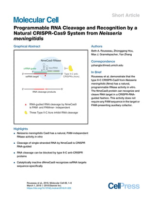
  • 1. Short Article Programmable RNA Cleavage and Recognition by a Natural CRISPR-Cas9 System from Neisseria meningitidis Graphical Abstract Highlights d Neisseria meningitidis Cas9 has a natural, PAM-independent RNase activity in vitro d Cleavage of single-stranded RNA by NmeCas9 is CRISPR RNA-guided d RNA cleavage can be blocked by type II-C anti-CRISPR proteins d Catalytically inactive dNmeCas9 recognizes ssRNA targets sequence-specifically Authors Beth A. Rousseau, Zhonggang Hou, Max J. Gramelspacher, Yan Zhang Correspondence yzhangbc@med.umich.edu In Brief Rousseau et al. demonstrate that the type II-C CRISPR-Cas9 from Neisseria meningitidis (Nme) has a natural, programmable RNase activity in vitro. The NmeCas9 protein can recognize and cleave RNA target in a CRISPR-RNA- guided fashion. This activity does not require any PAM sequence in the target or PAM-presenting auxiliary cofactor. Rousseau et al., 2018, Molecular Cell 69, 1–9 March 1, 2018 ª 2018 Elsevier Inc. https://doi.org/10.1016/j.molcel.2018.01.025
  • 2. Molecular Cell Short Article Programmable RNA Cleavage and Recognition by a Natural CRISPR-Cas9 System from Neisseria meningitidis Beth A. Rousseau,1,2 Zhonggang Hou,1,2 Max J. Gramelspacher,1 and Yan Zhang1,3,* 1Department of Biological Chemistry, University of Michigan, Ann Arbor, MI 48109, USA 2These authors contributed equally 3Lead Contact *Correspondence: yzhangbc@med.umich.edu https://doi.org/10.1016/j.molcel.2018.01.025 SUMMARY The microbial CRISPR systems enable adaptive defense against mobile elements and also provide formidable tools for genome engineering. The Cas9 proteins are type II CRISPR-associated, RNA-guided DNA endonucleases that identify double-stranded DNA targets by sequence complementarity and pro- tospacer adjacent motif (PAM) recognition. Here we report that the type II-C CRISPR-Cas9 from Neisseria meningitidis (Nme) is capable of programmable, RNA-guided, site-specific cleavage and recognition of single-stranded RNA targets and that this ribonu- clease activity is independent of the PAM sequence. We define the mechanistic feature and specificity constraint for RNA cleavage by NmeCas9 and also show that nuclease null dNmeCas9 binds to RNA target complementary to CRISPR RNA. Finally, we demonstrate that NmeCas9-catalyzed RNA cleav- age can be blocked by three families of type II-C anti-CRISPR proteins. These results fundamentally expand the targeting capacities of CRISPR-Cas9 and highlight the potential utility of NmeCas9 as a single platform to target both RNA and DNA. INTRODUCTION Clustered, regularly interspaced, short, palindromic repeats (CRISPR) loci and their associated (cas) genes constitute an adaptive defense system that is widespread in bacteria and archea and that limits horizontal genetic transfer (Barrangou et al., 2007; Marraffini and Sontheimer, 2008). CRISPR spacers, which are acquired from invaders’ genomes and integrated between CRISPR repeats, specify the nucleic acid targets for CRISPR interference (Barrangou et al., 2007; Marraffini, 2015). The CRISPR locus is transcribed and processed into small RNAs called CRISPR RNAs (crRNAs), which guide the Cas pro- tein effectors to destroy complementary targets (Brouns et al., 2008; Hale et al., 2009; Wright et al., 2016). Based upon cas gene content, the diverse CRISPR-Cas systems are categorized into two classes, six major types, and nearly thirty subtypes (Koonin et al., 2017), and the majority of these systems confer interference by DNA targeting (Garneau et al., 2010; Marraffini and Sontheimer, 2008; Wright et al., 2016). The Cas9 proteins, which are the single protein effectors of type II CRISPRs, generally function as RNA-guided DNA endo- nucleases (Gasiunas et al., 2012; Jinek et al., 2012) and provide revolutionary tools for programmable genome engineering in eu- karyotes and prokaryotes, with the type II-A SpyCas9 being the most commonly used (Cho et al., 2013; Cong et al., 2013; Hwang et al., 2013; Jiang et al., 2013; Jinek et al., 2013; Mali et al., 2013). When programmed by crRNA and another RNA cofactor called tracrRNA (Deltcheva et al., 2011), SpyCas9 targets double- stranded DNA (dsDNA) through the recognition of protospacer adjacent motif (PAM) sequence (Jinek et al., 2012), DNA unwind- ing, R-loop formation, and the triggering of DNA scission (Stern- berg et al., 2014). The HNH and RuvC nuclease domains of Cas9 cleave the crRNA-complementary and non-complementary target strands, respectively (Gasiunas et al., 2012; Jinek et al., 2012). Catalytically inactive ‘‘dead’’ Cas9s (dCas9s), which can bind to DNA targets without inducing any DNA breaks (Jinek et al., 2012), have also been harnessed as eukaryotic genome- binding platforms to deliver effector domains for the regulation, imaging, or modification of the specific chromosomal loci (Wright et al., 2016). In addition, the newly discovered type II anti-CRISPR (Acr) proteins, encoded by mobile genetic ele- ments (MGEs), are potent Cas9 inhibitors and can block Cas9- mediated DNA cleavage and gene editing (Harrington et al., 2017; Pawluk et al., 2016; Rauch et al., 2017). These and other Cas9-based DNA manipulation tools are transforming biomed- ical research. There are a few CRISPR systems capable of RNA targeting. For example, the type VI effector Cas13 is a promiscuous RNase that, upon activation by crRNA-guided RNA recognition, de- grades nearby RNAs non-specifically (Abudayyeh et al., 2016; East-Seletsky et al., 2016). The type II-B Cas9 from Francisella novicida (Fno) employs the tracrRNA, rather than crRNA, as a guide to silence an endogenous transcript, yet the protein en- coding the ribonuclease activity remains unidentified (Sampson et al., 2013). Type III CRISPRs use large multi-protein complexes to confer immunity through transcription-dependent co-degra- dation of the DNA and its transcripts (Elmore et al., 2016; Samai et al., 2015). A separate type-III-associated RNase, Csm6, Molecular Cell 69, 1–9, March 1, 2018 ª 2018 Elsevier Inc. 1 Please cite this article in press as: Rousseau et al., Programmable RNA Cleavage and Recognition by a Natural CRISPR-Cas9 System from Neisseria meningitidis, Molecular Cell (2018), https://doi.org/10.1016/j.molcel.2018.01.025
  • 3. provides signal-activated, non-specific RNA clearance (Jiang et al., 2016; Kazlauskiene et al., 2017; Niewoehner et al., 2017). Several reports have indicated that Cas9s lack crRNA-guided RNA cleavage activity (Gasiunas et al., 2012; Ma et al., 2015). SpyCas9, however, can be tricked into cleaving RNAs in vitro by an exogenously supplied PAMmer DNA oligo that hybridizes to the ssRNA target-flanking region and presents the DNA PAM on the opposing strand. Specific binding of SpyCas9 to the RNA target can also be achieved with a longer PAMmer that extends into the crRNA-complementary region (O’Connell et al., 2014). These in vitro findings open up doors to develop CRISPR-based RNA-targeting tools to recognize and manipulate specific RNAs in vitro and in vivo. Indeed, a PAMmer- and dSpyCas9-based strategy has enabled the pull-down of RNAs from cell extracts (O’Connell et al., 2014) and the visualization of stress granule RNAs in mammalian cells (Nelles et al., 2016). In addition, dSpyCas9 was repurposed to eliminate disease-associated, toxic repetitive RNAs (Batra et al., 2017), and more recently, the Cas13 RNase wasadopted for invivoknockdown and editing ofmamma- lian transcripts (Abudayyeh et al., 2017; Cox et al., 2017). Despite these advances, it is unclear whether any other Cas9 ortholog has PAMmer-inducible or even intrinsic crRNA-pro- grammable RNase activity. Here we explore the RNA-targeting potential of Nme CRISPR-Cas9, a type II-C system that has pre- viously been shown to limit DNA natural transformation in its native context N. meningitidis (Zhang et al., 2013) and has been adopted as an eukaryotic gene-editing platform (Esvelt et al., 2013; Hou et al., 2013; Lee et al., 2016; Zhang, 2017). We find that NmeCas9 has a natural PAMmer-oligo-independent and programmable RNase activity in vitro. We describe the func- tional determinants, mechanistic features, and specificity con- straints for RNA cleavage by NmeCas9 and show that this activ- ity can be blocked by three families of type II-C Acr proteins. We also find that catalytically inert dNmeCas9 binds to RNA targets in a sequence-specific manner. RESULTS A Natural RNA-Guided Ribonuclease Activity of NmeCas9 In Vitro The type II-C CRISPR-cas locus from N. meningitidis strain 8013 consists of three cas genes (cas1, cas2, and cas9), a tracrRNA locus, and a CRISPR array (Zhang et al., 2013; Figure 1A). We expressed recombinant, FLAG-tagged NmeCas9 in E. coli and isolated it by heparin, ion exchange, and size exclusion chroma- tography (Figure S1A). In vitro RNA cleavage assays were per- formed using purified protein, in vitro-transcribed tracrRNA and crRNA, and a fluorescent end-labeled ssRNA oligonucleo- tide bearing a target region complementary to the spacer of crRNA-sp25 (Figure 1B). We found that NmeCas9 efficiently cat- alyzes in vitro cleavage of the RNA substrate, resulting in one prominent labeled cleaved product, and that this reaction re- quires the cognate crRNA, the tracrRNA, and Mg2+ or Mn2+ (Fig- ures 1C and S1B). A DNA guide containing sequences identical to crRNA-sp25 cannot support RNA cleavage (Figure S1C), indi- cating that NmeCas9’s RNase activity is strictly RNA guided. Strikingly, unlike SpyCas9, NmeCas9 catalyzed efficient RNA cleavage without any PAMmer oligo co-factor (Figure 1C), reflecting a fundamental distinction between the RNase activities of these two Cas9 orthologs. We also tested whether a similar PAMmer strategy would modulate RNA cleavage by NmeCas9 by pre-annealing the same ssRNA target to various top-strand partners (Figure 1D, lower, and Figure S1D) and analyzing the resulting substrates. RNA cleavage was minimally enhanced by a 22-nucleotide (nt) DNA PAMmer but greatly impeded or completely abrogated on a fully annealed DNA-RNA heterodu- plex or on a double-stranded RNA substrate (Figure 1D, upper). These results reveal that NmeCas9-mediated RNA cleavage is specific for ssRNA target and is independent of any PAMmer provided in trans. Next, we tested whether NmeCas9’s ribonuclease activity requires a PAM equivalent in the target RNA (termed rPAM, 50 -AAUCN4-30 for NmeCas9) by assaying a mutant RNA sub- strate with 3-nt mutations (AAUC to AUAG) introduced into the rPAM (Figure 1E). The same triple mutation was sufficient to abolish dsDNA cleavage by NmeCas9 (Zhang et al., 2015). This rPAM mutant RNA substrate was cleaved as efficiently as the wild-type counterpart (Figure 1E), indicating that RNA cleav- age by NmeCas9 is rPAM-independent. All Cas9 enzymes described to date employ the HNH and RuvC domains to cut the crRNA-complementary and non-complementary strands of the dsDNA targets, respectively (Gasiunas et al., 2012; Jinek et al., 2012). For NmeCas9, active site residues D16 in the RuvC domain and H588 in the HNH domain were previously shown to be essential for dsDNA targeting in vivo and in vitro (Zhang et al., 2013, 2015). To test the involvement of these two active sites in RNA cleavage, we purified and analyzed three mutant NmeCas9 proteins (D16A, H588A, and the double mutant [dm, D16A+H588A]) (Figures S1A and S2B). RNA cleavage was abolished for the H588A and dm proteins but not for the D16A mutant (Figure 1F), suggesting that the HNH domain of NmeCas9 mediates the ssRNA cleavage and therefore is capable of both DNA and RNA scission. In addition, a time-course experiment revealed that RNA cleavage at 37 C became detectable within one minute and pla- teaued after 30 minutes and that it occurs more slowly at room temperature (Figure S1E). RNA cleavage was robust under a wide range of NmeCas9-RNA ribonucleoprotein (RNP) concen- trations (Figure S1F) or monovalent salt concentrations (KCl, Fig- ure S1G). By analyzing serial crRNA mutants with 50 truncations (in the 24-nt spacer portion) or 30 truncations (in the 24-nt repeat portion), we found that RNA cleavage is reduced with a 18-nt to 20-nt spacer and completely lost with a 16-nt spacer (Figure S3A) and that the first 8 nts of the crRNA repeat are sufficient to sup- port robust RNA cleavage by NmeCas9 (Figure S3B). RNA-Guided RNA Cleavage by NmeCas9 Is Programmable To assess the programmability of the NmeCas9 ribonuclease, we first attempted to redirect RNA cleavage to different positions within the same ssRNA target. We created two variants of the crRNA-sp25, walkÀ2 and walk+3, which match different regions of the same target (Figure 2A). These two variants were analyzed alongside the wild-type counterpart in a cleavage site mapping experiment. Both variants directed in vitro RNA cleavage, and importantly their cleavage sites moved in concert with the 2 Molecular Cell 69, 1–9, March 1, 2018 Please cite this article in press as: Rousseau et al., Programmable RNA Cleavage and Recognition by a Natural CRISPR-Cas9 System from Neisseria meningitidis, Molecular Cell (2018), https://doi.org/10.1016/j.molcel.2018.01.025
  • 4. Figure 1. NmeCas9 Possesses a Natural RNA-Guided Ribonuclease Activity In Vitro (A) Schematics depicting the CRISPR-Cas9 of N. meningitidis strain 8013 and domain organization of NmeCas9. Individual elements are not drawn to scale. Black rectangles, CRISPR repeats; yellow diamonds, CRISPR spacers; gray boxes, cas1, cas2, cas9, and tracrRNA genes; black arrows, transcription driven by repeat-embedded promoters. CTD, C-terminal domain; R, arginine-rich motif; REC, recognition domains; HNH, HNH domain; RuvC, RuvC domains. (B) A schematic for the complex of the crRNA-sp25, tracrRNA, and ssRNA target 25. Yellow, crRNA spacer; gray, crRNA repeat; bold, rPAM; green, FAM label. (C) NmeCas9 cleaves ssRNA target efficiently in vitro, and this reaction requires the cognate crRNA, the tracrRNA, and divalent metal Mg2+ . Non-cog crRNA, crRNA-sp23. The uncleaved probe and cleavage products are indicated. (D) An exogenously supplied PAMmer DNA oligo has a modest effect on NmeCas9-catalyzed ssRNA cleavage. The panel of pre-annealed nucleic acid substrates (depicted at the bottom) were assayed for in vitro RNA cleavage as in (C). Black, RNA; gray, DNA; red, crRNA-complementary region; yellow, PAM or rPAM; green, FAM label. (E) NmeCas9 cleaves ssRNA in an rPAM-independent manner. An rPAM mutant RNA substrate was analyzed for cleavage as in (C). Red, mutated sequences in the rPAM. (F) An intact HNH domain is required for RNA cleavage by NmeCas9. Nuclease domain active site mutants of NmeCas9 were tested as in (C). Dm, double mutant (D16A +H588A). Molecular Cell 69, 1–9, March 1, 2018 3 Please cite this article in press as: Rousseau et al., Programmable RNA Cleavage and Recognition by a Natural CRISPR-Cas9 System from Neisseria meningitidis, Molecular Cell (2018), https://doi.org/10.1016/j.molcel.2018.01.025
  • 5. Figure 2. NmeCas9-Catalyzed RNA Cleavage Is Programmable (A) A schematic depicting the ssRNA target 25 and three matching crRNAs, (crRNA-sp25-wt, walkÀ2, and walk+3). Yellow, crRNA spacer; gray, crRNA repeat; bold, rPAM; green, FAM label; red arrows, predominant RNA cleavage sites mapped out in (B). (B) RNA cleavage site mapping experiment. SsRNA target 25 and the three crRNAs used are shown in (A). The NmeCas9 cleavage products and RNase T1 and hydrolysis ladders were subjected to 30 dephosphorylation by T4 polynucleotide kinase and separated by 15% denaturing PAGE. T1, RNase T1 ladder; OH- , hydrolysis ladder. À2, walkÀ2; +3, walk+3; wt, crRNA-sp25. Sites of G cleavage by RNase T1 are indicated; sites of NmeCas9 cleavage (G30, A27, G25 for the three crRNAs, respectively) are marked by red arrows. (C) Serial single-nt mutants of crRNA-sp25 were analyzed for NmeCas9-catalyzed RNA cleavage. In M1 through M19, single-nt mutation is introduced into every other position of the crRNA spacer. The location and sequence of each mutation (in red) are shown at the top. Yellow, crRNA spacer; gray, crRNA repeat; red arrow, RNA cleavage site. Non-cog, crRNA-sp23. (D) A schematic depicting the ssRNA target 9 and the two matching crRNAs (crRNA-sp9-wt and walk-1). Yellow, crRNA spacer; gray, crRNA repeat; bold, rPAM; red arrows, predicted RNA cleavage sites; green, FAM label; magenta, Cy5 label. (E) NmeCas9’s ribonuclease activity is re-programmable on a different RNA substrate. The two crRNAs shown in (D) were assayed for in vitro cleavage on ssRNA target 9. The same denaturing gel is subjected to FAM (left) and Cy5 (right) detection. 4 Molecular Cell 69, 1–9, March 1, 2018 Please cite this article in press as: Rousseau et al., Programmable RNA Cleavage and Recognition by a Natural CRISPR-Cas9 System from Neisseria meningitidis, Molecular Cell (2018), https://doi.org/10.1016/j.molcel.2018.01.025
  • 6. guide-target complementarity (Figure 2B). By comparing the NmeCas9 cleavage products with RNase T1 and hydrolysis ladders, we found that the wild-type crRNA-sp25 and the two variants predominantly directed cuts after A27, G25, and G30, respectively (Figure 2B), indicating that NmeCas9 catalyzes RNA scission between the 3rd and the 4th nts of the crRNA-paired target region proximal to the 50 end of the target (Figure 2A). This is consistent with the DNA double-strand breaks (DSBs) be- tween the 3rd and the 4th nts generated by NmeCas9 (Zhang et al., 2015) and other Cas9 orthologs (Gasiunas et al., 2012; Jinek et al., 2012). The fact that the two variants of crRNA- sp25 effectively changed target-flanking sequence in the RNA substrate corroborated earlier finding in Figure 1E that RNA cleavage is rPAM-independent. Next, we sought to investigate the specificity rule governing NmeCas9’s tolerance for mismatches in the crRNA-RNA target complementarity. We created and tested serial crRNA mutants each bearing a single-nt deviation from the wild-type sequence at every odd position within the spacer (Figure 2C, upper). Only the two single mismatches at the 3rd and 5th nt, which are close to the cleavage site, abrogated in vitro RNA cleavage; the other mutations either did not affect cleavage or only caused modest defects (e.g., at the 9th or 17th nt) (Figure 2C, lower). We also analyzed crRNA mutants with multiple mismatches and found that RNA cleavage was diminished by short (2–4 nts) mu- tations clustered around the cleavage site (Figure S3C, lanes 4 and 8), but was only partially reduced by 2–3 nts of mismatches in regions away from the cleavage site (Figure S3C, lanes 3 and 5–7). CrRNAs with 4 or more nts of mismatches all exhibited severe cleavage defects (Figure S3C, lanes 8–10). Overall, the NmeCas9 ribonuclease has certain degree of tolerance for guide-target mismatches that are not next to the cleavage site. Programmable RNA cleavage by NmeCas9 was also observed with a different dual-fluorophore-labeled RNA substrate bearing the target sequence for spacer 9 of N. meningitis strain 8013 (Figure 2D). RNA cleavage guided by crRNA-sp9 resulted in one predominant 50 product and one major 30 product (Figure 2E, Cy5 and FAM labeled, respectively). For a variant of crRNA-sp9, walk-1, which has the guide-target pairing region shifted by 1 nt, the cleavage site moved in concert (Figures 2D and 2E). Collec- tively, these results demonstrate that NmeCas9 ribonuclease is crRNA-guided and programmable. Type II-C Anti-CRISPRs Inhibit In Vitro RNA Cleavage by NmeCas9 In light of the recent discovery of type II Acrs that inhibit Cas9- mediated genetic interference, DNA cleavage, and genome edit- ing (Harrington et al., 2017; Hynes et al., 2017; Pawluk et al., 2016; Rauch et al., 2017), we wondered whether the three families of type II-C Acrs also inhibit NmeCas9-catalyzed RNA cleavage. We purified four Acr proteins—AcrIIC1Nme, AcrIIC2Nme, AcrIIC3Nme and AcrIIA4 (Figure 3A)—and analyzed them in our RNA cleavage assay. Notably, pre-incubation of NmeCas9 with increasing amounts of AcrIIC1Nme, AcrIIC2Nme, or AcrIIC3Nme all resulted in blockage of RNA cleavage dose dependently, and near-complete inhibition was achieved with all the three Acr proteins at or above 3-fold molar excess over NmeCas9 (Figure 3B). In contrast, cleavage was not affected by increasing amounts of AcrIIA4 (Figure 3B), a control Acr that specifically blocks DNA cleavage by two type II-A Cas9s, SpyCas9 and Listeria monocytogenes (Lmo) Cas9 (Rauch et al., 2017). In a quality-control plasmid cleavage experiment, the three type II-C Acrs all prevented linearization of a target plasmid DNA by NmeCas9 (Figure 3C), consistent with previous Figure 3. Type II-C Anti-CRISPRs Block RNA Cleavage by NmeCas9 (A) A Coomassie-stained 15% SDS-PAGE of pu- rified Acr proteins. The predicted molecular weight of AcrIIC1Nme, AcrIIC2Nme, Flag-AcrIIC3Nme, and AcrIIA4 (a type II-A control Acr) are about 9.8, 14.4, 14.6, and 10.4 kDa, respectively. Molecular weight markers are indicated. (B) Three type II-C anti-CRISPR proteins AcrIIC1, AcrIIC2, and AcrIIC3, but not AcrIIA4, inhibit NmeCas9-catalyzed RNA cleavage. NmeCas9 was incubated with increasing amounts of various Acr proteins for 10 minutes, assembled with the tracrRNA and crRNA for another 10 minutes, and then mixed with fluorescently labeled RNA sub- strate (25 nM) to license in vitro RNA cleavage. Each Acr protein was used at 0.75-, 1.5-, 3-, and 6-fold molar equivalents relative to NmeCas9 RNP (500 nM). The cleavage reactions were analyzed by denaturing PAGE. (C) All three purified type II-C Acr proteins, but not AcrIIA4, are potent inhibitors for NmeCas9-medi- ated plasmid DNA cleavage. N, nicked; L, linear- ized plasmid, about 3.6 kb; SC: supercoiled. Mo- lecular size markers are indicated. Reactions were done as in (B) except that 8 nM of plasmid DNA was added as the substrate, and the reactions were analyzed by agarose gel and SYBR Safe staining. Molecular Cell 69, 1–9, March 1, 2018 5 Please cite this article in press as: Rousseau et al., Programmable RNA Cleavage and Recognition by a Natural CRISPR-Cas9 System from Neisseria meningitidis, Molecular Cell (2018), https://doi.org/10.1016/j.molcel.2018.01.025
  • 7. reports (Harrington et al., 2017; Pawluk et al., 2016; Rauch et al., 2017). In summary, these results showed that, in addition to their known roles in blocking DNA targeting, AcrIIC1Nme, AcrIIC2Nme, and AcrIIC3Nme can all block NmeCas9-catalyzed in vitro RNA cleavage as well. RNA-Guided, Sequence-Specific Binding of NmeCas9 to RNA Targets In Vitro To investigate how NmeCas9 engages ssRNA target, we turned to electrophoretic mobility shift assays (EMSAs). Divalent metals were omitted from the reactions to render NmeCas9 catalytically inactive. We observed robust mobility shifts forming on the fluo- rescently labeled RNA target only when NmeCas9, a matching crRNA, and the tracrRNA were included (Figure 4A, lane 2 from the left, top bands). Importantly, these shifts were greatly reduced when a non-cognate crRNA was used instead (Fig- ure 4A, lane 3 from the left), indicative of sequence-specific, sta- ble binding of NmeCas9 RNP to the ssRNA target. There were a few other shifts with intermediate mobility in all the binding reac- tions containing the cognate crRNA-sp25 (Figure 4A, lanes 2, 4, 7, and 8 from the left), likely reflecting binding events mediated by RNA-RNA interactions only. The same rPAM mutant RNA substrate used in Figure 1E was also analyzed in EMSA experi- ments and exhibited no binding defects at all (Figure 4B), suggesting that NmeCas9 recognizes its RNA target in an rPAM-independent way. This is in stark contrast to Cas9-medi- ated dsDNA binding, in which PAM recognition is a prerequisite (Jinek et al., 2012; Sternberg et al., 2014). The NmeCas9-mediated retarded migration in EMSAs occurred in a dose-dependent manner (Figure 4C), and the bind- ing was not affected by active site mutations in either the HNH or the RuvC domains (Figure 4D); this suggests that the recognition of RNA substrate can happen independently of RNA scission. It is reported that SpyCas9 is a single turnover enzyme that holds on to all four ends of the products after cleaving the duplexed DNA target (Richardson et al., 2016; Sternberg et al., 2014). We investigated whether the RNA cleavage products here are released from the NmeCas9 RNP. To this end, we performed Figure 4. dNmeCas9 Binds ssRNA Target In Vitro in a crRNA-Guided Manner (A) CrRNA-guided, sequence-specific recognition of RNA target by NmeCas9. Electrophoretic mobility shift assays (EMSAs) were performed using the 50 FAM-labeled ssRNA target 25 (25 nM), NmeCas9 (500 nM), and various small RNAs (500 nM) as indicated. Binding was performed in cleavage reaction buffer (but with Mg2+ omitted) at 37 C for half an hour. Non cog, non-cognate crRNA-sp23. (B) The association of NmeCas9 with RNA target is rPAM-independent. The same rPAM mutant RNA substrate used in Figure 1E was analyzed here using EMSAs. Binding reactions were done as in (A). Red, mutated sequences in the rPAM; non- cog, crRNA-sp23 (C) Dose-dependent binding to the RNA target by NmeCas9-RNP complex. Binding reactions were done as in (A) except that 10 nM substrate is used. The concentrations of NmeCas9 RNP used were indicated. (D) The wild-type NmeCas9 and its nuclease domain active site mutants (D16A, H588A, and dm) were assayed for sequence-specific binding to the RNA target, as in (A). Dm, double mutant; non-cog, crRNA-sp23. (E) The 30 , but not the 50 , product of RNA cleavage remained bound by the NmeCas9 RNP. NmeCas9-catalyzed RNA cleavage re- actions were done as in Figure 1C for two sepa- rate fluorescent RNA substrates, target 25 and target 9. Half of the reactions were analyzed by 6% native PAGE (upper), and the same native gel was visualized by FAM (left) and Cy5 (right) detection. The other half of the reactions were quenched by proteinase K treatment and form- amide loading dye, then analyzed by 15% dena- turing PAGE (lower), and the same denaturing gel was visualized by FAM (left) or Cy5 (right). Schematics of RNA substrates used were at the top. Green, FAM label; magenta, Cy5 label. Note that the 50 cleavage product dissociates from the NmeCas9 (upper right). 6 Molecular Cell 69, 1–9, March 1, 2018 Please cite this article in press as: Rousseau et al., Programmable RNA Cleavage and Recognition by a Natural CRISPR-Cas9 System from Neisseria meningitidis, Molecular Cell (2018), https://doi.org/10.1016/j.molcel.2018.01.025
  • 8. standard in vitro RNA cleavage assay and analyzed the reactions using two parallel approaches. Half of reactions were quenched and analyzed by denaturing PAGE (Figure 4E, lower), and the other half were analyzed by native PAGE (Figure 4E, upper). While robust cleavage was observed on both ssRNA target 25 and target 9 (Figure 4E, lower), the 50 cleavage products (FAM- labeled on target 25, Cy5-labeled on target 9) were released from the NmeCas9 RNP (Figure 4E, upper). In contrast, the 30 cleavage products (FAM-labeled on target 9), which contain 20 nts of sequence complementary to the cognate crRNA, were present only in higher molecular-weight shifts on native PAGE, suggesting that they were largely still bound by the NmeCas9 RNP (Figure 4E, upper panel, left). We also did similar analysis using dsDNA substrates for NmeCas9. DNA target- strand or non-target-strand oligonucleotides that were fluores- cently labeled at either the 30 or 50 end were annealed with appropriate partner strands (Figure S2A), and the resulting DNA duplexes were all robustly cleaved by NmeCas9 (Fig- ure S2D). Native PAGE analysis revealed that all but one of the four ends of DNA cleavage products remain largely bound by NmeCas9 (Figure S2C, left), whereas the 50 product of non-target DNA strand was released (Figure S2C, right). Taken together, these results suggest that NmeCas9 is likely a single turnover enzyme for both ssRNA cleavage and dsDNA cleavage. DISCUSSION Expanded Targeting Capacities for CRISPR-Cas9 Systems Central to the CRISPR genome editing revolution are the Cas9 DNA endonucleases, which can be easily programmed to cut any dsDNA target of interest through PAM recognition and guide-target base pairing (Wright et al., 2016). Our work here re- vealed that the type II-C Cas9 from N. meningitidis is capable of programmable, RNA-guided, sequence-specific cleavage and recognition of ssRNA target (Figures 1, 2, and 4). Importantly, un- like the type II-A SpyCas9 (O’Connell et al., 2014), NmeCas9’s RNase activity is independent of PAMmer DNA oligo auxiliary factor (Figures 1C and 1D). While the revision of this paper was under consideration, Strutt et al. (2018) reported similar natural RNase activity for the Cas9s of Staphylococcus aureus (Sau) and Campylobacter jejuni (Cje). Therefore, NmeCas9 represents one of the first native Cas9 endoribonucleases that expand the targeting capacities of CRISPR-Cas9. It is also tempting to speculate that additional natural Cas9 RNase may exist in the divergent Cas9 family. The rPAM- and PAMmer-independent nature of this ribonu- clease activity implies that the selection of ssRNA substrate is achieved mainly through RNA-RNA pairing between the guide and the target, without a requirement for rPAM recognition by NmeCas9. This feature may help enable the manipulation of cellular messenger RNAs while avoiding collateral cleavage or binding to the corresponding genomic sites lacking the PAMs. In fact, our in vitro assay revealed that NmeCas9 cleaves a no- rPAM RNA target without affecting the corresponding dsDNA substrate, which contains no correct PAM sequence (Figure S4, lanes 7–12 from left). Moreover, the same HNH nuclease domain of NmeCas9 that cleaves the target DNA strand also cleaves the ssRNA substrate. This is not surprising, given the existence of HNH motifs in homing endonucleases that can cleave both DNA and RNA molecules (Pommer et al., 2001). Future structural studies are needed to understand how the NmeCas9 and its HNH domain accommodate various kinds of nucleic acid targets. Acr proteins inhibit DNA targeting by Cas9s through distinct strategies (Dong et al., 2017; Harrington et al., 2017; Pawluk et al., 2016; Rauch et al., 2017), and these mechanistic informa- tion could be illuminating in the context of NmeCas9-catalyzed RNA cleavage. For example, AcrIIA4 structurally mimics a PAM for SpyCas9 (Dong et al., 2017), so an analogous Acr for NmeCas9 would not be expected to interfere with RNA cleavage, which is an rPAM- and PAMmer-independent process. AcrIIC1 is a broad-spectrum inhibitor that disarms diverse type II-C Cas9 orthologs by direct binding to the HNH domain (Harrington et al., 2017); therefore, it is not surprising that AcrIIC1 blocks RNA cleavage that relies on the HNH domain of NmeCas9. Potential RNA-Targeting Applications RNA plays critical and diverse biological roles, and RNA-target- ing tools have the potential to transform research and medicine. There are existing platforms such as RNA interference, antisense oligonucleotides, and designer RNA-binding proteins (e.g., Pumilios) that can recognize specific transcripts or exogenous RNA tags (Nelles et al., 2015). Recently, the CRISPR-Cas13 sys- tem that targets RNA was repurposed as a new tool to knock down or edit specific mammalian transcripts (Abudayyeh et al., 2017; Cox et al., 2017), and dSpyCas9 can also help to remove repetitive RNAs in human cells (Batra et al., 2017). These pro- grammable RNA-targeting tools will revolutionize how we modu- late RNA metabolism in the cells. NmeCas9 can potentially provide a unique single platform to achieve both dsDNA targeting and RNA targeting tasks, whereas Cas13 exclusively targetsRNA.The PAMmer-independent nature of its RNase activity makes NmeCas9 a desired system to circum- vent challenges that come with the delivery and toxicity of ssDNA PAMmer oligos. In addition, the fact that NmeCas9, SpyCas9, and Cas13s use orthogonal guide RNAs (Abudayyeh et al., 2016; Esvelt et al., 2013) can be exploited to achieve distinct but multiplexed RNA-targeting tasks. Moreover, the three families of type II-C Acr proteins can provide off switches to enable precise control of NmeCas9-based RNA cleavage applications. Biological Implications of NmeCas9 RNA Targeting Species of the genus Neisseria rely on natural transformation for frequent genetic exchange (Hamilton and Dillard, 2006). In CRISPR-containing N. meningitidis strains, dsDNA cleavage by Cas9 provides a barrier to genetic material transfer through nat- ural transformation (Zhang et al., 2013). NmeCas9, along with several other type II-C Cas9s, are also found to be capable of robust ssDNA cleavage in vitro, a feature proposed to play an evolutionarily ancestral role in restricting the ssDNA genome of filamentous bacteriophages or transforming ssDNAs (Ma et al., 2015; Zhang et al., 2015). As for the RNase activity of NmeCas9, the physiological rele- vance in Neisseria cells remains to be determined. One possibil- ity is that RNA targeting plays an auxiliary role in defense by help- ing the clearance of transcripts derived from transforming DNAs Molecular Cell 69, 1–9, March 1, 2018 7 Please cite this article in press as: Rousseau et al., Programmable RNA Cleavage and Recognition by a Natural CRISPR-Cas9 System from Neisseria meningitidis, Molecular Cell (2018), https://doi.org/10.1016/j.molcel.2018.01.025
  • 9. or bacteriophages. Another possibility is that NmeCas9 may target endogenous Neisseria RNAs in a crRNA-guided fashion. We bioinformatically examined potential matches between all the twenty-five spacers from native mature crRNAs and the self-chromosome of N. meningitidis strain 8013. BLASTN searches revealed no perfect or near-perfect hits (up to 4 nts of mismatches allowed), consistent with an earlier finding that Neisseria CRISPR spacers primarily match to the genomes of other strains or species (Zhang et al., 2013). The best matches for all but one crRNA have 7 or more nts of mismatches spread out in regions both proximal and distal to the presumed RNA cleavage site. This degree of mispairing, according to our in vitro mutagenesis study (Figures 2C and S3C), would largely abolish NmeCas9-catalyzed RNA cleavage. Therefore, we cautiously speculate that robust cleavage of endogenous Neisseria transcripts by NmeCas9 and existing CRISPR spacers is not very likely but could arise. Nonetheless, future work is needed to determine if crRNA-directed RNA binding or cleavage of endogenous transcripts by NmeCas9 occurs in Neisseria, and if so, what biological consequences could result. STAR+METHODS Detailed methods are provided in the online version of this paper and include the following: d KEY RESOURCES TABLE d CONTACT FOR REAGENT AND RESOURCE SHARING d EXPERIMENTAL MODEL AND SUBJECT DETAILS B Escherichia coli BL21 (DE3) B Escherichia coli JM109 d METHOD DETAILS B Plasmid construction B In vitro transcription (IVT) and purification of small RNAs B In vitro cleavage assay B RNA ladders and cleavage site mapping B Expression and purification of NmeCas9 protein B Expression and purification of Acr proteins B Electrophoretic mobility shift assay (EMSA) d QUANTIFICATION AND STATISTICAL ANALYSIS SUPPLEMENTAL INFORMATION Supplemental Information includes four figures and one table and can be found with this article online at https://doi.org/10.1016/j.molcel.2018.01.025. ACKNOWLEDGMENTS The authors would like to thank Erik Sontheimer, Hank Seifert, Daniel Goldman, and Michael Sheets for critical reading of the manuscript. We thank Chase Beisel and Cynthia Sharma for communicating unpublished work, Tianmin Fu (Harvard Medical School) for advice on protein purification, and Xufei Zhou for graphical assistance. This work was supported by the National Institutes of Health (GM117268 to Y.Z.) and University of Michigan institutional funds to Y.Z. AUTHOR CONTRIBUTIONS B.A.R, Z.H. and Y.Z. designed and conducted experiments. M.J.G. and Z.H. purified proteins. Z.H. and Y.Z. wrote the manuscript. All authors edited the manuscript. DECLARATION OF INTERESTS The authors declare no competing interests. Received: November 20, 2017 Revised: December 18, 2017 Accepted: January 18, 2018 Published: February 15, 2018 REFERENCES Abudayyeh, O.O., Gootenberg, J.S., Konermann, S., Joung, J., Slaymaker, I.M., Cox, D.B., Shmakov, S., Makarova, K.S., Semenova, E., Minakhin, L., et al. (2016). C2c2 is a single-component programmable RNA-guided RNA- targeting CRISPR effector. Science 353, aaf5573. Abudayyeh, O.O., Gootenberg, J.S., Essletzbichler, P., Han, S., Joung, J., Belanto, J.J., Verdine, V., Cox, D.B.T., Kellner, M.J., Regev, A., et al. (2017). RNA targeting with CRISPR-Cas13. Nature 550, 280–284. Barrangou, R., Fremaux, C., Deveau, H., Richards, M., Boyaval, P., Moineau, S., Romero, D.A., and Horvath, P. (2007). CRISPR provides acquired resis- tance against viruses in prokaryotes. Science 315, 1709–1712. Batra, R., Nelles, D.A., Pirie, E., Blue, S.M., Marina, R.J., Wang, H., Chaim, I.A., Thomas, J.D., Zhang, N., Nguyen, V., et al. (2017). Elimination of toxic microsatellite repeat expansion RNA by RNA-targeting Cas9. Cell 170, 899– 912.e10. Brouns, S.J., Jore, M.M., Lundgren, M., Westra, E.R., Slijkhuis, R.J., Snijders, A.P., Dickman, M.J., Makarova, K.S., Koonin, E.V., and van der Oost, J. (2008). Small CRISPR RNAs guide antiviral defense in prokaryotes. Science 321, 960–964. Cho, S.W., Kim, S., Kim, J.M., and Kim, J.S. (2013). Targeted genome engineering in human cells with the Cas9 RNA-guided endonuclease. Nat. Biotechnol. 31, 230–232. Cong, L., Ran, F.A., Cox, D., Lin, S., Barretto, R., Habib, N., Hsu, P.D., Wu, X., Jiang, W., Marraffini, L.A., and Zhang, F. (2013). Multiplex genome engineering using CRISPR/Cas systems. Science 339, 819–823. Cox, D.B.T., Gootenberg, J.S., Abudayyeh, O.O., Franklin, B., Kellner, M.J., Joung, J., and Zhang, F. (2017). RNA editing with CRISPR-Cas13. Science 358, 1019–1027. Deltcheva, E., Chylinski, K., Sharma, C.M., Gonzales, K., Chao, Y., Pirzada, Z.A., Eckert, M.R., Vogel, J., and Charpentier, E. (2011). CRISPR RNA matura- tion by trans-encoded small RNA and host factor RNase III. Nature 471, 602–607. Dong, D., Guo, M., Wang, S., Zhu, Y., Wang, S., Xiong, Z., Yang, J., Xu, Z., and Huang, Z. (2017). Structural basis of CRISPR-SpyCas9 inhibition by an anti- CRISPR protein. Nature 546, 436–439. East-Seletsky, A., O’Connell, M.R., Knight, S.C., Burstein, D., Cate, J.H., Tjian, R., and Doudna, J.A. (2016). Two distinct RNase activities of CRISPR-C2c2 enable guide-RNA processing and RNA detection. Nature 538, 270–273. Elmore, J.R., Sheppard, N.F., Ramia, N., Deighan, T., Li, H., Terns, R.M., and Terns, M.P. (2016). Bipartite recognition of target RNAs activates DNA cleav- age by the Type III-B CRISPR-Cas system. Genes Dev. 30, 447–459. Esvelt, K.M., Mali, P., Braff, J.L., Moosburner, M., Yaung, S.J., and Church, G.M. (2013). Orthogonal Cas9 proteins for RNA-guided gene regulation and editing. Nat. Methods 10, 1116–1121. Garneau, J.E., Dupuis, M.E., Villion, M., Romero, D.A., Barrangou, R., Boyaval, P., Fremaux, C., Horvath, P., Magada´ n, A.H., and Moineau, S. (2010). The CRISPR/Cas bacterial immune system cleaves bacteriophage and plasmid DNA. Nature 468, 67–71. Gasiunas, G., Barrangou, R., Horvath, P., and Siksnys, V. (2012). Cas9-crRNA ribonucleoprotein complex mediates specific DNA cleavage for adaptive immunity in bacteria. Proc. Natl. Acad. Sci. USA 109, E2579–E2586. Hale, C.R., Zhao, P., Olson, S., Duff, M.O., Graveley, B.R., Wells, L., Terns, R.M., and Terns, M.P. (2009). RNA-guided RNA cleavage by a CRISPR RNA-Cas protein complex. Cell 139, 945–956. 8 Molecular Cell 69, 1–9, March 1, 2018 Please cite this article in press as: Rousseau et al., Programmable RNA Cleavage and Recognition by a Natural CRISPR-Cas9 System from Neisseria meningitidis, Molecular Cell (2018), https://doi.org/10.1016/j.molcel.2018.01.025
  • 10. Hamilton, H.L., and Dillard, J.P. (2006). Natural transformation of Neisseria gonorrhoeae: from DNA donation to homologous recombination. Mol. Microbiol. 59, 376–385. Harrington, L.B., Doxzen, K.W., Ma, E., Liu, J.J., Knott, G.J., Edraki, A., Garcia, B., Amrani, N., Chen, J.S., Cofsky, J.C., et al. (2017). A broad-spectrum inhib- itor of CRISPR-Cas9. Cell 170, 1224–1233.e15.. Hou, Z., Zhang, Y., Propson, N.E., Howden, S.E., Chu, L.F., Sontheimer, E.J., and Thomson, J.A. (2013). Efficient genome engineering in human pluripotent stem cells using Cas9 from Neisseria meningitidis. Proc. Natl. Acad. Sci. USA 110, 15644–15649. Hwang, W.Y., Fu, Y., Reyon, D., Maeder, M.L., Tsai, S.Q., Sander, J.D., Peterson, R.T., Yeh, J.R., and Joung, J.K. (2013). Efficient genome editing in zebrafish using a CRISPR-Cas system. Nat. Biotechnol. 31, 227–229. Hynes, A.P., Rousseau, G.M., Lemay, M.L., Horvath, P., Romero, D.A., Fremaux, C., and Moineau, S. (2017). An anti-CRISPR from a virulent strepto- coccal phage inhibits Streptococcus pyogenes Cas9. Nat Microbiol. 2, 1374–1380. Jiang, W., Bikard, D., Cox, D., Zhang, F., and Marraffini, L.A. (2013). RNA- guided editing of bacterial genomes using CRISPR-Cas systems. Nat. Biotechnol. 31, 233–239. Jiang, W., Samai, P., and Marraffini, L.A. (2016). Degradation of phage tran- scripts by CRISPR-associated RNases enables type III CRISPR-Cas immu- nity. Cell 164, 710–721. Jinek, M., Chylinski, K., Fonfara, I., Hauer, M., Doudna, J.A., and Charpentier, E. (2012). A programmable dual-RNA-guided DNA endonuclease in adaptive bacterial immunity. Science 337, 816–821. Jinek, M., East, A., Cheng, A., Lin, S., Ma, E., and Doudna, J. (2013). RNA-pro- grammed genome editing in human cells. eLife 2, e00471. Kazlauskiene, M., Kostiuk, G., Venclovas, C., Tamulaitis, G., and Siksnys, V. (2017). A cyclic oligonucleotide signaling pathway in type III CRISPR-Cas sys- tems. Science 357, 605–609. Koonin, E.V., Makarova, K.S., and Zhang, F. (2017). Diversity, classification and evolution of CRISPR-Cas systems. Curr. Opin. Microbiol. 37, 67–78. Lee, C.M., Cradick, T.J., and Bao, G. (2016). The Neisseria meningitidis CRISPR-Cas9 system enables specific genome editing in mammalian cells. Mol. Ther. 24, 645–654. Ma, E., Harrington, L.B., O’Connell, M.R., Zhou, K., and Doudna, J.A. (2015). Single-Stranded DNA Cleavage by Divergent CRISPR-Cas9 Enzymes. Mol. Cell 60, 398–407. Mali, P., Yang, L., Esvelt, K.M., Aach, J., Guell, M., DiCarlo, J.E., Norville, J.E., and Church, G.M. (2013). RNA-guided human genome engineering via Cas9. Science 339, 823–826. Marraffini, L.A. (2015). CRISPR-Cas immunity in prokaryotes. Nature 526, 55–61. Marraffini, L.A., and Sontheimer, E.J. (2008). CRISPR interference limits horizontal gene transfer in staphylococci by targeting DNA. Science 322, 1843–1845. Nelles, D.A., Fang, M.Y., Aigner, S., and Yeo, G.W. (2015). Applications of Cas9 as an RNA-programmed RNA-binding protein. BioEssays 37, 732–739. Nelles, D.A., Fang, M.Y., O’Connell, M.R., Xu, J.L., Markmiller, S.J., Doudna, J.A., and Yeo, G.W. (2016). Programmable RNA Tracking in Live Cells with CRISPR/Cas9. Cell 165, 488–496. Niewoehner, O., Garcia-Doval, C., Rostøl, J.T., Berk, C., Schwede, F., Bigler, L., Hall, J., Marraffini, L.A., and Jinek, M. (2017). Type III CRISPR-Cas systems produce cyclic oligoadenylate second messengers. Nature 548, 543–548. O’Connell, M.R., Oakes, B.L., Sternberg, S.H., East-Seletsky, A., Kaplan, M., and Doudna, J.A. (2014). Programmable RNA recognition and cleavage by CRISPR/Cas9. Nature 516, 263–266. Pawluk, A., Amrani, N., Zhang, Y., Garcia, B., Hidalgo-Reyes, Y., Lee, J., Edraki, A., Shah, M., Sontheimer, E.J., Maxwell, K.L., et al. (2016). Naturally occurring off-switches for CRISPR-Cas9. Cell 167, 1829–1838.e9.. Pommer, A.J., Cal, S., Keeble, A.H., Walker, D., Evans, S.J., K€uhlmann, U.C., Cooper, A., Connolly, B.A., Hemmings, A.M., Moore, G.R., et al. (2001). Mechanism and cleavage specificity of the H-N-H endonuclease colicin E9. J. Mol. Biol. 314, 735–749. Rauch, B.J., Silvis, M.R., Hultquist, J.F., Waters, C.S., McGregor, M.J., Krogan, N.J., and Bondy-Denomy, J. (2017). Inhibition of CRISPR-Cas9 with Bacteriophage Proteins. Cell 168, 150–158.e10. Richardson, C.D., Ray, G.J., DeWitt, M.A., Curie, G.L., and Corn, J.E. (2016). Enhancing homology-directed genome editing by catalytically active and inactive CRISPR-Cas9 using asymmetric donor DNA. Nat. Biotechnol. 34, 339–344. Samai, P., Pyenson, N., Jiang, W., Goldberg, G.W., Hatoum-Aslan, A., and Marraffini, L.A. (2015). Co-transcriptional DNA and RNA Cleavage during Type III CRISPR-Cas Immunity. Cell 161, 1164–1174. Sampson, T.R., Saroj, S.D., Llewellyn, A.C., Tzeng, Y.L., and Weiss, D.S. (2013). A CRISPR/Cas system mediates bacterial innate immune evasion and virulence. Nature 497, 254–257. Sternberg, S.H., Redding, S., Jinek, M., Greene, E.C., and Doudna, J.A. (2014). DNA interrogation by the CRISPR RNA-guided endonuclease Cas9. Nature 507, 62–67. Strutt, S.C., Torrez, R.M., Kaya, E., Negrete, O.A., and Doudna, J.A. (2018). RNA-dependent RNA targeting by CRISPR-Cas9. eLife 7, e32724. Wright, A.V., Nun˜ ez, J.K., and Doudna, J.A. (2016). Biology and Applications of CRISPR Systems: Harnessing Nature’s Toolbox for Genome Engineering. Cell 164, 29–44. Zhang, Y. (2017). The CRISPR-Cas9 system in Neisseria spp. Pathog. Dis. 75. Zhang, Y., Heidrich, N., Ampattu, B.J., Gunderson, C.W., Seifert, H.S., Schoen, C., Vogel, J., and Sontheimer, E.J. (2013). Processing-independent CRISPR RNAs limit natural transformation in Neisseria meningitidis. Mol. Cell 50, 488–503. Zhang, Y., Rajan, R., Seifert, H.S., Mondrago´ n, A., and Sontheimer, E.J. (2015). DNase H activity of Neisseria meningitidis Cas9. Mol. Cell 60, 242–255. Molecular Cell 69, 1–9, March 1, 2018 9 Please cite this article in press as: Rousseau et al., Programmable RNA Cleavage and Recognition by a Natural CRISPR-Cas9 System from Neisseria meningitidis, Molecular Cell (2018), https://doi.org/10.1016/j.molcel.2018.01.025
  • 11. STAR+METHODS KEY RESOURCES TABLE REAGENT or RESOURCE SOURCE IDENTIFIER Chemicals, Peptides, and Recombinant Proteins Urea Sigma Aldrich #U5378-1KG Trizma base Sigma Aldrich #T1503-5KG Boric acid Sigma Aldrich #B6768-5KG EDTA Sigma Aldrich #EDS-500G DTT Thermo Fisher Scientific #BP17225 HEPES Sigma Aldrich #H4034-100G Proteinase K Thermo Fisher Scientific #FEREO0491 Magnesium chloride hexahydrate Sigma Aldrich #M2670-500G Potassium chloride Sigma Aldrich #P9541-500G Sodium chloride Thermo Fisher Scientific #S271-3 Sodium acetate Sigma Aldrich #S2889-250G Sodium bicarbonate Sigma Aldrich #S5761-500G Heparin Sigma Aldrich #H4784-250MG Glycogen Sigma Aldrich #10901393001 Q5 Hot Start High-Fidelity DNA Polymerase New England Biolabs #M0493L Invitrogen SYBR Safe DNA Gel Stain Thermo Fisher Scientific #S33102 Invitrogen Ambion Gel Loading Buffer II Thermo Fisher Scientific #AM8547 Formamide deionized Sigma Aldrich #F9037-100ML Formaldehyde solution Sigma Aldrich #F1635-25ML Orange G Sigma Aldrich #O3756-25G Glycerol Thermo Fisher Scientific #BP229-4 40% Acrylamide/Bis solution, 19:1 Bio-Rad #1610145 30% Acrylamide/Bis solution, 37.5:1 Bio-Rad #1610158 Ammonium persulfate Bio-Rad #A3678-100G TEMED Sigma Aldrich #T9281-50ml Coomassie Brilliant Blue G-250 Amresco #0615-10G LB Broth (Lennox) Sigma Aldrich #L3022-1KG Hydrochloric acid Thermo Fisher Scientific #A14-500 Agar Sigma Aldrich #A1296-1KG IPTG Thermo Fisher Scientific #BP1755-10 FastBreak Cell Lysis Reagent Promega #V8573 Imidazole Sigma Aldrich #I202-500G Bond-Breaker Neutral pH TCEP Solution Thermo Fisher Scientific #PI77720 Tobacco Etch Virus (TEV) protease Ryan Baldridge lab N/A T5 Exonuclease New England Biolabs #M0363S Taq DNA Ligase New England Biolabs #M0208L b-Nicotinamide adenine dinucleotide (NAD+) New England Biolabs #B9007S T4 Polynucleotide Kinase New England Biolabs #M0201S DpnI New England Biolabs #R0176S T4 DNA Ligase New England Biolabs #M0202L Yeast tRNA (10 mg/mL) Thermo Fisher Scientific #AM7119 RNase T1 Thermo Fisher Scientific #EN0541 (Continued on next page) e1 Molecular Cell 69, 1–9.e1–e4, March 1, 2018 Please cite this article in press as: Rousseau et al., Programmable RNA Cleavage and Recognition by a Natural CRISPR-Cas9 System from Neisseria meningitidis, Molecular Cell (2018), https://doi.org/10.1016/j.molcel.2018.01.025
  • 12. CONTACT FOR REAGENT AND RESOURCE SHARING Please direct any requests for further information or reagents to the Lead Contact, Yan Zhang (yzhangbc@med.umich.edu). EXPERIMENTAL MODEL AND SUBJECT DETAILS Escherichia coli BL21 (DE3) E. coli BL21 (DE3) cells were used for protein production for in vitro experiments. Cells were grown at 18 C in Lysogeny Broth (LB) medium supplemented with 50 mg/mL kanamycin. Escherichia coli JM109 This strain was used for cloning of plasmids. Cells were grown at 37 C in LB medium supplemented with 50 mg/mL kanamycin. METHOD DETAILS Plasmid construction The NmeCas9 gene was amplified from plasmid pGCC2/p-NmeCas9 (Zhang et al., 2013) and cloned via Gibson assembly into pET28b digested with NcoI and NotI. A flag tag was then inserted at the C terminus of NmeCas9 using Q5 site-directed mutagenesis. To create expression constructs for NmeCas9 active site mutants, a $2 kb fragment containing the active site regions were amplified from templates pGCC2/p-NmeCas9 D16A, H588, and dm (Zhang et al., 2013) and subcloned into the pET28/NmeCas9-Flag backbone using Gibson assembly. To create the target plasmid used in Figure 3C, an annealed oligo pair containing validated target sequence for spacer 25 was clone into the BamHI and HindIII sites of pCDF-1. See Table S1 for all primers used. The four anti-CRISPR genes AcrIIC1Nme, AcrIIC2Nme, AcrIIC3Nme, and AcrIIA4 were amplified from pGCC2/p-AcrIIC1Nme, pGCC2/ p-AcrIIC2Nme, pGCC2/p-AcrIIC3Nme (Pawluk et al., 2016) and pCSW21 (Addgene #86836) respectively, and cloned via the NdeI and Continued REAGENT or RESOURCE SOURCE IDENTIFIER Critical Commercial Assays AmpliScribe T7-Flash Transcription Kit Epicenter #ASF3507 QIAprep Spin Miniprep Kit QIAGEN #27106 QIAquick Gel Extraction Kit QIAGEN #28706 Ni-NTA agarose QIAGEN #30210 HiTrap Heparin HP GE Healthcare #17-0407-03 HiTrap SP HP GE Healthcare #7115201 HiPrep Sephacryl S-200 HR GE Healthcare #17-1166-01 Experimental Models: Organisms/Strains Escherichia coli JM109 Promega #L2005 Escherichia coli BL21 (DE3) EMD Millipore-Sigma #70235 Oligonucleotides See Table S1 for sequences of oligonucleotides used in this study. N/A N/A Recombinant DNA pET28b EMD Millipore-Sigma #69865-3 pET28/NmeCas9-Flag This paper N/A pET28/NmeCas9-Flag D16A This paper N/A pET28/NmeCas9-Flag H588A This paper N/A pET28/NmeCas9-Flag D16A H588A This paper N/A pET28/His-TEV-AcrIIC1Nme This paper N/A pET28/His-TEV-AcrIIC2Nme This paper N/A pET28/His-TEV-Flag-AcrIIC3Nme This paper N/A pET28/His-TEV-AcrIIA4 This paper N/A pCDF-1b Novagen #71330-10UG pCDF-1b/protospacer 25 This paper N/A pSTblue/U6-Nme sgRNA(Tdtomato) Hou et al., 2013 #47871, Addgene Molecular Cell 69, 1–9.e1–e4, March 1, 2018 e2 Please cite this article in press as: Rousseau et al., Programmable RNA Cleavage and Recognition by a Natural CRISPR-Cas9 System from Neisseria meningitidis, Molecular Cell (2018), https://doi.org/10.1016/j.molcel.2018.01.025
  • 13. HindIII sites into pET28b. A TEV protease site between His tag and the Acr gene was introduced via PCR primers. A flag tag was also added to the beginning of AcrIIC3Nme to improve solubility, as described before (Pawluk et al., 2016). See Table S1 for all primers used. In vitro transcription (IVT) and purification of small RNAs The crRNAs and tracrRNA were generated by in vitro transcription (AmpliScribe T7-Flash Transcription Kit), and gel purified by 15% denaturing PAGE. Gel slices were eluted with agitation overnight in 0.3M NaCl-TE (10 mM Tris, 1 mM EDTA, pH7.5), and RNAs were recovered by isopropanol precipitation. Fluorescent-labeled RNA and DNA oligonucleotides (IDT) were gel purified by 15% dena- turing PAGE. Transcription templates used in this study were either annealed DNA oligos (annealing buffer is 100 mM NaCl, 10 mM Tris, pH 8.5) or gel-purified PCR products. See Table S1 for all oligonucleotides used. In vitro cleavage assay All cleavage reactions were carried out in 1X cleavage buffer [20mM HEPES pH 7.5, 50 mM KCl, 0.1 mM EDTA, 0.5 mM DTT, and 10 mM MgCl2] at 37 C for 30 minutes, unless otherwise indicated. NmeCas9 (500 nM) was mixed with crRNA and tracrRNA (500 nM each) in buffer, and then fluorescently labeled RNA or DNA substrates were added to a final concentration of 25 or 50 nM. The finished reactions were treated with proteinase K (2 mg/ml) at 37 C for 15 min, quenched with either equal volume of 2x Gel loading buffer II (Invitrogen) or two volumes of 1.5x formaldehyde-formamide loading dye (1.5xTBE, 2.3 M formaldehyde, 53% formamide, 20 mM EDTA pH 8, 2.5 mg/mL Orange G), and then analyzed on 15% denaturing PAGE and visualized on Biorad Chemidoc MP imager. Plasmid cleavage by NmeCas9 in Figure 3C is carried out similarly, except that plasmid DNA was added to a final concentration of 5 nM. After proteinase K treatment, the reactions were separated on 1% agarose gel, stained by SYBR Safe (Invitrogen). For Acr inhibition experiments, NmeCas9 (500 nM) was pre-mixed with 0.35, 0.75, 1.5, and 3 mM of purified Acr proteins in 1X cleav- age buffer for 10 minutes at room temperature, tracrRNA and crRNA (500 nM ea) were then added to the incubation for 10 minutes at room temperature. Next, fluorescently labeled RNA or DNA substrates (or plasmids DNA) were added to a final concentration of 25 or 50 nM (or 8 nM for plasmid), and the reactions were carried out and analyzed as described above. RNA ladders and cleavage site mapping RNA ladders in Figure 2B were generated by alkaline hydrolysis and RNase T1 digestion of 8 pmole fluorescently-labeled ssRNA target 25, followed by 30 de-phosphorylation by T4 polynucleotide kinase (NEB). The reactions were then phenol: chloroform ex- tracted and ethanol precipitated before 10% of each was analyzed by 15% UREA PAGE. Alkaline hydrolysis ladder was prepared by incubating the RNA target in 50 mM NaHCO3, pH 9.2, 1mM EDTA and 6.75 ng/mL yeast tRNA (Thermo Fisher Scientific) at 95 C for 20 minutes. RNase T1 ladder was prepared by combing the RNA target with 4U of RNase T1 (Thermo Fisher Scientific) in a 50 mL reaction (50 mM Tris, pH 7.5, 2 mM EDTA, and 3 mg/ml yeast tRNA) for 5 minutes at 37 C. The NmeCas9-catalyzed RNA cleavage reactions were treated by T4 polynucleotide kinase, phenol: chloroform extraction and ethanol precipitation, then analyzed alongside the RNA ladders on a 15% sequencing PAGE gel. Expression and purification of NmeCas9 protein For NmeCas9, E. coli BL21 (DE3) cells were grown in LB medium at 37 C to OD600 0.4-0.6. Protein expression was induced with 0.5 mM IPTG at 18 C for 16 hr. Cells were harvested, resuspended in lysis buffer (1xPBS, 350 mM NaCl, 0.5 mM TCEP, 1xFastBreak lysis reagent) and lysed by sonication. Clarified lysates were loaded onto a heparin column (HiTrap Heparin HP, GE Healthcare), and eluted with a step gradient of NaCl (1xPBS with 600 mM, 850 mM, and 2M of NaCl). Fractions containing Cas9 were pooled, diluted 10-fold using 1xPBS, loaded onto an ion exchange column (HiTrap SP HP, GE Healthcare), and eluted with a step gradient of NaCl (1xPBS with 600 mM, 850 mM, and 2M of NaCl). Cas9 containing fractions were pooled, concentrated to 1ml, and further purified using a size exclusion column (HiPrep Sephacryl S-200). S200 column fractions containing monomeric Cas9 were pooled, concen- trated and stored at À80 C. Expression and purification of Acr proteins Acr proteins were purified using E. coli BL21 (DE3) as described previously (Pawluk et al., 2016) with minor modifications. Briefly, BL21 (DE3) cells were grown in LB medium at 37 C to OD600 0.4-0.6. Protein expression was induced with 0.5 mM IPTG either at 37 C for 3hr for AcrIIC1, AcrIIC2 and AcrIIA4, or at 18 C for 16hr for Flag-AcrIIC3. Cells were pelleted and resuspended in lysis buffer (20 mM HEPES pH7.5, 300 mM NaCl, 20 mM imidazole, 0.5 mM TCEP, 1x FastBreak lysis reagent) and lysed by sonication. Clarified lysate was purified with Ni-NTA resin, and His-tagged ACR proteins were eluted with 20 mM HEPES pH7.5, 300 mM NaCl, and 500 mM imidazole. The elution was mixed with His-tagged TEV protease and dialyzed into 20 mM HEPES pH 7.5, 300 mM NaCl, and 0.5mM DTT overnight at 4 C. Cleaved, untagged Acr proteins were purified using new Ni-NTA resin by collecting the unbound fraction with the exception of Flag-ACRIIC3, which was purified after TEV cleavage on sizing column. Purified proteins were concen- trated and flash frozen in small aliquots and stored at À80 C. e3 Molecular Cell 69, 1–9.e1–e4, March 1, 2018 Please cite this article in press as: Rousseau et al., Programmable RNA Cleavage and Recognition by a Natural CRISPR-Cas9 System from Neisseria meningitidis, Molecular Cell (2018), https://doi.org/10.1016/j.molcel.2018.01.025
  • 14. Electrophoretic mobility shift assay (EMSA) The in vitro binding reactions were assembled the same way as for cleavage reactions, except that 30 mg/ml heparin is included and MgCl2 is omitted from the buffer. Binding was carried out at 37 C for 30 minutes, then the reactions were supplemented with 10% glycerol and resolved on 5% native TBE-PAGE, and gel visualized using Biorad Chemidoc MP imager. For the cleavage-gel shift experiments in Figures 4E and S2, regular cleavage reaction was performed (without heparin, with Mg2+ ), then half of the reaction was supplemented with 10% glycerol and analyzed by 5% native TBE-PAGE, the other half treated with proteinase K and resolved on 15% denaturing TBE-PAGE. QUANTIFICATION AND STATISTICAL ANALYSIS All in vitro experiments were repeated three times, and representative gel images were shown. Quantification for RNA cleavage experiments in Figures S1E and S3C was done using the Image lab software (Bio-Rad). Molecular Cell 69, 1–9.e1–e4, March 1, 2018 e4 Please cite this article in press as: Rousseau et al., Programmable RNA Cleavage and Recognition by a Natural CRISPR-Cas9 System from Neisseria meningitidis, Molecular Cell (2018), https://doi.org/10.1016/j.molcel.2018.01.025