SlideShare una empresa de Scribd logo
1 de 61
EMG Changes During Fatigue and
Contraction
IAMR ( CCSU)
Ghaziabad
INTRODUCTIO
N
is a diagnostic procedure to assess the health of muscles and the nerve cells that control
them (motor neurons). EMG results can reveal nerve dysfunction, muscle dysfunction or
problems with nerve-to-muscle signal transmission.
Motor neurons transmit electrical signals that cause muscles to contract. An EMG uses
tiny devices called electrodes to translate these signals into graphs, sounds or numerical
values that are then interpreted by a specialist.
During a needle EMG, a needle electrode inserted directly into a muscle records
the electrical activity in that muscle.
conti:
A nerve conduction study, another part of an EMG, uses electrode stickers applied
to the skin (surface electrodes) to measure the speed and strength of signals
traveling between two or more points.
conti:
Electromyography (EMG), the recording of electrical activity in muscle, should be regarded as
an extension of the clinical examination. It can distinguish myopathic from neurogenic
muscle wasting and weakness. It can detect abnormalities such as chronic denervation or
fasciculations in clinically normal muscle. It can, by determining the distribution of neurogenic
abnormalities, differentiate focal nerve, plexus, or radicular pathology; and it can provide
supportive evidence of the pathophysiology of peripheral neuropathy, either axonal degeneration
emonstrateor demyelination. EMG is an obligatory investigation in motor neurone disease to d
the widespread denervation and fasciculation required for secure diagnosis.
conti
Electromyography tests the integrity of the entire motor
system, which consists of upper and lower motor
neurons, the neuromuscular junction, and muscle.
•Electromyography (EMG) is used to evaluate the scope
of neuromuscular disease or trauma, and kinesiological
electromyography is used to study
muscle function. As the examination procedure, clinical EMG involves the
detection and recording of electrical potentials from skeletal
muscles fibers.
•
conti:
Electromyographer must first learn physiologic mechanisms of
normal muscle contraction to understand the various
abnormalities that characterize disorders of the motor system.
• Multiple factors affect the outcome of recordings.
1. age of patients.
2. the particular properties of the muscle under study.
3.the electrical specifications of the needle electrodes and
recording apparatus.
Concept of
EMGEMG is the recording of the electrical activity
of muscles and in essence, the study of motor
unit activity.
The single axon conducts an impulse to all its
muscle fibers, causing them to depolarize at
relatively the same time.
conti:
Types of Electromyography:
1. Diagnostic or clinical electromyography
2. Kinesiological electromyography
Diagnostic or clinical electromyography:
It is used for the study of diseases of muscles,
neuromuscular junctions and nerves. It is used for the purpose of electrodiagnosis. The
electric potentials from the skeletal muscle fibers are recorded and analysed for the study
of some disease processes. Diseases in which the structure and function of the motor
unit is affected, the motor unit action potential may have an abnormal configuration and
the pattern of motor unit activity during voluntary contraction may be altered. Healthy
muscle fibers contract only when they are activated by neurons and hence under normal
conditions, only the motor unit action potentials are seen. In neuromuscular disease, single
muscle fiber may contract apparently spontaneously and this may be recognized by the
action potential derived from small group of fibers.
Kinesiological electromyography:
It is used in the study of muscle activity and to
establish the role of various muscles in specific activities. Kinesiological EMG is beneficial
for producing the objective means for documenting the effects of treatment on muscle
impairments. It is used to examine the muscle function during the specific, purposeful
tasks or therapeutic regimen.
Motor
What electomyography can tell us :
Whether muscle is active or inactive in a given task
• When does the muscle turn on and off?
•Is there a temporal relationship between the
muscles of interest?
•Is the magnitude of the EMG
activity greater
(implying higher stress)?
•
Conti
Is the muscle fatigued?
•is the muscle injured or diseased? (some
pathological profiles can be extracted from the EMG
signature; e.g. MS)
EMG METHODOLOGY
Recordings are made with a disposable concentric needle electrode inserted into the muscle.A
fine wire in the axis of the needle is insulated from the shaft, the end of the needle being cut at an
acute angle. The area of the recording surface determines the volume of muscle that the needle
can ‘‘see’’. Conventional EMG needles record from a hemisphere of radius of about 1 mm. Within
this volume there are some 100 muscle fibres. The many hundreds of muscle fibres belonging to
one motor unit are distributed widely throughout the cross section of the muscle and, therefore,
within the pick-up region of the needle there may be just 4–6 fibres of a single motor unit.
Analysis of the waveforms and firing rates of single motor or multiple motor units can give
diagnostic information.
conti:
Electromyographers are skilled at interpreting both the appearance of muscle activity and
the sound of the activity transmitted through a loud speaker. Normal resting muscle is silent.
Patients often have difficulty completely relaxing a muscle. The motor unit activity associated
with incomplete relaxation is distinguished from abnormal spontaneous activity by its
rhythmicity. Motor units when first recruited or on the point of being de-recruited fire regularly
at 6–10 spikes per second. Voluntary firing caused by incomplete relaxation can often be silenced
by passively changing the posture of the limb or by slight activation of the antagonist. Voluntary
motor units never fire as single isolated discharges, a useful point in distinguishing them from
fasciculation
Factor affecting the EMG
Intrinsic Factors:–
Muscle fiber diameter involved
in the signal
–Number of muscle fibers
involved in the signal
– Muscle fiber conduction velocity
– Muscle fiber type
– Muscle fiber location
– Motor unit firing rate
Conti:
Muscle blood flow
–Distance from the electrode to
the muscle fiber
–Amount and type of tissue
surrounding the muscle
– Hydration state of the muscle
– Number of active motor units
– Fatigue
– Temperature
Extrinsic factors
– Characteristics of the
electrode-skin interface
– Signal conditioning
– Inter-Electrode spacing
– Ambient noise
• Power line hum
• Machinery
Conti:
• Cross – talk (signals
from sources other
than what is studied)
– Distance between the
electrode and the motor
point
Human
Movement
Component of EMG
:The components of electromyography apparatus are:
•Electrodes
•Amplifier
•Filter
•Display method
Electrodes
For clinical Electromyography following
Needle electrodes :
-Bipolar concentric
-Bipolar coaxial -
-Bipolar single
-Fibre macro electrode
Concentric Bipolar Needle
•This electrode consists of a cannula with 2 wires within it
•It records the potential difference b/w the two wires, as
one acts as active and other as reference
•It records from a very localised area, activity from only few
muscle fibres is picked up
•Amplitudes of MUPs are reduced due to reduced area
Monopolar electrode
•Electrode inserted into muscle acts as active
electrode, reference electrode is placed over the
surface
•Due to wide separation of the electrodes the
resolution of the low amplitude signals is better,
how ever the noise also gets amplified
Single fibre EMG needle
•It has smaller leading edge to record from single muscle
fibre rather than motor unit
•Like concentric bipolar needle it has a cannula with a wire
inside it but the wire is bent towards the side of the
cannula few mm behind the tip
Macro electrode
•It is suited for recording both from the single fibre and motor unit
Amplifiers
•Bioelectrical potentials recorded will be in the range of 1μV to
1mV these signals need to be amplified by 1million to
thousand times for deflection of 1cm in 1v/cm recording
•Differential amplifiers increases the amplitude of the desired
response while rejecting unwanted noise
•Amplifiers ability to reject common signals is known
as its common mode rejection ratio (CMRR). The higher
the CMRR, the better the rejection Gain
•Amplifier gain describes the extent to which the input signal
is increased in voltage.
Display sensitivity
•Describes the visible waveform and is expressed as volts per
division or volts per centimeter
•Usually kept at 50-200μV/cm
Filters
•They are used to selectively attenuate the noise preserving
the signal
•Band pass filters extending from 10HZ to 10KHZ is Display
•Once the wave form is recorded and processed it is
displayed for visual analysis
•As the EMG potentials have distinct auditory
characteristics presenting them as audible sounds also
helps in differentiating various responses
commonly used
Clinical
EMG
The EMG Examination : testing usually involves
observation of muscle action potentials form several
muscles in different stages of muscle contraction.
•The EMG signals is only part of a complete
examination, however which will include a thorough
understanding of the patients history and clinical
findings.
Insertional activity
initially, the patient is asked to
relax the muscle to be examined during insertion of the
needle electrode.
•At this time, a spontaneous burst of potentials is
observed, which is possibly caused by the needle
breaking through muscle fiber membranes.
•This is called insertional activity and normally lasts less
than 300msec.
•Insertional activity can be describe as normal, reduced,
absent, increased, or prolonged.
conti:
Muscle at Rest
following cessation of insertional
activity, a normal relaxed muscle will exhibit electrical
silence, which is the absence of electrical potentials.
•It is often difficult for the patient to relax sufficiently to
observe complete electrical silence.
•However, the potential seen will be distinct motor unit
potential, whereas spontaneous potential can be
differentiated by their distinct characteristics related to
amplitude, shape, frequency, waveform and sound.
Motor Unit Action Potentional
after observing
the muscle at rest, the patient is asked to contract the
muscle minimally.
•This weak voluntary effort should cause individual
motor unit to fire.
•These motor unit potential are examined with respect
to amplitude, duration, shape, sound, and frequency.
Conti:
conti:
Prepare the patient
•Prior to the test Patient should be briefly explained about
the procedure and insertion of needle would cause some
discomfort
•Wipe the skin over the each puncture site with spirit before
needle is inserted
•Though most patients tolerate the pain some may require
oral analgesic
Selecting the muscle
•It is done on the basis of clinical indication
•Ideally muscle selected should be superficial, easily
palpated, Located away from major blood vessels and nerve
trunks
Abductor pollices brevis
Needle insertion:at mid point of
1st metacarpel
Abductor digiti minimi
Needle insertion at mid
point of 5th metacarpal
Needle Insertion
•Prior to needle insertion the muscle should be palpated during
intermittent contraction to localise its borders
•Skin over the puncture site is made taut and needle is inserted
smoothly into superficial layers of the muscle
•When testing the small muscles needle should be inserted
obliquely to increase the needles path
Needle movement
•Needle is moved along a straight line in to the muscle in short steps
of 0.5-1mm as large movements are more painful
•Needle is advanced in 5-30 such steps with brief pause b/w each
Step Once the diameter of the muscle is traversed needle is
withdrawn till subcutaneous plain and reinserted from a
different angle at same location
•All the 4 quadrants should be sampled for achieving good
Precuations
•For patient with bleeding disorders or those on anticoagulants
INR should be <2
Platelet count >20,000
•Caution should be taken in patients with skin infection, cellulitis
•Patient s with prosthetic heart valves may have risk of infective
Endocarditis
Finding in normal Emg Insertional activity
•Burst of high frequency positive or negative spikes
occurring during the movement of the needle electrode
•It occurs due to stimulation of muscle fibres due to
mechanical irritation/injury by the penetrating needle
•The level of response depends on magnitude and speed
of needle movement
•It lasts for about few hundred milliseconds
•Though it is a normal response exaggeration/attenuation
of this response may suggest pathology
End plate
Noise•It is frequent irregular low amplitude (10-50μv )negative
waveform with duration of 1-2ms
•It correspond to miniature end plate potential
•It occurs with the release of acetylcholin due to irritation of
intramuscular nerve terminals by the needle tip at the end
plate region
•Sounds like seashell held to the ear
•Following botulinum inj analysis of end plate noise helps to
evaluate the neuromuscular transmission
End plate
Spike
•It is irregular high amplitude(100-200μv)negative
waveform with duration of 3-4ms
•It occurs due to stimulation of the single muscle fibre by
the tip of the needle at the end plate
Small irregular positive discharges may also occur at the
end plate particularly with concentric needles, these are
considered to be normal
Fasiculation Potentials
•They are similar to motor unit action Potentials occurs due to
spontaneous activation of the muscle fibres of individual motor
units.
•Stimulus can originate at any level from anterior horn cell to
axon terminal
•About 77% of normal individuals can have fasciculations
•Association with fibrillations, positive sharp waves suggest
pathological fasciculations
•Generally Benign fasciculations fire at higher frequency(1-2Hz)
than pathological fasciculations(<1Hz), however it is difficult to
differentiate benign from pathological
Motor Unit Action Potential (MUAP)
The motor unit action potential is a compound potential
representing the sum of the individual action potentials
generated in the few muscle fibres of the unit that are within
the pick-up range of the recording electrode
Component
•Amplitude
•Duration
•Rise time
•Phases
Area
Amplitude
•It is measured between the greatest positive and the
greatest negative deflections of the potentials.
•When recorded by a concentric needle electrode, it is
usually between 200 μV and 3 mV
Factor Influencing the Amplitude of MUP
•Predominantly determined by the action potentials of
fibres that lie close to the recording electrode
•Slight movement of the electrode has significant effect on
amplitude
•Temporal dispersion of the individual action potentials
also affects to some extent
Rise time of
MUP
•It is the time lag from the initial positive peak to the
subsequent negative peak of the MUP.
•It reflects the distance between the recording electrode and
the muscle fibres of the motor unit
•Rise time less than 500μs indicate appropriate position of
the electrode within the motor unit territory
Duration of MUP
Measured from the initial deflection from the base line to the
final return to the base line
•It indicate the synchrony among various fibres of a motor
unit
•It is influenced by fibres in the recording region that may
extend to about 2-2.5mm radius from the needle
•Normally varies from 5-15ms
Area of
MUP
•It depends on the number of muscle fibres with in
2mm radius of the recording electrode
•Movement of the electrode has significant effect on
area
•Ratio of amplitude to area is stable and less affected
by electrode movement
•Helps to differentiate neuropathy from myopat
Phase of
MUP
It is determined by counting the number of base line crossings
of MUP plus one
•It indicates the synchrony among the individual muscle fibres
of a motor unit
•Usually MUP has 2-4 phases, when >4 it is called polyphasic
•In normal limb muscles about 12 percent may have five or
more phases (polyphasic)
Physiological Factor Influencing of MUPs
● Patient age,
Increasing age from infancy to adulthood there is an increase in
the mean duration of motor unit action potentials in limb
muscles
● Intramuscular temperature,
As temperature declines Mean duration of motor unit
potentials and the number of polyphasic potentials also
increase
•The site of the recording electrode within the muscle, and
Particular muscle under examination
EMG changes during fatigue and contarction
Measurements were made from the human adductor pollicis muscle of force, contractile speed, and
electromyographic activity (EMG) before, during, and after maximal isometric voluntary contractions sustained for
60 s. The use of brief test periods of maximal nerve stimulation with single shocks or trains of shocks enabled
various muscle mechanical properties to be studied throughout each contraction. Electrical activity was measured
after rectification and smoothing of the surface potentials and also by counting the total number of potentials per
unit time from a population of motor units using fine wire intramuscular electrodes. During a 60-s maximal
voluntary contraction, the force fell by 30-50%. Throughout the experiment the voluntary force matched that
produced by supramaximal tetanic nerve stimulation. This indicated that, with sufficient practice, full muscle
activation could be maintained by voluntary effort. However, the amplitude of the smoothed, rectifed EMG and the
rate of spike counts declined. Since no evidence for neuromuscular block was found, the decline in EMG and
spike counts was attributed to a progressive reduction of the neural drive from the central nervous system, despite
maintained maximum effort. After the prolonged voluntary contractions twitch duration was prolonged, mainly as a
result of slowing in relaxation rate. Twitch summation in unfused tetani increased.
Both the maximum rate of relaxation and the time course of force decay declined by
50-70%. Similar changes were seen in both voluntary contractions and in test periods of
stimulation. The percentage change in muscle contractile speed measured by these
parameters approximately equaled the percentage change in the surface EMG measured
simultaneously. It is concluded that 1) during a 60-s sustained maximal voluntary
contraction there is a progressive slowing of contraction speed such that the excitation
rate required to give maximal force generation is reduced, 2) the simultaneous decline in
EMG may be due to a continuous reduction in motoneuron discharge rate, and 3) the
EMG decline may not necessarily contribute to force
EMG changes during
Fatigue
When a muscle cell fires an action potential due to a motor neuron
command, this causes a release of calcium (Ca2+) inside the
muscle fiber from the sarcoplasmic reticulum. The Ca2+ then flows
into the area where the actin and myosin is (the sarcomere),
initiating a complex cellular reaction with ATP that allows the
myosin to pull on the actin. The movement of myosin pulling on
actin in the sarcomeres is called a "sliding filament model" and
consists of 4 steps.
Conti:
conti:
As long as calcium and ATP are available, the actin and myosin will continue pulling on each
other and the twitching will continue. Note that the calcium is rapidly transported back into
the sarcoplasmic reticulum where the process must be initiated again by the muscle firing an
action potential to cause another twitch. The summing together of many of these incredibly
tiny "pulling events" produces a twitch (a very tiny, very fast force). When many twitches
occur in a row, the twitches sum together and produce a larger force. ATP is continually
provided in the muscle by breaking down glucose (see our "Oxygen Experiment" for an
explanation of this metabolism. If glucose isn't available, fatty acids can be used to make
pyruvate and keep the Krebs cycle and the oxidative phosphorylation pathway operating. As
long as oxygen (O2) is present and can be readily transported to the muscle cell, the
oxidative phosphorylation pathway can produce ATP at incredible rates. This is called
aerobic contraction, meaning "using oxygen."
Dr Akshay Raj Chandra PT
Consultant Pediatric Physiotherapist
BPT MPT(Neurology) MIAP
DCPT
Email :physio.akshyarajchandra@gmail.com
Phone No : +917827068869
Thank
you

Más contenido relacionado

La actualidad más candente

Muscle plasticity in response to electrical stimulation
Muscle plasticity in response to electrical stimulationMuscle plasticity in response to electrical stimulation
Muscle plasticity in response to electrical stimulationDrPriyanka PT
 
Voluntary Control and Assessment Physiotherapy Perspective.pptx
Voluntary Control and Assessment Physiotherapy Perspective.pptxVoluntary Control and Assessment Physiotherapy Perspective.pptx
Voluntary Control and Assessment Physiotherapy Perspective.pptxSusan Jose
 
Motor control Assessment
Motor control AssessmentMotor control Assessment
Motor control AssessmentNeha234666
 
Biomechanics of ligaments &amp; tendons
Biomechanics of ligaments &amp; tendonsBiomechanics of ligaments &amp; tendons
Biomechanics of ligaments &amp; tendonsKyat Andrew
 
Biomechanics of Soft Tissue
Biomechanics of Soft TissueBiomechanics of Soft Tissue
Biomechanics of Soft TissueJyoti1801
 
Neurodynamics, mobilization of nervous system, neural mobilization
Neurodynamics, mobilization of nervous system, neural mobilizationNeurodynamics, mobilization of nervous system, neural mobilization
Neurodynamics, mobilization of nervous system, neural mobilizationSaurab Sharma
 
Chapter 3 - Assessment of Posture.ppt
Chapter 3 - Assessment of Posture.pptChapter 3 - Assessment of Posture.ppt
Chapter 3 - Assessment of Posture.pptDr.Shivani Salian
 
Environmental influence on performance
Environmental influence on performance Environmental influence on performance
Environmental influence on performance Sukanya1411
 
ANATOMY & PHYSIOLOGY OF NERVE, MUSCLE AND NMJ
ANATOMY & PHYSIOLOGY OF NERVE, MUSCLE AND NMJANATOMY & PHYSIOLOGY OF NERVE, MUSCLE AND NMJ
ANATOMY & PHYSIOLOGY OF NERVE, MUSCLE AND NMJDr. Nithin Nair (PT)
 
Electrical stimulation
Electrical stimulationElectrical stimulation
Electrical stimulationApatel99094
 

La actualidad más candente (20)

Neurodynamics- I
Neurodynamics- INeurodynamics- I
Neurodynamics- I
 
Biophysics Basics in Electrotherapy
Biophysics Basics in ElectrotherapyBiophysics Basics in Electrotherapy
Biophysics Basics in Electrotherapy
 
Muscle plasticity in response to electrical stimulation
Muscle plasticity in response to electrical stimulationMuscle plasticity in response to electrical stimulation
Muscle plasticity in response to electrical stimulation
 
Voluntary Control and Assessment Physiotherapy Perspective.pptx
Voluntary Control and Assessment Physiotherapy Perspective.pptxVoluntary Control and Assessment Physiotherapy Perspective.pptx
Voluntary Control and Assessment Physiotherapy Perspective.pptx
 
Emg presentation
Emg presentationEmg presentation
Emg presentation
 
Motor control Assessment
Motor control AssessmentMotor control Assessment
Motor control Assessment
 
Biomechanics of ligaments &amp; tendons
Biomechanics of ligaments &amp; tendonsBiomechanics of ligaments &amp; tendons
Biomechanics of ligaments &amp; tendons
 
Biomechanics of Soft Tissue
Biomechanics of Soft TissueBiomechanics of Soft Tissue
Biomechanics of Soft Tissue
 
Biomechanics of ADL-I
Biomechanics of ADL-IBiomechanics of ADL-I
Biomechanics of ADL-I
 
Gait analysis
Gait analysisGait analysis
Gait analysis
 
Neurodynamics, mobilization of nervous system, neural mobilization
Neurodynamics, mobilization of nervous system, neural mobilizationNeurodynamics, mobilization of nervous system, neural mobilization
Neurodynamics, mobilization of nervous system, neural mobilization
 
Electromyography (EMG)
Electromyography (EMG)Electromyography (EMG)
Electromyography (EMG)
 
Muscle plasticity
Muscle plasticityMuscle plasticity
Muscle plasticity
 
Posture analysis
Posture analysisPosture analysis
Posture analysis
 
Chapter 3 - Assessment of Posture.ppt
Chapter 3 - Assessment of Posture.pptChapter 3 - Assessment of Posture.ppt
Chapter 3 - Assessment of Posture.ppt
 
Environmental influence on performance
Environmental influence on performance Environmental influence on performance
Environmental influence on performance
 
Electromyogram
ElectromyogramElectromyogram
Electromyogram
 
ANATOMY & PHYSIOLOGY OF NERVE, MUSCLE AND NMJ
ANATOMY & PHYSIOLOGY OF NERVE, MUSCLE AND NMJANATOMY & PHYSIOLOGY OF NERVE, MUSCLE AND NMJ
ANATOMY & PHYSIOLOGY OF NERVE, MUSCLE AND NMJ
 
Coordination
CoordinationCoordination
Coordination
 
Electrical stimulation
Electrical stimulationElectrical stimulation
Electrical stimulation
 

Similar a Emg changes during fatigue and contraction

Electro diagnostic tests ppt
Electro diagnostic tests pptElectro diagnostic tests ppt
Electro diagnostic tests pptKanchan Sharma
 
Myoelectric Leg for Transfemoral Amputee
Myoelectric Leg for Transfemoral AmputeeMyoelectric Leg for Transfemoral Amputee
Myoelectric Leg for Transfemoral Amputeeijsrd.com
 
ELECTROMYOGRAPHY.pptx
ELECTROMYOGRAPHY.pptxELECTROMYOGRAPHY.pptx
ELECTROMYOGRAPHY.pptxBatul Dawoodi
 
Electromyography: Dr. Anand Heggannavar,
Electromyography: Dr. Anand Heggannavar, Electromyography: Dr. Anand Heggannavar,
Electromyography: Dr. Anand Heggannavar, Radhika Chintamani
 
Electromyography.pptx for electrical diagnosed
Electromyography.pptx for electrical diagnosedElectromyography.pptx for electrical diagnosed
Electromyography.pptx for electrical diagnosedAhsanAli479495
 
Electromyography Analysis for Person Identification
Electromyography Analysis for Person IdentificationElectromyography Analysis for Person Identification
Electromyography Analysis for Person IdentificationCSCJournals
 
Medical Instrumentation- EMG,ERG,EOG,PCG
Medical Instrumentation- EMG,ERG,EOG,PCGMedical Instrumentation- EMG,ERG,EOG,PCG
Medical Instrumentation- EMG,ERG,EOG,PCGPoornima D
 
Emg and ncs slides chapter 1 and 2
Emg and ncs slides chapter 1 and 2Emg and ncs slides chapter 1 and 2
Emg and ncs slides chapter 1 and 2romegonzal
 
EMG Final year bpT.pptx
EMG Final year bpT.pptxEMG Final year bpT.pptx
EMG Final year bpT.pptxDrYeshaVashi
 
Intraoperative Electromyography (EMG)
Intraoperative Electromyography (EMG)Intraoperative Electromyography (EMG)
Intraoperative Electromyography (EMG)Anurag Tewari MD
 

Similar a Emg changes during fatigue and contraction (20)

Electro diagnostic tests ppt
Electro diagnostic tests pptElectro diagnostic tests ppt
Electro diagnostic tests ppt
 
Myoelectric Leg for Transfemoral Amputee
Myoelectric Leg for Transfemoral AmputeeMyoelectric Leg for Transfemoral Amputee
Myoelectric Leg for Transfemoral Amputee
 
Electromyogram
ElectromyogramElectromyogram
Electromyogram
 
L1- EMG+MNCV.ppt
L1- EMG+MNCV.pptL1- EMG+MNCV.ppt
L1- EMG+MNCV.ppt
 
ELECTROMYOGRAPHY.pptx
ELECTROMYOGRAPHY.pptxELECTROMYOGRAPHY.pptx
ELECTROMYOGRAPHY.pptx
 
EMG
EMGEMG
EMG
 
Electromyography: Dr. Anand Heggannavar,
Electromyography: Dr. Anand Heggannavar, Electromyography: Dr. Anand Heggannavar,
Electromyography: Dr. Anand Heggannavar,
 
Electromyography.pptx for electrical diagnosed
Electromyography.pptx for electrical diagnosedElectromyography.pptx for electrical diagnosed
Electromyography.pptx for electrical diagnosed
 
EMG.pdf
EMG.pdfEMG.pdf
EMG.pdf
 
EMG.pptx
EMG.pptxEMG.pptx
EMG.pptx
 
Electromyography
ElectromyographyElectromyography
Electromyography
 
Interpretation of NCS and EMG
Interpretation of NCS and EMG Interpretation of NCS and EMG
Interpretation of NCS and EMG
 
Basics of emg
Basics of emgBasics of emg
Basics of emg
 
Electromyography Analysis for Person Identification
Electromyography Analysis for Person IdentificationElectromyography Analysis for Person Identification
Electromyography Analysis for Person Identification
 
Medical Instrumentation- EMG,ERG,EOG,PCG
Medical Instrumentation- EMG,ERG,EOG,PCGMedical Instrumentation- EMG,ERG,EOG,PCG
Medical Instrumentation- EMG,ERG,EOG,PCG
 
Sec 4 emg
Sec 4 emgSec 4 emg
Sec 4 emg
 
Emg and ncs slides chapter 1 and 2
Emg and ncs slides chapter 1 and 2Emg and ncs slides chapter 1 and 2
Emg and ncs slides chapter 1 and 2
 
G1103034042
G1103034042G1103034042
G1103034042
 
EMG Final year bpT.pptx
EMG Final year bpT.pptxEMG Final year bpT.pptx
EMG Final year bpT.pptx
 
Intraoperative Electromyography (EMG)
Intraoperative Electromyography (EMG)Intraoperative Electromyography (EMG)
Intraoperative Electromyography (EMG)
 

Último

EMS and Extrication: Coordinating Critical Care
EMS and Extrication: Coordinating Critical CareEMS and Extrication: Coordinating Critical Care
EMS and Extrication: Coordinating Critical CareRommie Duckworth
 
Call Girl Service ITPL - [ Cash on Delivery ] Contact 7001305949 Escorts Service
Call Girl Service ITPL - [ Cash on Delivery ] Contact 7001305949 Escorts ServiceCall Girl Service ITPL - [ Cash on Delivery ] Contact 7001305949 Escorts Service
Call Girl Service ITPL - [ Cash on Delivery ] Contact 7001305949 Escorts Servicenarwatsonia7
 
VIP Call Girls Hyderabad Megha 9907093804 Independent Escort Service Hyderabad
VIP Call Girls Hyderabad Megha 9907093804 Independent Escort Service HyderabadVIP Call Girls Hyderabad Megha 9907093804 Independent Escort Service Hyderabad
VIP Call Girls Hyderabad Megha 9907093804 Independent Escort Service Hyderabaddelhimodelshub1
 
Russian Call Girls in Raipur 9873940964 Book Hot And Sexy Girls
Russian Call Girls in Raipur 9873940964 Book Hot And Sexy GirlsRussian Call Girls in Raipur 9873940964 Book Hot And Sexy Girls
Russian Call Girls in Raipur 9873940964 Book Hot And Sexy Girlsddev2574
 
Book Call Girls in Noida Pick Up Drop With Cash Payment 9711199171 Call Girls
Book Call Girls in Noida Pick Up Drop With Cash Payment 9711199171 Call GirlsBook Call Girls in Noida Pick Up Drop With Cash Payment 9711199171 Call Girls
Book Call Girls in Noida Pick Up Drop With Cash Payment 9711199171 Call GirlsCall Girls Noida
 
Experience learning - lessons from 25 years of ATACC - Mark Forrest and Halde...
Experience learning - lessons from 25 years of ATACC - Mark Forrest and Halde...Experience learning - lessons from 25 years of ATACC - Mark Forrest and Halde...
Experience learning - lessons from 25 years of ATACC - Mark Forrest and Halde...scanFOAM
 
Russian Call Girl Chandapura Dommasandra Road - 7001305949 Escorts Service 50...
Russian Call Girl Chandapura Dommasandra Road - 7001305949 Escorts Service 50...Russian Call Girl Chandapura Dommasandra Road - 7001305949 Escorts Service 50...
Russian Call Girl Chandapura Dommasandra Road - 7001305949 Escorts Service 50...narwatsonia7
 
Gurgaon Sector 45 Call Girls ( 9873940964 ) Book Hot And Sexy Girls In A Few ...
Gurgaon Sector 45 Call Girls ( 9873940964 ) Book Hot And Sexy Girls In A Few ...Gurgaon Sector 45 Call Girls ( 9873940964 ) Book Hot And Sexy Girls In A Few ...
Gurgaon Sector 45 Call Girls ( 9873940964 ) Book Hot And Sexy Girls In A Few ...vrvipin164
 
Globalny raport: „Prawdziwe piękno 2024" od Dove
Globalny raport: „Prawdziwe piękno 2024" od DoveGlobalny raport: „Prawdziwe piękno 2024" od Dove
Globalny raport: „Prawdziwe piękno 2024" od Doveagatadrynko
 
College Call Girls Hyderabad Sakshi 9907093804 Independent Escort Service Hyd...
College Call Girls Hyderabad Sakshi 9907093804 Independent Escort Service Hyd...College Call Girls Hyderabad Sakshi 9907093804 Independent Escort Service Hyd...
College Call Girls Hyderabad Sakshi 9907093804 Independent Escort Service Hyd...delhimodelshub1
 
Call Girls Service Chandigarh Grishma ❤️🍑 9907093804 👄🫦 Independent Escort Se...
Call Girls Service Chandigarh Grishma ❤️🍑 9907093804 👄🫦 Independent Escort Se...Call Girls Service Chandigarh Grishma ❤️🍑 9907093804 👄🫦 Independent Escort Se...
Call Girls Service Chandigarh Grishma ❤️🍑 9907093804 👄🫦 Independent Escort Se...High Profile Call Girls Chandigarh Aarushi
 
Call Girl Gurgaon Saloni 9711199012 Independent Escort Service Gurgaon
Call Girl Gurgaon Saloni 9711199012 Independent Escort Service GurgaonCall Girl Gurgaon Saloni 9711199012 Independent Escort Service Gurgaon
Call Girl Gurgaon Saloni 9711199012 Independent Escort Service GurgaonCall Girls Service Gurgaon
 
Russian Call Girls in Goa Samaira 7001305949 Independent Escort Service Goa
Russian Call Girls in Goa Samaira 7001305949 Independent Escort Service GoaRussian Call Girls in Goa Samaira 7001305949 Independent Escort Service Goa
Russian Call Girls in Goa Samaira 7001305949 Independent Escort Service Goanarwatsonia7
 
Call Girl Hyderabad Madhuri 9907093804 Independent Escort Service Hyderabad
Call Girl Hyderabad Madhuri 9907093804 Independent Escort Service HyderabadCall Girl Hyderabad Madhuri 9907093804 Independent Escort Service Hyderabad
Call Girl Hyderabad Madhuri 9907093804 Independent Escort Service Hyderabaddelhimodelshub1
 
Call Girls Hsr Layout Whatsapp 7001305949 Independent Escort Service
Call Girls Hsr Layout Whatsapp 7001305949 Independent Escort ServiceCall Girls Hsr Layout Whatsapp 7001305949 Independent Escort Service
Call Girls Hsr Layout Whatsapp 7001305949 Independent Escort Servicenarwatsonia7
 
9711199012 Najafgarh Call Girls ₹5.5k With COD Free Home Delivery
9711199012 Najafgarh Call Girls ₹5.5k With COD Free Home Delivery9711199012 Najafgarh Call Girls ₹5.5k With COD Free Home Delivery
9711199012 Najafgarh Call Girls ₹5.5k With COD Free Home Deliverymarshasaifi
 
2025 Inpatient Prospective Payment System (IPPS) Proposed Rule
2025 Inpatient Prospective Payment System (IPPS) Proposed Rule2025 Inpatient Prospective Payment System (IPPS) Proposed Rule
2025 Inpatient Prospective Payment System (IPPS) Proposed RuleShelby Lewis
 

Último (20)

EMS and Extrication: Coordinating Critical Care
EMS and Extrication: Coordinating Critical CareEMS and Extrication: Coordinating Critical Care
EMS and Extrication: Coordinating Critical Care
 
Call Girl Service ITPL - [ Cash on Delivery ] Contact 7001305949 Escorts Service
Call Girl Service ITPL - [ Cash on Delivery ] Contact 7001305949 Escorts ServiceCall Girl Service ITPL - [ Cash on Delivery ] Contact 7001305949 Escorts Service
Call Girl Service ITPL - [ Cash on Delivery ] Contact 7001305949 Escorts Service
 
VIP Call Girls Hyderabad Megha 9907093804 Independent Escort Service Hyderabad
VIP Call Girls Hyderabad Megha 9907093804 Independent Escort Service HyderabadVIP Call Girls Hyderabad Megha 9907093804 Independent Escort Service Hyderabad
VIP Call Girls Hyderabad Megha 9907093804 Independent Escort Service Hyderabad
 
Russian Call Girls in Raipur 9873940964 Book Hot And Sexy Girls
Russian Call Girls in Raipur 9873940964 Book Hot And Sexy GirlsRussian Call Girls in Raipur 9873940964 Book Hot And Sexy Girls
Russian Call Girls in Raipur 9873940964 Book Hot And Sexy Girls
 
Book Call Girls in Noida Pick Up Drop With Cash Payment 9711199171 Call Girls
Book Call Girls in Noida Pick Up Drop With Cash Payment 9711199171 Call GirlsBook Call Girls in Noida Pick Up Drop With Cash Payment 9711199171 Call Girls
Book Call Girls in Noida Pick Up Drop With Cash Payment 9711199171 Call Girls
 
Experience learning - lessons from 25 years of ATACC - Mark Forrest and Halde...
Experience learning - lessons from 25 years of ATACC - Mark Forrest and Halde...Experience learning - lessons from 25 years of ATACC - Mark Forrest and Halde...
Experience learning - lessons from 25 years of ATACC - Mark Forrest and Halde...
 
Russian Call Girl Chandapura Dommasandra Road - 7001305949 Escorts Service 50...
Russian Call Girl Chandapura Dommasandra Road - 7001305949 Escorts Service 50...Russian Call Girl Chandapura Dommasandra Road - 7001305949 Escorts Service 50...
Russian Call Girl Chandapura Dommasandra Road - 7001305949 Escorts Service 50...
 
Call Girls in Lucknow Esha 🔝 8923113531 🔝 🎶 Independent Escort Service Lucknow
Call Girls in Lucknow Esha 🔝 8923113531  🔝 🎶 Independent Escort Service LucknowCall Girls in Lucknow Esha 🔝 8923113531  🔝 🎶 Independent Escort Service Lucknow
Call Girls in Lucknow Esha 🔝 8923113531 🔝 🎶 Independent Escort Service Lucknow
 
Gurgaon Sector 45 Call Girls ( 9873940964 ) Book Hot And Sexy Girls In A Few ...
Gurgaon Sector 45 Call Girls ( 9873940964 ) Book Hot And Sexy Girls In A Few ...Gurgaon Sector 45 Call Girls ( 9873940964 ) Book Hot And Sexy Girls In A Few ...
Gurgaon Sector 45 Call Girls ( 9873940964 ) Book Hot And Sexy Girls In A Few ...
 
Globalny raport: „Prawdziwe piękno 2024" od Dove
Globalny raport: „Prawdziwe piękno 2024" od DoveGlobalny raport: „Prawdziwe piękno 2024" od Dove
Globalny raport: „Prawdziwe piękno 2024" od Dove
 
College Call Girls Hyderabad Sakshi 9907093804 Independent Escort Service Hyd...
College Call Girls Hyderabad Sakshi 9907093804 Independent Escort Service Hyd...College Call Girls Hyderabad Sakshi 9907093804 Independent Escort Service Hyd...
College Call Girls Hyderabad Sakshi 9907093804 Independent Escort Service Hyd...
 
Russian Call Girls South Delhi 9711199171 discount on your booking
Russian Call Girls South Delhi 9711199171 discount on your bookingRussian Call Girls South Delhi 9711199171 discount on your booking
Russian Call Girls South Delhi 9711199171 discount on your booking
 
Call Girls Guwahati Aaradhya 👉 7001305949👈 🎶 Independent Escort Service Guwahati
Call Girls Guwahati Aaradhya 👉 7001305949👈 🎶 Independent Escort Service GuwahatiCall Girls Guwahati Aaradhya 👉 7001305949👈 🎶 Independent Escort Service Guwahati
Call Girls Guwahati Aaradhya 👉 7001305949👈 🎶 Independent Escort Service Guwahati
 
Call Girls Service Chandigarh Grishma ❤️🍑 9907093804 👄🫦 Independent Escort Se...
Call Girls Service Chandigarh Grishma ❤️🍑 9907093804 👄🫦 Independent Escort Se...Call Girls Service Chandigarh Grishma ❤️🍑 9907093804 👄🫦 Independent Escort Se...
Call Girls Service Chandigarh Grishma ❤️🍑 9907093804 👄🫦 Independent Escort Se...
 
Call Girl Gurgaon Saloni 9711199012 Independent Escort Service Gurgaon
Call Girl Gurgaon Saloni 9711199012 Independent Escort Service GurgaonCall Girl Gurgaon Saloni 9711199012 Independent Escort Service Gurgaon
Call Girl Gurgaon Saloni 9711199012 Independent Escort Service Gurgaon
 
Russian Call Girls in Goa Samaira 7001305949 Independent Escort Service Goa
Russian Call Girls in Goa Samaira 7001305949 Independent Escort Service GoaRussian Call Girls in Goa Samaira 7001305949 Independent Escort Service Goa
Russian Call Girls in Goa Samaira 7001305949 Independent Escort Service Goa
 
Call Girl Hyderabad Madhuri 9907093804 Independent Escort Service Hyderabad
Call Girl Hyderabad Madhuri 9907093804 Independent Escort Service HyderabadCall Girl Hyderabad Madhuri 9907093804 Independent Escort Service Hyderabad
Call Girl Hyderabad Madhuri 9907093804 Independent Escort Service Hyderabad
 
Call Girls Hsr Layout Whatsapp 7001305949 Independent Escort Service
Call Girls Hsr Layout Whatsapp 7001305949 Independent Escort ServiceCall Girls Hsr Layout Whatsapp 7001305949 Independent Escort Service
Call Girls Hsr Layout Whatsapp 7001305949 Independent Escort Service
 
9711199012 Najafgarh Call Girls ₹5.5k With COD Free Home Delivery
9711199012 Najafgarh Call Girls ₹5.5k With COD Free Home Delivery9711199012 Najafgarh Call Girls ₹5.5k With COD Free Home Delivery
9711199012 Najafgarh Call Girls ₹5.5k With COD Free Home Delivery
 
2025 Inpatient Prospective Payment System (IPPS) Proposed Rule
2025 Inpatient Prospective Payment System (IPPS) Proposed Rule2025 Inpatient Prospective Payment System (IPPS) Proposed Rule
2025 Inpatient Prospective Payment System (IPPS) Proposed Rule
 

Emg changes during fatigue and contraction

  • 1. EMG Changes During Fatigue and Contraction IAMR ( CCSU) Ghaziabad
  • 2.
  • 3. INTRODUCTIO N is a diagnostic procedure to assess the health of muscles and the nerve cells that control them (motor neurons). EMG results can reveal nerve dysfunction, muscle dysfunction or problems with nerve-to-muscle signal transmission. Motor neurons transmit electrical signals that cause muscles to contract. An EMG uses tiny devices called electrodes to translate these signals into graphs, sounds or numerical values that are then interpreted by a specialist. During a needle EMG, a needle electrode inserted directly into a muscle records the electrical activity in that muscle.
  • 4. conti: A nerve conduction study, another part of an EMG, uses electrode stickers applied to the skin (surface electrodes) to measure the speed and strength of signals traveling between two or more points.
  • 5. conti: Electromyography (EMG), the recording of electrical activity in muscle, should be regarded as an extension of the clinical examination. It can distinguish myopathic from neurogenic muscle wasting and weakness. It can detect abnormalities such as chronic denervation or fasciculations in clinically normal muscle. It can, by determining the distribution of neurogenic abnormalities, differentiate focal nerve, plexus, or radicular pathology; and it can provide supportive evidence of the pathophysiology of peripheral neuropathy, either axonal degeneration emonstrateor demyelination. EMG is an obligatory investigation in motor neurone disease to d the widespread denervation and fasciculation required for secure diagnosis.
  • 6. conti Electromyography tests the integrity of the entire motor system, which consists of upper and lower motor neurons, the neuromuscular junction, and muscle. •Electromyography (EMG) is used to evaluate the scope of neuromuscular disease or trauma, and kinesiological electromyography is used to study muscle function. As the examination procedure, clinical EMG involves the detection and recording of electrical potentials from skeletal muscles fibers. •
  • 7. conti: Electromyographer must first learn physiologic mechanisms of normal muscle contraction to understand the various abnormalities that characterize disorders of the motor system. • Multiple factors affect the outcome of recordings. 1. age of patients. 2. the particular properties of the muscle under study. 3.the electrical specifications of the needle electrodes and recording apparatus.
  • 8. Concept of EMGEMG is the recording of the electrical activity of muscles and in essence, the study of motor unit activity. The single axon conducts an impulse to all its muscle fibers, causing them to depolarize at relatively the same time.
  • 10. Types of Electromyography: 1. Diagnostic or clinical electromyography 2. Kinesiological electromyography Diagnostic or clinical electromyography: It is used for the study of diseases of muscles, neuromuscular junctions and nerves. It is used for the purpose of electrodiagnosis. The electric potentials from the skeletal muscle fibers are recorded and analysed for the study of some disease processes. Diseases in which the structure and function of the motor unit is affected, the motor unit action potential may have an abnormal configuration and the pattern of motor unit activity during voluntary contraction may be altered. Healthy muscle fibers contract only when they are activated by neurons and hence under normal conditions, only the motor unit action potentials are seen. In neuromuscular disease, single muscle fiber may contract apparently spontaneously and this may be recognized by the action potential derived from small group of fibers.
  • 11. Kinesiological electromyography: It is used in the study of muscle activity and to establish the role of various muscles in specific activities. Kinesiological EMG is beneficial for producing the objective means for documenting the effects of treatment on muscle impairments. It is used to examine the muscle function during the specific, purposeful tasks or therapeutic regimen. Motor
  • 12. What electomyography can tell us : Whether muscle is active or inactive in a given task • When does the muscle turn on and off? •Is there a temporal relationship between the muscles of interest? •Is the magnitude of the EMG activity greater (implying higher stress)? •
  • 13. Conti Is the muscle fatigued? •is the muscle injured or diseased? (some pathological profiles can be extracted from the EMG signature; e.g. MS)
  • 14. EMG METHODOLOGY Recordings are made with a disposable concentric needle electrode inserted into the muscle.A fine wire in the axis of the needle is insulated from the shaft, the end of the needle being cut at an acute angle. The area of the recording surface determines the volume of muscle that the needle can ‘‘see’’. Conventional EMG needles record from a hemisphere of radius of about 1 mm. Within this volume there are some 100 muscle fibres. The many hundreds of muscle fibres belonging to one motor unit are distributed widely throughout the cross section of the muscle and, therefore, within the pick-up region of the needle there may be just 4–6 fibres of a single motor unit. Analysis of the waveforms and firing rates of single motor or multiple motor units can give diagnostic information.
  • 15. conti: Electromyographers are skilled at interpreting both the appearance of muscle activity and the sound of the activity transmitted through a loud speaker. Normal resting muscle is silent. Patients often have difficulty completely relaxing a muscle. The motor unit activity associated with incomplete relaxation is distinguished from abnormal spontaneous activity by its rhythmicity. Motor units when first recruited or on the point of being de-recruited fire regularly at 6–10 spikes per second. Voluntary firing caused by incomplete relaxation can often be silenced by passively changing the posture of the limb or by slight activation of the antagonist. Voluntary motor units never fire as single isolated discharges, a useful point in distinguishing them from fasciculation
  • 16. Factor affecting the EMG Intrinsic Factors:– Muscle fiber diameter involved in the signal –Number of muscle fibers involved in the signal – Muscle fiber conduction velocity – Muscle fiber type – Muscle fiber location – Motor unit firing rate
  • 17. Conti: Muscle blood flow –Distance from the electrode to the muscle fiber –Amount and type of tissue surrounding the muscle – Hydration state of the muscle – Number of active motor units – Fatigue – Temperature
  • 18. Extrinsic factors – Characteristics of the electrode-skin interface – Signal conditioning – Inter-Electrode spacing – Ambient noise • Power line hum • Machinery
  • 19. Conti: • Cross – talk (signals from sources other than what is studied) – Distance between the electrode and the motor point Human Movement
  • 20. Component of EMG :The components of electromyography apparatus are: •Electrodes •Amplifier •Filter •Display method
  • 21. Electrodes For clinical Electromyography following Needle electrodes : -Bipolar concentric -Bipolar coaxial - -Bipolar single -Fibre macro electrode
  • 22. Concentric Bipolar Needle •This electrode consists of a cannula with 2 wires within it •It records the potential difference b/w the two wires, as one acts as active and other as reference •It records from a very localised area, activity from only few muscle fibres is picked up •Amplitudes of MUPs are reduced due to reduced area
  • 23. Monopolar electrode •Electrode inserted into muscle acts as active electrode, reference electrode is placed over the surface •Due to wide separation of the electrodes the resolution of the low amplitude signals is better, how ever the noise also gets amplified
  • 24. Single fibre EMG needle •It has smaller leading edge to record from single muscle fibre rather than motor unit •Like concentric bipolar needle it has a cannula with a wire inside it but the wire is bent towards the side of the cannula few mm behind the tip Macro electrode •It is suited for recording both from the single fibre and motor unit
  • 25. Amplifiers •Bioelectrical potentials recorded will be in the range of 1μV to 1mV these signals need to be amplified by 1million to thousand times for deflection of 1cm in 1v/cm recording •Differential amplifiers increases the amplitude of the desired response while rejecting unwanted noise •Amplifiers ability to reject common signals is known as its common mode rejection ratio (CMRR). The higher the CMRR, the better the rejection Gain •Amplifier gain describes the extent to which the input signal is increased in voltage.
  • 26. Display sensitivity •Describes the visible waveform and is expressed as volts per division or volts per centimeter •Usually kept at 50-200μV/cm
  • 27. Filters •They are used to selectively attenuate the noise preserving the signal •Band pass filters extending from 10HZ to 10KHZ is Display •Once the wave form is recorded and processed it is displayed for visual analysis •As the EMG potentials have distinct auditory characteristics presenting them as audible sounds also helps in differentiating various responses commonly used
  • 28. Clinical EMG The EMG Examination : testing usually involves observation of muscle action potentials form several muscles in different stages of muscle contraction. •The EMG signals is only part of a complete examination, however which will include a thorough understanding of the patients history and clinical findings.
  • 29. Insertional activity initially, the patient is asked to relax the muscle to be examined during insertion of the needle electrode. •At this time, a spontaneous burst of potentials is observed, which is possibly caused by the needle breaking through muscle fiber membranes. •This is called insertional activity and normally lasts less than 300msec. •Insertional activity can be describe as normal, reduced, absent, increased, or prolonged.
  • 31. Muscle at Rest following cessation of insertional activity, a normal relaxed muscle will exhibit electrical silence, which is the absence of electrical potentials. •It is often difficult for the patient to relax sufficiently to observe complete electrical silence. •However, the potential seen will be distinct motor unit potential, whereas spontaneous potential can be differentiated by their distinct characteristics related to amplitude, shape, frequency, waveform and sound.
  • 32. Motor Unit Action Potentional after observing the muscle at rest, the patient is asked to contract the muscle minimally. •This weak voluntary effort should cause individual motor unit to fire. •These motor unit potential are examined with respect to amplitude, duration, shape, sound, and frequency.
  • 35. Prepare the patient •Prior to the test Patient should be briefly explained about the procedure and insertion of needle would cause some discomfort •Wipe the skin over the each puncture site with spirit before needle is inserted •Though most patients tolerate the pain some may require oral analgesic
  • 36. Selecting the muscle •It is done on the basis of clinical indication •Ideally muscle selected should be superficial, easily palpated, Located away from major blood vessels and nerve trunks
  • 37. Abductor pollices brevis Needle insertion:at mid point of 1st metacarpel
  • 38. Abductor digiti minimi Needle insertion at mid point of 5th metacarpal
  • 39. Needle Insertion •Prior to needle insertion the muscle should be palpated during intermittent contraction to localise its borders •Skin over the puncture site is made taut and needle is inserted smoothly into superficial layers of the muscle •When testing the small muscles needle should be inserted obliquely to increase the needles path
  • 40. Needle movement •Needle is moved along a straight line in to the muscle in short steps of 0.5-1mm as large movements are more painful •Needle is advanced in 5-30 such steps with brief pause b/w each Step Once the diameter of the muscle is traversed needle is withdrawn till subcutaneous plain and reinserted from a different angle at same location •All the 4 quadrants should be sampled for achieving good
  • 41. Precuations •For patient with bleeding disorders or those on anticoagulants INR should be <2 Platelet count >20,000 •Caution should be taken in patients with skin infection, cellulitis •Patient s with prosthetic heart valves may have risk of infective Endocarditis
  • 42. Finding in normal Emg Insertional activity •Burst of high frequency positive or negative spikes occurring during the movement of the needle electrode •It occurs due to stimulation of muscle fibres due to mechanical irritation/injury by the penetrating needle •The level of response depends on magnitude and speed of needle movement •It lasts for about few hundred milliseconds •Though it is a normal response exaggeration/attenuation of this response may suggest pathology
  • 43. End plate Noise•It is frequent irregular low amplitude (10-50μv )negative waveform with duration of 1-2ms •It correspond to miniature end plate potential •It occurs with the release of acetylcholin due to irritation of intramuscular nerve terminals by the needle tip at the end plate region •Sounds like seashell held to the ear •Following botulinum inj analysis of end plate noise helps to evaluate the neuromuscular transmission
  • 44. End plate Spike •It is irregular high amplitude(100-200μv)negative waveform with duration of 3-4ms •It occurs due to stimulation of the single muscle fibre by the tip of the needle at the end plate Small irregular positive discharges may also occur at the end plate particularly with concentric needles, these are considered to be normal
  • 45. Fasiculation Potentials •They are similar to motor unit action Potentials occurs due to spontaneous activation of the muscle fibres of individual motor units. •Stimulus can originate at any level from anterior horn cell to axon terminal •About 77% of normal individuals can have fasciculations •Association with fibrillations, positive sharp waves suggest pathological fasciculations •Generally Benign fasciculations fire at higher frequency(1-2Hz) than pathological fasciculations(<1Hz), however it is difficult to differentiate benign from pathological
  • 46. Motor Unit Action Potential (MUAP) The motor unit action potential is a compound potential representing the sum of the individual action potentials generated in the few muscle fibres of the unit that are within the pick-up range of the recording electrode
  • 48. Amplitude •It is measured between the greatest positive and the greatest negative deflections of the potentials. •When recorded by a concentric needle electrode, it is usually between 200 μV and 3 mV
  • 49. Factor Influencing the Amplitude of MUP •Predominantly determined by the action potentials of fibres that lie close to the recording electrode •Slight movement of the electrode has significant effect on amplitude •Temporal dispersion of the individual action potentials also affects to some extent
  • 50. Rise time of MUP •It is the time lag from the initial positive peak to the subsequent negative peak of the MUP. •It reflects the distance between the recording electrode and the muscle fibres of the motor unit •Rise time less than 500μs indicate appropriate position of the electrode within the motor unit territory
  • 51. Duration of MUP Measured from the initial deflection from the base line to the final return to the base line •It indicate the synchrony among various fibres of a motor unit •It is influenced by fibres in the recording region that may extend to about 2-2.5mm radius from the needle •Normally varies from 5-15ms
  • 52. Area of MUP •It depends on the number of muscle fibres with in 2mm radius of the recording electrode •Movement of the electrode has significant effect on area •Ratio of amplitude to area is stable and less affected by electrode movement •Helps to differentiate neuropathy from myopat
  • 53. Phase of MUP It is determined by counting the number of base line crossings of MUP plus one •It indicates the synchrony among the individual muscle fibres of a motor unit •Usually MUP has 2-4 phases, when >4 it is called polyphasic •In normal limb muscles about 12 percent may have five or more phases (polyphasic)
  • 54. Physiological Factor Influencing of MUPs ● Patient age, Increasing age from infancy to adulthood there is an increase in the mean duration of motor unit action potentials in limb muscles ● Intramuscular temperature, As temperature declines Mean duration of motor unit potentials and the number of polyphasic potentials also increase •The site of the recording electrode within the muscle, and Particular muscle under examination
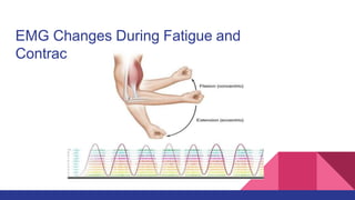
  • 55. EMG changes during fatigue and contarction Measurements were made from the human adductor pollicis muscle of force, contractile speed, and electromyographic activity (EMG) before, during, and after maximal isometric voluntary contractions sustained for 60 s. The use of brief test periods of maximal nerve stimulation with single shocks or trains of shocks enabled various muscle mechanical properties to be studied throughout each contraction. Electrical activity was measured after rectification and smoothing of the surface potentials and also by counting the total number of potentials per unit time from a population of motor units using fine wire intramuscular electrodes. During a 60-s maximal voluntary contraction, the force fell by 30-50%. Throughout the experiment the voluntary force matched that produced by supramaximal tetanic nerve stimulation. This indicated that, with sufficient practice, full muscle activation could be maintained by voluntary effort. However, the amplitude of the smoothed, rectifed EMG and the rate of spike counts declined. Since no evidence for neuromuscular block was found, the decline in EMG and spike counts was attributed to a progressive reduction of the neural drive from the central nervous system, despite maintained maximum effort. After the prolonged voluntary contractions twitch duration was prolonged, mainly as a result of slowing in relaxation rate. Twitch summation in unfused tetani increased.
  • 56. Both the maximum rate of relaxation and the time course of force decay declined by 50-70%. Similar changes were seen in both voluntary contractions and in test periods of stimulation. The percentage change in muscle contractile speed measured by these parameters approximately equaled the percentage change in the surface EMG measured simultaneously. It is concluded that 1) during a 60-s sustained maximal voluntary contraction there is a progressive slowing of contraction speed such that the excitation rate required to give maximal force generation is reduced, 2) the simultaneous decline in EMG may be due to a continuous reduction in motoneuron discharge rate, and 3) the EMG decline may not necessarily contribute to force
  • 57. EMG changes during Fatigue When a muscle cell fires an action potential due to a motor neuron command, this causes a release of calcium (Ca2+) inside the muscle fiber from the sarcoplasmic reticulum. The Ca2+ then flows into the area where the actin and myosin is (the sarcomere), initiating a complex cellular reaction with ATP that allows the myosin to pull on the actin. The movement of myosin pulling on actin in the sarcomeres is called a "sliding filament model" and consists of 4 steps.
  • 59. conti: As long as calcium and ATP are available, the actin and myosin will continue pulling on each other and the twitching will continue. Note that the calcium is rapidly transported back into the sarcoplasmic reticulum where the process must be initiated again by the muscle firing an action potential to cause another twitch. The summing together of many of these incredibly tiny "pulling events" produces a twitch (a very tiny, very fast force). When many twitches occur in a row, the twitches sum together and produce a larger force. ATP is continually provided in the muscle by breaking down glucose (see our "Oxygen Experiment" for an explanation of this metabolism. If glucose isn't available, fatty acids can be used to make pyruvate and keep the Krebs cycle and the oxidative phosphorylation pathway operating. As long as oxygen (O2) is present and can be readily transported to the muscle cell, the oxidative phosphorylation pathway can produce ATP at incredible rates. This is called aerobic contraction, meaning "using oxygen."
  • 60. Dr Akshay Raj Chandra PT Consultant Pediatric Physiotherapist BPT MPT(Neurology) MIAP DCPT Email :physio.akshyarajchandra@gmail.com Phone No : +917827068869