SlideShare una empresa de Scribd logo
1 de 22
Descargar para leer sin conexión
Journal of Manipulative and Physiological Therapeutics   623
                                                                                             Volume 23 • Number 9 • November/December 2000
                                                                                        0161-4754/2000/$12.00 + 0 76/1/110941 © 2000 JMPT


   COMMENTARY

                              A Normal Spinal Position: It’s Time to Accept the Evidence


INTRODUCTION                                                                           Inasmuch as Haas et al7 had many different
   Recent trends in our chiropractic profes-                                            topics in their commentary and did provide
sion seem to be leading away from well-                                                  section titles, it is convenient to respond
ness care into an exclusive focus on short-                                              with reference to those section titles. It is
term care for relief of symptoms, especially                                            noted that some of their section titles are
pain.1 In contrast, some recent articles                                               obscure and certainly not mainstream (eg,
authored by CBP Nonprofit, Inc, researchers                                            their reference to Sackett); nonetheless, the
express an interest in spinal reconstruction,                                      titles are useful as objects for debate. First,
structural outcomes, and care beyond the mere                                  however, we present a logical approach to move-
relief of symptoms.2-6 In a recent commentary, Haas et                   ments in upright posture, from which much about a nor-
al7 have taken exception to this approach.                         mal upright position can immediately be derived.
   A commentary by Haas et al7 concerning one of our                  It is a basic theorem of physics and engineering that the
recent papers8 expressed a paradigm for chiropractic science       movement of any object can be decomposed into rotation,
and patient treatment that is different from that expressed in     translation, and deformation.20 Whereas White and Panjabi21
our recent literature reviews and original publications. Their     have used this theorem to describe the 6 degrees of freedom
views on normal spinal position, radiograph usage, radi-           (DOF) of individual spinal segments (rigid bodies), we have
ograph reliability, and spinal rehabilitation of normal struc-     used this theorem to express all possible movements of the
ture, as expressed in their commentary, did not include            human head, thoracic cage, and pelvis in 3 dimensions.10-11
mechanical engineering principles, which we believe neces-         Figs 1 and 2 are reprinted from a previous article in the
sary for understanding the stresses and strains in abnormal        Journal of Manipulative and Physiological Therapeutics.22
or asymmetric loading of spinal tissues.                           These movements will form the basis on which we illumi-
   In 1998, we had discussed a number critical flaws in 8          nate a normal postural position.
commonly held beliefs espoused by some diplomate chiro-               After providing a review of normal upright position in
practic radiologists.8 Thus, given the fact that the “conven-      terms of the engineering principles and literature reviews to
tional wisdom” of chiropractic radiologists was challenged,        be presented below, analysis of chiropractic manipulations
it was not surprising that there were a total of 8 authors and     (which are mostly torsional loads) will lead the reader to con-
consultants who contributed to the rebuttal commentary of          clude that diversified manipulation is inadequate for obtain-
Haas et al.7 What was not expected was the divergence into a       ing a structural change in the neutral resting posture. Thus,
critical analysis of Chiropractic Biophysics (CBP) methods         precise postural setups (such as those used in the CBP tech-
and the Harrison spinal model,9 which is used as an anatom-        nique) are recommended for the sake of obtaining structural
ical outcome for patients receiving CBP-based treatment.           correction in a patient’s spine after the relief of symptoms.
However, we are pleased to both address those raised con-
cerns and present our rebuttal to Haas et al’s misconceptions      Biologic Plausibility and Validity
about the use of radiography in chiropractic clinical practice.       Haas et al1 defined “biologic plausibility” for us. They
   Because this normal spinal model was only self-published        appear to have assumed that the only important “biologic
until 1992,10-12 some have denied the existence of the             process” is back pain, and on the basis of that view they
Harrison normal spinal model and its implications for physi-       assume that it is unnecessary to address the upright spinal
ology. These implications were discussed in a short review         configuration under gravity. In addition, they state that our
of Wolff’s law (bone remodels to stress) and Davis’ law (soft      model is “merely a mathematical description of optimal
tissue remodels to stress) for abnormal sagittal spinal config-     stress on a static system”.7 We now reply that back pain is a
urations.3 Because this Harrison model has recently been           multifactorial condition. The process of spinal degeneration
published in the indexed literature,13-19 its existence can no     and abnormal biomechanics’ causing mechanical distor-
longer be denied or ignored.                                       tions of the central nervous system (CNS) is better charac-
                                                                   terized as a degenerative disease process. Thus, symptoms
doi:10.1067/mmt.2000.110941                                        appear after the disease process is well advanced (as is the
624   Journal of Manipulative and Physiological Therapeutics
      Volume 23 • Number 9 • November/December 2000
      Commentary • Harrison et al


                                                           Fig 1. Rotational DOF of global body parts.


  case in heart disease, cancer, and hypertension). In such                       the entire oculovestibular, muscle spindle, and mechanore-
  processes, the emphasis is placed on controlling risk fac-                      ceptor systems perform this function from moment to
  tors. It is suggested that optimizing the spine’s position to                   moment in upright stance. To imply that this is an unimpor-
  resist the compressive force of gravity is a logical place at                   tant subject regarding spinal mechanics is to ignore a major
  which to address an “optimal stress” risk factor. Of course,                    function of the CNS.
Journal of Manipulative and Physiological Therapeutics   625
                                                                                        Volume 23 • Number 9 • November/December 2000
                                                                                                              Commentary • Harrison et al


                                         Fig 2. Translational DOF of global body parts.


  Haas et al7 believe that there is no compelling evidence to     ing the spine. A model is used to predict nature; it is a tool.
indicate that subluxation exists. However, subluxation has        Our model’s usefulness will be determined in future studies
been precisely defined through use of deviations from normal       of clinical significance. For such studies to be carried out,
upright posture and our normal spinal model.23 In addition,       the parameters of the model’s usage must first be established,
Haas et al do not appear to appreciate the concept of model-      and one of these parameters is a measurement method—ie,
626       Journal of Manipulative and Physiological Therapeutics
          Volume 23 • Number 9 • November/December 2000
          Commentary • Harrison et al


      A                                            B                                 C                               D


          Fig 3. A, Vertical position of normal AP posture as agreed on in literature. B, D, Proposed ideal lateral postures. C, Average lateral
          posture. Note that differences between Fig 3, B and Fig 3, C are slight.


  posture and radiography. Therefore, determining the reliabili-              ciated with asymmetric muscle efforts. Numerous elec-
  ty of the radiographic markings (and posture reliability) is a              tromyographic studies over the past 4 decades add support
  logical first step. Haas et al claim that we cannot address                 for this fact.24-34 Because many clinicians7 may be unfamiliar
  “clinically meaningful conditions,” but they want to stop us                with engineering principles and aspects of upright human
  from attempting such future research by negating our                        stance, 2 other “proofs” are presented.
  attempts to study the reliability of our methods at the begin-                 As a first discussion of normal posture, the readers and
  ning. Clinical significance can be determined only after one                 Haas et al7 are asked to consider that all chiropractic, osteo-
  has precise definitions to test and only after one has reliabili-            pathic, physical therapy, and medical colleges teach a plumb
  ty data established for the measuring methods.                              line analysis and global ranges of motion, as depicted for
     Because a normal postural position (outside alignment)                   ±Tx, ±Ry, and ±Rz in Figs 1 and 2. With regard to the AP view,
  and a normal spinal position (inside alignment) are funda-                  the following 3 questions come immediately to mind: (1)
  mental to our approach to “controlling risk factors” for spinal             axial rotation to the left and to the right—from where? (2)
  dysfunction, a review pertaining to each of these ideas is pro-             lateral flexion to the left and to the right—from where? (3)
  vided to enable the reader to determine whether Haas et al’s                lateral translation to the left and to the right—from where?
  paradigm matches the biomechanical/biomedical literature.                      Before trying to give an answer that is different from ver-
                                                                              tical alignment in the AP view, recall that these main postur-
  Normal Upright Posture                                                      al motions of the head and thoracic cage, ±Tx, ±Ry, and ±Rz,
     There are at least 3 approaches by which one can immedi-                 cause spinal coupling patterns for which the movements are
  ately discover a normal spinal alignment in the anteroposteri-              known.22,23,35,36 These vertebral coupling patterns are
  or (AP) view and the lateral view. Most rigorously, equilibri-              always described as movement away from a vertical normal
  um equations in the AP view can be used to prove that the                   spinal alignment. Our research team is the first to report
  postures of ±Tx, ±Ry, and ±Rz in Figs 1 and 2 result in abnor-              spinal coupling for ±Tx of the head and thoracic cage.35,36
  mal asymmetric loads on the spinal tissues and lower extrem-                   Some authors have argued that slight deviations of the
  ities when these postures are in the neutral resting posture of             upper thoracic spine are correlated to the side of the domi-
  the subject under investigation. These postures are also asso-              nant arm37 and that a true vertical spine in the AP view there-
Journal of Manipulative and Physiological Therapeutics   627
                                                                                          Volume 23 • Number 9 • November/December 2000
                                                                                                                Commentary • Harrison et al


fore cannot be considered normal. Either sight deviations             Therefore, the vertical alignment of the global centers
can be ignored because of the minimal amount of asymmet-           of mass in upright posture in both the AP and lateral views
ric loading, or unilateral upper extremity exercises on the        has been statistically established for young adults. Fig 3
nondominant side can be prescribed to correct these. In            illustrates this alignment with some additional references
addition, Haas et al7 claim that because of the natural asym-      applied to the location of the shoulders and lower extrem-
metry of the spinous processes, the sensibility of a true verti-   ities.
cal spine in the AP view is questionable. However, more
than 2 decades ago, Farfan38 found that the entire vertebral       Harrison Spinal Model
architecture in the lumbar spine will change and keep the             Through use of multiple studies and normal distributions
lamina junction in line with the structural center of the verte-   with an ideal mean normal center, normal upright posture
bral body. This means that the center of mass will remain          has been defined in the literature. This normal posture in the
approximately the same. Farfan38 writes (pages 34-5): “It          AP view correlates with a straight (vertically aligned) spine.
would appear that in the development of the vertebra, asym-        In the lateral view of upright normal posture, it is generally
metrical body growth is compensated for by asymmetric              accepted that there exist a cervical lordosis, a thoracic
growth of the neural arch.” Therefore, the centers of mass in      kyphosis, and a lumbar lordosis; however, neither the over-
the AP view will still be in line with vertical gravity to mini-   all sagittal curvatures of the cervical, thoracic, and lumbar
mize loads on the spinal tissues and to be the origin position     areas nor the segmental contributions are generally stated or
for postural movements.39-47                                       agreed on. Therefore, when normality is being debated, only
   Recently, Kiefer et al48 showed that T1, T12, and S1 must       the overall geometric shape and segmental angulation of the
be vertically aligned in the lateral view. In the lateral view,    cervical, thoracic, and lumbar sagittal curvatures are open to
the previous argument can be applied to the movements of           debate.
±Rx, ±Ty, and ±Tz in Figs 1 and 2. In fact, much research has         This is where our normal Harrison Spinal Model enters
been conducted on sagittal posture, especially anterior head       the literature. Both our average (400 subjects) and our ideal
translation (+Tz) and pelvic posture, by physical therapists.49-   normal sagittal cervical spine models were published in
61
   This research indicates that anterior head translation (pro-    1996.14 Our average (derived from 552 subjects) normal
trusion) is fundamentally accepted as abnormal and that, in        lumbar model was published in 1997.18 Our ideal normal
general, posture evaluation is repeatable and reliable.            lumbar model was published in 1998.13
   For a second analysis, a statistical evaluation is present-        At this point, it is important to point out that the
ed. As always, there is debate in the literature concerning        Harrison Spinal Model is the normal path of the posterior
average normal and ideal normal (upright posture).                 longitudinal ligament along the posterior vertebral bodies.
However, the differences in these upright positions are            This ideal and statistical average model has a mean and a
small. In 1973 and 1975, Beck and Killus62,63 used a com-          random error component (variation around the mean). It is
puter analysis to average upright lateral and upright AP           a model not of the entire anatomy of the spine but only of
postures of several hundred young adults. They reported            the curvature in the sagittal plane. Models of the entire
on the reliability of their digitizing methods and showed          spine require huge computer codes in which finite ele-
that posture was reproducible “even though the x-rays              ments methods are used.
were taken years apart.” They critically analyzed all of the          The paradigm presented by Haas et al7—that abnormal
previous reports of posture (such as those of Staffel, 64          posture has no effect on physiology—is quite puzzling. It is
Goff, 65 Leger, 66 Haglund, 67 Osgood, 68 and Wiles &              well documented that abnormal spinal loads (forces and
Lond, 69 which include such terms as flat back, round              moments) over time cause pathoses. This is taught in all chi-
back, and hollow back). Beck and Killus 62 found highly            ropractic college physiology courses as Wolff’s law (bone
pathologic postures, small sample sizes, incorrectly               remodels to stress) and Davis’ law (soft tissues remodel to
applied statistics, and lack of an origin for measurement          stress).
included in previous categories of posture. Using a digi-             In the light of the foregoing preceding background data
tizer, they plotted, in graphic form, the coordinates (paths)      and ideas, a response to each of Haas et al’s topics7 can now
of the vertebral bodies of all subjects in the AP and lateral      be presented.
views. With 1500 subjects in their 1975 report, 63 they
noted that they had “one normal distribution in every point        Clinical Sensibility and Validity
of abscissa.” They write:                                              Cervical kyphosis is not a normal variant. In their commentary,
                                                                   Haas et al7 repeat the common misconception that cervi-
     It follows that there is only one ideal type of spinal col-
  umn. According to our findings the theory of constitutional      cal kyphosis is a normal variant. In general, the studies
  types (of posture) cannot be confirmed.62                         cited by Haas et al 7 are inadequate, being characterized
                                                                   by inappropriate methods, flawed statistical analysis (if
  Beck and Killus provided separate graphs of their maxi-          any), and/or unsupported conclusions. It is our intent to
mum points of their normal distributions of posture. The           review the 6 studies 70-75 mentioned by Haas et al and to
spine had the 3 spinal curves with vertical alignment of T1,       present an updated literature review focused on cervical
T12, and S1 in the lateral view.                                   kyphosis.
628   Journal of Manipulative and Physiological Therapeutics
      Volume 23 • Number 9 • November/December 2000
      Commentary • Harrison et al


     To begin, Haas et al7 refer to a 1986 study by Gore et al.70    less than 3 weeks’ duration before the past 2 months or had
  In 200 randomly selected asymptomatic subjects, Gore et al         pain in the 2-month period immediately before the study. In
  found an average of 22 degrees of lordosis using Ruth              addition, the sample size was extremely small and subjects
  Jackson’s angle from C2-C7; they found that a minority             in the two groups were not matched for age, weight, height,
  (9%) of these subjects demonstrated the presence of a cervi-       or sex.
  cal kyphosis. In contrast, Harrison et al76 found that 35% of         Furthermore, Haas et al7 state that “in a study of 180 sub-
  250 symptomatic subjects had kyphotic deformities. These           jects, Borden et al concluded that a straight cervical spine is
  data indicate a dramatic increase in cervical kyphosis in          abnormal only in conjunction with decreased cervical flexi-
  symptomatic subjects in comparison with asymptomatic               bility.” In actuality, the study by Borden et al73 found that of
  subjects. An additional concern is the cross-sectional study       180 asymptomatic subjects, 3 (1.7%) had a reversed cervical
  design used by Gore et al. It is common knowledge among            curve and 13 (7.2%) had a straight cervical spine. Of the 13
  health care providers that several life-threatening illnesses      subjects with a straight cervical spine, 11 (85.0%) had con-
  and diseases remain asymptomatic throughout their course           comitant degenerative changes. In the 3 subjects with cervi-
  or are associated with no overt signs or symptoms until the        cal kyphosis, “there were generalized hypertrophic degener-
  end stages. Harrison et al3 have previously reviewed the           ative changes and fusion.”73 Borden et al concluded that
  shortcomings of the study by Gore et al.70                         “roentgenographically demonstrated loss of the cervical
     The second article used by Haas et al7 in their assertion       curve, combined with normal thoracic kyphosis and lumbar
  that kyphosis is a normal variant is the study by Maimaris et      lordosis, may be indicative of abnormality of the cervical
  al,71 who retrospectively assessed 67 asymptomatic and 35          spine.”73 In the light of this information, one might wonder
  symptomatic whiplash-injured patients. Follow-up was               how Haas et al7 could possibly believe that this reference
  attained an average of 2 years after the injury. Maimaris et al    supports the position that cervical hypolordosis or kyphosis
  concluded that “reversal of the cervical spine to a kyphotic       is a normal variant. Perhaps they were performing secondary
  position was not associated with prolonged disability."            referencing and did not read the original paper.
  However, there are several problems with this study. First,           Likewise, Haas et al7 believe that the study by Marshall
  only 39 (58%) of the 67 asymptomatic subjects were "clini-         and Tuchin74 indicates “that spinal curve alteration itself
  cally examined," whereas 30 (86%) of the 35 symptomatic            may not require treatment.” However, there are several
  subjects were examined. This is a large difference in the per-     potential problems with the method used by Marshall and
  centage of subjects examined in each group, and it can lead        Tuchin.74 The most serious of these is their choice of the C1-
  to inaccurate data analysis between the groups. Second, the        C7 Cobb angle for lateral cervical curve mensuration. The 4-
  lateral cervical spine radiographs were not measured and           line C1-C7 Cobb angle is not a valid representation of the
  quantified; they were merely classified into lordotic, straight,     cervical lordosis.8,77 Therefore, without a segmental analy-
  or kyphotic. Therefore, no quantifiable statistical analysis       sis, no conclusions can be made concerning the shape and/or
  was performed to actually address the issue of the amount or       magnitude of the cervical lordosis and its clinical signifi-
  type of lateral cervical curvature as it relates to the presence   cance in that study.
  of symptoms. As an example, on page 396, Maimaris et al               Last, Haas et al7 rely on the conclusions of Gay.75 In a
  present two figures, one representing a kyphotic cervical          1993 review, Gay concluded that cervical kyphosis is a nor-
  curve and the other representing "loss of the normal lordo-        mal variant. However, before Haas et al’s7 commentary, the
  sis." If one looks at these two radiographs, it is readily         vast majority of the references cited by Gay had previously
  apparent that both of these are kyphotic curvatures. Last, in      been critically analyzed and had been found to be inappro-
  their Table III on page 395, Maimaris et al present the per-       priate by us in 1998.8 Thus, no weight can be applied to
  centage of subjects in each group with radiographic abnor-         Gay’s75 review.
  malities. Clinically significant differences are noted between         Recent and past mechanical, clinical, and surgical studies
  the symptomatic and asymptomatic groups, with the symp-            concerning the cervical lordosis do not support the position
  tomatic group having higher percentages of straight and            that cervical kyphosis is a normal variant. Haas et al7 seem
  kyphotic curves and the asymptomatic group having the              to insinuate that the only supporting reference that we8
  highest percentage of lordotic curves.                             offered for the clinical significance of a normal cervical lor-
     Similarly, Haas et al7 make the claim that Pedersen72           dosis was by Cote et al78 (our old reference 1). This clearly
  “observed that cervical hypolordsis may be a normal vari-          was not the case (see references 9-17 in our original manu-
  ant.” However, the study by Pedersen is of insufficient quali-      script8). However, because Haas et al and many others con-
  ty for the drawing of such a conclusion. Pedersen compared         tinue to debate this issue, a thorough review of the literature
  a “treatment” group of only 9 patients with a “control” group      on cervical kyphosis and its effects is presented here.
  of 17 patients. The criterion for inclusion in the control            It is relevant that several studies have investigated and
  group was as follows: “The subject must not have had any           linked altered cervical curve configurations to the presence
  history of cervical or cervico-thoracic symptoms prior to 2        of chronic headache pain.79-81 In a survey of more than 6000
  months before entry (lasting continuously for more than 3          cases of chronic headache-sufferers, Braaf and Rosner79
  weeks).” This designation was a poor choice, inasmuch as           found that “complete or segmental loss or reversal of the
  subjects in the control group could either have had pain of        normal lordotic curve of the cervical spine is the most con-
Journal of Manipulative and Physiological Therapeutics   629
                                                                                        Volume 23 • Number 9 • November/December 2000
                                                                                                              Commentary • Harrison et al


sistent characteristic feature and very often is the only          method is important because the standard error of measure-
abnormality found.” In 47 subjects with tension and                ment (SEM) is much lower (<3) and the segmental analysis
migraine headaches, Vernon et al80 found a high incidence          finds alterations within the curve—ie, it is not just evalua-
of hypolordosis, straightened cervical curve configurations,        tion of the end segments.
and reversed cervical curve configurations. Nagasawa et al81           According to Haas et al,7 the surgical studies just cited,
compared 372 patients with tension headaches and 225 con-          all of which indicate the clinical significance of the cervical
trol subjects matched for age and sex; they found statistical-     lordosis, are not appropriate because surgical patients are
ly significant differences between the two groups, the             “not typical chiropractic patients who require conservative
headache patients having straightened cervical curve config-        care.” But perhaps this is merely an excuse for not accepting
urations and low set shoulders. In addition, with increasing       the facts.
age, the headache patients’ cervical curve was straight more          However, there are several nonsurgical studies indicating
frequently. This information contrasts nicely with the find-       cervical kyphosis as a factor predictive of poor results.98-100
ings of Gore et al,70 who noted that in asymptomatic sub-          In a long-term follow-up of 146 whiplash-injured patients,
jects, the cervical curve increased with age.                      Hohl98 identified cervical kyphosis as factor predicting a
   Looking into the matter further, we note that when poor out-    poor outcome. Norris and Watt99 followed 61 patients
comes after surgery are considered, several studies have found     involved in motor vehicle accidents for a minimum of 6
a good cervical lordosis to be a significant factor in preventing   months; they found abnormal neck curves to be “more com-
neurologic deficit and/or compromise.82-93 For example, Goto        mon in patients with a poor outcome.”99
and Kita82 state that “postoperative kyphotic malalignment or         In addition to clinical outcomes of pain and neurologic
angulation are factors strongly affecting the outcome of           deficits, several papers focusing on cervical biomechanics
surgery.” Yamazaki et al83 compared 2 groups of patients with      indicate that cervical kyphosis is abnormal.101-103 Using an
ossification of the posterior longitudinal ligament (PLL) post-     engineering analysis of slope and bending moment,
operatively. Group I consisted of patients with no anterior        Matsunaga et al101 proved that degeneration and consequent
“compression” of the spinal cord, and group II comprised           subluxation will develop in abnormal kyphotic configura-
patients with residual cord “compression.” The investigators       tions, which they termed buckling alignments. “In the case
found a lordosis of less than 10.0 degrees or cervical kyphosis    of the S-shaped spine,” they write, “the spine is in a ‘buck-
correlated with the presence of postoperative spinal cord com-     ling’ condition, which corresponds to an unreasonable sup-
pression; this result was statistically significant.                porting function.”
   Similarly, recent surgical studies have emphasized that            With regard to ossification of the PLL, Matsunaga et al102
operations that leave the patients’ cervical spines with less      followed 101 patients for a 5-year period. Using an engi-
lordosis are associated with increased neck, upper thoracic,       neering analysis of strain, they found that ossification of
and shoulder pain and overall poorer health outcomes.94-97         PLL progressed in the areas showing uneven and abnormal
Lowry94 used the average lordosis of 22 degrees found by           strain at the adjacent intervertebral disk and PLL. Spe-
Gore et al70 to gauge a successful surgical outcome; he            cifically, strain in the tensile direction was found to corre-
claimed that “a fixed kyphotic posture of the mid-cervical         late with ossification of the PLL. It is important to note that
spine will lead to overall sagittal imbalance manifested by        the only way to increase the tensile strain in the PLL is to
axial pain in the upper to mid-thoracic region as well as in       have concomitant hypolordosis or kyphosis.104
the posterior cervical spine.” Katsuura et al95 compared 44           Several other studies have indicated that loss of cervical
cases treated with anterior cervical plating (a method of          lordosis or kyphosis is a cause of or is associated with a
ensuring a stable lordotic correction) to 30 patients receiv-      high incidence of degenerative changes in the disk and ver-
ing anterior fusion without plating. These patients were fol-      tebral bodies.70,73,98,105 Because degenerative changes of the
lowed for an average of 38 months postoperatively. The             cervical spine are a known cause of morbidity,98,99,106 it can
magnitude of cervical lordosis was assessed through use of         be stated with confidence that cervical kyphosis is not a nor-
Ruth Jackson’s angle (posterior tangents from C2-C7), and          mal variant.
intersegmental (2-line) Cobb angles were used for the align-          In a recent 1997 text on the spine, statements by
ment of the fused segments. Katsuura et al found statistical-      Frymoyer et al109 are clinically significant about cervical
ly significant differences in the incidence of neck pain           kyphosis as a normal variant. In fact, they assert that
between the two groups, the plated group having less neck          “kyphosis, straightening, and hyperlordosis of the cervical
pain and a statistically significant increase in cervical lordo-    spine may all be seen in conjunction with spondylotic
sis. Likewise, Kawakami et al96 retrospectively evaluated 60       myelopathy, whereas in other patients the normal curvature
patients postsurgically. Cervical lordosis was measured with       is preserved” and that “kyphosis and hyperlordosis may
a 4-line Cobb’s method from C2-C7 and a segmental (2-              coexist and thus this gives rise to an S-shaped deformity of
line) Cobb angle at the fused level. Segmental kyphosis at         the spine, causing greater instability and management prob-
the level of the fusion and less lordotic curvature were found     lems.”109 Furthermore, in a 1996 review on the cervical
to statistically predict which patients would develop “axial       spine surgeries, Lowery94 states that “an additional feature
symptoms” (neck pain, shoulder pain, and/or stiff neck).           of any plate system, whether anterior or posterior, should be
The use of 2-line Cobb methods rather than the 4-line Cobb         to establish a lordotic cervical curvature.” The evidence
630   Journal of Manipulative and Physiological Therapeutics
      Volume 23 • Number 9 • November/December 2000
      Commentary • Harrison et al


  against76-107 cervical kyphosis as a normal variant far out-      subjects, Bernhardt and Bridwell123 reported a mean Cobb
  weighs the evidence to the contrary.7,70-75                       for T3-T12 of ~36 degrees and a mean Cobb for T1-T12 of
     Finally, with regard to the cervical spine, it is noted that   ~40 degrees. Jackson and McManus124 investigated sagittal
  many authors (including Haas et al) have neglected to inves-      balance in 100 patients and 100 volunteers; they reported the
  tigate cervical lordosis and child development. It should not     same means and ranges for males and females and for
  have been necessary to present the preceding review indicat-      patients and volunteers (mean T1-T12: ~43 ± 9 degrees). In
  ing that cervical kyphosis is not a normal variant, inasmuch      1998, Vedantam et al125 investigated standing sagittal align-
  as Bagnall et al108 showed in 1977 that the cervical lordotic     ment in 88 adolescents (aged 10-18 years); they reported a
  curve is formed in intrauterine life (9.5 weeks). Cervical lor-   “striking similarity in regional thoracic kyphosis and lumbar
  dosis could thus be considered a primary curve, like the tho-     lordosis between adolescents and adults.”
  racic kyphosis. Any loss of cervical lordosis is due to trau-        In 1980, Fon et al126 reported that in 316 subjects, thoracic
  ma, repetitive play, or work postural loadings during and/or      kyphosis increased with age. In 1993, Cutler et al127 reported
  after birth. Importantly, using skin markers or surface visual-   that hyperkyphosis in women was associated with altered
  ization does not correlate with the shape, magnitude, or          vertebral body shape (anterior wedging), loss of bone densi-
  direction of the sagittal cervical spine.109,110 Therefore, the   ty, loss of fitness, decreased muscle strength, and reduced
  use of radiography for identification of any abnormal lateral      survival; the investigators suggested a correlation between
  cervical configuration is absolutely mandatory.                    abnormal upright thoracic posture and changing architecture
                                                                    of the spinal column (Wolff’s law). In 1996, Kolessar et al128
  Thoracic Kyphosis                                                 reported that 69 patients had kyphotic Cobb T5-T12 mea-
     In addition to the work of Beck and Killus,62 whose nor-       surements of >33 degrees, whereas all 24 volunteers had
  mal graph used a statistical average, multiple studies have       Cobb T5-T12 < 33 degrees.
  investigated thoracic kyphosis. As in cervical skin contour          It is interesting to note that studies on thoracic kyphosis
  versus radiographic measurements, D’Osualdo et al111 found        with large population groups provide nearly normal distri-
  considerable disagreement between radiographic findings           butions. Thus, as noted by Beck and Killus,62-63 statistically
  and results obtained with an Arcometer. In 1983, Willner and      there would be one ideal normal at the mean of these distrib-
  Johnson112 used spinal pantographs based on skin contour to       utions. With the few reported values for Cobb T1-T12 and
  suggest that kyphosis varied in girls and boys. However,          with ratios of values from Cobb T2-T12, T3-T12, T4-T12,
  using an ISIS scanner for evaluating back shape, Carr et al113    and T5-T12, the mean thoracic kyphotic value seems to be
  reported no statistically significant differences with respect    between 40 and 50 degrees for a Cobb angle from the top of
  to age and sex in children, no statistically significant differ-   T1 to the bottom endplate of T12.
  ences with respect to age and sex in adults, and no differ-
  ences between children and adults. Using radiography,             Lumbar Lordosis
  Voutsinas and MacEwen114 reported in 1986 that “the differ-          Haas et al7 have neglected a large body of literature sup-
  ences between boys and girls were not significant in any          porting the clinical significance of a normal lumbar lordo-
  group.”                                                           sis.13,18,19,116,129-158 To date, there have been 10 separate
     Some studies have reported a large range of values for         studies in which a similar method was used to investigate
  thoracic kyphosis—from 20 degrees to 40 degrees.115-120           the normal shape and magnitude of the human lumbar lor-
  One problem in deciphering a normal average thoracic              dosis.18,115-117,122,129-133 These 10 studies used strict criteria
  kyphosis is the inability to see T1-T4 on most lateral tho-       in categorizing men and women as normal subjects. The
  racic views; this has caused different levels of Cobb angle       age range for the subjects was large—between 10 and 80
  analysis in the literature—with different angles of kyphosis,     years of age. All 10 of these investigations used radi-
  of course. For example, the following levels have been used:      ographic analysis in the neutral upright posture, and total
  T5-T12, T4-T12, T3-T12, T2-T12, and T1-T12. This causes           as well as segmental lumbar lordotic angles were mea-
  wide ranges of values. For example, Propst-Proctor and            sured. In all, a total of 755 normal subjects from 10 sepa-
  Bleck117 measured the inferior endplate of T5 to the superior     rate studies were included in the analysis. All of these stud-
  endplate of T12 in 104 normal children who were referred          ies18,115-117,122,129-133 indicate that the upper lumbar curve,
  for suspected screening of scoliosis; they reported a normal      T12-L3, is minimally lordotic whereas the distal lumbar
  distribution with a range of 21 to 33 degrees and a mean of       curve, L4-S1, accounts for approximately 65.0% of the
  27 degrees.                                                       lumbar lordosis.
     In 22 cases, Singer et al120 reported no difference in tho-       Haas et al7 claim (believe) that different patient popula-
  racic kyphosis in subjects before and after death (T4-T12:        tions (races) have different optimal spinal configurations and
  ~49 degrees).120 Milne and Williamson121 reported no sig-         that “given the natural variation and asymmetry in the
  nificant change in thoracic curvature on follow-up radi-          human musculoskeletal system,” an ideal spinal model is not
  ographs of 261 subjects aged 62-90 years. In 99 Greek men         plausible.
  and women, Korovessis et al122 reported that thoracic kypho-         Their beliefs are not supported by recent anthropometric
  sis was not gender related, though kyphosis was found to          clinical and experimental data on the lumbar spine. For exam-
  increase with age (mean T4-T12: ~36 ± 9 degrees). In 102          ple, several studies addressing the shape and magnitude of the
Journal of Manipulative and Physiological Therapeutics   631
                                                                                           Volume 23 • Number 9 • November/December 2000
                                                                                                                 Commentary • Harrison et al


lumbar lordosis found no differences with respect to geo-          adult patients with symptomatic degenerative lumbar disk
graphic location and/or ethnicity.133-137 In a radiographic        disease, 30 patients with low-grade L5-S1 isthmic spondy-
study of the lumbar lordosis in 16 healthy Chinese men,            lolisthesis, and 30 patients with idiopathic or degenerative
Chen133 concluded that “no obvious interracial differences         scoliosis. Again, statistically significant differences
were found in the geometric data found in this study.”             between the groups were found in the radiographic mea-
Mosner et al134 compared the lumbar lordotic spinal radi-          surements.138
ographs of 25 black women with those of 27 white women;               Adams et al142 followed 403 volunteer health care work-
no significant differences were found in the lumbar lordosis,       ers for a 3-year period. A decreased lumbar lordosis was
as measured with spinal radiographs, though differences            found to be one of the personal risk factors in predisposing
were found in external skin contours. This difference in skin      subjects to a first-time episode of low back pain requiring
contour “gives a false impression of increased lumbar lordo-       medical evaluation. Itoi143 analyzed lateral lumbar radi-
sis”134 in black versus white women. In a 1985 study of 973        ographs in 100 postmenopausal female patients with spinal
adults, Fernand and Fox135 reported that “black and white          osteoporosis; a statistically significant increase in low back
populations demonstrated no significant difference in the          pain was found in patients with a rounded thoracic spine
mean lumbar lordotic angle.” Korovessis et al136 studied the       and lower acute lumbar kyphosis. This lower lumbar kypho-
sagittal configuration of the lumbar spine in 120 asympto-         sis has been shown to cause an increase in low back pain,
matic Greek volunteers. “We have shown that there are no           perhaps as a result of increased electromyographic activity
differences in the sagittal profile across geographically          of the paraspinal musculature.144
and ethnically different populations,” they write. As regards         Many surgical papers have identified both lumbar hyper-
the “natural variation” of human anatomy, Janik et al13 have       lordosis145 and hypolordosis or kyphosis146-152 as risk fac-
shown that reported small differences in vertebral                 tors for low back pain requiring surgical correction. Last,
body–to–disk height ratios, found in different studies, have       several investigators have found statistically significant
no effect on their elliptical model of the lumbar lordosis (ie,    increases or associations between loss of lumbar lordosis
none of the segmental angles, T12-S1, were found to change         and degeneration of the disk, vertebral body, and lig-
more than 1 degree).                                               aments.152-156
   As regards low back pain and lumbar lordosis, 6 separate           With regard to lumbar lordosis, most experts agree that
studies have found statistically significant differences           “the assessment of spinal curvature is a part of an evalua-
between patients with low back pain and matched normal             tion in patients with back problems”.157 Because surface
control subjects.19,136,138-141 Korovessis et al136 compared       measurement of spinal curvature represents neither seg-
homogenous groups of 120 volunteers and 120 patients with          mental nor total lumbar lordotic alignment, radiographic
chronic low back pain; subjects were matched for age, sex,         analysis is a mandatory part of the routine examination of
and ethnicity. Lordosis was measured through use of a 4-line       the lumbar spine.134,158 We believe that Haas et al’s7 nega-
Cobb’s method from T12 to S1, and segmental angles (2-line         tion of the importance of the structural alignment of the
Cobb) were measured through use of endplate lines relative         spine stems from a neglect of mathematics and engineering
to horizontal. Statistically significant differences were found     analysis. They seem to be reading published papers with
between the two groups, the chronic low back pain subjects         just rudimentary 4-line Cobb angle geometry while ignor-
having a decreased distal lordosis, L4-S1, in comparison           ing papers that analyze spinal alignment with a slightly
with the controls.                                                 higher level of mathematics. Papers that use segmental
   Harrison et al19 used elliptical modeling to discriminate       analysis (see the references above), some analytic geome-
between a rigorously defined normal group and 3 separate           try,13,19 calculus,103,120,153 curve fitting,9,159-161 and finite
groups of patients with low back pain; that study included         elements162-164 have shown the importance of normal
(1) 50 normal subjects from a pre-employment physical              alignment of the spine.
screening, (2) 50 patients with acute low back pain (first
occurrence; less than 6 weeks’ duration), (3) 50 patients with     Pelvis Orientation
chronic low back pain (pain of more than 6 weeks’ duration           Plaugher et al165 have investigated pelvic radiographic
or a history of recurrent disabling low back pain), and (4) 24     measurements and found reproducibility in pelvic position-
subjects with low back pain and radiographically verified          ing and radiographic marking. Troyanovich et al166 have
degenerative disorders or pathoses. These 4 groups were            established high intraclass coefficients (ICCs) and low
matched for age, weight, height, and sex. The investigators        SEMs for pelvic tilt on the lateral lumbopelvic view.
found that “of 11 angles, 2 distances, and 2 ratios, statistical   Jackson et al138 have reported high Pearson correlation
analysis was significantly different across groups for 12 of       coefficients for pelvic measurements (in degrees) on lateral
these measurements.”19 The chronic low back pain and lum-          full spine views; they reported significant differences in
bar pathosis group had a decreased lordosis, whereas the           pelvic measurements for normal subjects versus 3 patient
acute low back pain group had an increased lordosis in com-        groups.
parison with the normal subjects.                                    In 1995, Saji et al167 studied 61 adolescents with idio-
   In a similar study, Jackson et al138 compared sagittal lum-     pathic scoliosis and 33 normal subjects for femoral neck
bar radiographs of 50 normal volunteers with those of 50           shaft angles and asymmetry of the hips. An angle was
632      Journal of Manipulative and Physiological Therapeutics
         Volume 23 • Number 9 • November/December 2000
         Commentary • Harrison et al


      Table. Partial review of x-ray reliability studies: ICCs and standard errors of measurement for intraobserver error and interobserver
      error (in degrees)

                                                                  Standard error of measurement (degrees)
                                                  ICC                   4-line Cobb               2-line Cobb         Mod. Risser-Ferg     Post. tangent
       Area/Reference                     Intra         Inter          Intra       Inter          Intra     Inter     Intra      Inter      Intra   Inter
       AP cervical                          —         —                 —           —              —         —         —         —           —       —
        Troyanovich et al                 >0.90     >0.90               —           —              —         —        <2.0      <1.5         —       —
       AP thoracic                          —         —                 —           —              —         —         —         —           —       —
        Pruijs et al (1994)               (Spearman > 0.98)             —        3.2, 2.0*         —         —         —         —           —       —
        Sevastikoglou, Bergquist            —         —                 —           3.1            —         —         —         —           —       —
        Beekman, Hall                       —         —                 —          4.2*            —         —         —         —           —       —
        Gross et al (p176)                  —         —                 3.2         2.3            —         —         —         —           —       —
        Morrissy et al                      —         —              4.9, 3.8,   7.2, 6.3,         —         —         —         —           —       —
                                                                        2.8         6.3
        Carman et al                        —           —               —          3.8*            —         —         —         —           —       —
        Goldberg et al                      —           —               1.9         2.5            —         —         —         —           —       —
        Desmet et al                        —           —               —        1.1, 1.3*         —         —         —         —           —       —
       AP lumbar                            —           —               —           —              —         —         —         —           —       —
        Troyanovich et al                 >0.70       >0.80             —           —              —         —        <2.0      <2.0         —       —
       Lateral cervical                     —           —               —           —              —         —         —         —           —       —
        Phillips et al (1999)               —         >0.83             —           —              —       <2.7†       —         —           —       —
        Singer et al5                       —           —               4.5         —              —         —         —         —           —       —
        Plaugher et al                      —           —              2.9†        4.2†            —         —         —         —           —       —
        Cote et al                          —         >0.90             —        9.1, 8.3‡         —         —         —         —           —       —
        Jackson et al                     (Pearson r > .80)             —           —              —         —         —         —           1.2     1.2
        Harrison et alSpine               >0.70       >0.80             —           —            3.6, 3.2 2.2, 2.7‡    —         —           2.3     2.4
       Lateral thoracic                     —           —               —           —              —         —         —         —           —       —
        Carman et al                        —           —               —          3.3*            —         —         —         —           —       —
        Voutsinas, MacEwen                  —           —              2.2*         —              —         —         —         —           —       —
        Jackson et al (1998)              (Pearson r > .82)             —           —              —         —         —         —           —       —
       Lateral lumbar                       —           —               —           —              —         —         —         —           —       —
        Jackson et al (1998)              (Pearson r > .73)             —           —              —         —         —         —           —       —
        Polly et al (1996)                >0.81       >0.83            10.0        10.0            —         —         —         —           —       —
        Voutsinas, MacEwen                  —           —              2.1*         —              —         —         —         —           —       —
        Gelb et al                          —           —               —           —             3.0*      3.0*       —         —           —       —
        Wood et al                          —           —               —           —              —        1.1†       —         —           —       —
        Troyanovich et al                 >0.70       >0.80             —           —              —         —         —         —           1.5     1.7
        Troyanovich et al                 >0.70       >0.80             —           —              —         —         —         —           1.7     1.8

        ICC, Intraclass coefficient; Intra, intraobserver error; Inter, interobserver error.
        *Only average difference was reported (not standard error of measurement).
        †Standard error of measurement was reported.
        ‡First value is for C1-C7; second value is for C2-C7.


  formed by lines through the femoral shaft and neck. Intra-                                 ic analysis of spinal displacement.” We have published 5
  observer and interobserver reliability were assessed. There                                radiographic line drawing analysis reliability studies and
  were no significant differences between the measurements                                   have another in review.77,166,169-172 In other chiropractic pub-
  of a single observer or between the measurements of differ-                                lications, the reliability of line drawing analysis on the nasi-
  ent observers. Standard errors of measurement were small                                   um view and pelvis, additional AP, and lateral views have
  (intraobserver, 1.6 degrees; interobserver, 1.9 degrees). Saji                             been reported.165,173-176 Curiously, in regard to the nasium
  et al discovered that a normal femoral neck-shaft angle was                                and AP lumbopelvic radiographic views, Haas et al make the
  approximately 135 degrees, the scoliosis subjects having a                                 following inaccurate statement: “There are no grounds for
  much higher value for this angle (straightened femur).                                     generalizing from studies on pelvic height and the cranium
     Levine and Whittle168 studied the effects of pelvic tilt on                             to the repeatability of analyzing spinal curves and segmental
  lumbar lordosis. They reported that flexion of the pelvis                                  displacement.”7 Perhaps Haas et al are unaware that the nasi-
  increased lumbar lordosis and that extension of the pelvis                                 um radiograph includes the cervical spine and that its align-
  was associated with hypolordosis. Similarly, Harrison et al19                              ment in comparison with the “cranium” has been subjected
  reported that their subjects with acute low back pain had                                  to several reliability studies.173,174
  flexed pelvises and lumbar hyperlordosis whereas their sub-                                    For the various Cobb methods (2-line and 4-line), numer-
  jects with chronic pain had extended pelvic positions and                                  ous reliability studies have been done on AP lumbar, AP tho-
  lumbar hypolordosis.                                                                       racolumbar, and AP thoracic, lateral lumbar, lateral thoracic,
                                                                                             and lateral cervical views.9,159-161,177-192 Furthermore, the
  Reliability                                                                                measurement of pelvic rotation and sagittal segmental trans-
     Haas et al7 make the following statement: “To date, there                               lations has been reported as reliable.193-195 In 1992, Capasso
  is little convincing evidence for the reliability of radiograph-                           et al196 reported high Pearson correlation (r > 0.90) for both
Journal of Manipulative and Physiological Therapeutics   633
                                                                                      Volume 23 • Number 9 • November/December 2000
                                                                                                            Commentary • Harrison et al


“intra and intergroup correlations among orthopedic sur-            Recently, computer-aided spinal radiographic measure-
geons” for 3 types of spondylolisthesis radiographic mea-        ments have been shown to be highly reliable. For example,
surements. Similar to Tallroth et al’s194 findings for lumbar    in 1996, Rosol et al207 reported a high Pearson correlation
flexion/extension, the findings of Lind et al197 for cervical    for vertebral morphometry (r = 0.96) and low mean coeffi-
flexion/extension were high examiner reliability and low         cients of intraobserver variability (4%) and interobserver
intraobserver error of measurement (<1.8 degrees) for later-     variability (2%).
al cervical segmental flexion angles. Gross et al198 (1983)         We have already mentioned the recent (1998) study of
and Goldberg et al199 (1988) revisited AP Cobb angle relia-      sagittal balance with normal subjects and 3 patient groups
bility and reported smaller errors of measurement (between       conducted by Jackson et al.138 They reported high Pearson
1.9 and 3.2 degrees) than most studies. Similarly, DeSmet et     correlation coefficients in the vast majority of their interob-
al200 reported small interobserver errors of the differences     server and intraobserver reliability measures (13 measures
(<1.2 degrees) for Cobb angles.                                  in their Tables 8 and 9, most being greater than 0.75).138 In
   The multitude of studies just cited (a total of 35) report-   addition to the pelvic measurements, they also reported sig-
ing high radiographic reliability (those with ICCs report the    nificant differences in the normal subjects and the 3 patient
majority of ICCs to be in the >0.75 range) is sufficient to      groups for a multitude of lateral spine measurements. In
establish the “reliability of radiographic analysis of spinal    1997, Hardacker et al208 studied lateral cervical reliability
displacement” to the satisfaction of any well-informed           with segmental Cobb angles and reported only 3 values of
reader.                                                          interobserver and intraoberver Pearson correlations less
   Perhaps the high variability of SEM (ranging between 3        than 0.83 for 8 measurements of segmental lordosis (0.64 <
and 10 degrees) with the 4-line Cobb methods are of con-         r < 0.92).
cern. Inasmuch as the Harrison posterior tangent method has         In 1994, Pruijs et al209 used a Spearman correlation coef-
smaller SEMs (<2 degrees), this method is suggested to           ficient for measurements of Cobb angles on AP thoracic
Haas et al7 as well as to clinicians and other researchers for   radiographs. They reported high coefficients (>0.98) for
the analysis of the sagittal spine.                              both interobserver and intraobserver variation with small
   However, until recently (the 1990s), most investigators       SDs of differences (<3.2 degrees).
reported only means, SDs, and SDs of differences in read-           In 1996, Levine and Whittle168 measured pelvic tilt and
ings and rereadings (Table). Because the SEM has SD in its       lumbar lordosis using a television/computer system that
formula, the low means and SDs for measurement differ-           obtained 3-dimensional (3-D) coordinates of markers on the
ences indicate good-to-excellent reliability. However, before    pelvis and spine at 20-ms intervals. ICCs for 3 readings of
1990, authors rarely provided proper statistical analyses        pelvic tilt and lumbar lordosis were 0.78 < ICCs < 0.95. The
with Pearson r, intrarater ICC, interrater ICC, 95% CI, and      investigators reported that altering the pelvic tilt significant-
SEM values. The vast majority of medical investigators have      ly changed the angle of lumbar lordosis.
stated that the low means and SDs for differences in drawing        In 1995, Saji et al167 studied 61 idiopathic scoliosis ado-
and redrawing radiographic lines (Table) suffice for a claim      lescents with 33 normal subjects for femoral neck shaft
of adequate reliability. After all, it is merely Euclidean       angles and asymmetry of the hips. An angle was formed by
geometry!                                                        lines through the femoral shaft and neck. They also per-
   In 1999, Phillips et al201 reported high correlation coeffi-   formed Cobb angle measurements. Intraobserver reliability
cients (0.83, 0.91, 0.89, 0.91, 0.88, 0.87) and low SEMs         and interobserver reliability were assessed; there were no
(2.45 degrees, 2.71 degrees, 1.69 degrees; 1.22 mm, 1.32         significant differences between the measurements of a single
mm, 1.48 mm) when measuring the occipital-C2 angle and           observer or between the measurements of different
distances on neutral, flexion, and extension radiographs. For     observers. SEMs were small (intraobserver, 1.6 degrees;
measuring spinal canal dimensions and cord size in a 1999        interobserver, 1.9 degrees). In addition, the investigators
review article, Rao and Fehlings202 reported that 7 studies      reported a significant difference in the obtuse femoral angles
provided interobserver and intraobserver reliability of radi-    in normal versus scoliotic subjects, the latter having much
ographic measurements (0.82 < ICC < 0.99). In 1997, with         larger obtuse angles. In 1985, Tibrewal and Pearcy210
means and SDs for measurement differences on AP thoracic         assessed measurement of disk heights from lateral radi-
views, it was reported that the rib vertebral angle could be     ographs. They reported a maximum error of 0.7 mm for
reliably measured (intraobserver mean, 4.4 degrees; interob-     intraobserver and interobserver measurements.
server mean, 3.6 degrees).203                                       Since 1990, many chiropractic studies have included sta-
   In 1996, Omeroglu et al204 reported that “both intraobserv-   tistical analyses that are extensive in comparison with those
er and interobserver error risks were insignificant” for use of   of previous studies. For our 6 CBP radiography reliability
the Perdriolle torsionmeter to measure vertebral axial rota-     studies, we consulted with Burt Holland, PhD, a professor in
tion in scoliosis on AP radiographs.204 In 1994, Gilliam et      the Department of Statistics at Temple University. With his
al205 reported high ICCs for both intertester (ICC > 0.86) and   aid, our studies have reported complete statistical analyses
intratester (ICC > 0.92) pelvic radiographic measurements.       with a majority of intraobserver and interobserver ICCs
Also in 1993, Hamberg et al206 reported that “the x-ray mea-     greater than 0.75 and small SEMs (<2 degrees). It is interest-
surements showed high reliability” for pelvic tilt analysis.     ing to note that ICC > 0.60 is the set of values that Haas et
634   Journal of Manipulative and Physiological Therapeutics
      Volume 23 • Number 9 • November/December 2000
      Commentary • Harrison et al


                                                          Fig 4. Four types of loads with inherent stresses.


  al7 have declared to be in the good-to-excellent range for                       more than Euclidean geometry, which is the basis of every
  chiropractic studies.211 This range is close to the excellent                    human-made structure in our industrial lives.
  range suggested by Shrout and Fleiss212 (poor < 0.4; 0.4 <
  fair to good < 0.75; excellent > 0.75).                                          Posture Reproducibility, Reliability, and Validity
     It would be a great surprise to every mathematician,                             There is in the literature a plethora of posture reliability
  physicist, and engineer to find that radiographic line draw-                     and reproducibility studies performed by ergonomists, med-
  ings of all types are not highly reliable. After all, it is nothing              ical doctors, chiropractors, and physical therapists. Posture
Journal of Manipulative and Physiological Therapeutics   635
                                                                                               Volume 23 • Number 9 • November/December 2000
                                                                                                                     Commentary • Harrison et al


A                                                                                    B
              Fig 5. A, Diversified technique is limited; lumbar roll is a torsional load applied to lumbar segments by
              twisting pelvis relative to fixed thoracic cage. B, Cervical rotary-break is a torsional load applied to head in
              lateral flexed and forward flexed positions. These loads are applied to every subject; patient’s abnormal
              pretreatment posture is disregarded.


has been studied with patients lying,213 sitting,214 stand-               Last, inasmuch as Fialka-Moser et al255 reported that
ing,215 lifting,216 working,217 walking,218 engaging in daily          adolescents’ postural faults have been established as the
routine performance,219 and running.220 Postural stress has            cause of pain syndromes in adulthood, we are bewildered
been correlated with scoliosis,221 office work,222 work-relat-          that chiropractic state licensing boards are being influenced
ed lifting injuries,223 driving,224 sitting,225 space flight,226       by the paradigm promoted by Haas et al7 and others, as
sports injuries,227 and back pain.228 Posture as a valid out-          cited in a recent review.8 For example, in August 1999 the
come of care is universally accepted by almost all health care         Nevada Board of Chiropractic Examiners told practitioners
sciences.229-231 Virtually every body part has been studied for        that it was against Board ruling to take radiographs of chil-
its posture in normal subjects versus injured individuals;             dren and that posttreatment radiographs were unwarranted.
included are the postures of the head,232 shoulders,233 upper          The selective information (countered in our preceding
extemity,234 trunk,235 pelvis,236 lower extremity,237 and short        review) generally generated and disseminated by chiroprac-
leg,238 as well as the whole body posture.239-251 The vast             tic radiologists and academicians is adversely affecting chi-
majority of these studies report high interobserver and                ropractic clinicians’ ability to provide the best possible ser-
intraobserver reliability. In 1990, in a study of 86 Chinese           vice to the patients who seek our care.
subjects, Dai and Gu252 found no significant differences in
posture for males and females and no significant differences            Clinical Utility and Appropriateness
in posture between groups of old-aged subjects and middle-                Haas et al’s7 negation of structure (spinal alignment) as
aged subjects with eyes open, though they did find a differ-           a clinical outcome seems to be based on a few controlled
ence in posture between groups of old-aged subjects and                clinical trials that failed to demonstrate the effectiveness
middle-aged subjects with eyes closed. In a 1997 study of              of chiropractic manipulation in conditions such as asth-
160 men and women, Raine and Twomey253 found no signif-                ma260 and nocturnal enuresis.261 These studies used spinal
icant gender differences for each of head and shoulder pos-            manipulation as the intervention (treatment). Because
ture measurements. For a review of the normal alignment of             manipulation seemed to be beneficial for headaches, neck
the lower extremities, the reader is referred to the 1996              pain, and back pain but not for other diseases or spinal dis-
review by Riegger-Krugh and Keysor,254 who noted that                  placement, Haas et al prefer to see chiropractic as merely a
pathology of structures results from skeletal malalignment.            temporary treatment regimen for the relief of spinal aches
   Adolescents’ postural faults have been established as the           and pains.
cause of pain syndromes in adulthood.255 Anatomical short                 There are many flaws in this line of reasoning, and most
leg inequality has received much attention in the literature.          of these flaws are derived from a complete void in an engi-
True anatomical leg length inequality causes a recogniz-               neering education. At least two topics can be applied to
able abnormal posture. For a review, the reader is referred            Haas et al’s faulty reasoning: (1) exclusive use of torsional
to the 1983 review by the American Academy of Osteo-                   loads in spinal manipulation and (2) neglect of CNS biome-
pathy256 and to recent articles by Beaudoin et al257 and               chanics. This whole discussion of posture and radiographic
Keppler et al.258                                                      line drawing reliability/validity omits a very important clin-
   In terms of validity, posture as a valid outcome in the             ical factor when any of the postural rotations and transla-
health care sciences is well accepted. In fact, the editor of          tions in Figs 2 and 3 are present for long periods, especially
this Journal has made the following statement: “That pos-              flexed segments in the cervical and lumbar areas.
ture can lead to low back pain will come as a surprise to no           Deformations of the CNS, which result from abnormal pos-
one.”259                                                               tural loads, were recently reviewed in this Journal.262-264
636       Journal of Manipulative and Physiological Therapeutics
          Volume 23 • Number 9 • November/December 2000
          Commentary • Harrison et al


      A                                                                                    B


      Fig 6. In these examples, torsional loads make subjects’ posture worse. A, Patient’s initial posture is left head lateral flexion with con-
      tralateral right thoracic bending (low shoulder at right). B, Patient’s head is right laterally translated with contralateral translation of
      thoracic cage. Y-axis torsional loadings (seen in Fig 5) will not correct this posture and will in fact add other abnormal postural loads.

  Thus, of the two topics, only that of torsional loads in spinal             loads. After being exposed to some anatomy and ranges of
  manipulation needs to be presented here.                                    motion, any engineer can visualize the stresses (forces/area)
                                                                              and strains (measurements of deformation) in these postures.
  Torsional Loads                                                             For example, in lateral flexion, the flexural stresses are com-
     Let us revisit the postural DOF in Figs 1 and 2, which are               pression on the concave fibers and tension on the convex
  based on the engineering concepts of rotations and transla-                 fibers. For the paraspinal tissues, these stresses/strains are
  tions in 3 dimensions. There are 12 single movements in 6                   directly associated with Wolff’s law and Davis’ law, especial-
  DOF for each of the head, thoracic cage, and pelvis.                        ly for the application of these abnormal loads over long peri-
  Through use of permutations in probability theory, it can be                ods. The previous discussion now illuminates a glaring defi-
  determined that there are 728 possible postures (singles,                   ciency in applying diversified loadings to patients’ spines.
  doubles, triples, . . . sextuples) of each of the head, thoracic               The vast majority of diversified maneuvers are torsional
  cage, and pelvis in 3 dimensions, for a total of 7283 or                    loads (Fig 5). It might seem that these loadings will have a
  385,828,352 possible upright human postures!12 This is such                 probability of being only 11% effective (of the 18 DOF illus-
  an overwhelming number that it is best to restrict our discus-              trated in Figs 1 and 2, head and thoracic torsions are only 2
  sion to the individual postures depicted in Figs 1 and 2.                   DOF). However, when considering the total permutations of
  These 36 postures (12 singles each for head, thoracic cage,                 posture, these diversified torsional loads are 4 out of
  and pelvis) can be recategorized into the 4 types of loads                  385,828,352 possible human postures, which amounts to
  taught in junior-level engineering courses on the mechanics                 approximately 0% of specificity in matching the patient’s
  of materials (Fig 4).                                                       abnormal posture. Haas et al7 seemingly do not understand
     In mechanics of materials, engineers study the material                  the necessity of matching the patient’s individual posture
  properties, design, loadings, deflections, stresses, and strains             because they do not have a realization that these (rotated and
  of structures.265 The y-axis rotations in Fig 1 are caused by               translated postures) are mathematical functions in linear
  torsional loads, whereas the x-axis (flexion/extension) and z-               algebra, which is fundamental to an engineering education.
  axis (lateral flexion) rotations are examples of pure bending.                  Linear algebra is the study of linear transformations.266
  The y-axis translations are examples of axial loads, whereas                There are 4 important linear transformations in 3-D geometry:
  the x-axis and z-axis translations are examples of transverse               (1) rotations, (2) reflections, (3) projections, and (4) transla-
Journal of Manipulative and Physiological Therapeutics   637
                                                                                          Volume 23 • Number 9 • November/December 2000
                                                                                                                Commentary • Harrison et al


tions. Because of a complete void in chiropractic education,          However, the complete void of mechanical engineering
clinicians and academicians are unaware that rotations, reflec-     in their reasoning has led them to a paradigm of chiropractic
tions, and translations have unique inverses (ie, only one load    treatment that we do not accept. Their paradigm of treat-
will correct them) and that rotations, translations, and reflec-    ment, based on torsional loads, is detrimental to the
tions are the only 3-D rigid body movers (this is a theorem in     patient’s well-being in the longitudinal sense. They seem to
mathematics). From linear algebra, one deduces that there          be interested only in temporary “patient happiness out-
must be a normal (origin) from which to measure the displace-      comes”7 of chiropractic care, whereas we are interested in
ments caused by rotations, reflections, and translations.           the long-term effects of abnormal postural loads. We find it
   From the study of linear algebra, it becomes apparent that      paramount to measure the patient’s structure on the outside
y-axis torsional loads are inappropriate interventions in the      (posture) and use radiography for the segmental alignments
vast majority of patients. Actually, in most cases (ie, most       on the inside (spine). The measurements gathered are used
postural positions of the patient), y-axis torsional loads make    to uniquely determine the specific interventions chosen for
the patient’s posture worse (adding a new position not pre-        each individual patient. We cannot accept the paradigm of
sent in the initial posture). For the sake of brevity, we pro-     applying general torsional loads to every patient.
vide only 2 examples.                                                                                     Deed E. Harrison, DC
   Suppose the new patient has either head lateral flexion                                       Private Practice of Chiropractic
to the right and thoracic lateral flexion to the left or con-                                                  123 Second Street
tralateral lateral translations of the head and rib cage (Fig                                                   Elko, NV 89801
6). A cervical rotary break, as in Fig 5, A, will cause                                           Donald D. Harrison, DC, PhD
increased lateral flexion and the addition of head rotation,
                                                                                                    Stephan J. Troyanovich, DC
whereas a lumbar roll will not correct the thoracic lateral
bending to the left but will add a y-axis rotation posture to                                           Stacy Harmon, DC, MD
the patient. The torsion stresses cause deformation of the
mechanoreceptors in the disks, ligaments, and muscle               REFERENCES
spindles (these reflexes reduce pain), but they do not alter         1. Haldeman D, Peterson D, Chapman-Smith D. Guidelines for
the initial and ongoing postural loads on the subject’s                 chiropractic quality assurance and standards of practice.
paraspinal tissues. (This simple example, like the preced-              Gaithersburg, Md: Aspen Publishers; 1992.
ing discussion on outside posture, neglects the configura-           2. Harrison DD, Jackson BL, Troyanovich SJ, Robertson GA,
tions of the sagittal cervical, thoracic, and lumbar curves             DeGeorge D, Barker WF. The efficacy of cervical exten-
                                                                        sion-compression traction combined with diversified
on the inside.)                                                         manipulation and drop table adjustments in the rehabilita-
                                                                        tion of cervical lordosis. J Manipulative Physiol Ther
CONCLUSION                                                              1994;17:454-64.
   A thorough review of normal upright posture, normal               3. Harrison DD, Troyanovich SJ, Harrison DE, Janik TJ, Murphy
spinal modeling, structural spinal alignment as a clinical              DJ. A normal sagittal spinal configuration: a desirable clinical
                                                                        outcome. J Manipulative Physiol Ther 1996;19:398-405.
outcome, and radiograph reliability has been provided. It            4. Troyanovich SJ, Harrison DD. Chiropractic biophysics
is up to the reader to evaluate whether it is the present               (CBP) technique. Chiropr Technique 1996;8:1-6.
authors or Haas et al7 who (1) have been guilty of “omit-            5. Troyanovich SJ, Harrison DE, Harrison DD. Review of the sci-
ting of relevant studies, the misrepresentation of evidence,            entific literature relevant to structural rehabilitation of the spine
and the disregard of the rules of evidence”7 and/or (2) may             and posture: rationale for treatment beyond the resolution of
                                                                        symptoms. J Manipulative Physiol Ther 1998;21:37-50.
have presented a “‘bits and pieces’ approach to the litera-          6. Troyanovich SJ, Harrison DD, Harrison DE. Low back pain
ture, selective extracting and manipulating that which                  and the lumbar intervertebral disc: clinical considerations for
could be construed to be supportive of their position and               the doctor of chiropractic. J Manipulative Physiol Ther
ignoring the rest.”267 The reader may compare the litera-               1999;22:96-104.
ture cited by Haas et al7 and Morgan267 (a total of 49 refer-        7. Haas M, Taylor JAM, Gillete RG. The routine use of radi-
                                                                        ographic spinal displacement analysis: a dissent. J Mani-
ences) with the literature cited in this review and a 1998              pulative Physiol Ther 1999;22:254-9.
review 8 (a total of 434 references) to determine whether            8. Harrison DE, Harrison DD, Troyanovich SJ. Reliability of
published studies have been misrepresented and/or                       spinal displacement analysis on plane X-rays: a review of
neglected.                                                              commonly accepted facts and fallacies with implications for
   A detailed response to the criticisms of Haas et al con-             chiropractic education and technique. J Manipulative Physiol
                                                                        Ther 1998;21:252-66.
cerning biological plausibility, clinical sensibility, and so on     9. Harrison DD, Janik TJ, Harrison GR, Troyanovich SJ,
has been provided here. Finally, Haas et al and Morgan have             Harrison DE, Harrison SO. Chiropractic biophysics tech-
questioned our integrity and have become personal in their              nique: a linear algebra approach to posture in chiropractic. J
attack on our credibility. For example, Morgan267 claims that           Manipulative Physiol Ther 1996;19:525-35.
“the Harrison et al approach to the topic of spinal displace-       10. Harrison DD. CBP technique: the physics of spinal cor-
                                                                        rection. National Library of Medicine #WE 725 4318C,
ment appears much less as research and much more as pro-                1982-97.
motion.” Science requires critique of ideas, but personal           11. Harrison DD. Spinal biomechanics: a chiropractic perspective.
attacks have no place in a scientific debate.                            National Library of Medicine #WE 725 4318C, 1982-97.
638    Journal of Manipulative and Physiological Therapeutics
       Volume 23 • Number 9 • November/December 2000
       Commentary • Harrison et al


      12. Harrison DD. Abnormal postural permutations calculated as          32. Joseph J, Nightingale A. Electromyography of muscles of
          rotations and translations from an ideal normal upright static         posture: thigh muscles in males. J Physiol 1954;126:81-5.
          spine. In: Sweere JJ, editor. Chiropractic family practice.        33. Joseph J, Nightingale A, Williams PL. A detailed study of the
          Gaithersburg, Md: Aspen Publishers; 1992. p 6-1:1-22.                  electric potentials recorded over some postural muscles while
      13. Janik TJ, Harrison DD, Cailliet R, Troyanovich SJ, Harrison            relaxed and standing. J Physiol 1955;127:617-25.
          DE. Can the sagittal lumbar curvature be closely approximat-       34. Joseph J, Nightingale A. Electromyography of muscles of
          ed by an ellipse? J Orthop Res 1998;16:766-70.                         posture: leg and thigh muscles in women, including the
      14. Harrison DD, Janik TJ, Troyanovich SJ, Holland B.                      effects of high heels. J Physiol 1956;132:465-8.
          Comparisons of lordotic cervical spine curvatures to a theo-       35. Harrison DE, Cailliet R, Harrison DD, Janik TJ, Troyanovich
          retical ideal model of the static sagittal cervical spine. Spine       SJ, Coleman RR. Lumbar coupling during lateral translations
          1996;21:667-75.                                                        of the thoracic cage relative to a fixed pelvis. Clin Biomech
      15. Harrison DD, Janik TJ, Troyanovich SJ, Harrison DE,                    1999;14:704-9.
          Colloca CJ. Evaluations of the assumptions used to derive an       36. Harrison DE, Cailliet R, Harrison DD, Janik TJ, Troyanovich
          ideal normal cervical spine model. J Manipulative Physiol              SJ. Cervical coupling during lateral translations of the head.
          Ther 1997;20:246-56.                                                   Clin Biomech 2000;15:436-40.
      16. Harrison DD, Janik TJ. Clinical validation of an ideal normal      37. Williams PL, Warwick R. Grays anatomy. 36th ed.
          static cervical spine model. In: Witten M, editor.                     Philadelphia: WB Saunders; 1980.
          Computational medicine, public health, and biotechnology, 2.       38. Farfan HP. Mech disorders of the low back. Philadelphia: Lea
          Austin, Tex: World Scientific Publishing; 1995. p 1047-55.              & Febiger; 1973. p 34-5.
      17. Janik TJ, Harrison DD. Prediction of 2-D static normal posi-       39. Woodhull AM, Maltrud K, Mello BL. Alignment of the
          tion of the cervical spine from mathematical modeling. In:             human body in standing. Eur J Appl Physiol 1985;54:109-15.
          Witten M, editor. Computational medicine, public health, and       40. Wells KF, Luttgens K. Kinesiology: scientific basis of human
          biotechnology, 2. Austin, Tex: World Scientific Publishing;            motion. 6th ed. Philadelphia: WB Saunders; 1976.
          1995. p 1036-46.                                                   41. Hellebrandt FA, Tepper RH, Braun CL, Elliott MC. The loca-
      18. Troyanovich SJ, Cailliet R, Janik TJ, Harrison DD, Harrison            tion of the cardinal anatomical orientation planes passing
          DE. Radiographic mensuration characteristics of the sagittal           through the center of weight in young adult women. Am J
          lumbar spine from a normal population with a method to                 Physiol 1938;121:465-70.
          synthesize prior studies of lordosis. J Spinal Disord 1997;        42. Hellebrandt FA, Hirt S, Fries EC. Centre of gravity of the
          10:380-6.                                                              human body. Arch Phys Ther 1944;25:465-70.
      19. Harrison DD, Cailliet R, Janik TJ, Troyanovich SJ, Harrison        43. May J. The placement of the gravity line on the human body
                                                                                 in the anterior-posterior plane and its relationship to posture
          DE, Holland B. Elliptical modeling of the sagittal lumbar lor-
                                                                                 by roentgenoscopic study [unpublished thesis]. Ames, Iowa:
          dosis and segmental rotation angles as a method to discrimi-
                                                                                 State University of Iowa; 1955.
          nate between normal and low back pain subjects. J Spinal
                                                                             44. Smidt GL, Day JW, Gerleman DG. Iowa anatomical position
          Disord 1998;11:430-9.
                                                                                 system: a method of assessing posture. Eur J Appl Physiol
      20. Cowin S. Bone mechanics. Boca Raton, Fla: CRC Press;
                                                                                 1984;52:407-13.
          1989. p 37.
                                                                             45. Kapandji AI. The physiology of the joints. Vol 3. New York:
      21. White AA, Panjabi MM. Clinical biomechanics. Phila-
                                                                                 Churchill Livingstone; 1974. p 15.
          delphia: Lippincott; 1978.                                         46. Kendall HO, Kendall FP, Boynton DA. Posture and pain.
      22. Harrison DE, Harrison DD, Troyanovich SJ. Three-dimen-                 Baltimore: Williams & Wilkins; 1952.
          sional spinal coupling mechanics, I: a review of the literature.   47. Kuchera M. Gravitational stress, musculoligamentous strain,
          J Manipulative Physiol Ther 1998;21:101-13.                            and postural alignment. Spine: State of the Art Reviews
      23. Harrison DE, Harrison DD, Troyanovich SJ. Three-dimen-                 1995;9:463-89.
          sional spinal coupling mechanics, II: implications for chiro-      48. Kiefer A, Shirazi-Adl A, Parnianpour M. Synergy of the
          practic theories and practice. J Manipulative Physiol Ther             human spine in neutral postures. Eur Spine J 1998;7:471-9.
          1998;21:177-86.                                                    49. Hanten WP, Lucio RM, Russell JL, Brunt D. Assessment of
      24. Carlsoo S. The static muscle load in different work positions:         total head excursion and resting head posture. Arch Phys Med
          an electromyographic study. Ergonomics 1961;4:193-211.                 Rehabil 1991;72:877-80.
      25. Klausen K. The form and function of the loaded human spine.        50. Less M, Eihelberg W. Force changes in neck vertebrae and
          Acta Physiol Scand 1965;65:176-90.                                     muscles. In: Komi P, editor. Biomechanics. Vol VA. Baltimore:
      26. Nachemson A, Elfstrom G. Intravital dynamic pressure mea-              University Park Press; 1976. p 530-36.
          surements in lumbar discs: a study of common movements,            51. Ferguson D. Posture aching and body build in telephonists. J
          maneuvers and exercises. Scand J Rehabil Med 1970;1                    Hum Ergol 1976;5:183-86.
          (Suppl):1-40.                                                      52. Biraune W, Fischer O. On the center of gravity of the human
      27. Andersson BJG, Ortengren R, Nachemson A, Elfstrom G.                   body. New York: Springer-Verlag; 1985.
          Lumbar disc pressure and myoelectric back muscle activity          53. Vital JM, Senegas J. Anatomical bases of the study of the
          during sitting, I: studies on an experimental chair. Scand J           constraints to which the cervical spine is subject in the sagit-
          Rehabil Med 1974;6:104-14.                                             tal plane: a study of the center of gravity of the head. Surg
      28. Basmajian JV, Bentzon JW. Electromyographic study of cer-              Radiol Anat 1986;8:169-73.
          tain muscles of the leg and foot in the standing position. Surg    54. McKenzie RA. The cervical and thoracic spine: mechanical
          Gynecol Obstet 1954;98:662-6.                                          diagnosis and therapy. Waikanae, New Zealand: Spinal
      29. Basmajian JV. Electromyography of iliopsoas. Anat Rec                  Publications, Ltd; 1990.
          1958;130:267.                                                      55. Garrett TR, Youdas JW, Madson TJ. Reliability of measuring
      30. Floyd WF, Silver PHS. The function of the erector spinae               forward head posture in a clinical setting. J Orthop Sports
          muscles in certain movements and postures in man. J Physiol            Phys Ther 1993;17:155-60.
          1955;129:184-203.                                                  56. Lundstrom F, Lundstrom A. Natural head position as a basis
      31. Joseph J, Nightingale A. Electromyography of muscles of                for cephalometric analysis. Am J Orthod Dentofacial Orthop
          posture: leg muscles in males. J Physiol 1952;117:484-91.              1992;101:244-7.
A Normal Spinal Position: It's Time to Accept the Evidence

Más contenido relacionado

La actualidad más candente

Physiotherapy theory and practice
Physiotherapy theory and practicePhysiotherapy theory and practice
Physiotherapy theory and practiceUdelas Chiriqui
 
Pro disc l replacement vs circumferential arthrodesis of spine
Pro disc l replacement vs  circumferential arthrodesis of spinePro disc l replacement vs  circumferential arthrodesis of spine
Pro disc l replacement vs circumferential arthrodesis of spineQueen Mary Hospital
 
Does a standard outpatient physiotherapy regime improve the range of knee mot...
Does a standard outpatient physiotherapy regime improve the range of knee mot...Does a standard outpatient physiotherapy regime improve the range of knee mot...
Does a standard outpatient physiotherapy regime improve the range of knee mot...FUAD HAZIME
 
Analysis of Spinal Decompression via Surgical Methods and Traction Therapy
Analysis of Spinal Decompression via Surgical Methods and Traction TherapyAnalysis of Spinal Decompression via Surgical Methods and Traction Therapy
Analysis of Spinal Decompression via Surgical Methods and Traction TherapyPaige Barrett
 
Aquatic CAP-DCB-NRM-DCB
Aquatic CAP-DCB-NRM-DCBAquatic CAP-DCB-NRM-DCB
Aquatic CAP-DCB-NRM-DCBNicole Miela
 
Rischke_Viscoelastic Disc Arthroplasty Provides Superior Back and Leg Pain Re...
Rischke_Viscoelastic Disc Arthroplasty Provides Superior Back and Leg Pain Re...Rischke_Viscoelastic Disc Arthroplasty Provides Superior Back and Leg Pain Re...
Rischke_Viscoelastic Disc Arthroplasty Provides Superior Back and Leg Pain Re...Kari Zimmers
 
Early Outcome of Discectomy with Interspinous Process Distraction Device a Re...
Early Outcome of Discectomy with Interspinous Process Distraction Device a Re...Early Outcome of Discectomy with Interspinous Process Distraction Device a Re...
Early Outcome of Discectomy with Interspinous Process Distraction Device a Re...CrimsonPublishersOPROJ
 
12 aaom reeves workshop apr 19 research summary
12 aaom reeves  workshop apr 19   research summary12 aaom reeves  workshop apr 19   research summary
12 aaom reeves workshop apr 19 research summaryNomienredes
 
Aquatic CAP Poster-DCB-NRM
Aquatic CAP Poster-DCB-NRMAquatic CAP Poster-DCB-NRM
Aquatic CAP Poster-DCB-NRMNicole Miela
 
Restorative Invection Therapy.ppt
Restorative Invection Therapy.pptRestorative Invection Therapy.ppt
Restorative Invection Therapy.pptsobramid
 
Pmr buzz magazine april 2021
Pmr buzz magazine april 2021Pmr buzz magazine april 2021
Pmr buzz magazine april 2021mrinal joshi
 
Maladaptive movement and motor control impairments as underlying mechanism
Maladaptive movement and motor control impairments as underlying mechanismMaladaptive movement and motor control impairments as underlying mechanism
Maladaptive movement and motor control impairments as underlying mechanismMeziat
 
Lorangerie 23 10 12 00 Leonardo Kapural (SIMPOSIO OPTIKA).ppt
Lorangerie  23 10  12 00  Leonardo Kapural (SIMPOSIO OPTIKA).pptLorangerie  23 10  12 00  Leonardo Kapural (SIMPOSIO OPTIKA).ppt
Lorangerie 23 10 12 00 Leonardo Kapural (SIMPOSIO OPTIKA).pptsobramid
 
Results of Mini-Open Technique
Results of Mini-Open TechniqueResults of Mini-Open Technique
Results of Mini-Open TechniqueWenjay Sung
 
11. Frozen shoulder and Hydroplasty
11.  Frozen shoulder and Hydroplasty11.  Frozen shoulder and Hydroplasty
11. Frozen shoulder and Hydroplastydrajun
 

La actualidad más candente (20)

Physiotherapy theory and practice
Physiotherapy theory and practicePhysiotherapy theory and practice
Physiotherapy theory and practice
 
Pro disc l replacement vs circumferential arthrodesis of spine
Pro disc l replacement vs  circumferential arthrodesis of spinePro disc l replacement vs  circumferential arthrodesis of spine
Pro disc l replacement vs circumferential arthrodesis of spine
 
Does a standard outpatient physiotherapy regime improve the range of knee mot...
Does a standard outpatient physiotherapy regime improve the range of knee mot...Does a standard outpatient physiotherapy regime improve the range of knee mot...
Does a standard outpatient physiotherapy regime improve the range of knee mot...
 
Analysis of Spinal Decompression via Surgical Methods and Traction Therapy
Analysis of Spinal Decompression via Surgical Methods and Traction TherapyAnalysis of Spinal Decompression via Surgical Methods and Traction Therapy
Analysis of Spinal Decompression via Surgical Methods and Traction Therapy
 
Aquatic CAP-DCB-NRM-DCB
Aquatic CAP-DCB-NRM-DCBAquatic CAP-DCB-NRM-DCB
Aquatic CAP-DCB-NRM-DCB
 
Rischke_Viscoelastic Disc Arthroplasty Provides Superior Back and Leg Pain Re...
Rischke_Viscoelastic Disc Arthroplasty Provides Superior Back and Leg Pain Re...Rischke_Viscoelastic Disc Arthroplasty Provides Superior Back and Leg Pain Re...
Rischke_Viscoelastic Disc Arthroplasty Provides Superior Back and Leg Pain Re...
 
Early Outcome of Discectomy with Interspinous Process Distraction Device a Re...
Early Outcome of Discectomy with Interspinous Process Distraction Device a Re...Early Outcome of Discectomy with Interspinous Process Distraction Device a Re...
Early Outcome of Discectomy with Interspinous Process Distraction Device a Re...
 
Artrodesis hallux
Artrodesis halluxArtrodesis hallux
Artrodesis hallux
 
12 aaom reeves workshop apr 19 research summary
12 aaom reeves  workshop apr 19   research summary12 aaom reeves  workshop apr 19   research summary
12 aaom reeves workshop apr 19 research summary
 
Injury 2014 Guerra
Injury 2014 GuerraInjury 2014 Guerra
Injury 2014 Guerra
 
Aquatic CAP Poster-DCB-NRM
Aquatic CAP Poster-DCB-NRMAquatic CAP Poster-DCB-NRM
Aquatic CAP Poster-DCB-NRM
 
Restorative Invection Therapy.ppt
Restorative Invection Therapy.pptRestorative Invection Therapy.ppt
Restorative Invection Therapy.ppt
 
Pmr buzz magazine april 2021
Pmr buzz magazine april 2021Pmr buzz magazine april 2021
Pmr buzz magazine april 2021
 
GOODAY.ARTICLE.Final
GOODAY.ARTICLE.FinalGOODAY.ARTICLE.Final
GOODAY.ARTICLE.Final
 
Terapia manual e hidratação dos discos
Terapia manual e hidratação dos discosTerapia manual e hidratação dos discos
Terapia manual e hidratação dos discos
 
Maladaptive movement and motor control impairments as underlying mechanism
Maladaptive movement and motor control impairments as underlying mechanismMaladaptive movement and motor control impairments as underlying mechanism
Maladaptive movement and motor control impairments as underlying mechanism
 
Lorangerie 23 10 12 00 Leonardo Kapural (SIMPOSIO OPTIKA).ppt
Lorangerie  23 10  12 00  Leonardo Kapural (SIMPOSIO OPTIKA).pptLorangerie  23 10  12 00  Leonardo Kapural (SIMPOSIO OPTIKA).ppt
Lorangerie 23 10 12 00 Leonardo Kapural (SIMPOSIO OPTIKA).ppt
 
Abstract
AbstractAbstract
Abstract
 
Results of Mini-Open Technique
Results of Mini-Open TechniqueResults of Mini-Open Technique
Results of Mini-Open Technique
 
11. Frozen shoulder and Hydroplasty
11.  Frozen shoulder and Hydroplasty11.  Frozen shoulder and Hydroplasty
11. Frozen shoulder and Hydroplasty
 

Destacado (7)

Evidence Based Protocol for Structural Rehab of Spine
Evidence Based Protocol for Structural Rehab of SpineEvidence Based Protocol for Structural Rehab of Spine
Evidence Based Protocol for Structural Rehab of Spine
 
Hormesis
HormesisHormesis
Hormesis
 
What is Spine Remodeling?
What is Spine Remodeling?What is Spine Remodeling?
What is Spine Remodeling?
 
The challenging issue of low dose radiation hormesis and adaptive response- ...
The challenging issue of low dose radiation  hormesis and adaptive response- ...The challenging issue of low dose radiation  hormesis and adaptive response- ...
The challenging issue of low dose radiation hormesis and adaptive response- ...
 
Stop Suffering From Foot, Knee, Hip, and Back Pain!
Stop Suffering From Foot, Knee, Hip, and Back Pain!Stop Suffering From Foot, Knee, Hip, and Back Pain!
Stop Suffering From Foot, Knee, Hip, and Back Pain!
 
Nuclear Disasters
Nuclear DisastersNuclear Disasters
Nuclear Disasters
 
Fukushima Nuclear Disaster
Fukushima Nuclear DisasterFukushima Nuclear Disaster
Fukushima Nuclear Disaster
 

Similar a A Normal Spinal Position: It's Time to Accept the Evidence

Tensión y Deslizamiento
Tensión y DeslizamientoTensión y Deslizamiento
Tensión y Deslizamientolichugojavier
 
The stabilizing system_of_the_spine_part_1
The stabilizing system_of_the_spine_part_1The stabilizing system_of_the_spine_part_1
The stabilizing system_of_the_spine_part_1alguiz
 
Research 4801-Winter 2015-Sahijwani, Raj-Final
Research 4801-Winter 2015-Sahijwani, Raj-FinalResearch 4801-Winter 2015-Sahijwani, Raj-Final
Research 4801-Winter 2015-Sahijwani, Raj-FinalRaj Sahijwani
 
Steadman Hawkins Sports Medicine Lecture Series: Chiropractic (2006)
Steadman Hawkins Sports Medicine Lecture Series: Chiropractic  (2006)Steadman Hawkins Sports Medicine Lecture Series: Chiropractic  (2006)
Steadman Hawkins Sports Medicine Lecture Series: Chiropractic (2006)Mark J Pitcher DC, Msc CCSP, EMT
 
January 2012 report
January 2012  reportJanuary 2012  report
January 2012 reportecupresident
 
Neuroanatomy in Clinical Context -1 Duane E. Haines (9th Edition).pdf
Neuroanatomy in Clinical Context -1 Duane E. Haines (9th Edition).pdfNeuroanatomy in Clinical Context -1 Duane E. Haines (9th Edition).pdf
Neuroanatomy in Clinical Context -1 Duane E. Haines (9th Edition).pdfDrConan1
 
Cervical Myelopathy Differential Diagnosis
Cervical Myelopathy Differential DiagnosisCervical Myelopathy Differential Diagnosis
Cervical Myelopathy Differential DiagnosisPhysical Therapy Central
 
Paradigm shift in spinal manual therapy
Paradigm shift in spinal manual therapyParadigm shift in spinal manual therapy
Paradigm shift in spinal manual therapyDr.Kannabiran Bhojan
 
Mcq Companionto Applied Radiological Anatomy3 Haxap
Mcq Companionto Applied Radiological Anatomy3 HaxapMcq Companionto Applied Radiological Anatomy3 Haxap
Mcq Companionto Applied Radiological Anatomy3 Haxappgr224
 
Intervertebral Disc Replacement
Intervertebral Disc ReplacementIntervertebral Disc Replacement
Intervertebral Disc ReplacementDr Madhusudhan NC
 
THE ORTHOPAEDIC PHYSICAL EXAMINATION. 2 Ed..pdf
THE ORTHOPAEDIC  PHYSICAL EXAMINATION. 2 Ed..pdfTHE ORTHOPAEDIC  PHYSICAL EXAMINATION. 2 Ed..pdf
THE ORTHOPAEDIC PHYSICAL EXAMINATION. 2 Ed..pdfkeyly fabiana
 
Artificial disc replacement vs ACDF
Artificial disc replacement vs ACDFArtificial disc replacement vs ACDF
Artificial disc replacement vs ACDFPonnilavan Ponz
 

Similar a A Normal Spinal Position: It's Time to Accept the Evidence (20)

Tensión y Deslizamiento
Tensión y DeslizamientoTensión y Deslizamiento
Tensión y Deslizamiento
 
The stabilizing system_of_the_spine_part_1
The stabilizing system_of_the_spine_part_1The stabilizing system_of_the_spine_part_1
The stabilizing system_of_the_spine_part_1
 
Top 10 research breakthroughs to support chiropractic care in the last decade...
Top 10 research breakthroughs to support chiropractic care in the last decade...Top 10 research breakthroughs to support chiropractic care in the last decade...
Top 10 research breakthroughs to support chiropractic care in the last decade...
 
Research 4801-Winter 2015-Sahijwani, Raj-Final
Research 4801-Winter 2015-Sahijwani, Raj-FinalResearch 4801-Winter 2015-Sahijwani, Raj-Final
Research 4801-Winter 2015-Sahijwani, Raj-Final
 
Jospt.1998.27.4
Jospt.1998.27.4Jospt.1998.27.4
Jospt.1998.27.4
 
Steadman Hawkins Sports Medicine Lecture Series: Chiropractic (2006)
Steadman Hawkins Sports Medicine Lecture Series: Chiropractic  (2006)Steadman Hawkins Sports Medicine Lecture Series: Chiropractic  (2006)
Steadman Hawkins Sports Medicine Lecture Series: Chiropractic (2006)
 
January 2012 report
January 2012  reportJanuary 2012  report
January 2012 report
 
Neuroanatomy in Clinical Context -1 Duane E. Haines (9th Edition).pdf
Neuroanatomy in Clinical Context -1 Duane E. Haines (9th Edition).pdfNeuroanatomy in Clinical Context -1 Duane E. Haines (9th Edition).pdf
Neuroanatomy in Clinical Context -1 Duane E. Haines (9th Edition).pdf
 
Cervical Myelopathy Differential Diagnosis
Cervical Myelopathy Differential DiagnosisCervical Myelopathy Differential Diagnosis
Cervical Myelopathy Differential Diagnosis
 
Chiro Cervical Adjustment
Chiro Cervical AdjustmentChiro Cervical Adjustment
Chiro Cervical Adjustment
 
Paradigm shift in spinal manual therapy
Paradigm shift in spinal manual therapyParadigm shift in spinal manual therapy
Paradigm shift in spinal manual therapy
 
PostureTek Full Version
PostureTek Full VersionPostureTek Full Version
PostureTek Full Version
 
PostureTek PPT
PostureTek PPTPostureTek PPT
PostureTek PPT
 
Mcq Companionto Applied Radiological Anatomy3 Haxap
Mcq Companionto Applied Radiological Anatomy3 HaxapMcq Companionto Applied Radiological Anatomy3 Haxap
Mcq Companionto Applied Radiological Anatomy3 Haxap
 
Peoria chiropractor founder
Peoria chiropractor founderPeoria chiropractor founder
Peoria chiropractor founder
 
Intervertebral Disc Replacement
Intervertebral Disc ReplacementIntervertebral Disc Replacement
Intervertebral Disc Replacement
 
THE ORTHOPAEDIC PHYSICAL EXAMINATION. 2 Ed..pdf
THE ORTHOPAEDIC  PHYSICAL EXAMINATION. 2 Ed..pdfTHE ORTHOPAEDIC  PHYSICAL EXAMINATION. 2 Ed..pdf
THE ORTHOPAEDIC PHYSICAL EXAMINATION. 2 Ed..pdf
 
Artificial disc replacement vs ACDF
Artificial disc replacement vs ACDFArtificial disc replacement vs ACDF
Artificial disc replacement vs ACDF
 
Phys ther 2007-cott-925-6
Phys ther 2007-cott-925-6Phys ther 2007-cott-925-6
Phys ther 2007-cott-925-6
 
Curriculum Vitae
Curriculum VitaeCurriculum Vitae
Curriculum Vitae
 

Último

Book Call Girls in Kasavanahalli - 7001305949 with real photos and phone numbers
Book Call Girls in Kasavanahalli - 7001305949 with real photos and phone numbersBook Call Girls in Kasavanahalli - 7001305949 with real photos and phone numbers
Book Call Girls in Kasavanahalli - 7001305949 with real photos and phone numbersnarwatsonia7
 
Call Girls Service Chennai Jiya 7001305949 Independent Escort Service Chennai
Call Girls Service Chennai Jiya 7001305949 Independent Escort Service ChennaiCall Girls Service Chennai Jiya 7001305949 Independent Escort Service Chennai
Call Girls Service Chennai Jiya 7001305949 Independent Escort Service ChennaiNehru place Escorts
 
Hematology and Immunology - Leukocytes Functions
Hematology and Immunology - Leukocytes FunctionsHematology and Immunology - Leukocytes Functions
Hematology and Immunology - Leukocytes FunctionsMedicoseAcademics
 
Call Girls Thane Just Call 9910780858 Get High Class Call Girls Service
Call Girls Thane Just Call 9910780858 Get High Class Call Girls ServiceCall Girls Thane Just Call 9910780858 Get High Class Call Girls Service
Call Girls Thane Just Call 9910780858 Get High Class Call Girls Servicesonalikaur4
 
Russian Call Girls Gunjur Mugalur Road : 7001305949 High Profile Model Escort...
Russian Call Girls Gunjur Mugalur Road : 7001305949 High Profile Model Escort...Russian Call Girls Gunjur Mugalur Road : 7001305949 High Profile Model Escort...
Russian Call Girls Gunjur Mugalur Road : 7001305949 High Profile Model Escort...narwatsonia7
 
Call Girls Whitefield Just Call 7001305949 Top Class Call Girl Service Available
Call Girls Whitefield Just Call 7001305949 Top Class Call Girl Service AvailableCall Girls Whitefield Just Call 7001305949 Top Class Call Girl Service Available
Call Girls Whitefield Just Call 7001305949 Top Class Call Girl Service Availablenarwatsonia7
 
Call Girl Bangalore Nandini 7001305949 Independent Escort Service Bangalore
Call Girl Bangalore Nandini 7001305949 Independent Escort Service BangaloreCall Girl Bangalore Nandini 7001305949 Independent Escort Service Bangalore
Call Girl Bangalore Nandini 7001305949 Independent Escort Service Bangalorenarwatsonia7
 
Noida Sector 135 Call Girls ( 9873940964 ) Book Hot And Sexy Girls In A Few C...
Noida Sector 135 Call Girls ( 9873940964 ) Book Hot And Sexy Girls In A Few C...Noida Sector 135 Call Girls ( 9873940964 ) Book Hot And Sexy Girls In A Few C...
Noida Sector 135 Call Girls ( 9873940964 ) Book Hot And Sexy Girls In A Few C...rajnisinghkjn
 
call girls in green park DELHI 🔝 >༒9540349809 🔝 genuine Escort Service 🔝✔️✔️
call girls in green park  DELHI 🔝 >༒9540349809 🔝 genuine Escort Service 🔝✔️✔️call girls in green park  DELHI 🔝 >༒9540349809 🔝 genuine Escort Service 🔝✔️✔️
call girls in green park DELHI 🔝 >༒9540349809 🔝 genuine Escort Service 🔝✔️✔️saminamagar
 
Call Girl Nagpur Sia 7001305949 Independent Escort Service Nagpur
Call Girl Nagpur Sia 7001305949 Independent Escort Service NagpurCall Girl Nagpur Sia 7001305949 Independent Escort Service Nagpur
Call Girl Nagpur Sia 7001305949 Independent Escort Service NagpurRiya Pathan
 
Mumbai Call Girls Service 9910780858 Real Russian Girls Looking Models
Mumbai Call Girls Service 9910780858 Real Russian Girls Looking ModelsMumbai Call Girls Service 9910780858 Real Russian Girls Looking Models
Mumbai Call Girls Service 9910780858 Real Russian Girls Looking Modelssonalikaur4
 
call girls in Connaught Place DELHI 🔝 >༒9540349809 🔝 genuine Escort Service ...
call girls in Connaught Place  DELHI 🔝 >༒9540349809 🔝 genuine Escort Service ...call girls in Connaught Place  DELHI 🔝 >༒9540349809 🔝 genuine Escort Service ...
call girls in Connaught Place DELHI 🔝 >༒9540349809 🔝 genuine Escort Service ...saminamagar
 
Call Girls In Andheri East Call 9920874524 Book Hot And Sexy Girls
Call Girls In Andheri East Call 9920874524 Book Hot And Sexy GirlsCall Girls In Andheri East Call 9920874524 Book Hot And Sexy Girls
Call Girls In Andheri East Call 9920874524 Book Hot And Sexy Girlsnehamumbai
 
Call Girls Electronic City Just Call 7001305949 Top Class Call Girl Service A...
Call Girls Electronic City Just Call 7001305949 Top Class Call Girl Service A...Call Girls Electronic City Just Call 7001305949 Top Class Call Girl Service A...
Call Girls Electronic City Just Call 7001305949 Top Class Call Girl Service A...narwatsonia7
 
Call Girls Service Noida Maya 9711199012 Independent Escort Service Noida
Call Girls Service Noida Maya 9711199012 Independent Escort Service NoidaCall Girls Service Noida Maya 9711199012 Independent Escort Service Noida
Call Girls Service Noida Maya 9711199012 Independent Escort Service NoidaPooja Gupta
 
Call Girl Service Bidadi - For 7001305949 Cheap & Best with original Photos
Call Girl Service Bidadi - For 7001305949 Cheap & Best with original PhotosCall Girl Service Bidadi - For 7001305949 Cheap & Best with original Photos
Call Girl Service Bidadi - For 7001305949 Cheap & Best with original Photosnarwatsonia7
 
97111 47426 Call Girls In Delhi MUNIRKAA
97111 47426 Call Girls In Delhi MUNIRKAA97111 47426 Call Girls In Delhi MUNIRKAA
97111 47426 Call Girls In Delhi MUNIRKAAjennyeacort
 
Call Girls Frazer Town Just Call 7001305949 Top Class Call Girl Service Avail...
Call Girls Frazer Town Just Call 7001305949 Top Class Call Girl Service Avail...Call Girls Frazer Town Just Call 7001305949 Top Class Call Girl Service Avail...
Call Girls Frazer Town Just Call 7001305949 Top Class Call Girl Service Avail...narwatsonia7
 
Kolkata Call Girls Services 9907093804 @24x7 High Class Babes Here Call Now
Kolkata Call Girls Services 9907093804 @24x7 High Class Babes Here Call NowKolkata Call Girls Services 9907093804 @24x7 High Class Babes Here Call Now
Kolkata Call Girls Services 9907093804 @24x7 High Class Babes Here Call NowNehru place Escorts
 
Call Girls Hebbal Just Call 7001305949 Top Class Call Girl Service Available
Call Girls Hebbal Just Call 7001305949 Top Class Call Girl Service AvailableCall Girls Hebbal Just Call 7001305949 Top Class Call Girl Service Available
Call Girls Hebbal Just Call 7001305949 Top Class Call Girl Service Availablenarwatsonia7
 

Último (20)

Book Call Girls in Kasavanahalli - 7001305949 with real photos and phone numbers
Book Call Girls in Kasavanahalli - 7001305949 with real photos and phone numbersBook Call Girls in Kasavanahalli - 7001305949 with real photos and phone numbers
Book Call Girls in Kasavanahalli - 7001305949 with real photos and phone numbers
 
Call Girls Service Chennai Jiya 7001305949 Independent Escort Service Chennai
Call Girls Service Chennai Jiya 7001305949 Independent Escort Service ChennaiCall Girls Service Chennai Jiya 7001305949 Independent Escort Service Chennai
Call Girls Service Chennai Jiya 7001305949 Independent Escort Service Chennai
 
Hematology and Immunology - Leukocytes Functions
Hematology and Immunology - Leukocytes FunctionsHematology and Immunology - Leukocytes Functions
Hematology and Immunology - Leukocytes Functions
 
Call Girls Thane Just Call 9910780858 Get High Class Call Girls Service
Call Girls Thane Just Call 9910780858 Get High Class Call Girls ServiceCall Girls Thane Just Call 9910780858 Get High Class Call Girls Service
Call Girls Thane Just Call 9910780858 Get High Class Call Girls Service
 
Russian Call Girls Gunjur Mugalur Road : 7001305949 High Profile Model Escort...
Russian Call Girls Gunjur Mugalur Road : 7001305949 High Profile Model Escort...Russian Call Girls Gunjur Mugalur Road : 7001305949 High Profile Model Escort...
Russian Call Girls Gunjur Mugalur Road : 7001305949 High Profile Model Escort...
 
Call Girls Whitefield Just Call 7001305949 Top Class Call Girl Service Available
Call Girls Whitefield Just Call 7001305949 Top Class Call Girl Service AvailableCall Girls Whitefield Just Call 7001305949 Top Class Call Girl Service Available
Call Girls Whitefield Just Call 7001305949 Top Class Call Girl Service Available
 
Call Girl Bangalore Nandini 7001305949 Independent Escort Service Bangalore
Call Girl Bangalore Nandini 7001305949 Independent Escort Service BangaloreCall Girl Bangalore Nandini 7001305949 Independent Escort Service Bangalore
Call Girl Bangalore Nandini 7001305949 Independent Escort Service Bangalore
 
Noida Sector 135 Call Girls ( 9873940964 ) Book Hot And Sexy Girls In A Few C...
Noida Sector 135 Call Girls ( 9873940964 ) Book Hot And Sexy Girls In A Few C...Noida Sector 135 Call Girls ( 9873940964 ) Book Hot And Sexy Girls In A Few C...
Noida Sector 135 Call Girls ( 9873940964 ) Book Hot And Sexy Girls In A Few C...
 
call girls in green park DELHI 🔝 >༒9540349809 🔝 genuine Escort Service 🔝✔️✔️
call girls in green park  DELHI 🔝 >༒9540349809 🔝 genuine Escort Service 🔝✔️✔️call girls in green park  DELHI 🔝 >༒9540349809 🔝 genuine Escort Service 🔝✔️✔️
call girls in green park DELHI 🔝 >༒9540349809 🔝 genuine Escort Service 🔝✔️✔️
 
Call Girl Nagpur Sia 7001305949 Independent Escort Service Nagpur
Call Girl Nagpur Sia 7001305949 Independent Escort Service NagpurCall Girl Nagpur Sia 7001305949 Independent Escort Service Nagpur
Call Girl Nagpur Sia 7001305949 Independent Escort Service Nagpur
 
Mumbai Call Girls Service 9910780858 Real Russian Girls Looking Models
Mumbai Call Girls Service 9910780858 Real Russian Girls Looking ModelsMumbai Call Girls Service 9910780858 Real Russian Girls Looking Models
Mumbai Call Girls Service 9910780858 Real Russian Girls Looking Models
 
call girls in Connaught Place DELHI 🔝 >༒9540349809 🔝 genuine Escort Service ...
call girls in Connaught Place  DELHI 🔝 >༒9540349809 🔝 genuine Escort Service ...call girls in Connaught Place  DELHI 🔝 >༒9540349809 🔝 genuine Escort Service ...
call girls in Connaught Place DELHI 🔝 >༒9540349809 🔝 genuine Escort Service ...
 
Call Girls In Andheri East Call 9920874524 Book Hot And Sexy Girls
Call Girls In Andheri East Call 9920874524 Book Hot And Sexy GirlsCall Girls In Andheri East Call 9920874524 Book Hot And Sexy Girls
Call Girls In Andheri East Call 9920874524 Book Hot And Sexy Girls
 
Call Girls Electronic City Just Call 7001305949 Top Class Call Girl Service A...
Call Girls Electronic City Just Call 7001305949 Top Class Call Girl Service A...Call Girls Electronic City Just Call 7001305949 Top Class Call Girl Service A...
Call Girls Electronic City Just Call 7001305949 Top Class Call Girl Service A...
 
Call Girls Service Noida Maya 9711199012 Independent Escort Service Noida
Call Girls Service Noida Maya 9711199012 Independent Escort Service NoidaCall Girls Service Noida Maya 9711199012 Independent Escort Service Noida
Call Girls Service Noida Maya 9711199012 Independent Escort Service Noida
 
Call Girl Service Bidadi - For 7001305949 Cheap & Best with original Photos
Call Girl Service Bidadi - For 7001305949 Cheap & Best with original PhotosCall Girl Service Bidadi - For 7001305949 Cheap & Best with original Photos
Call Girl Service Bidadi - For 7001305949 Cheap & Best with original Photos
 
97111 47426 Call Girls In Delhi MUNIRKAA
97111 47426 Call Girls In Delhi MUNIRKAA97111 47426 Call Girls In Delhi MUNIRKAA
97111 47426 Call Girls In Delhi MUNIRKAA
 
Call Girls Frazer Town Just Call 7001305949 Top Class Call Girl Service Avail...
Call Girls Frazer Town Just Call 7001305949 Top Class Call Girl Service Avail...Call Girls Frazer Town Just Call 7001305949 Top Class Call Girl Service Avail...
Call Girls Frazer Town Just Call 7001305949 Top Class Call Girl Service Avail...
 
Kolkata Call Girls Services 9907093804 @24x7 High Class Babes Here Call Now
Kolkata Call Girls Services 9907093804 @24x7 High Class Babes Here Call NowKolkata Call Girls Services 9907093804 @24x7 High Class Babes Here Call Now
Kolkata Call Girls Services 9907093804 @24x7 High Class Babes Here Call Now
 
Call Girls Hebbal Just Call 7001305949 Top Class Call Girl Service Available
Call Girls Hebbal Just Call 7001305949 Top Class Call Girl Service AvailableCall Girls Hebbal Just Call 7001305949 Top Class Call Girl Service Available
Call Girls Hebbal Just Call 7001305949 Top Class Call Girl Service Available
 

A Normal Spinal Position: It's Time to Accept the Evidence

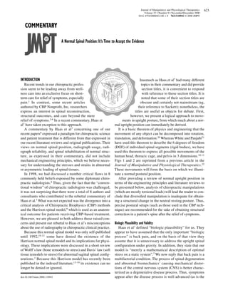
  • 1. Journal of Manipulative and Physiological Therapeutics 623 Volume 23 • Number 9 • November/December 2000 0161-4754/2000/$12.00 + 0 76/1/110941 © 2000 JMPT COMMENTARY A Normal Spinal Position: It’s Time to Accept the Evidence INTRODUCTION Inasmuch as Haas et al7 had many different Recent trends in our chiropractic profes- topics in their commentary and did provide sion seem to be leading away from well- section titles, it is convenient to respond ness care into an exclusive focus on short- with reference to those section titles. It is term care for relief of symptoms, especially noted that some of their section titles are pain.1 In contrast, some recent articles obscure and certainly not mainstream (eg, authored by CBP Nonprofit, Inc, researchers their reference to Sackett); nonetheless, the express an interest in spinal reconstruction, titles are useful as objects for debate. First, structural outcomes, and care beyond the mere however, we present a logical approach to move- relief of symptoms.2-6 In a recent commentary, Haas et ments in upright posture, from which much about a nor- al7 have taken exception to this approach. mal upright position can immediately be derived. A commentary by Haas et al7 concerning one of our It is a basic theorem of physics and engineering that the recent papers8 expressed a paradigm for chiropractic science movement of any object can be decomposed into rotation, and patient treatment that is different from that expressed in translation, and deformation.20 Whereas White and Panjabi21 our recent literature reviews and original publications. Their have used this theorem to describe the 6 degrees of freedom views on normal spinal position, radiograph usage, radi- (DOF) of individual spinal segments (rigid bodies), we have ograph reliability, and spinal rehabilitation of normal struc- used this theorem to express all possible movements of the ture, as expressed in their commentary, did not include human head, thoracic cage, and pelvis in 3 dimensions.10-11 mechanical engineering principles, which we believe neces- Figs 1 and 2 are reprinted from a previous article in the sary for understanding the stresses and strains in abnormal Journal of Manipulative and Physiological Therapeutics.22 or asymmetric loading of spinal tissues. These movements will form the basis on which we illumi- In 1998, we had discussed a number critical flaws in 8 nate a normal postural position. commonly held beliefs espoused by some diplomate chiro- After providing a review of normal upright position in practic radiologists.8 Thus, given the fact that the “conven- terms of the engineering principles and literature reviews to tional wisdom” of chiropractic radiologists was challenged, be presented below, analysis of chiropractic manipulations it was not surprising that there were a total of 8 authors and (which are mostly torsional loads) will lead the reader to con- consultants who contributed to the rebuttal commentary of clude that diversified manipulation is inadequate for obtain- Haas et al.7 What was not expected was the divergence into a ing a structural change in the neutral resting posture. Thus, critical analysis of Chiropractic Biophysics (CBP) methods precise postural setups (such as those used in the CBP tech- and the Harrison spinal model,9 which is used as an anatom- nique) are recommended for the sake of obtaining structural ical outcome for patients receiving CBP-based treatment. correction in a patient’s spine after the relief of symptoms. However, we are pleased to both address those raised con- cerns and present our rebuttal to Haas et al’s misconceptions Biologic Plausibility and Validity about the use of radiography in chiropractic clinical practice. Haas et al1 defined “biologic plausibility” for us. They Because this normal spinal model was only self-published appear to have assumed that the only important “biologic until 1992,10-12 some have denied the existence of the process” is back pain, and on the basis of that view they Harrison normal spinal model and its implications for physi- assume that it is unnecessary to address the upright spinal ology. These implications were discussed in a short review configuration under gravity. In addition, they state that our of Wolff’s law (bone remodels to stress) and Davis’ law (soft model is “merely a mathematical description of optimal tissue remodels to stress) for abnormal sagittal spinal config- stress on a static system”.7 We now reply that back pain is a urations.3 Because this Harrison model has recently been multifactorial condition. The process of spinal degeneration published in the indexed literature,13-19 its existence can no and abnormal biomechanics’ causing mechanical distor- longer be denied or ignored. tions of the central nervous system (CNS) is better charac- terized as a degenerative disease process. Thus, symptoms doi:10.1067/mmt.2000.110941 appear after the disease process is well advanced (as is the
  • 2. 624 Journal of Manipulative and Physiological Therapeutics Volume 23 • Number 9 • November/December 2000 Commentary • Harrison et al Fig 1. Rotational DOF of global body parts. case in heart disease, cancer, and hypertension). In such the entire oculovestibular, muscle spindle, and mechanore- processes, the emphasis is placed on controlling risk fac- ceptor systems perform this function from moment to tors. It is suggested that optimizing the spine’s position to moment in upright stance. To imply that this is an unimpor- resist the compressive force of gravity is a logical place at tant subject regarding spinal mechanics is to ignore a major which to address an “optimal stress” risk factor. Of course, function of the CNS.
  • 3. Journal of Manipulative and Physiological Therapeutics 625 Volume 23 • Number 9 • November/December 2000 Commentary • Harrison et al Fig 2. Translational DOF of global body parts. Haas et al7 believe that there is no compelling evidence to ing the spine. A model is used to predict nature; it is a tool. indicate that subluxation exists. However, subluxation has Our model’s usefulness will be determined in future studies been precisely defined through use of deviations from normal of clinical significance. For such studies to be carried out, upright posture and our normal spinal model.23 In addition, the parameters of the model’s usage must first be established, Haas et al do not appear to appreciate the concept of model- and one of these parameters is a measurement method—ie,
  • 4. 626 Journal of Manipulative and Physiological Therapeutics Volume 23 • Number 9 • November/December 2000 Commentary • Harrison et al A B C D Fig 3. A, Vertical position of normal AP posture as agreed on in literature. B, D, Proposed ideal lateral postures. C, Average lateral posture. Note that differences between Fig 3, B and Fig 3, C are slight. posture and radiography. Therefore, determining the reliabili- ciated with asymmetric muscle efforts. Numerous elec- ty of the radiographic markings (and posture reliability) is a tromyographic studies over the past 4 decades add support logical first step. Haas et al claim that we cannot address for this fact.24-34 Because many clinicians7 may be unfamiliar “clinically meaningful conditions,” but they want to stop us with engineering principles and aspects of upright human from attempting such future research by negating our stance, 2 other “proofs” are presented. attempts to study the reliability of our methods at the begin- As a first discussion of normal posture, the readers and ning. Clinical significance can be determined only after one Haas et al7 are asked to consider that all chiropractic, osteo- has precise definitions to test and only after one has reliabili- pathic, physical therapy, and medical colleges teach a plumb ty data established for the measuring methods. line analysis and global ranges of motion, as depicted for Because a normal postural position (outside alignment) ±Tx, ±Ry, and ±Rz in Figs 1 and 2. With regard to the AP view, and a normal spinal position (inside alignment) are funda- the following 3 questions come immediately to mind: (1) mental to our approach to “controlling risk factors” for spinal axial rotation to the left and to the right—from where? (2) dysfunction, a review pertaining to each of these ideas is pro- lateral flexion to the left and to the right—from where? (3) vided to enable the reader to determine whether Haas et al’s lateral translation to the left and to the right—from where? paradigm matches the biomechanical/biomedical literature. Before trying to give an answer that is different from ver- tical alignment in the AP view, recall that these main postur- Normal Upright Posture al motions of the head and thoracic cage, ±Tx, ±Ry, and ±Rz, There are at least 3 approaches by which one can immedi- cause spinal coupling patterns for which the movements are ately discover a normal spinal alignment in the anteroposteri- known.22,23,35,36 These vertebral coupling patterns are or (AP) view and the lateral view. Most rigorously, equilibri- always described as movement away from a vertical normal um equations in the AP view can be used to prove that the spinal alignment. Our research team is the first to report postures of ±Tx, ±Ry, and ±Rz in Figs 1 and 2 result in abnor- spinal coupling for ±Tx of the head and thoracic cage.35,36 mal asymmetric loads on the spinal tissues and lower extrem- Some authors have argued that slight deviations of the ities when these postures are in the neutral resting posture of upper thoracic spine are correlated to the side of the domi- the subject under investigation. These postures are also asso- nant arm37 and that a true vertical spine in the AP view there-
  • 5. Journal of Manipulative and Physiological Therapeutics 627 Volume 23 • Number 9 • November/December 2000 Commentary • Harrison et al fore cannot be considered normal. Either sight deviations Therefore, the vertical alignment of the global centers can be ignored because of the minimal amount of asymmet- of mass in upright posture in both the AP and lateral views ric loading, or unilateral upper extremity exercises on the has been statistically established for young adults. Fig 3 nondominant side can be prescribed to correct these. In illustrates this alignment with some additional references addition, Haas et al7 claim that because of the natural asym- applied to the location of the shoulders and lower extrem- metry of the spinous processes, the sensibility of a true verti- ities. cal spine in the AP view is questionable. However, more than 2 decades ago, Farfan38 found that the entire vertebral Harrison Spinal Model architecture in the lumbar spine will change and keep the Through use of multiple studies and normal distributions lamina junction in line with the structural center of the verte- with an ideal mean normal center, normal upright posture bral body. This means that the center of mass will remain has been defined in the literature. This normal posture in the approximately the same. Farfan38 writes (pages 34-5): “It AP view correlates with a straight (vertically aligned) spine. would appear that in the development of the vertebra, asym- In the lateral view of upright normal posture, it is generally metrical body growth is compensated for by asymmetric accepted that there exist a cervical lordosis, a thoracic growth of the neural arch.” Therefore, the centers of mass in kyphosis, and a lumbar lordosis; however, neither the over- the AP view will still be in line with vertical gravity to mini- all sagittal curvatures of the cervical, thoracic, and lumbar mize loads on the spinal tissues and to be the origin position areas nor the segmental contributions are generally stated or for postural movements.39-47 agreed on. Therefore, when normality is being debated, only Recently, Kiefer et al48 showed that T1, T12, and S1 must the overall geometric shape and segmental angulation of the be vertically aligned in the lateral view. In the lateral view, cervical, thoracic, and lumbar sagittal curvatures are open to the previous argument can be applied to the movements of debate. ±Rx, ±Ty, and ±Tz in Figs 1 and 2. In fact, much research has This is where our normal Harrison Spinal Model enters been conducted on sagittal posture, especially anterior head the literature. Both our average (400 subjects) and our ideal translation (+Tz) and pelvic posture, by physical therapists.49- normal sagittal cervical spine models were published in 61 This research indicates that anterior head translation (pro- 1996.14 Our average (derived from 552 subjects) normal trusion) is fundamentally accepted as abnormal and that, in lumbar model was published in 1997.18 Our ideal normal general, posture evaluation is repeatable and reliable. lumbar model was published in 1998.13 For a second analysis, a statistical evaluation is present- At this point, it is important to point out that the ed. As always, there is debate in the literature concerning Harrison Spinal Model is the normal path of the posterior average normal and ideal normal (upright posture). longitudinal ligament along the posterior vertebral bodies. However, the differences in these upright positions are This ideal and statistical average model has a mean and a small. In 1973 and 1975, Beck and Killus62,63 used a com- random error component (variation around the mean). It is puter analysis to average upright lateral and upright AP a model not of the entire anatomy of the spine but only of postures of several hundred young adults. They reported the curvature in the sagittal plane. Models of the entire on the reliability of their digitizing methods and showed spine require huge computer codes in which finite ele- that posture was reproducible “even though the x-rays ments methods are used. were taken years apart.” They critically analyzed all of the The paradigm presented by Haas et al7—that abnormal previous reports of posture (such as those of Staffel, 64 posture has no effect on physiology—is quite puzzling. It is Goff, 65 Leger, 66 Haglund, 67 Osgood, 68 and Wiles & well documented that abnormal spinal loads (forces and Lond, 69 which include such terms as flat back, round moments) over time cause pathoses. This is taught in all chi- back, and hollow back). Beck and Killus 62 found highly ropractic college physiology courses as Wolff’s law (bone pathologic postures, small sample sizes, incorrectly remodels to stress) and Davis’ law (soft tissues remodel to applied statistics, and lack of an origin for measurement stress). included in previous categories of posture. Using a digi- In the light of the foregoing preceding background data tizer, they plotted, in graphic form, the coordinates (paths) and ideas, a response to each of Haas et al’s topics7 can now of the vertebral bodies of all subjects in the AP and lateral be presented. views. With 1500 subjects in their 1975 report, 63 they noted that they had “one normal distribution in every point Clinical Sensibility and Validity of abscissa.” They write: Cervical kyphosis is not a normal variant. In their commentary, Haas et al7 repeat the common misconception that cervi- It follows that there is only one ideal type of spinal col- umn. According to our findings the theory of constitutional cal kyphosis is a normal variant. In general, the studies types (of posture) cannot be confirmed.62 cited by Haas et al 7 are inadequate, being characterized by inappropriate methods, flawed statistical analysis (if Beck and Killus provided separate graphs of their maxi- any), and/or unsupported conclusions. It is our intent to mum points of their normal distributions of posture. The review the 6 studies 70-75 mentioned by Haas et al and to spine had the 3 spinal curves with vertical alignment of T1, present an updated literature review focused on cervical T12, and S1 in the lateral view. kyphosis.
  • 6. 628 Journal of Manipulative and Physiological Therapeutics Volume 23 • Number 9 • November/December 2000 Commentary • Harrison et al To begin, Haas et al7 refer to a 1986 study by Gore et al.70 less than 3 weeks’ duration before the past 2 months or had In 200 randomly selected asymptomatic subjects, Gore et al pain in the 2-month period immediately before the study. In found an average of 22 degrees of lordosis using Ruth addition, the sample size was extremely small and subjects Jackson’s angle from C2-C7; they found that a minority in the two groups were not matched for age, weight, height, (9%) of these subjects demonstrated the presence of a cervi- or sex. cal kyphosis. In contrast, Harrison et al76 found that 35% of Furthermore, Haas et al7 state that “in a study of 180 sub- 250 symptomatic subjects had kyphotic deformities. These jects, Borden et al concluded that a straight cervical spine is data indicate a dramatic increase in cervical kyphosis in abnormal only in conjunction with decreased cervical flexi- symptomatic subjects in comparison with asymptomatic bility.” In actuality, the study by Borden et al73 found that of subjects. An additional concern is the cross-sectional study 180 asymptomatic subjects, 3 (1.7%) had a reversed cervical design used by Gore et al. It is common knowledge among curve and 13 (7.2%) had a straight cervical spine. Of the 13 health care providers that several life-threatening illnesses subjects with a straight cervical spine, 11 (85.0%) had con- and diseases remain asymptomatic throughout their course comitant degenerative changes. In the 3 subjects with cervi- or are associated with no overt signs or symptoms until the cal kyphosis, “there were generalized hypertrophic degener- end stages. Harrison et al3 have previously reviewed the ative changes and fusion.”73 Borden et al concluded that shortcomings of the study by Gore et al.70 “roentgenographically demonstrated loss of the cervical The second article used by Haas et al7 in their assertion curve, combined with normal thoracic kyphosis and lumbar that kyphosis is a normal variant is the study by Maimaris et lordosis, may be indicative of abnormality of the cervical al,71 who retrospectively assessed 67 asymptomatic and 35 spine.”73 In the light of this information, one might wonder symptomatic whiplash-injured patients. Follow-up was how Haas et al7 could possibly believe that this reference attained an average of 2 years after the injury. Maimaris et al supports the position that cervical hypolordosis or kyphosis concluded that “reversal of the cervical spine to a kyphotic is a normal variant. Perhaps they were performing secondary position was not associated with prolonged disability." referencing and did not read the original paper. However, there are several problems with this study. First, Likewise, Haas et al7 believe that the study by Marshall only 39 (58%) of the 67 asymptomatic subjects were "clini- and Tuchin74 indicates “that spinal curve alteration itself cally examined," whereas 30 (86%) of the 35 symptomatic may not require treatment.” However, there are several subjects were examined. This is a large difference in the per- potential problems with the method used by Marshall and centage of subjects examined in each group, and it can lead Tuchin.74 The most serious of these is their choice of the C1- to inaccurate data analysis between the groups. Second, the C7 Cobb angle for lateral cervical curve mensuration. The 4- lateral cervical spine radiographs were not measured and line C1-C7 Cobb angle is not a valid representation of the quantified; they were merely classified into lordotic, straight, cervical lordosis.8,77 Therefore, without a segmental analy- or kyphotic. Therefore, no quantifiable statistical analysis sis, no conclusions can be made concerning the shape and/or was performed to actually address the issue of the amount or magnitude of the cervical lordosis and its clinical signifi- type of lateral cervical curvature as it relates to the presence cance in that study. of symptoms. As an example, on page 396, Maimaris et al Last, Haas et al7 rely on the conclusions of Gay.75 In a present two figures, one representing a kyphotic cervical 1993 review, Gay concluded that cervical kyphosis is a nor- curve and the other representing "loss of the normal lordo- mal variant. However, before Haas et al’s7 commentary, the sis." If one looks at these two radiographs, it is readily vast majority of the references cited by Gay had previously apparent that both of these are kyphotic curvatures. Last, in been critically analyzed and had been found to be inappro- their Table III on page 395, Maimaris et al present the per- priate by us in 1998.8 Thus, no weight can be applied to centage of subjects in each group with radiographic abnor- Gay’s75 review. malities. Clinically significant differences are noted between Recent and past mechanical, clinical, and surgical studies the symptomatic and asymptomatic groups, with the symp- concerning the cervical lordosis do not support the position tomatic group having higher percentages of straight and that cervical kyphosis is a normal variant. Haas et al7 seem kyphotic curves and the asymptomatic group having the to insinuate that the only supporting reference that we8 highest percentage of lordotic curves. offered for the clinical significance of a normal cervical lor- Similarly, Haas et al7 make the claim that Pedersen72 dosis was by Cote et al78 (our old reference 1). This clearly “observed that cervical hypolordsis may be a normal vari- was not the case (see references 9-17 in our original manu- ant.” However, the study by Pedersen is of insufficient quali- script8). However, because Haas et al and many others con- ty for the drawing of such a conclusion. Pedersen compared tinue to debate this issue, a thorough review of the literature a “treatment” group of only 9 patients with a “control” group on cervical kyphosis and its effects is presented here. of 17 patients. The criterion for inclusion in the control It is relevant that several studies have investigated and group was as follows: “The subject must not have had any linked altered cervical curve configurations to the presence history of cervical or cervico-thoracic symptoms prior to 2 of chronic headache pain.79-81 In a survey of more than 6000 months before entry (lasting continuously for more than 3 cases of chronic headache-sufferers, Braaf and Rosner79 weeks).” This designation was a poor choice, inasmuch as found that “complete or segmental loss or reversal of the subjects in the control group could either have had pain of normal lordotic curve of the cervical spine is the most con-
  • 7. Journal of Manipulative and Physiological Therapeutics 629 Volume 23 • Number 9 • November/December 2000 Commentary • Harrison et al sistent characteristic feature and very often is the only method is important because the standard error of measure- abnormality found.” In 47 subjects with tension and ment (SEM) is much lower (<3) and the segmental analysis migraine headaches, Vernon et al80 found a high incidence finds alterations within the curve—ie, it is not just evalua- of hypolordosis, straightened cervical curve configurations, tion of the end segments. and reversed cervical curve configurations. Nagasawa et al81 According to Haas et al,7 the surgical studies just cited, compared 372 patients with tension headaches and 225 con- all of which indicate the clinical significance of the cervical trol subjects matched for age and sex; they found statistical- lordosis, are not appropriate because surgical patients are ly significant differences between the two groups, the “not typical chiropractic patients who require conservative headache patients having straightened cervical curve config- care.” But perhaps this is merely an excuse for not accepting urations and low set shoulders. In addition, with increasing the facts. age, the headache patients’ cervical curve was straight more However, there are several nonsurgical studies indicating frequently. This information contrasts nicely with the find- cervical kyphosis as a factor predictive of poor results.98-100 ings of Gore et al,70 who noted that in asymptomatic sub- In a long-term follow-up of 146 whiplash-injured patients, jects, the cervical curve increased with age. Hohl98 identified cervical kyphosis as factor predicting a Looking into the matter further, we note that when poor out- poor outcome. Norris and Watt99 followed 61 patients comes after surgery are considered, several studies have found involved in motor vehicle accidents for a minimum of 6 a good cervical lordosis to be a significant factor in preventing months; they found abnormal neck curves to be “more com- neurologic deficit and/or compromise.82-93 For example, Goto mon in patients with a poor outcome.”99 and Kita82 state that “postoperative kyphotic malalignment or In addition to clinical outcomes of pain and neurologic angulation are factors strongly affecting the outcome of deficits, several papers focusing on cervical biomechanics surgery.” Yamazaki et al83 compared 2 groups of patients with indicate that cervical kyphosis is abnormal.101-103 Using an ossification of the posterior longitudinal ligament (PLL) post- engineering analysis of slope and bending moment, operatively. Group I consisted of patients with no anterior Matsunaga et al101 proved that degeneration and consequent “compression” of the spinal cord, and group II comprised subluxation will develop in abnormal kyphotic configura- patients with residual cord “compression.” The investigators tions, which they termed buckling alignments. “In the case found a lordosis of less than 10.0 degrees or cervical kyphosis of the S-shaped spine,” they write, “the spine is in a ‘buck- correlated with the presence of postoperative spinal cord com- ling’ condition, which corresponds to an unreasonable sup- pression; this result was statistically significant. porting function.” Similarly, recent surgical studies have emphasized that With regard to ossification of the PLL, Matsunaga et al102 operations that leave the patients’ cervical spines with less followed 101 patients for a 5-year period. Using an engi- lordosis are associated with increased neck, upper thoracic, neering analysis of strain, they found that ossification of and shoulder pain and overall poorer health outcomes.94-97 PLL progressed in the areas showing uneven and abnormal Lowry94 used the average lordosis of 22 degrees found by strain at the adjacent intervertebral disk and PLL. Spe- Gore et al70 to gauge a successful surgical outcome; he cifically, strain in the tensile direction was found to corre- claimed that “a fixed kyphotic posture of the mid-cervical late with ossification of the PLL. It is important to note that spine will lead to overall sagittal imbalance manifested by the only way to increase the tensile strain in the PLL is to axial pain in the upper to mid-thoracic region as well as in have concomitant hypolordosis or kyphosis.104 the posterior cervical spine.” Katsuura et al95 compared 44 Several other studies have indicated that loss of cervical cases treated with anterior cervical plating (a method of lordosis or kyphosis is a cause of or is associated with a ensuring a stable lordotic correction) to 30 patients receiv- high incidence of degenerative changes in the disk and ver- ing anterior fusion without plating. These patients were fol- tebral bodies.70,73,98,105 Because degenerative changes of the lowed for an average of 38 months postoperatively. The cervical spine are a known cause of morbidity,98,99,106 it can magnitude of cervical lordosis was assessed through use of be stated with confidence that cervical kyphosis is not a nor- Ruth Jackson’s angle (posterior tangents from C2-C7), and mal variant. intersegmental (2-line) Cobb angles were used for the align- In a recent 1997 text on the spine, statements by ment of the fused segments. Katsuura et al found statistical- Frymoyer et al109 are clinically significant about cervical ly significant differences in the incidence of neck pain kyphosis as a normal variant. In fact, they assert that between the two groups, the plated group having less neck “kyphosis, straightening, and hyperlordosis of the cervical pain and a statistically significant increase in cervical lordo- spine may all be seen in conjunction with spondylotic sis. Likewise, Kawakami et al96 retrospectively evaluated 60 myelopathy, whereas in other patients the normal curvature patients postsurgically. Cervical lordosis was measured with is preserved” and that “kyphosis and hyperlordosis may a 4-line Cobb’s method from C2-C7 and a segmental (2- coexist and thus this gives rise to an S-shaped deformity of line) Cobb angle at the fused level. Segmental kyphosis at the spine, causing greater instability and management prob- the level of the fusion and less lordotic curvature were found lems.”109 Furthermore, in a 1996 review on the cervical to statistically predict which patients would develop “axial spine surgeries, Lowery94 states that “an additional feature symptoms” (neck pain, shoulder pain, and/or stiff neck). of any plate system, whether anterior or posterior, should be The use of 2-line Cobb methods rather than the 4-line Cobb to establish a lordotic cervical curvature.” The evidence
  • 8. 630 Journal of Manipulative and Physiological Therapeutics Volume 23 • Number 9 • November/December 2000 Commentary • Harrison et al against76-107 cervical kyphosis as a normal variant far out- subjects, Bernhardt and Bridwell123 reported a mean Cobb weighs the evidence to the contrary.7,70-75 for T3-T12 of ~36 degrees and a mean Cobb for T1-T12 of Finally, with regard to the cervical spine, it is noted that ~40 degrees. Jackson and McManus124 investigated sagittal many authors (including Haas et al) have neglected to inves- balance in 100 patients and 100 volunteers; they reported the tigate cervical lordosis and child development. It should not same means and ranges for males and females and for have been necessary to present the preceding review indicat- patients and volunteers (mean T1-T12: ~43 ± 9 degrees). In ing that cervical kyphosis is not a normal variant, inasmuch 1998, Vedantam et al125 investigated standing sagittal align- as Bagnall et al108 showed in 1977 that the cervical lordotic ment in 88 adolescents (aged 10-18 years); they reported a curve is formed in intrauterine life (9.5 weeks). Cervical lor- “striking similarity in regional thoracic kyphosis and lumbar dosis could thus be considered a primary curve, like the tho- lordosis between adolescents and adults.” racic kyphosis. Any loss of cervical lordosis is due to trau- In 1980, Fon et al126 reported that in 316 subjects, thoracic ma, repetitive play, or work postural loadings during and/or kyphosis increased with age. In 1993, Cutler et al127 reported after birth. Importantly, using skin markers or surface visual- that hyperkyphosis in women was associated with altered ization does not correlate with the shape, magnitude, or vertebral body shape (anterior wedging), loss of bone densi- direction of the sagittal cervical spine.109,110 Therefore, the ty, loss of fitness, decreased muscle strength, and reduced use of radiography for identification of any abnormal lateral survival; the investigators suggested a correlation between cervical configuration is absolutely mandatory. abnormal upright thoracic posture and changing architecture of the spinal column (Wolff’s law). In 1996, Kolessar et al128 Thoracic Kyphosis reported that 69 patients had kyphotic Cobb T5-T12 mea- In addition to the work of Beck and Killus,62 whose nor- surements of >33 degrees, whereas all 24 volunteers had mal graph used a statistical average, multiple studies have Cobb T5-T12 < 33 degrees. investigated thoracic kyphosis. As in cervical skin contour It is interesting to note that studies on thoracic kyphosis versus radiographic measurements, D’Osualdo et al111 found with large population groups provide nearly normal distri- considerable disagreement between radiographic findings butions. Thus, as noted by Beck and Killus,62-63 statistically and results obtained with an Arcometer. In 1983, Willner and there would be one ideal normal at the mean of these distrib- Johnson112 used spinal pantographs based on skin contour to utions. With the few reported values for Cobb T1-T12 and suggest that kyphosis varied in girls and boys. However, with ratios of values from Cobb T2-T12, T3-T12, T4-T12, using an ISIS scanner for evaluating back shape, Carr et al113 and T5-T12, the mean thoracic kyphotic value seems to be reported no statistically significant differences with respect between 40 and 50 degrees for a Cobb angle from the top of to age and sex in children, no statistically significant differ- T1 to the bottom endplate of T12. ences with respect to age and sex in adults, and no differ- ences between children and adults. Using radiography, Lumbar Lordosis Voutsinas and MacEwen114 reported in 1986 that “the differ- Haas et al7 have neglected a large body of literature sup- ences between boys and girls were not significant in any porting the clinical significance of a normal lumbar lordo- group.” sis.13,18,19,116,129-158 To date, there have been 10 separate Some studies have reported a large range of values for studies in which a similar method was used to investigate thoracic kyphosis—from 20 degrees to 40 degrees.115-120 the normal shape and magnitude of the human lumbar lor- One problem in deciphering a normal average thoracic dosis.18,115-117,122,129-133 These 10 studies used strict criteria kyphosis is the inability to see T1-T4 on most lateral tho- in categorizing men and women as normal subjects. The racic views; this has caused different levels of Cobb angle age range for the subjects was large—between 10 and 80 analysis in the literature—with different angles of kyphosis, years of age. All 10 of these investigations used radi- of course. For example, the following levels have been used: ographic analysis in the neutral upright posture, and total T5-T12, T4-T12, T3-T12, T2-T12, and T1-T12. This causes as well as segmental lumbar lordotic angles were mea- wide ranges of values. For example, Propst-Proctor and sured. In all, a total of 755 normal subjects from 10 sepa- Bleck117 measured the inferior endplate of T5 to the superior rate studies were included in the analysis. All of these stud- endplate of T12 in 104 normal children who were referred ies18,115-117,122,129-133 indicate that the upper lumbar curve, for suspected screening of scoliosis; they reported a normal T12-L3, is minimally lordotic whereas the distal lumbar distribution with a range of 21 to 33 degrees and a mean of curve, L4-S1, accounts for approximately 65.0% of the 27 degrees. lumbar lordosis. In 22 cases, Singer et al120 reported no difference in tho- Haas et al7 claim (believe) that different patient popula- racic kyphosis in subjects before and after death (T4-T12: tions (races) have different optimal spinal configurations and ~49 degrees).120 Milne and Williamson121 reported no sig- that “given the natural variation and asymmetry in the nificant change in thoracic curvature on follow-up radi- human musculoskeletal system,” an ideal spinal model is not ographs of 261 subjects aged 62-90 years. In 99 Greek men plausible. and women, Korovessis et al122 reported that thoracic kypho- Their beliefs are not supported by recent anthropometric sis was not gender related, though kyphosis was found to clinical and experimental data on the lumbar spine. For exam- increase with age (mean T4-T12: ~36 ± 9 degrees). In 102 ple, several studies addressing the shape and magnitude of the
  • 9. Journal of Manipulative and Physiological Therapeutics 631 Volume 23 • Number 9 • November/December 2000 Commentary • Harrison et al lumbar lordosis found no differences with respect to geo- adult patients with symptomatic degenerative lumbar disk graphic location and/or ethnicity.133-137 In a radiographic disease, 30 patients with low-grade L5-S1 isthmic spondy- study of the lumbar lordosis in 16 healthy Chinese men, lolisthesis, and 30 patients with idiopathic or degenerative Chen133 concluded that “no obvious interracial differences scoliosis. Again, statistically significant differences were found in the geometric data found in this study.” between the groups were found in the radiographic mea- Mosner et al134 compared the lumbar lordotic spinal radi- surements.138 ographs of 25 black women with those of 27 white women; Adams et al142 followed 403 volunteer health care work- no significant differences were found in the lumbar lordosis, ers for a 3-year period. A decreased lumbar lordosis was as measured with spinal radiographs, though differences found to be one of the personal risk factors in predisposing were found in external skin contours. This difference in skin subjects to a first-time episode of low back pain requiring contour “gives a false impression of increased lumbar lordo- medical evaluation. Itoi143 analyzed lateral lumbar radi- sis”134 in black versus white women. In a 1985 study of 973 ographs in 100 postmenopausal female patients with spinal adults, Fernand and Fox135 reported that “black and white osteoporosis; a statistically significant increase in low back populations demonstrated no significant difference in the pain was found in patients with a rounded thoracic spine mean lumbar lordotic angle.” Korovessis et al136 studied the and lower acute lumbar kyphosis. This lower lumbar kypho- sagittal configuration of the lumbar spine in 120 asympto- sis has been shown to cause an increase in low back pain, matic Greek volunteers. “We have shown that there are no perhaps as a result of increased electromyographic activity differences in the sagittal profile across geographically of the paraspinal musculature.144 and ethnically different populations,” they write. As regards Many surgical papers have identified both lumbar hyper- the “natural variation” of human anatomy, Janik et al13 have lordosis145 and hypolordosis or kyphosis146-152 as risk fac- shown that reported small differences in vertebral tors for low back pain requiring surgical correction. Last, body–to–disk height ratios, found in different studies, have several investigators have found statistically significant no effect on their elliptical model of the lumbar lordosis (ie, increases or associations between loss of lumbar lordosis none of the segmental angles, T12-S1, were found to change and degeneration of the disk, vertebral body, and lig- more than 1 degree). aments.152-156 As regards low back pain and lumbar lordosis, 6 separate With regard to lumbar lordosis, most experts agree that studies have found statistically significant differences “the assessment of spinal curvature is a part of an evalua- between patients with low back pain and matched normal tion in patients with back problems”.157 Because surface control subjects.19,136,138-141 Korovessis et al136 compared measurement of spinal curvature represents neither seg- homogenous groups of 120 volunteers and 120 patients with mental nor total lumbar lordotic alignment, radiographic chronic low back pain; subjects were matched for age, sex, analysis is a mandatory part of the routine examination of and ethnicity. Lordosis was measured through use of a 4-line the lumbar spine.134,158 We believe that Haas et al’s7 nega- Cobb’s method from T12 to S1, and segmental angles (2-line tion of the importance of the structural alignment of the Cobb) were measured through use of endplate lines relative spine stems from a neglect of mathematics and engineering to horizontal. Statistically significant differences were found analysis. They seem to be reading published papers with between the two groups, the chronic low back pain subjects just rudimentary 4-line Cobb angle geometry while ignor- having a decreased distal lordosis, L4-S1, in comparison ing papers that analyze spinal alignment with a slightly with the controls. higher level of mathematics. Papers that use segmental Harrison et al19 used elliptical modeling to discriminate analysis (see the references above), some analytic geome- between a rigorously defined normal group and 3 separate try,13,19 calculus,103,120,153 curve fitting,9,159-161 and finite groups of patients with low back pain; that study included elements162-164 have shown the importance of normal (1) 50 normal subjects from a pre-employment physical alignment of the spine. screening, (2) 50 patients with acute low back pain (first occurrence; less than 6 weeks’ duration), (3) 50 patients with Pelvis Orientation chronic low back pain (pain of more than 6 weeks’ duration Plaugher et al165 have investigated pelvic radiographic or a history of recurrent disabling low back pain), and (4) 24 measurements and found reproducibility in pelvic position- subjects with low back pain and radiographically verified ing and radiographic marking. Troyanovich et al166 have degenerative disorders or pathoses. These 4 groups were established high intraclass coefficients (ICCs) and low matched for age, weight, height, and sex. The investigators SEMs for pelvic tilt on the lateral lumbopelvic view. found that “of 11 angles, 2 distances, and 2 ratios, statistical Jackson et al138 have reported high Pearson correlation analysis was significantly different across groups for 12 of coefficients for pelvic measurements (in degrees) on lateral these measurements.”19 The chronic low back pain and lum- full spine views; they reported significant differences in bar pathosis group had a decreased lordosis, whereas the pelvic measurements for normal subjects versus 3 patient acute low back pain group had an increased lordosis in com- groups. parison with the normal subjects. In 1995, Saji et al167 studied 61 adolescents with idio- In a similar study, Jackson et al138 compared sagittal lum- pathic scoliosis and 33 normal subjects for femoral neck bar radiographs of 50 normal volunteers with those of 50 shaft angles and asymmetry of the hips. An angle was
  • 10. 632 Journal of Manipulative and Physiological Therapeutics Volume 23 • Number 9 • November/December 2000 Commentary • Harrison et al Table. Partial review of x-ray reliability studies: ICCs and standard errors of measurement for intraobserver error and interobserver error (in degrees) Standard error of measurement (degrees) ICC 4-line Cobb 2-line Cobb Mod. Risser-Ferg Post. tangent Area/Reference Intra Inter Intra Inter Intra Inter Intra Inter Intra Inter AP cervical — — — — — — — — — — Troyanovich et al >0.90 >0.90 — — — — <2.0 <1.5 — — AP thoracic — — — — — — — — — — Pruijs et al (1994) (Spearman > 0.98) — 3.2, 2.0* — — — — — — Sevastikoglou, Bergquist — — — 3.1 — — — — — — Beekman, Hall — — — 4.2* — — — — — — Gross et al (p176) — — 3.2 2.3 — — — — — — Morrissy et al — — 4.9, 3.8, 7.2, 6.3, — — — — — — 2.8 6.3 Carman et al — — — 3.8* — — — — — — Goldberg et al — — 1.9 2.5 — — — — — — Desmet et al — — — 1.1, 1.3* — — — — — — AP lumbar — — — — — — — — — — Troyanovich et al >0.70 >0.80 — — — — <2.0 <2.0 — — Lateral cervical — — — — — — — — — — Phillips et al (1999) — >0.83 — — — <2.7† — — — — Singer et al5 — — 4.5 — — — — — — — Plaugher et al — — 2.9† 4.2† — — — — — — Cote et al — >0.90 — 9.1, 8.3‡ — — — — — — Jackson et al (Pearson r > .80) — — — — — — 1.2 1.2 Harrison et alSpine >0.70 >0.80 — — 3.6, 3.2 2.2, 2.7‡ — — 2.3 2.4 Lateral thoracic — — — — — — — — — — Carman et al — — — 3.3* — — — — — — Voutsinas, MacEwen — — 2.2* — — — — — — — Jackson et al (1998) (Pearson r > .82) — — — — — — — — Lateral lumbar — — — — — — — — — — Jackson et al (1998) (Pearson r > .73) — — — — — — — — Polly et al (1996) >0.81 >0.83 10.0 10.0 — — — — — — Voutsinas, MacEwen — — 2.1* — — — — — — — Gelb et al — — — — 3.0* 3.0* — — — — Wood et al — — — — — 1.1† — — — — Troyanovich et al >0.70 >0.80 — — — — — — 1.5 1.7 Troyanovich et al >0.70 >0.80 — — — — — — 1.7 1.8 ICC, Intraclass coefficient; Intra, intraobserver error; Inter, interobserver error. *Only average difference was reported (not standard error of measurement). †Standard error of measurement was reported. ‡First value is for C1-C7; second value is for C2-C7. formed by lines through the femoral shaft and neck. Intra- ic analysis of spinal displacement.” We have published 5 observer and interobserver reliability were assessed. There radiographic line drawing analysis reliability studies and were no significant differences between the measurements have another in review.77,166,169-172 In other chiropractic pub- of a single observer or between the measurements of differ- lications, the reliability of line drawing analysis on the nasi- ent observers. Standard errors of measurement were small um view and pelvis, additional AP, and lateral views have (intraobserver, 1.6 degrees; interobserver, 1.9 degrees). Saji been reported.165,173-176 Curiously, in regard to the nasium et al discovered that a normal femoral neck-shaft angle was and AP lumbopelvic radiographic views, Haas et al make the approximately 135 degrees, the scoliosis subjects having a following inaccurate statement: “There are no grounds for much higher value for this angle (straightened femur). generalizing from studies on pelvic height and the cranium Levine and Whittle168 studied the effects of pelvic tilt on to the repeatability of analyzing spinal curves and segmental lumbar lordosis. They reported that flexion of the pelvis displacement.”7 Perhaps Haas et al are unaware that the nasi- increased lumbar lordosis and that extension of the pelvis um radiograph includes the cervical spine and that its align- was associated with hypolordosis. Similarly, Harrison et al19 ment in comparison with the “cranium” has been subjected reported that their subjects with acute low back pain had to several reliability studies.173,174 flexed pelvises and lumbar hyperlordosis whereas their sub- For the various Cobb methods (2-line and 4-line), numer- jects with chronic pain had extended pelvic positions and ous reliability studies have been done on AP lumbar, AP tho- lumbar hypolordosis. racolumbar, and AP thoracic, lateral lumbar, lateral thoracic, and lateral cervical views.9,159-161,177-192 Furthermore, the Reliability measurement of pelvic rotation and sagittal segmental trans- Haas et al7 make the following statement: “To date, there lations has been reported as reliable.193-195 In 1992, Capasso is little convincing evidence for the reliability of radiograph- et al196 reported high Pearson correlation (r > 0.90) for both
  • 11. Journal of Manipulative and Physiological Therapeutics 633 Volume 23 • Number 9 • November/December 2000 Commentary • Harrison et al “intra and intergroup correlations among orthopedic sur- Recently, computer-aided spinal radiographic measure- geons” for 3 types of spondylolisthesis radiographic mea- ments have been shown to be highly reliable. For example, surements. Similar to Tallroth et al’s194 findings for lumbar in 1996, Rosol et al207 reported a high Pearson correlation flexion/extension, the findings of Lind et al197 for cervical for vertebral morphometry (r = 0.96) and low mean coeffi- flexion/extension were high examiner reliability and low cients of intraobserver variability (4%) and interobserver intraobserver error of measurement (<1.8 degrees) for later- variability (2%). al cervical segmental flexion angles. Gross et al198 (1983) We have already mentioned the recent (1998) study of and Goldberg et al199 (1988) revisited AP Cobb angle relia- sagittal balance with normal subjects and 3 patient groups bility and reported smaller errors of measurement (between conducted by Jackson et al.138 They reported high Pearson 1.9 and 3.2 degrees) than most studies. Similarly, DeSmet et correlation coefficients in the vast majority of their interob- al200 reported small interobserver errors of the differences server and intraobserver reliability measures (13 measures (<1.2 degrees) for Cobb angles. in their Tables 8 and 9, most being greater than 0.75).138 In The multitude of studies just cited (a total of 35) report- addition to the pelvic measurements, they also reported sig- ing high radiographic reliability (those with ICCs report the nificant differences in the normal subjects and the 3 patient majority of ICCs to be in the >0.75 range) is sufficient to groups for a multitude of lateral spine measurements. In establish the “reliability of radiographic analysis of spinal 1997, Hardacker et al208 studied lateral cervical reliability displacement” to the satisfaction of any well-informed with segmental Cobb angles and reported only 3 values of reader. interobserver and intraoberver Pearson correlations less Perhaps the high variability of SEM (ranging between 3 than 0.83 for 8 measurements of segmental lordosis (0.64 < and 10 degrees) with the 4-line Cobb methods are of con- r < 0.92). cern. Inasmuch as the Harrison posterior tangent method has In 1994, Pruijs et al209 used a Spearman correlation coef- smaller SEMs (<2 degrees), this method is suggested to ficient for measurements of Cobb angles on AP thoracic Haas et al7 as well as to clinicians and other researchers for radiographs. They reported high coefficients (>0.98) for the analysis of the sagittal spine. both interobserver and intraobserver variation with small However, until recently (the 1990s), most investigators SDs of differences (<3.2 degrees). reported only means, SDs, and SDs of differences in read- In 1996, Levine and Whittle168 measured pelvic tilt and ings and rereadings (Table). Because the SEM has SD in its lumbar lordosis using a television/computer system that formula, the low means and SDs for measurement differ- obtained 3-dimensional (3-D) coordinates of markers on the ences indicate good-to-excellent reliability. However, before pelvis and spine at 20-ms intervals. ICCs for 3 readings of 1990, authors rarely provided proper statistical analyses pelvic tilt and lumbar lordosis were 0.78 < ICCs < 0.95. The with Pearson r, intrarater ICC, interrater ICC, 95% CI, and investigators reported that altering the pelvic tilt significant- SEM values. The vast majority of medical investigators have ly changed the angle of lumbar lordosis. stated that the low means and SDs for differences in drawing In 1995, Saji et al167 studied 61 idiopathic scoliosis ado- and redrawing radiographic lines (Table) suffice for a claim lescents with 33 normal subjects for femoral neck shaft of adequate reliability. After all, it is merely Euclidean angles and asymmetry of the hips. An angle was formed by geometry! lines through the femoral shaft and neck. They also per- In 1999, Phillips et al201 reported high correlation coeffi- formed Cobb angle measurements. Intraobserver reliability cients (0.83, 0.91, 0.89, 0.91, 0.88, 0.87) and low SEMs and interobserver reliability were assessed; there were no (2.45 degrees, 2.71 degrees, 1.69 degrees; 1.22 mm, 1.32 significant differences between the measurements of a single mm, 1.48 mm) when measuring the occipital-C2 angle and observer or between the measurements of different distances on neutral, flexion, and extension radiographs. For observers. SEMs were small (intraobserver, 1.6 degrees; measuring spinal canal dimensions and cord size in a 1999 interobserver, 1.9 degrees). In addition, the investigators review article, Rao and Fehlings202 reported that 7 studies reported a significant difference in the obtuse femoral angles provided interobserver and intraobserver reliability of radi- in normal versus scoliotic subjects, the latter having much ographic measurements (0.82 < ICC < 0.99). In 1997, with larger obtuse angles. In 1985, Tibrewal and Pearcy210 means and SDs for measurement differences on AP thoracic assessed measurement of disk heights from lateral radi- views, it was reported that the rib vertebral angle could be ographs. They reported a maximum error of 0.7 mm for reliably measured (intraobserver mean, 4.4 degrees; interob- intraobserver and interobserver measurements. server mean, 3.6 degrees).203 Since 1990, many chiropractic studies have included sta- In 1996, Omeroglu et al204 reported that “both intraobserv- tistical analyses that are extensive in comparison with those er and interobserver error risks were insignificant” for use of of previous studies. For our 6 CBP radiography reliability the Perdriolle torsionmeter to measure vertebral axial rota- studies, we consulted with Burt Holland, PhD, a professor in tion in scoliosis on AP radiographs.204 In 1994, Gilliam et the Department of Statistics at Temple University. With his al205 reported high ICCs for both intertester (ICC > 0.86) and aid, our studies have reported complete statistical analyses intratester (ICC > 0.92) pelvic radiographic measurements. with a majority of intraobserver and interobserver ICCs Also in 1993, Hamberg et al206 reported that “the x-ray mea- greater than 0.75 and small SEMs (<2 degrees). It is interest- surements showed high reliability” for pelvic tilt analysis. ing to note that ICC > 0.60 is the set of values that Haas et
  • 12. 634 Journal of Manipulative and Physiological Therapeutics Volume 23 • Number 9 • November/December 2000 Commentary • Harrison et al Fig 4. Four types of loads with inherent stresses. al7 have declared to be in the good-to-excellent range for more than Euclidean geometry, which is the basis of every chiropractic studies.211 This range is close to the excellent human-made structure in our industrial lives. range suggested by Shrout and Fleiss212 (poor < 0.4; 0.4 < fair to good < 0.75; excellent > 0.75). Posture Reproducibility, Reliability, and Validity It would be a great surprise to every mathematician, There is in the literature a plethora of posture reliability physicist, and engineer to find that radiographic line draw- and reproducibility studies performed by ergonomists, med- ings of all types are not highly reliable. After all, it is nothing ical doctors, chiropractors, and physical therapists. Posture
  • 13. Journal of Manipulative and Physiological Therapeutics 635 Volume 23 • Number 9 • November/December 2000 Commentary • Harrison et al A B Fig 5. A, Diversified technique is limited; lumbar roll is a torsional load applied to lumbar segments by twisting pelvis relative to fixed thoracic cage. B, Cervical rotary-break is a torsional load applied to head in lateral flexed and forward flexed positions. These loads are applied to every subject; patient’s abnormal pretreatment posture is disregarded. has been studied with patients lying,213 sitting,214 stand- Last, inasmuch as Fialka-Moser et al255 reported that ing,215 lifting,216 working,217 walking,218 engaging in daily adolescents’ postural faults have been established as the routine performance,219 and running.220 Postural stress has cause of pain syndromes in adulthood, we are bewildered been correlated with scoliosis,221 office work,222 work-relat- that chiropractic state licensing boards are being influenced ed lifting injuries,223 driving,224 sitting,225 space flight,226 by the paradigm promoted by Haas et al7 and others, as sports injuries,227 and back pain.228 Posture as a valid out- cited in a recent review.8 For example, in August 1999 the come of care is universally accepted by almost all health care Nevada Board of Chiropractic Examiners told practitioners sciences.229-231 Virtually every body part has been studied for that it was against Board ruling to take radiographs of chil- its posture in normal subjects versus injured individuals; dren and that posttreatment radiographs were unwarranted. included are the postures of the head,232 shoulders,233 upper The selective information (countered in our preceding extemity,234 trunk,235 pelvis,236 lower extremity,237 and short review) generally generated and disseminated by chiroprac- leg,238 as well as the whole body posture.239-251 The vast tic radiologists and academicians is adversely affecting chi- majority of these studies report high interobserver and ropractic clinicians’ ability to provide the best possible ser- intraobserver reliability. In 1990, in a study of 86 Chinese vice to the patients who seek our care. subjects, Dai and Gu252 found no significant differences in posture for males and females and no significant differences Clinical Utility and Appropriateness in posture between groups of old-aged subjects and middle- Haas et al’s7 negation of structure (spinal alignment) as aged subjects with eyes open, though they did find a differ- a clinical outcome seems to be based on a few controlled ence in posture between groups of old-aged subjects and clinical trials that failed to demonstrate the effectiveness middle-aged subjects with eyes closed. In a 1997 study of of chiropractic manipulation in conditions such as asth- 160 men and women, Raine and Twomey253 found no signif- ma260 and nocturnal enuresis.261 These studies used spinal icant gender differences for each of head and shoulder pos- manipulation as the intervention (treatment). Because ture measurements. For a review of the normal alignment of manipulation seemed to be beneficial for headaches, neck the lower extremities, the reader is referred to the 1996 pain, and back pain but not for other diseases or spinal dis- review by Riegger-Krugh and Keysor,254 who noted that placement, Haas et al prefer to see chiropractic as merely a pathology of structures results from skeletal malalignment. temporary treatment regimen for the relief of spinal aches Adolescents’ postural faults have been established as the and pains. cause of pain syndromes in adulthood.255 Anatomical short There are many flaws in this line of reasoning, and most leg inequality has received much attention in the literature. of these flaws are derived from a complete void in an engi- True anatomical leg length inequality causes a recogniz- neering education. At least two topics can be applied to able abnormal posture. For a review, the reader is referred Haas et al’s faulty reasoning: (1) exclusive use of torsional to the 1983 review by the American Academy of Osteo- loads in spinal manipulation and (2) neglect of CNS biome- pathy256 and to recent articles by Beaudoin et al257 and chanics. This whole discussion of posture and radiographic Keppler et al.258 line drawing reliability/validity omits a very important clin- In terms of validity, posture as a valid outcome in the ical factor when any of the postural rotations and transla- health care sciences is well accepted. In fact, the editor of tions in Figs 2 and 3 are present for long periods, especially this Journal has made the following statement: “That pos- flexed segments in the cervical and lumbar areas. ture can lead to low back pain will come as a surprise to no Deformations of the CNS, which result from abnormal pos- one.”259 tural loads, were recently reviewed in this Journal.262-264
  • 14. 636 Journal of Manipulative and Physiological Therapeutics Volume 23 • Number 9 • November/December 2000 Commentary • Harrison et al A B Fig 6. In these examples, torsional loads make subjects’ posture worse. A, Patient’s initial posture is left head lateral flexion with con- tralateral right thoracic bending (low shoulder at right). B, Patient’s head is right laterally translated with contralateral translation of thoracic cage. Y-axis torsional loadings (seen in Fig 5) will not correct this posture and will in fact add other abnormal postural loads. Thus, of the two topics, only that of torsional loads in spinal loads. After being exposed to some anatomy and ranges of manipulation needs to be presented here. motion, any engineer can visualize the stresses (forces/area) and strains (measurements of deformation) in these postures. Torsional Loads For example, in lateral flexion, the flexural stresses are com- Let us revisit the postural DOF in Figs 1 and 2, which are pression on the concave fibers and tension on the convex based on the engineering concepts of rotations and transla- fibers. For the paraspinal tissues, these stresses/strains are tions in 3 dimensions. There are 12 single movements in 6 directly associated with Wolff’s law and Davis’ law, especial- DOF for each of the head, thoracic cage, and pelvis. ly for the application of these abnormal loads over long peri- Through use of permutations in probability theory, it can be ods. The previous discussion now illuminates a glaring defi- determined that there are 728 possible postures (singles, ciency in applying diversified loadings to patients’ spines. doubles, triples, . . . sextuples) of each of the head, thoracic The vast majority of diversified maneuvers are torsional cage, and pelvis in 3 dimensions, for a total of 7283 or loads (Fig 5). It might seem that these loadings will have a 385,828,352 possible upright human postures!12 This is such probability of being only 11% effective (of the 18 DOF illus- an overwhelming number that it is best to restrict our discus- trated in Figs 1 and 2, head and thoracic torsions are only 2 sion to the individual postures depicted in Figs 1 and 2. DOF). However, when considering the total permutations of These 36 postures (12 singles each for head, thoracic cage, posture, these diversified torsional loads are 4 out of and pelvis) can be recategorized into the 4 types of loads 385,828,352 possible human postures, which amounts to taught in junior-level engineering courses on the mechanics approximately 0% of specificity in matching the patient’s of materials (Fig 4). abnormal posture. Haas et al7 seemingly do not understand In mechanics of materials, engineers study the material the necessity of matching the patient’s individual posture properties, design, loadings, deflections, stresses, and strains because they do not have a realization that these (rotated and of structures.265 The y-axis rotations in Fig 1 are caused by translated postures) are mathematical functions in linear torsional loads, whereas the x-axis (flexion/extension) and z- algebra, which is fundamental to an engineering education. axis (lateral flexion) rotations are examples of pure bending. Linear algebra is the study of linear transformations.266 The y-axis translations are examples of axial loads, whereas There are 4 important linear transformations in 3-D geometry: the x-axis and z-axis translations are examples of transverse (1) rotations, (2) reflections, (3) projections, and (4) transla-
  • 15. Journal of Manipulative and Physiological Therapeutics 637 Volume 23 • Number 9 • November/December 2000 Commentary • Harrison et al tions. Because of a complete void in chiropractic education, However, the complete void of mechanical engineering clinicians and academicians are unaware that rotations, reflec- in their reasoning has led them to a paradigm of chiropractic tions, and translations have unique inverses (ie, only one load treatment that we do not accept. Their paradigm of treat- will correct them) and that rotations, translations, and reflec- ment, based on torsional loads, is detrimental to the tions are the only 3-D rigid body movers (this is a theorem in patient’s well-being in the longitudinal sense. They seem to mathematics). From linear algebra, one deduces that there be interested only in temporary “patient happiness out- must be a normal (origin) from which to measure the displace- comes”7 of chiropractic care, whereas we are interested in ments caused by rotations, reflections, and translations. the long-term effects of abnormal postural loads. We find it From the study of linear algebra, it becomes apparent that paramount to measure the patient’s structure on the outside y-axis torsional loads are inappropriate interventions in the (posture) and use radiography for the segmental alignments vast majority of patients. Actually, in most cases (ie, most on the inside (spine). The measurements gathered are used postural positions of the patient), y-axis torsional loads make to uniquely determine the specific interventions chosen for the patient’s posture worse (adding a new position not pre- each individual patient. We cannot accept the paradigm of sent in the initial posture). For the sake of brevity, we pro- applying general torsional loads to every patient. vide only 2 examples. Deed E. Harrison, DC Suppose the new patient has either head lateral flexion Private Practice of Chiropractic to the right and thoracic lateral flexion to the left or con- 123 Second Street tralateral lateral translations of the head and rib cage (Fig Elko, NV 89801 6). A cervical rotary break, as in Fig 5, A, will cause Donald D. Harrison, DC, PhD increased lateral flexion and the addition of head rotation, Stephan J. Troyanovich, DC whereas a lumbar roll will not correct the thoracic lateral bending to the left but will add a y-axis rotation posture to Stacy Harmon, DC, MD the patient. The torsion stresses cause deformation of the mechanoreceptors in the disks, ligaments, and muscle REFERENCES spindles (these reflexes reduce pain), but they do not alter 1. Haldeman D, Peterson D, Chapman-Smith D. Guidelines for the initial and ongoing postural loads on the subject’s chiropractic quality assurance and standards of practice. paraspinal tissues. (This simple example, like the preced- Gaithersburg, Md: Aspen Publishers; 1992. ing discussion on outside posture, neglects the configura- 2. Harrison DD, Jackson BL, Troyanovich SJ, Robertson GA, tions of the sagittal cervical, thoracic, and lumbar curves DeGeorge D, Barker WF. The efficacy of cervical exten- sion-compression traction combined with diversified on the inside.) manipulation and drop table adjustments in the rehabilita- tion of cervical lordosis. J Manipulative Physiol Ther CONCLUSION 1994;17:454-64. A thorough review of normal upright posture, normal 3. Harrison DD, Troyanovich SJ, Harrison DE, Janik TJ, Murphy spinal modeling, structural spinal alignment as a clinical DJ. A normal sagittal spinal configuration: a desirable clinical outcome. J Manipulative Physiol Ther 1996;19:398-405. outcome, and radiograph reliability has been provided. It 4. Troyanovich SJ, Harrison DD. Chiropractic biophysics is up to the reader to evaluate whether it is the present (CBP) technique. Chiropr Technique 1996;8:1-6. authors or Haas et al7 who (1) have been guilty of “omit- 5. Troyanovich SJ, Harrison DE, Harrison DD. Review of the sci- ting of relevant studies, the misrepresentation of evidence, entific literature relevant to structural rehabilitation of the spine and the disregard of the rules of evidence”7 and/or (2) may and posture: rationale for treatment beyond the resolution of symptoms. J Manipulative Physiol Ther 1998;21:37-50. have presented a “‘bits and pieces’ approach to the litera- 6. Troyanovich SJ, Harrison DD, Harrison DE. Low back pain ture, selective extracting and manipulating that which and the lumbar intervertebral disc: clinical considerations for could be construed to be supportive of their position and the doctor of chiropractic. J Manipulative Physiol Ther ignoring the rest.”267 The reader may compare the litera- 1999;22:96-104. ture cited by Haas et al7 and Morgan267 (a total of 49 refer- 7. Haas M, Taylor JAM, Gillete RG. The routine use of radi- ographic spinal displacement analysis: a dissent. J Mani- ences) with the literature cited in this review and a 1998 pulative Physiol Ther 1999;22:254-9. review 8 (a total of 434 references) to determine whether 8. Harrison DE, Harrison DD, Troyanovich SJ. Reliability of published studies have been misrepresented and/or spinal displacement analysis on plane X-rays: a review of neglected. commonly accepted facts and fallacies with implications for A detailed response to the criticisms of Haas et al con- chiropractic education and technique. J Manipulative Physiol Ther 1998;21:252-66. cerning biological plausibility, clinical sensibility, and so on 9. Harrison DD, Janik TJ, Harrison GR, Troyanovich SJ, has been provided here. Finally, Haas et al and Morgan have Harrison DE, Harrison SO. Chiropractic biophysics tech- questioned our integrity and have become personal in their nique: a linear algebra approach to posture in chiropractic. J attack on our credibility. For example, Morgan267 claims that Manipulative Physiol Ther 1996;19:525-35. “the Harrison et al approach to the topic of spinal displace- 10. Harrison DD. CBP technique: the physics of spinal cor- rection. National Library of Medicine #WE 725 4318C, ment appears much less as research and much more as pro- 1982-97. motion.” Science requires critique of ideas, but personal 11. Harrison DD. Spinal biomechanics: a chiropractic perspective. attacks have no place in a scientific debate. National Library of Medicine #WE 725 4318C, 1982-97.
  • 16. 638 Journal of Manipulative and Physiological Therapeutics Volume 23 • Number 9 • November/December 2000 Commentary • Harrison et al 12. Harrison DD. Abnormal postural permutations calculated as 32. Joseph J, Nightingale A. Electromyography of muscles of rotations and translations from an ideal normal upright static posture: thigh muscles in males. J Physiol 1954;126:81-5. spine. In: Sweere JJ, editor. Chiropractic family practice. 33. Joseph J, Nightingale A, Williams PL. A detailed study of the Gaithersburg, Md: Aspen Publishers; 1992. p 6-1:1-22. electric potentials recorded over some postural muscles while 13. Janik TJ, Harrison DD, Cailliet R, Troyanovich SJ, Harrison relaxed and standing. J Physiol 1955;127:617-25. DE. Can the sagittal lumbar curvature be closely approximat- 34. Joseph J, Nightingale A. Electromyography of muscles of ed by an ellipse? J Orthop Res 1998;16:766-70. posture: leg and thigh muscles in women, including the 14. Harrison DD, Janik TJ, Troyanovich SJ, Holland B. effects of high heels. J Physiol 1956;132:465-8. Comparisons of lordotic cervical spine curvatures to a theo- 35. Harrison DE, Cailliet R, Harrison DD, Janik TJ, Troyanovich retical ideal model of the static sagittal cervical spine. Spine SJ, Coleman RR. Lumbar coupling during lateral translations 1996;21:667-75. of the thoracic cage relative to a fixed pelvis. Clin Biomech 15. Harrison DD, Janik TJ, Troyanovich SJ, Harrison DE, 1999;14:704-9. Colloca CJ. Evaluations of the assumptions used to derive an 36. Harrison DE, Cailliet R, Harrison DD, Janik TJ, Troyanovich ideal normal cervical spine model. J Manipulative Physiol SJ. Cervical coupling during lateral translations of the head. Ther 1997;20:246-56. Clin Biomech 2000;15:436-40. 16. Harrison DD, Janik TJ. Clinical validation of an ideal normal 37. Williams PL, Warwick R. Grays anatomy. 36th ed. static cervical spine model. In: Witten M, editor. Philadelphia: WB Saunders; 1980. Computational medicine, public health, and biotechnology, 2. 38. Farfan HP. Mech disorders of the low back. Philadelphia: Lea Austin, Tex: World Scientific Publishing; 1995. p 1047-55. & Febiger; 1973. p 34-5. 17. Janik TJ, Harrison DD. Prediction of 2-D static normal posi- 39. Woodhull AM, Maltrud K, Mello BL. Alignment of the tion of the cervical spine from mathematical modeling. In: human body in standing. Eur J Appl Physiol 1985;54:109-15. Witten M, editor. Computational medicine, public health, and 40. Wells KF, Luttgens K. Kinesiology: scientific basis of human biotechnology, 2. Austin, Tex: World Scientific Publishing; motion. 6th ed. Philadelphia: WB Saunders; 1976. 1995. p 1036-46. 41. Hellebrandt FA, Tepper RH, Braun CL, Elliott MC. The loca- 18. Troyanovich SJ, Cailliet R, Janik TJ, Harrison DD, Harrison tion of the cardinal anatomical orientation planes passing DE. Radiographic mensuration characteristics of the sagittal through the center of weight in young adult women. Am J lumbar spine from a normal population with a method to Physiol 1938;121:465-70. synthesize prior studies of lordosis. J Spinal Disord 1997; 42. Hellebrandt FA, Hirt S, Fries EC. Centre of gravity of the 10:380-6. human body. Arch Phys Ther 1944;25:465-70. 19. Harrison DD, Cailliet R, Janik TJ, Troyanovich SJ, Harrison 43. May J. The placement of the gravity line on the human body in the anterior-posterior plane and its relationship to posture DE, Holland B. Elliptical modeling of the sagittal lumbar lor- by roentgenoscopic study [unpublished thesis]. Ames, Iowa: dosis and segmental rotation angles as a method to discrimi- State University of Iowa; 1955. nate between normal and low back pain subjects. J Spinal 44. Smidt GL, Day JW, Gerleman DG. Iowa anatomical position Disord 1998;11:430-9. system: a method of assessing posture. Eur J Appl Physiol 20. Cowin S. Bone mechanics. Boca Raton, Fla: CRC Press; 1984;52:407-13. 1989. p 37. 45. Kapandji AI. The physiology of the joints. Vol 3. New York: 21. White AA, Panjabi MM. Clinical biomechanics. Phila- Churchill Livingstone; 1974. p 15. delphia: Lippincott; 1978. 46. Kendall HO, Kendall FP, Boynton DA. Posture and pain. 22. Harrison DE, Harrison DD, Troyanovich SJ. Three-dimen- Baltimore: Williams & Wilkins; 1952. sional spinal coupling mechanics, I: a review of the literature. 47. Kuchera M. Gravitational stress, musculoligamentous strain, J Manipulative Physiol Ther 1998;21:101-13. and postural alignment. Spine: State of the Art Reviews 23. Harrison DE, Harrison DD, Troyanovich SJ. Three-dimen- 1995;9:463-89. sional spinal coupling mechanics, II: implications for chiro- 48. Kiefer A, Shirazi-Adl A, Parnianpour M. Synergy of the practic theories and practice. J Manipulative Physiol Ther human spine in neutral postures. Eur Spine J 1998;7:471-9. 1998;21:177-86. 49. Hanten WP, Lucio RM, Russell JL, Brunt D. Assessment of 24. Carlsoo S. The static muscle load in different work positions: total head excursion and resting head posture. Arch Phys Med an electromyographic study. Ergonomics 1961;4:193-211. Rehabil 1991;72:877-80. 25. Klausen K. The form and function of the loaded human spine. 50. Less M, Eihelberg W. Force changes in neck vertebrae and Acta Physiol Scand 1965;65:176-90. muscles. In: Komi P, editor. Biomechanics. Vol VA. Baltimore: 26. Nachemson A, Elfstrom G. Intravital dynamic pressure mea- University Park Press; 1976. p 530-36. surements in lumbar discs: a study of common movements, 51. Ferguson D. Posture aching and body build in telephonists. J maneuvers and exercises. Scand J Rehabil Med 1970;1 Hum Ergol 1976;5:183-86. (Suppl):1-40. 52. Biraune W, Fischer O. On the center of gravity of the human 27. Andersson BJG, Ortengren R, Nachemson A, Elfstrom G. body. New York: Springer-Verlag; 1985. Lumbar disc pressure and myoelectric back muscle activity 53. Vital JM, Senegas J. Anatomical bases of the study of the during sitting, I: studies on an experimental chair. Scand J constraints to which the cervical spine is subject in the sagit- Rehabil Med 1974;6:104-14. tal plane: a study of the center of gravity of the head. Surg 28. Basmajian JV, Bentzon JW. Electromyographic study of cer- Radiol Anat 1986;8:169-73. tain muscles of the leg and foot in the standing position. Surg 54. McKenzie RA. The cervical and thoracic spine: mechanical Gynecol Obstet 1954;98:662-6. diagnosis and therapy. Waikanae, New Zealand: Spinal 29. Basmajian JV. Electromyography of iliopsoas. Anat Rec Publications, Ltd; 1990. 1958;130:267. 55. Garrett TR, Youdas JW, Madson TJ. Reliability of measuring 30. Floyd WF, Silver PHS. The function of the erector spinae forward head posture in a clinical setting. J Orthop Sports muscles in certain movements and postures in man. J Physiol Phys Ther 1993;17:155-60. 1955;129:184-203. 56. Lundstrom F, Lundstrom A. Natural head position as a basis 31. Joseph J, Nightingale A. Electromyography of muscles of for cephalometric analysis. Am J Orthod Dentofacial Orthop posture: leg muscles in males. J Physiol 1952;117:484-91. 1992;101:244-7.