SlideShare una empresa de Scribd logo
1 de 35
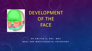
DEVELOPMENT
OF THE
FACE
D R A M I T H A G , B D S , M D S
O R A L A N D M A X I L L O F A C I A L P A T H O L O G Y
Face develops in humans between 4th – 10th
week of intrauterine life.
PRENATAL GROWTH OF THE MAXILLA
4th week of intrauterine life
- Formation of the head fold
- Following which the developing brain and the pericardium form 2 prominent bulges on
the ventral aspect of the embryo.
- The 2 bulges are separated from each other by  a shallow depression called
stomatoedum (corresponding to the primitive mouth).
- Floor of the stomatodeum is formed by the Buccopharyngeal membrane, which
separates the stomatodeum from the foregut.
- Soon, mesoderm covering the developing
forebrain proliferates, and forms a
downward projection that overlaps the
upper part of the stomatodeum – this
downward projection is called frontonasal
process.
- During the same time (4th week of intrauterine life) five
branchial arches form in the region of the future head and
neck region
- The first branchial arch is called – mandibular arch and play
an important role in the development of the nasomaxillary
region.
- The mandibular arch forms the lateral wall of the
stomatodeum.
- This arch gives off a bud from its dorsal end – this is called
maxillary process (4th week).
- It grows ventromedially cranial to the main part of the arch
- Main part of the arch is now called mandibular process.
- Therefore the face is derived from
- Frontonasal structures
- First pharyngeal (mandibular) arch of each side
5th week of intrauterine life
- The ectoderm overlying the frontonasal process soon shows bilateral
localized thickenings that are situated a little above (caudal or
raustral to) the stomatodeum. These are the nasal placodes or
olfactory placodes.
- The formation of these placodes is induced by the underlying
forebrain.
- The placodes soon sink below the surface to form the nasal pits.
- The nasal pits are continuous with the stomatodeum below.
- The edges of each pit are raised above the surface
- Medial raised edge is called – medial nasal process.
- Lateral raised edge – called lateral nasal process
- Now, all the primordial for the formation of lip and primary
palate are present
- Medial nasal process
- Lateral nasal formed
- Maxillary process
DEVELOPMENT OF UPPER LIP
- Maxillary process enlarges
- Maxillary process now grows medially and approaches the
lateral and medial nasal processes but remains separated
from them by distinct grooves (naso-optic groove and
bucco nasal groove)
- From each furrow or groove  a solid ectodermal rod of
cells sink below the surface and canalizes to form
nasolacrimal duct.
- Medial growth of the maxillary process further fuses
first with the lateral nasal process and then medial
nasal process.
- The medial and lateral nasal processes also fuse with
each other.
- This way the nasal pits  now called external nares
are cut off from the stomatodeum
- The maxillary process undergoes considerable growth
 therefore, pushes the medial nasal process towards
the midline, where it merges with its anatomic
counterpart from the opposite side.
- At the same time the frontonasal process becomes
much narrower from side to side, with the result that
the 2 external nares come close to each other.
6th week of intrauterine life
- 2 theories have been proposed for the continued development of the upper lip beyond the
6th week.
1st theory
- Mesenchyme of the maxillary process entirely overgrows the mesenchyme of the medial
nasal process to meet in the midline and thus contribute all the tissue for the upper lip.
- This is based on the fact
 Maxillary process  supplied by maxillary nerve
 Fronto nasal process  by opthalmic nerve
 Fully formed upper lip  supplied by infraorbital branch of the maxillary nerve)
2nd theory
- Maxillary process merely meets the medial nasal process without any overgrowth or
mesenchymal invasion.
- Therefore the middle third of the upper lip is derived from the merged medial nasal process
of frontonasal process.
- Histological evidence favours this
Mouth
- Stomatodeum is now bounded above by the Upper lip which is derived as follows
o Mesoderm of the lateral part of the lip is formed from the maxillary process.
o The overlying skin is derived from ectoderm covering this process
o Mesoderm of the median part of the lips (philtrum) is formed from the frontonasal
process.
o Ectoderm of the maxillary process overgrows this mesoderm to meet that of the opposite
maxillary process in the midline
o As a result skin of the entire upper lip is innervated by the maxillary nerves.
o Muscles of the face (including the lips)  are derived from the mesoderm of the second
branchial arch and are therefore supplied by facial nerve.
Development of lower lip
- At 6th week of embryo
- The mandibular process of the 2 sides grow towards each
other and fuse in the midline
- They form the lower margin of the stomatodeum.
- The fused mandibular process gives rise to lower lip and
the lower jaw
Development of nose
- Nose receives contributions from
 Frontonasal process
 Medial nasal process
 Lateral nasal process
- External nares are formed when the nasal pits are cut off from the stomatodeum by the fusion
of the maxillary process with medial nasal process.
- The external nares gradually approach each other as the maxillary process grows  pushing
the frontonasal process towards each other.
- Deeper part of the frontonasal process  forms nasal septum
- Mesoderm becomes heaped up in the median plane to form the prominence of the nose.
- Simultaneously a groove appear between the region of the nose and the bulging forebrain
(now called forehead)
- As the nose becomes prominent  the external nares comers to open downeards instead
of forwards.
- The external form of the nose is thus formed.
DEVELOPMENT OF CHEEKS
- After formation of the upper and lower lips, the stomatodeum (now called mouth) is very
broad.
- In its lateral part it is bounded
 Above by the maxillary process
 Below by the mandibular process
- These process undergo progressive fusion with each other to form cheeks
- The maxillary process fuses with the lateral nasal process  in the region of the lip + extends
from the stomatodeum to medial amgle of the developing eye this line of fusion is marked
by a groove called the naso optic furrow or nasolacrimal sulcus
- A strip of ectoderm becomes buried along this furrow and gives rise to the nasolacrimal duct.
POST NATAL GROWTH OF THE MAXILLA
- Growth of the nasomaxillary complex is produced by the following mechanisms
o Displacement
o Growth of the sutures
o Surface modeling
Displacement
- 2 types of displacement
 Primary displacement (bone is displaced by its own enlargement)
 Primary displacement of the maxilla  in a forwards direction
 This occurs by growth of the maxillary tuberosity in a posterior direction
 This results in the whole maxilla being carried anteriorly
 The amount of forwards displacement = amount of posterior lengthening
 Secondary or passive displacement (bone is not displaced by its own enlargement, rather a
passive displacement by the growth of the cranial base)
 Maxilla is attached to the cranial base by means of a number of sutures
 Therefore when the cranial base grows  produces a direct effect on the nasomaxillary
complex
 Cranial base grows  Middle cranial fossa grows anteriorly  therefore the passive
pressure produced by the cranial base  pushes the nasomaxillary complex  downwards
and forwards.
Growth at the sutures
- Maxilla is connected to the cranium and cranial base by a number of sutures
 Fronto nasal suture
 Fronto maxillary suture
 Zygomaticotemporal suture
 Zygomatico maxillary suture
 Pterygo palatine suture
- These sutures are oblique and almost parallel to each other
- This allows the downward and forward repositioning of the maxilla as growth occurs at
these sutures.
- Tension related bone formation occurs at sutures.
- Growth of the surrounding soft tissues  carries the maxilla downeards and forward 
leading to opening up of space at the sutural attachements  new bone is now formed on
either side of the suture  increasing the overall size of the bones on either side.
Surface remodeling
- Remodeling of bone by bone deposition and resorption occurs to bring about
 Increase in size
 Change in shape of the bone
 Change in functional relationship.
- Bone remodeling changes taking place in nasomaxillary complex are
 At the orbital rim
 Lateral surface  resorption
To compensate
 Medial surface of the rim  deposition
 External surface of the lateral rim  deposition
 At the Orbital floor
 Orbital floor faces superiorly, laterally and anteriorly
 Surface deposition occurs here and results in  growth in a superior, lateral and anterior direction
 At the maxillary tuberosity
 Bone deposition occurs along the posterior margin  causing the lengthening of the dental arch  and
enlargement of the antero posterior dimension of the entire maxillary body  helps accommodating the
erupting molars.
 At the nose
 Laterall wall of nose  resorption  leading to increase in size of the nasal cavity
 Floor of the nasal cavity
 Floor of the nasal cavity  resorption
To compensate
 Palatal aspect  deposition  therefore net downward shift occurs  leading to increase in maxillary height
 At the zygomatic bone
 Anterior surface  resorption
 Posterior surface  deposition
 The zygomatic bone  moves in a posterior direction.
 At zygomatic arch
 Lateral surface  deposition
 Medial surface  resorption
 Anterior nasal spine  bone deposition  becomes prominent
 Alveolar margins  As teeth erupts  deposition  increasing the maxillary height and
depth of the palate.
 Maxillary sinus  entire wall of the sinus except the mesial wall  undergoes resorption 
results in increase in size of maxillary antrum.
CLINICAL CONSIDERATIONS
- Since the formation of various parts of the face involves fusion of diverse components.
- Occasionally this fusion can be incomplete  give rise to various anomalies
HARE LIP
- Upper lip of the hare normally has a cleft
- Hence the term hare lip is used for defects of the lips.
- When 1 or both maxillary processes do not fuse with the
medial nasal process  this gives rise to defects in upper lip.
- This may vary in degree
- Unilateral or bilateral
b) Defective development of lowermost part of the
frontonasal process  give rise to a midline defect of ht
upper lip
c) When 2 mandibular process  do not fuse  with each
other  lower lip shows a defect in midline  the defect
usually extends to the jaw.
OBLIQUE FACIAL CLEFT
- Non fusion of the maxillary an dlateral nasal processes gives rise to a cleft running from the
medial angle of the eye to mouth.
- Nasolacrimal duct is also not formed.
MACROSTOMIA
- Inadequate fusion of mandibular and maxillary processes with each other  leads to
abnormally wide mouth  macrostomia
LATERAL FACIAL CLEFT
- Unilateral lack of fusion of mandibular and maxillary process  forms LATERAL FACIAL
CLEFT.
MICROSTOMIA
- Too much fusion of mandibular andmaxillary processes  may result  in small mouth
MANDIBULOFACIAL DYSOSTOSIS OR FIRST ARCH SYNDROME
- Entire first arch may remain underdeveloped on one or both sides, affecting
 Lower eyelid
 Maxilla
 Mandible
 External ear.
- Prominence of the cheek is absent
- Ear is displaced ventrally and caudally
HEMIFACIAL HYPERTROPHY
- One half of the face is overdeveloped
HEMIFACIAL ATROPHY
- One half of the face is under developed
RETROGNATHIA
- Mandible may be small compared to the rest of the face resulting in a receding chin.
HYPERTELORISM
- Eyes may be widely separated
PARAMEDIAN OR COMMISURAL PITS
- Lips showing congenital pits or fistula
DOUBLE LIP
- Lips are double
CONGENITAL TUMORS
- May be present in relation to the face.
ANOMALIES OF NOSE
Nose may be bifid
One half of nose is absent
Proboscis – nose forms a cylindrical projection jutting out from just below the forehead.
This anomaly may sometimes affect only one half of the nose and usually associated with
fusion of the eyes  Cyclops
THANK YOU

Más contenido relacionado

La actualidad más candente

La actualidad más candente (20)

development and growth of teeth
development and growth of teethdevelopment and growth of teeth
development and growth of teeth
 
oral mucous membrane
oral mucous membraneoral mucous membrane
oral mucous membrane
 
Amelogenesis Slideshare.pptx
Amelogenesis Slideshare.pptxAmelogenesis Slideshare.pptx
Amelogenesis Slideshare.pptx
 
Odontogenesis
Odontogenesis  Odontogenesis
Odontogenesis
 
Development of Face and Palate including Anomalies of palate
Development of Face and Palate including Anomalies of palate Development of Face and Palate including Anomalies of palate
Development of Face and Palate including Anomalies of palate
 
Dentinogenesis
DentinogenesisDentinogenesis
Dentinogenesis
 
Cementum
Cementum Cementum
Cementum
 
Tongue development, applied anatomy and prosthetic implications
Tongue development, applied anatomy and prosthetic implicationsTongue development, applied anatomy and prosthetic implications
Tongue development, applied anatomy and prosthetic implications
 
Development & Growth of Maxilla
Development & Growth of Maxilla  Development & Growth of Maxilla
Development & Growth of Maxilla
 
Maxillary artery
Maxillary arteryMaxillary artery
Maxillary artery
 
Anatomy of maxilla and mandible
Anatomy of maxilla and mandibleAnatomy of maxilla and mandible
Anatomy of maxilla and mandible
 
Tooth development
Tooth developmentTooth development
Tooth development
 
Mandibular nerve
Mandibular nerve Mandibular nerve
Mandibular nerve
 
Life cycle of ameloblast
Life cycle of ameloblastLife cycle of ameloblast
Life cycle of ameloblast
 
The maxillary nerve
The maxillary nerveThe maxillary nerve
The maxillary nerve
 
Tooth eruption
Tooth eruptionTooth eruption
Tooth eruption
 
Development of Mandible
Development of MandibleDevelopment of Mandible
Development of Mandible
 
Amelogenesis
Amelogenesis Amelogenesis
Amelogenesis
 
Histology of oral mucous membrane and gingiva
Histology of oral mucous membrane and gingivaHistology of oral mucous membrane and gingiva
Histology of oral mucous membrane and gingiva
 
Muscles of Mastication
Muscles of MasticationMuscles of Mastication
Muscles of Mastication
 

Similar a Development of Face

Growth and Development of maxilla and nasomaxillary complex
Growth and Development of maxilla and nasomaxillary complexGrowth and Development of maxilla and nasomaxillary complex
Growth and Development of maxilla and nasomaxillary complexRaahat Vikram singh
 
Development of face including nose ppt
Development of face including nose ppt Development of face including nose ppt
Development of face including nose ppt BharanidharanS5
 
Development of face, nose, palate by dr. noura 2014
Development of face, nose, palate by dr. noura 2014Development of face, nose, palate by dr. noura 2014
Development of face, nose, palate by dr. noura 2014Dr. Noura El Tahawy
 
PPT ............................CLG.pptx
PPT ............................CLG.pptxPPT ............................CLG.pptx
PPT ............................CLG.pptxHimanshu Goyal
 
Growth & Development of Maxilla
Growth & Development of MaxillaGrowth & Development of Maxilla
Growth & Development of MaxillaSaibel Farishta
 
Lecture 13 face, nose palate 22042010 pdf
Lecture 13 face, nose palate 22042010 pdfLecture 13 face, nose palate 22042010 pdf
Lecture 13 face, nose palate 22042010 pdfasyaari
 
Lecture 13 face, nose palate 22042010 pdf
Lecture 13 face, nose palate 22042010 pdfLecture 13 face, nose palate 22042010 pdf
Lecture 13 face, nose palate 22042010 pdfdhina raddy
 
Face development of a fetus
Face development of a fetusFace development of a fetus
Face development of a fetusalyssaazlan
 
Prenatal and postnatal growth & development of maxilla and palate presented b...
Prenatal and postnatal growth & development of maxilla and palate presented b...Prenatal and postnatal growth & development of maxilla and palate presented b...
Prenatal and postnatal growth & development of maxilla and palate presented b...Dr. Himanshu Gorawat
 
SURGICAL ANATOMY OF MID FACE.pptx
SURGICAL ANATOMY OF MID FACE.pptxSURGICAL ANATOMY OF MID FACE.pptx
SURGICAL ANATOMY OF MID FACE.pptxshalini sampreethi
 
EMBERYOLOGY AND ANATOMY OF EXTERNAL NOSE.pptx
EMBERYOLOGY AND ANATOMY OF EXTERNAL NOSE.pptxEMBERYOLOGY AND ANATOMY OF EXTERNAL NOSE.pptx
EMBERYOLOGY AND ANATOMY OF EXTERNAL NOSE.pptxAhlam Alzuway
 
Development-of-the-face-and-the-palate.pdf
Development-of-the-face-and-the-palate.pdfDevelopment-of-the-face-and-the-palate.pdf
Development-of-the-face-and-the-palate.pdfAmulyaSnr
 
Development of maxilla
Development of maxillaDevelopment of maxilla
Development of maxillaAnanthesh Rao
 
Growth and Development of Nasomaxillary complex PPT.ppt
Growth and Development of Nasomaxillary complex PPT.pptGrowth and Development of Nasomaxillary complex PPT.ppt
Growth and Development of Nasomaxillary complex PPT.pptchandrashekarpatil15
 
Growth & development of maxilla and mandible
Growth & development of maxilla and mandibleGrowth & development of maxilla and mandible
Growth & development of maxilla and mandibleRajesh Bariker
 
Development of face and palate
Development of face and palateDevelopment of face and palate
Development of face and palateRajvi Nahar
 
Formation of the palate
Formation of the palateFormation of the palate
Formation of the palateLheanne Tesoro
 
Osteology of Maxilla
Osteology of MaxillaOsteology of Maxilla
Osteology of MaxillaJenin N T
 

Similar a Development of Face (20)

Growth and Development of maxilla and nasomaxillary complex
Growth and Development of maxilla and nasomaxillary complexGrowth and Development of maxilla and nasomaxillary complex
Growth and Development of maxilla and nasomaxillary complex
 
Development of face including nose ppt
Development of face including nose ppt Development of face including nose ppt
Development of face including nose ppt
 
Development of face, nose, palate by dr. noura 2014
Development of face, nose, palate by dr. noura 2014Development of face, nose, palate by dr. noura 2014
Development of face, nose, palate by dr. noura 2014
 
PPT ............................CLG.pptx
PPT ............................CLG.pptxPPT ............................CLG.pptx
PPT ............................CLG.pptx
 
Growth & Development of Maxilla
Growth & Development of MaxillaGrowth & Development of Maxilla
Growth & Development of Maxilla
 
Maxxilla seminar ih
Maxxilla seminar  ihMaxxilla seminar  ih
Maxxilla seminar ih
 
Lecture 13 face, nose palate 22042010 pdf
Lecture 13 face, nose palate 22042010 pdfLecture 13 face, nose palate 22042010 pdf
Lecture 13 face, nose palate 22042010 pdf
 
Lecture 13 face, nose palate 22042010 pdf
Lecture 13 face, nose palate 22042010 pdfLecture 13 face, nose palate 22042010 pdf
Lecture 13 face, nose palate 22042010 pdf
 
Face development of a fetus
Face development of a fetusFace development of a fetus
Face development of a fetus
 
Prenatal and postnatal growth & development of maxilla and palate presented b...
Prenatal and postnatal growth & development of maxilla and palate presented b...Prenatal and postnatal growth & development of maxilla and palate presented b...
Prenatal and postnatal growth & development of maxilla and palate presented b...
 
SURGICAL ANATOMY OF MID FACE.pptx
SURGICAL ANATOMY OF MID FACE.pptxSURGICAL ANATOMY OF MID FACE.pptx
SURGICAL ANATOMY OF MID FACE.pptx
 
Development of palate
Development of palateDevelopment of palate
Development of palate
 
EMBERYOLOGY AND ANATOMY OF EXTERNAL NOSE.pptx
EMBERYOLOGY AND ANATOMY OF EXTERNAL NOSE.pptxEMBERYOLOGY AND ANATOMY OF EXTERNAL NOSE.pptx
EMBERYOLOGY AND ANATOMY OF EXTERNAL NOSE.pptx
 
Development-of-the-face-and-the-palate.pdf
Development-of-the-face-and-the-palate.pdfDevelopment-of-the-face-and-the-palate.pdf
Development-of-the-face-and-the-palate.pdf
 
Development of maxilla
Development of maxillaDevelopment of maxilla
Development of maxilla
 
Growth and Development of Nasomaxillary complex PPT.ppt
Growth and Development of Nasomaxillary complex PPT.pptGrowth and Development of Nasomaxillary complex PPT.ppt
Growth and Development of Nasomaxillary complex PPT.ppt
 
Growth & development of maxilla and mandible
Growth & development of maxilla and mandibleGrowth & development of maxilla and mandible
Growth & development of maxilla and mandible
 
Development of face and palate
Development of face and palateDevelopment of face and palate
Development of face and palate
 
Formation of the palate
Formation of the palateFormation of the palate
Formation of the palate
 
Osteology of Maxilla
Osteology of MaxillaOsteology of Maxilla
Osteology of Maxilla
 

Más de oral and maxillofacial pathology (20)

Saliva as a Diagnostic Tool
Saliva as a Diagnostic ToolSaliva as a Diagnostic Tool
Saliva as a Diagnostic Tool
 
TONGUE ppt
TONGUE pptTONGUE ppt
TONGUE ppt
 
SQUAMOUS CELL CARCINOMA - ORAL CANCER PPT
SQUAMOUS CELL CARCINOMA - ORAL CANCER PPTSQUAMOUS CELL CARCINOMA - ORAL CANCER PPT
SQUAMOUS CELL CARCINOMA - ORAL CANCER PPT
 
Levels of Lymph Nodes
Levels of Lymph NodesLevels of Lymph Nodes
Levels of Lymph Nodes
 
DEGLUTTITION (SWALLOWING)
DEGLUTTITION (SWALLOWING)DEGLUTTITION (SWALLOWING)
DEGLUTTITION (SWALLOWING)
 
TONGUE
TONGUETONGUE
TONGUE
 
Trigeminal Nerve
Trigeminal NerveTrigeminal Nerve
Trigeminal Nerve
 
PAGETS DISEASE
PAGETS DISEASEPAGETS DISEASE
PAGETS DISEASE
 
Dental Caries
Dental CariesDental Caries
Dental Caries
 
Lymph Nodes
Lymph NodesLymph Nodes
Lymph Nodes
 
Research Microscopes
Research MicroscopesResearch Microscopes
Research Microscopes
 
Maxillary Second Premolar
Maxillary Second PremolarMaxillary Second Premolar
Maxillary Second Premolar
 
Mandibular Canine
Mandibular CanineMandibular Canine
Mandibular Canine
 
Maxillary Central Incisor
Maxillary Central IncisorMaxillary Central Incisor
Maxillary Central Incisor
 
ENAMEL
ENAMELENAMEL
ENAMEL
 
Developmental Of The Mandible
Developmental Of The MandibleDevelopmental Of The Mandible
Developmental Of The Mandible
 
Developmental Disturbances of The Face
Developmental Disturbances of The FaceDevelopmental Disturbances of The Face
Developmental Disturbances of The Face
 
TMJ- 3
TMJ- 3TMJ- 3
TMJ- 3
 
TMJ DISORDER - 2
TMJ DISORDER - 2TMJ DISORDER - 2
TMJ DISORDER - 2
 
TMJ DISORDER- 1
TMJ DISORDER- 1TMJ DISORDER- 1
TMJ DISORDER- 1
 

Último

See the 2,456 pharmacies on the National E-Pharmacy Platform
See the 2,456 pharmacies on the National E-Pharmacy PlatformSee the 2,456 pharmacies on the National E-Pharmacy Platform
See the 2,456 pharmacies on the National E-Pharmacy PlatformKweku Zurek
 
Radiation Dosimetry Parameters and Isodose Curves.pptx
Radiation Dosimetry Parameters and Isodose Curves.pptxRadiation Dosimetry Parameters and Isodose Curves.pptx
Radiation Dosimetry Parameters and Isodose Curves.pptxDr. Dheeraj Kumar
 
world health day presentation ppt download
world health day presentation ppt downloadworld health day presentation ppt download
world health day presentation ppt downloadAnkitKumar311566
 
The next social challenge to public health: the information environment.pptx
The next social challenge to public health:  the information environment.pptxThe next social challenge to public health:  the information environment.pptx
The next social challenge to public health: the information environment.pptxTina Purnat
 
Statistical modeling in pharmaceutical research and development.
Statistical modeling in pharmaceutical research and development.Statistical modeling in pharmaceutical research and development.
Statistical modeling in pharmaceutical research and development.ANJALI
 
Apiculture Chapter 1. Introduction 2.ppt
Apiculture Chapter 1. Introduction 2.pptApiculture Chapter 1. Introduction 2.ppt
Apiculture Chapter 1. Introduction 2.pptkedirjemalharun
 
Introduction to Sports Injuries by- Dr. Anjali Rai
Introduction to Sports Injuries by- Dr. Anjali RaiIntroduction to Sports Injuries by- Dr. Anjali Rai
Introduction to Sports Injuries by- Dr. Anjali RaiGoogle
 
call girls in Dwarka Sector 21 Metro DELHI 🔝 >༒9540349809 🔝 genuine Escort Se...
call girls in Dwarka Sector 21 Metro DELHI 🔝 >༒9540349809 🔝 genuine Escort Se...call girls in Dwarka Sector 21 Metro DELHI 🔝 >༒9540349809 🔝 genuine Escort Se...
call girls in Dwarka Sector 21 Metro DELHI 🔝 >༒9540349809 🔝 genuine Escort Se...saminamagar
 
Primary headache and facial pain. (2024)
Primary headache and facial pain. (2024)Primary headache and facial pain. (2024)
Primary headache and facial pain. (2024)Mohamed Rizk Khodair
 
call girls in Connaught Place DELHI 🔝 >༒9540349809 🔝 genuine Escort Service ...
call girls in Connaught Place  DELHI 🔝 >༒9540349809 🔝 genuine Escort Service ...call girls in Connaught Place  DELHI 🔝 >༒9540349809 🔝 genuine Escort Service ...
call girls in Connaught Place DELHI 🔝 >༒9540349809 🔝 genuine Escort Service ...saminamagar
 
POST NATAL EXERCISES AND ITS IMPACT.pptx
POST NATAL EXERCISES AND ITS IMPACT.pptxPOST NATAL EXERCISES AND ITS IMPACT.pptx
POST NATAL EXERCISES AND ITS IMPACT.pptxvirengeeta
 
SWD (Short wave diathermy)- Physiotherapy.ppt
SWD (Short wave diathermy)- Physiotherapy.pptSWD (Short wave diathermy)- Physiotherapy.ppt
SWD (Short wave diathermy)- Physiotherapy.pptMumux Mirani
 
SYNDESMOTIC INJURY- ANATOMICAL REPAIR.pptx
SYNDESMOTIC INJURY- ANATOMICAL REPAIR.pptxSYNDESMOTIC INJURY- ANATOMICAL REPAIR.pptx
SYNDESMOTIC INJURY- ANATOMICAL REPAIR.pptxdrashraf369
 
Presentation on Parasympathetic Nervous System
Presentation on Parasympathetic Nervous SystemPresentation on Parasympathetic Nervous System
Presentation on Parasympathetic Nervous SystemPrerana Jadhav
 
History and Development of Pharmacovigilence.pdf
History and Development of Pharmacovigilence.pdfHistory and Development of Pharmacovigilence.pdf
History and Development of Pharmacovigilence.pdfSasikiranMarri
 
Biomechanics- Shoulder Joint!!!!!!!!!!!!
Biomechanics- Shoulder Joint!!!!!!!!!!!!Biomechanics- Shoulder Joint!!!!!!!!!!!!
Biomechanics- Shoulder Joint!!!!!!!!!!!!ibtesaam huma
 
epilepsy and status epilepticus for undergraduate.pptx
epilepsy and status epilepticus  for undergraduate.pptxepilepsy and status epilepticus  for undergraduate.pptx
epilepsy and status epilepticus for undergraduate.pptxMohamed Rizk Khodair
 
Glomerular Filtration and determinants of glomerular filtration .pptx
Glomerular Filtration and  determinants of glomerular filtration .pptxGlomerular Filtration and  determinants of glomerular filtration .pptx
Glomerular Filtration and determinants of glomerular filtration .pptxDr.Nusrat Tariq
 
April 2024 ONCOLOGY CARTOON by DR KANHU CHARAN PATRO
April 2024 ONCOLOGY CARTOON by  DR KANHU CHARAN PATROApril 2024 ONCOLOGY CARTOON by  DR KANHU CHARAN PATRO
April 2024 ONCOLOGY CARTOON by DR KANHU CHARAN PATROKanhu Charan
 
PNEUMOTHORAX AND ITS MANAGEMENTS.pdf
PNEUMOTHORAX   AND  ITS  MANAGEMENTS.pdfPNEUMOTHORAX   AND  ITS  MANAGEMENTS.pdf
PNEUMOTHORAX AND ITS MANAGEMENTS.pdfDolisha Warbi
 

Último (20)

See the 2,456 pharmacies on the National E-Pharmacy Platform
See the 2,456 pharmacies on the National E-Pharmacy PlatformSee the 2,456 pharmacies on the National E-Pharmacy Platform
See the 2,456 pharmacies on the National E-Pharmacy Platform
 
Radiation Dosimetry Parameters and Isodose Curves.pptx
Radiation Dosimetry Parameters and Isodose Curves.pptxRadiation Dosimetry Parameters and Isodose Curves.pptx
Radiation Dosimetry Parameters and Isodose Curves.pptx
 
world health day presentation ppt download
world health day presentation ppt downloadworld health day presentation ppt download
world health day presentation ppt download
 
The next social challenge to public health: the information environment.pptx
The next social challenge to public health:  the information environment.pptxThe next social challenge to public health:  the information environment.pptx
The next social challenge to public health: the information environment.pptx
 
Statistical modeling in pharmaceutical research and development.
Statistical modeling in pharmaceutical research and development.Statistical modeling in pharmaceutical research and development.
Statistical modeling in pharmaceutical research and development.
 
Apiculture Chapter 1. Introduction 2.ppt
Apiculture Chapter 1. Introduction 2.pptApiculture Chapter 1. Introduction 2.ppt
Apiculture Chapter 1. Introduction 2.ppt
 
Introduction to Sports Injuries by- Dr. Anjali Rai
Introduction to Sports Injuries by- Dr. Anjali RaiIntroduction to Sports Injuries by- Dr. Anjali Rai
Introduction to Sports Injuries by- Dr. Anjali Rai
 
call girls in Dwarka Sector 21 Metro DELHI 🔝 >༒9540349809 🔝 genuine Escort Se...
call girls in Dwarka Sector 21 Metro DELHI 🔝 >༒9540349809 🔝 genuine Escort Se...call girls in Dwarka Sector 21 Metro DELHI 🔝 >༒9540349809 🔝 genuine Escort Se...
call girls in Dwarka Sector 21 Metro DELHI 🔝 >༒9540349809 🔝 genuine Escort Se...
 
Primary headache and facial pain. (2024)
Primary headache and facial pain. (2024)Primary headache and facial pain. (2024)
Primary headache and facial pain. (2024)
 
call girls in Connaught Place DELHI 🔝 >༒9540349809 🔝 genuine Escort Service ...
call girls in Connaught Place  DELHI 🔝 >༒9540349809 🔝 genuine Escort Service ...call girls in Connaught Place  DELHI 🔝 >༒9540349809 🔝 genuine Escort Service ...
call girls in Connaught Place DELHI 🔝 >༒9540349809 🔝 genuine Escort Service ...
 
POST NATAL EXERCISES AND ITS IMPACT.pptx
POST NATAL EXERCISES AND ITS IMPACT.pptxPOST NATAL EXERCISES AND ITS IMPACT.pptx
POST NATAL EXERCISES AND ITS IMPACT.pptx
 
SWD (Short wave diathermy)- Physiotherapy.ppt
SWD (Short wave diathermy)- Physiotherapy.pptSWD (Short wave diathermy)- Physiotherapy.ppt
SWD (Short wave diathermy)- Physiotherapy.ppt
 
SYNDESMOTIC INJURY- ANATOMICAL REPAIR.pptx
SYNDESMOTIC INJURY- ANATOMICAL REPAIR.pptxSYNDESMOTIC INJURY- ANATOMICAL REPAIR.pptx
SYNDESMOTIC INJURY- ANATOMICAL REPAIR.pptx
 
Presentation on Parasympathetic Nervous System
Presentation on Parasympathetic Nervous SystemPresentation on Parasympathetic Nervous System
Presentation on Parasympathetic Nervous System
 
History and Development of Pharmacovigilence.pdf
History and Development of Pharmacovigilence.pdfHistory and Development of Pharmacovigilence.pdf
History and Development of Pharmacovigilence.pdf
 
Biomechanics- Shoulder Joint!!!!!!!!!!!!
Biomechanics- Shoulder Joint!!!!!!!!!!!!Biomechanics- Shoulder Joint!!!!!!!!!!!!
Biomechanics- Shoulder Joint!!!!!!!!!!!!
 
epilepsy and status epilepticus for undergraduate.pptx
epilepsy and status epilepticus  for undergraduate.pptxepilepsy and status epilepticus  for undergraduate.pptx
epilepsy and status epilepticus for undergraduate.pptx
 
Glomerular Filtration and determinants of glomerular filtration .pptx
Glomerular Filtration and  determinants of glomerular filtration .pptxGlomerular Filtration and  determinants of glomerular filtration .pptx
Glomerular Filtration and determinants of glomerular filtration .pptx
 
April 2024 ONCOLOGY CARTOON by DR KANHU CHARAN PATRO
April 2024 ONCOLOGY CARTOON by  DR KANHU CHARAN PATROApril 2024 ONCOLOGY CARTOON by  DR KANHU CHARAN PATRO
April 2024 ONCOLOGY CARTOON by DR KANHU CHARAN PATRO
 
PNEUMOTHORAX AND ITS MANAGEMENTS.pdf
PNEUMOTHORAX   AND  ITS  MANAGEMENTS.pdfPNEUMOTHORAX   AND  ITS  MANAGEMENTS.pdf
PNEUMOTHORAX AND ITS MANAGEMENTS.pdf
 

Development of Face

  • 1. DEVELOPMENT OF THE FACE D R A M I T H A G , B D S , M D S O R A L A N D M A X I L L O F A C I A L P A T H O L O G Y
  • 2. Face develops in humans between 4th – 10th week of intrauterine life.
  • 3. PRENATAL GROWTH OF THE MAXILLA 4th week of intrauterine life - Formation of the head fold - Following which the developing brain and the pericardium form 2 prominent bulges on the ventral aspect of the embryo. - The 2 bulges are separated from each other by  a shallow depression called stomatoedum (corresponding to the primitive mouth). - Floor of the stomatodeum is formed by the Buccopharyngeal membrane, which separates the stomatodeum from the foregut.
  • 4. - Soon, mesoderm covering the developing forebrain proliferates, and forms a downward projection that overlaps the upper part of the stomatodeum – this downward projection is called frontonasal process.
  • 5. - During the same time (4th week of intrauterine life) five branchial arches form in the region of the future head and neck region - The first branchial arch is called – mandibular arch and play an important role in the development of the nasomaxillary region. - The mandibular arch forms the lateral wall of the stomatodeum. - This arch gives off a bud from its dorsal end – this is called maxillary process (4th week). - It grows ventromedially cranial to the main part of the arch - Main part of the arch is now called mandibular process.
  • 6. - Therefore the face is derived from - Frontonasal structures - First pharyngeal (mandibular) arch of each side
  • 7. 5th week of intrauterine life - The ectoderm overlying the frontonasal process soon shows bilateral localized thickenings that are situated a little above (caudal or raustral to) the stomatodeum. These are the nasal placodes or olfactory placodes. - The formation of these placodes is induced by the underlying forebrain. - The placodes soon sink below the surface to form the nasal pits. - The nasal pits are continuous with the stomatodeum below. - The edges of each pit are raised above the surface - Medial raised edge is called – medial nasal process. - Lateral raised edge – called lateral nasal process
  • 8. - Now, all the primordial for the formation of lip and primary palate are present - Medial nasal process - Lateral nasal formed - Maxillary process
  • 9. DEVELOPMENT OF UPPER LIP - Maxillary process enlarges - Maxillary process now grows medially and approaches the lateral and medial nasal processes but remains separated from them by distinct grooves (naso-optic groove and bucco nasal groove) - From each furrow or groove  a solid ectodermal rod of cells sink below the surface and canalizes to form nasolacrimal duct.
  • 10. - Medial growth of the maxillary process further fuses first with the lateral nasal process and then medial nasal process. - The medial and lateral nasal processes also fuse with each other. - This way the nasal pits  now called external nares are cut off from the stomatodeum
  • 11. - The maxillary process undergoes considerable growth  therefore, pushes the medial nasal process towards the midline, where it merges with its anatomic counterpart from the opposite side. - At the same time the frontonasal process becomes much narrower from side to side, with the result that the 2 external nares come close to each other.
  • 12. 6th week of intrauterine life - 2 theories have been proposed for the continued development of the upper lip beyond the 6th week. 1st theory - Mesenchyme of the maxillary process entirely overgrows the mesenchyme of the medial nasal process to meet in the midline and thus contribute all the tissue for the upper lip. - This is based on the fact  Maxillary process  supplied by maxillary nerve  Fronto nasal process  by opthalmic nerve  Fully formed upper lip  supplied by infraorbital branch of the maxillary nerve)
  • 13. 2nd theory - Maxillary process merely meets the medial nasal process without any overgrowth or mesenchymal invasion. - Therefore the middle third of the upper lip is derived from the merged medial nasal process of frontonasal process. - Histological evidence favours this
  • 14. Mouth - Stomatodeum is now bounded above by the Upper lip which is derived as follows o Mesoderm of the lateral part of the lip is formed from the maxillary process. o The overlying skin is derived from ectoderm covering this process o Mesoderm of the median part of the lips (philtrum) is formed from the frontonasal process. o Ectoderm of the maxillary process overgrows this mesoderm to meet that of the opposite maxillary process in the midline o As a result skin of the entire upper lip is innervated by the maxillary nerves. o Muscles of the face (including the lips)  are derived from the mesoderm of the second branchial arch and are therefore supplied by facial nerve.
  • 15.
  • 16. Development of lower lip - At 6th week of embryo - The mandibular process of the 2 sides grow towards each other and fuse in the midline - They form the lower margin of the stomatodeum. - The fused mandibular process gives rise to lower lip and the lower jaw
  • 17. Development of nose - Nose receives contributions from  Frontonasal process  Medial nasal process  Lateral nasal process - External nares are formed when the nasal pits are cut off from the stomatodeum by the fusion of the maxillary process with medial nasal process. - The external nares gradually approach each other as the maxillary process grows  pushing the frontonasal process towards each other.
  • 18. - Deeper part of the frontonasal process  forms nasal septum - Mesoderm becomes heaped up in the median plane to form the prominence of the nose. - Simultaneously a groove appear between the region of the nose and the bulging forebrain (now called forehead) - As the nose becomes prominent  the external nares comers to open downeards instead of forwards. - The external form of the nose is thus formed.
  • 19. DEVELOPMENT OF CHEEKS - After formation of the upper and lower lips, the stomatodeum (now called mouth) is very broad. - In its lateral part it is bounded  Above by the maxillary process  Below by the mandibular process - These process undergo progressive fusion with each other to form cheeks - The maxillary process fuses with the lateral nasal process  in the region of the lip + extends from the stomatodeum to medial amgle of the developing eye this line of fusion is marked by a groove called the naso optic furrow or nasolacrimal sulcus - A strip of ectoderm becomes buried along this furrow and gives rise to the nasolacrimal duct.
  • 20. POST NATAL GROWTH OF THE MAXILLA - Growth of the nasomaxillary complex is produced by the following mechanisms o Displacement o Growth of the sutures o Surface modeling Displacement - 2 types of displacement  Primary displacement (bone is displaced by its own enlargement)  Primary displacement of the maxilla  in a forwards direction  This occurs by growth of the maxillary tuberosity in a posterior direction  This results in the whole maxilla being carried anteriorly  The amount of forwards displacement = amount of posterior lengthening
  • 21.  Secondary or passive displacement (bone is not displaced by its own enlargement, rather a passive displacement by the growth of the cranial base)  Maxilla is attached to the cranial base by means of a number of sutures  Therefore when the cranial base grows  produces a direct effect on the nasomaxillary complex  Cranial base grows  Middle cranial fossa grows anteriorly  therefore the passive pressure produced by the cranial base  pushes the nasomaxillary complex  downwards and forwards.
  • 22. Growth at the sutures - Maxilla is connected to the cranium and cranial base by a number of sutures  Fronto nasal suture  Fronto maxillary suture  Zygomaticotemporal suture  Zygomatico maxillary suture  Pterygo palatine suture
  • 23. - These sutures are oblique and almost parallel to each other - This allows the downward and forward repositioning of the maxilla as growth occurs at these sutures. - Tension related bone formation occurs at sutures. - Growth of the surrounding soft tissues  carries the maxilla downeards and forward  leading to opening up of space at the sutural attachements  new bone is now formed on either side of the suture  increasing the overall size of the bones on either side.
  • 24. Surface remodeling - Remodeling of bone by bone deposition and resorption occurs to bring about  Increase in size  Change in shape of the bone  Change in functional relationship. - Bone remodeling changes taking place in nasomaxillary complex are  At the orbital rim  Lateral surface  resorption To compensate  Medial surface of the rim  deposition  External surface of the lateral rim  deposition
  • 25.  At the Orbital floor  Orbital floor faces superiorly, laterally and anteriorly  Surface deposition occurs here and results in  growth in a superior, lateral and anterior direction  At the maxillary tuberosity  Bone deposition occurs along the posterior margin  causing the lengthening of the dental arch  and enlargement of the antero posterior dimension of the entire maxillary body  helps accommodating the erupting molars.  At the nose  Laterall wall of nose  resorption  leading to increase in size of the nasal cavity  Floor of the nasal cavity  Floor of the nasal cavity  resorption To compensate  Palatal aspect  deposition  therefore net downward shift occurs  leading to increase in maxillary height
  • 26.  At the zygomatic bone  Anterior surface  resorption  Posterior surface  deposition  The zygomatic bone  moves in a posterior direction.  At zygomatic arch  Lateral surface  deposition  Medial surface  resorption  Anterior nasal spine  bone deposition  becomes prominent  Alveolar margins  As teeth erupts  deposition  increasing the maxillary height and depth of the palate.  Maxillary sinus  entire wall of the sinus except the mesial wall  undergoes resorption  results in increase in size of maxillary antrum.
  • 27. CLINICAL CONSIDERATIONS - Since the formation of various parts of the face involves fusion of diverse components. - Occasionally this fusion can be incomplete  give rise to various anomalies
  • 28. HARE LIP - Upper lip of the hare normally has a cleft - Hence the term hare lip is used for defects of the lips. - When 1 or both maxillary processes do not fuse with the medial nasal process  this gives rise to defects in upper lip. - This may vary in degree - Unilateral or bilateral
  • 29. b) Defective development of lowermost part of the frontonasal process  give rise to a midline defect of ht upper lip c) When 2 mandibular process  do not fuse  with each other  lower lip shows a defect in midline  the defect usually extends to the jaw.
  • 30. OBLIQUE FACIAL CLEFT - Non fusion of the maxillary an dlateral nasal processes gives rise to a cleft running from the medial angle of the eye to mouth. - Nasolacrimal duct is also not formed.
  • 31. MACROSTOMIA - Inadequate fusion of mandibular and maxillary processes with each other  leads to abnormally wide mouth  macrostomia LATERAL FACIAL CLEFT - Unilateral lack of fusion of mandibular and maxillary process  forms LATERAL FACIAL CLEFT. MICROSTOMIA - Too much fusion of mandibular andmaxillary processes  may result  in small mouth
  • 32. MANDIBULOFACIAL DYSOSTOSIS OR FIRST ARCH SYNDROME - Entire first arch may remain underdeveloped on one or both sides, affecting  Lower eyelid  Maxilla  Mandible  External ear. - Prominence of the cheek is absent - Ear is displaced ventrally and caudally
  • 33. HEMIFACIAL HYPERTROPHY - One half of the face is overdeveloped HEMIFACIAL ATROPHY - One half of the face is under developed RETROGNATHIA - Mandible may be small compared to the rest of the face resulting in a receding chin. HYPERTELORISM - Eyes may be widely separated PARAMEDIAN OR COMMISURAL PITS - Lips showing congenital pits or fistula DOUBLE LIP - Lips are double
  • 34. CONGENITAL TUMORS - May be present in relation to the face. ANOMALIES OF NOSE Nose may be bifid One half of nose is absent Proboscis – nose forms a cylindrical projection jutting out from just below the forehead. This anomaly may sometimes affect only one half of the nose and usually associated with fusion of the eyes  Cyclops