
Recommended
Recommended
More Related Content
Similar to 8Hypersensitivity03.ppt
Similar to 8Hypersensitivity03.ppt (20)
More from AnguaniVictor
Recently uploaded
Recently uploaded (20)
8Hypersensitivity03.ppt
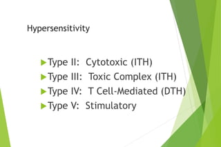
- 1. Hypersensitivity Type II: Cytotoxic (ITH) Type III: Toxic Complex (ITH) Type IV: T Cell-Mediated (DTH) Type V: Stimulatory
- 3. Characteristics of Cytotoxic Hypersensitivity Directed against cell surface or tissue antigen Characterized by complement cascade activation and various effector cells
- 4. Complement Formation of membrane attack complex (lytic enzymes) Activated C3 forms opsonin recognized by phagocytes Formation of chemotactic factors Effector cells possess Fc and complement receptors macrophages/monocytes neutrophils NK cells
- 5. Examples of Type II Hypersensitivity • Blood transfusion reactions • Hemolytic disease of the newborn (Rh disease) • Autoimmune hemolytic anemias • Drug reactions • Drug-induced loss of self-tolerance • Hyperacute graft rejection • Myasthenia gravis (acetylcholine receptor) • Sensitivity to tissue antigens
- 6. ABO Blood Group Antigens Precursor oligosaccharide H antigen B antigen NAcGA Gal NAG Fuc NAG Gal NAG Gal Fuc H Fuc Gal Gal NAG B A A antigen NAcGA (N-acetylgalactoseamine) Gal (galactose)
- 7. ABO Blood Group Reactivity blood group genotypes antigens antibodies to (phenotype) ABO in serum A AA, AO A anti-B B BB, BO B anti-A AB AB A and B none O OO H anti-A/B
- 8. Hemolytic Disease of the Newborn RhD positive red cells RhD negative mother RhD positive fetus Lysis Of RBC’s B cell anti-RhD first birth post partum subsequent anti-RhD RhD positive fetus
- 9. Drug-Induced Reactions: Adherence to Blood Components complement blood cell adsorbed drug or antigen drug metabolite antibody to drug lysis
- 10. Toxic Complex Hypersensitivity (Type III)
- 11. Diseases associated with immune complexes Persistent infection microbial antigens deposition of immune complexes in kidneys Autoimmunity self antigens deposition of immune complexes in kidneys, joints, arteries and skin Extrinsic factors environmental antigens deposition of immune complexes in lungs
- 12. Inflammatory Mechanisms in Type III • Complement activation • anaphylatoxins • Chemotactic factors • Neutrophils attracted • difficult to phagocytize tissue-trapped complexes • frustrated phagocytosis leads to tissue damage
- 13. Disease Models • Serum sickness • Arthus reaction
- 14. Serum Sickness
- 15. Arthus Reaction
- 16. T-Cell Mediated Hypersensitivity (Type IV / Delayed-Type)
- 17. Manifestations of T-Cell Mediated Hypersensitivity Allergic reactions to bacteria, viruses and fungi Contact dermatitis due to chemicals Rejection of tissue transplants
- 18. General Characteristics of DTH • An exaggerated interaction between antigen and normal CMI- mechanisms • Requires prior priming to antigen • Memory T-cells recognize antigen together with class II MHC molecules on antigen-presenting cells • Blast transformation and proliferation • Stimulated T-cells release soluble factors (cytokines) • Cytokines • attract and activate macrophages and/or eosinophils • help cytotoxic T-cells become killer cells, which cause tissue damage
- 19. Inducers of Type IV Hypersensitivity
- 20. Types of Delayed Hypersensitivity Delayed Reaction maximal reaction time Jones-Mote 24 hours Contact 48-72 hours tuberculin 48-72 hours granulomatous at least 14 days
- 21. Jones-Mote Hypersensitivity • Now referred to as “cutaneous basophil hypersensitivity” • Basophils are prominent as secondary infiltrating cells. • Basophilic infiltration of area under epidermis • Induced by soluble (weak) antigens • Transient dermal response • Prominent in reactions to viral antigens, in contact reactions, skin allograft rejections, reactions to tumor cells and in some cases of hypersensitivity pneumonitis (allergic alveolitis) • May be important in rejection of blood-feeding ticks on the skin surface
- 22. Contact Hypersensitivity • Usually maximal at 48 hours • Predominantly an epidermal reaction • Langerhans cells are the antigen presenting cells • a dendritic antigen presenting cell • carry antigen to lymph nodes draining skin • Associated with hapten-induced eczema • nickel salts in jewellry • picryl chloride • acrylates • p-Phenylene diamine in hair dyes • chromates • chemicals in rubber • poison ivy (urushiol)
- 24. Tuberculin Hypersensitivity • Maximum at 48-72 hours • Inflitration of lesion with mononuclear cells • First described as a reaction to the lipoprotein antigen of tubercle bacillus • Responsible for lesions associated with bacterial allergy • cavitation, caseation, general toxemia seen in TB • May progress to granulomatous reaction in unresolved infection
- 25. Granulomatous Hypersensitivity • Clinically, the most important form of DTH, since it causes many of the pathological effects in diseases which involve T cell-mediated immunity • Maximal at 14 days • Continual release of cytokines • Leads to accumulation of large numbers of macrophages • Granulomas can also arise from persistence of “indigestible” antigen such as talc (absence of lymphocytes in lesion)
- 26. Epitheloid Cell Granuloma Formation Large flattened cells with increased endoplasmic reticulum Multinucleate giant cells with little ER May see necrosis Damage due to killer T-cells recognizing antigen-coated macrophages, cytokine-activated macrophages Attempt by the body to wall-off site of persistent infection
- 28. Examples of Microbial-Induced DTH • Viruses (destructive skin rashes) • smallpox • measles • herpes simplex • Fungi • candidiasis • dematomycosis • coccidioidomycosis • histoplasmosis • Parasites (against enzymes from the eggs lodged in liver) • leishmaniasis • schistosomiasis
- 29. Type V Stimulatory Hypersensitivity Interaction of autoantibodies with cellular receptors Antibody binding mimics receptor-ligand interaction Examples thyroid stimulating antibody (mimics thyroid stimulating hormone [TSH] of pituitary binds to thyroid cell receptor activation of B-cell by anti-immunoglobulin
- 30. Innate Hypersensitivity Reactions • Toxic shock syndrome (S. aureus TSS toxin) • hypotension, hypoxia, oliguria and microvascular abnormalities • excessive release of TNF, IL-1, IL-6 • intravascular activation of complement • Septicemia - Septic Shock • primarily due to lipopolysaccharide • Adult respiratory distress syndrome • overwhelming accumulation of neutrophils in lung • Platelet aggregation/adherence to macrophages by gram-positive bacteria • Superantigens • Gram positive enterotoxins • react directly with T-cell receptors and induce massive cytokine release