Identify Acid-Fast Bacteria Using Staining Techniques
•
40 likes•25,816 views

- Acid-fast staining is used to differentiate acid-fast and non-acid fast bacteria by identifying organisms like Mycobacterium that have wax-like cell walls resistant to decolorizing acids. It was developed based on techniques by Ehrlich, Ziehl, and Neelsen and involves staining with carbol fuchsin and decolorizing non-acid fast cells with acid before counterstaining. - Mycobacterium and other acid-fast bacteria contain high amounts of mycolic acids in their cell walls, making them impermeable and resistant to drying and disinfectants. This waxy composition allows them to retain the red carbol fuchsin stain after acid treatment. -

Recommended
More Related Content
What's hot
What's hot (20)
Similar to Identify Acid-Fast Bacteria Using Staining Techniques
Similar to Identify Acid-Fast Bacteria Using Staining Techniques (20)
More from Anup Bajracharya
More from Anup Bajracharya (20)
Recently uploaded
Recently uploaded (20)
Identify Acid-Fast Bacteria Using Staining Techniques
- 1. Acid-fast staining Anup Muni Bajracharya
- 2. Acid-fast staining • is a differential staining • used to differentiate acid fast and non acid fast bacteria. • used to identify acid-fast organisms such as members of the genus Mycobacterium . • also known as Ziehl-Neelsen method • first introduced by Paul Ehrlich later modified by two German doctors bacteriologist Franz Ziehl (1859–1926) and the pathologist Friedrich Neelsen (1854–1898). A.B
- 3. History • In 1882 Robert Koch discovered the etiology of tuberculosis. • Soon after Koch’s discovery, Paul Ehrlich developed a stain for mycobacterium tuberculosis, called the alum hematoxylin stain. • Franz Ziehl then altered Ehrlich’s staining technique by using carbolic acid as the mordant. • Friedrich Neelsen kept Ziehl’s choice of mordant but changed the primary stain to carbol fuchsin. • Ziehl and Neelsen’s modifications together have developed the Ziehl-Neelsen stain. • Another acid-fast satin was developed by Joseph Kinyoun by using the Ziehl-Neelsen staining technique but removing the heating step from the procedure. • This new stain from Kinyoun was named the Kinyoun stain. A.B
- 4. What are acid fast bacteria? • those which have a high content of mycolic acids in their cell walls. • are characterized by • wax-like, nearly impermeable cell walls; • contain mycolic acid and large amounts of fatty acids, waxes, and complex lipids. • are highly resistant to disinfectants and dry conditions. • Due to their waxy cell wall components, Mycobacterium are acid fast; that is, they retain the red dye, carbol fuchsin, after rinsing with acid solvents. A.B
- 5. Acid fast organisms Acid-fast organisms like Mycobacterium contain large amounts of lipid substances,mycolic acids. which resist staining by ordinary methods such as a Gram stain. Require acid-fast staining that is used to screen for the Mycobacterium, Nocardia, and Legionella species in body tissues and fluids. Acid-fastness is an uncommon characteristic shared by the genera Mycobacterium and Nocardia (weakly acid-fast). Because of this feature, this stain is extremely helpful in identification in diseases caused by acid-fast bacteria, particularly tuberculosis and leprosy. A.B
- 6. Principle • Because the cell wall containing mycolic acid is so waxy and resistant to most compounds, acid-fast organisms require a special staining technique. • The primary stain used in acid-fast staining, carbol fuchsin, is lipid-soluble and contains phenol, which helps the stain penetrate the cell wall. This is further assisted by the addition of heat. • The smear is then rinsed with a very strong decolorizer, which strips the stain from all non-acid-fast cells but does not permeate the cell wall of acid-fast organisms. • The decolorized non-acid-fast cells then take up the counterstain. • Acid fast bacteria will be red, while nonacid fast bacteria will stain green. A.B
- 7. A.B
- 8. Requirements Reagents • Primary Stain: Carbol-fuchsin. • Decoloriser: (HCl+Ethanol) • Counterstain: Methylene blue. Materials Slides Sample Inoculating loop Bunsen burner Cotton Microscope A.B
- 9. Preparation of reagents • Primary Stain: 0.3% Carbol-fuchsin. • Dissolve 50 g phenol in 100 mL ethanol (95%). • Dissolve 3 g Basic fuchsin in the mixture and add distilled water to bring the volume to 1 L. Decolorization Solution: (HCl+Ethanol) • Add 30 mL hydrochloric acid to 1 L of 95% denatured alcohol. • Cool and mix well before use. Counterstain: 0.3% Methylene blue. • Dissolve 3 g methylene blue in 1 L distilled water. A.B
- 10. Procedure • Take a clean grease free slide. • Prepare the smear from provided sample. • Air dry and heat fixed the smear. • Place a small strip of filter paper over the top of smear and place the slide over a boiling hot water bath on a mesh surface. • Cover the filter paper with the primary stain, carbol fuchsin. • Leave the slide on the water bath for 5 minutes or more. • Note- Continue to apply stain if the filter paper begins to dry. • Remove the filter paper and rinse the slide with water until the solution runs clear. • Run acid-alcohol decolorizer over the slide for approximately 10 to 15 seconds. • Rinse the slide with water. • Cover the smear with the counterstain, methylene blue, for 1 minute. • Gently rinse the slide with water. • Blot the slide dry with bibulous paper. • Observe under microscope. A.B
- 11. Procedure A.B
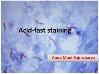
- 12. Observation under Microscope Acid fast: Red, straight or slightly curved rods, occurring singly or in small groups, may appear beaded Non-acid fast: Blue color; In addition, background material stain blue. A.B
- 13. Organisms stained by acid fast staining A.B
- 14. Kinyoun stain • Unlike the (Z-N stain), the Kinyoun method of staining does not require heating. • In the ZN stain, heat acts as a physical mordant while phenol (carbol of carbol fuschin) acts as the chemical mordant. • Since the Kinyoun stain is a cold method (no heat applied), the concentration of carbol fuschin used is increased. A.B